
Thermo Scientific Avidin-Biotin Technical Handbook
Thermo Scientific
Avidin-Biotin Technical Handbook
Table of Contents
EZ-Link Biotinylation Reagents Introduction
1
Biotin-Labeling Reagent Selection Guides
2-3
Selection Guide 1 – Reagent Selection
by Application
Selection Guide 2 – Reagent Selection
by Functional Group
3
Amine-Reactive Biotinylation Reagents
4-11
Introduction
Amine-Reactive Biotinylation Kits
Amine-Reactive Biotinylation Reagents
4
5
6-11
2
Protein Immunodetection
NeutrAvidin Products
Streptavidin Products
Avidin Products
ABC Staining Kits
Biotin Conjugates
Example Protocols for Biotinylation
Troubleshooting Guide for Biotinylation
with NHS-esters
Biotinylating Cell Surface Proteins
One-Step Biotinylation and Dialysis in a
Thermo Scientific Slide-A-Lyzer Cassette
Sulfhydryl-Reactive Biotinylation Reagents
12-15
Carboxyl-Reactive Biotinylation Reagents
16-17
Example Protocols for Affinity Purification
Based on Avidin-Biotin Binding
Carbohydrate/Aldehyde-Reactive
Biotinylation Reagents
18-19
Photo-Reactive Biotinylation Reagents
20-21
Specialty Biotinylation Reagents
22-25
Avidin-Biotin-Based Kits
26
Protein Labeling – Solid Phase Biotinylation Kits
27
Protein Extraction
Cell Surface Protein Isolation Kit
Far-Western Blotting
Pull-down Kit for Biotinylated Proteins
Avidin-Biotin Binding
Introduction
Immobilized Avidin Products
Immobilized Streptavidin Products
Immobilized NeutrAvidin Products
Immobilized Monomeric Avidin and Kit
Immobilized Iminobiotin
Thermo Scientific MagnaBind Beads
NeutrAvidin Coated Polystyrene Plates
NeutrAvidin High binding Capacity Coated Plates
Streptavidin Coated Polystyrene Plates
Streptavidin HBC Coated Plates
28-29
28
29
29
30-38
30
31
32
32
33
33
34
35
36
37
38
39-43
39
40
41
42
43
44-46
45
45
46
47-48
Introduction
47
Affinity Purification of Biotinylated Molecules
47
Afffinity Purification Using a Biotinylated Antibody
48
Immunoprecipitation Using a Biotinylated Antibody 48
Thermo Scientific EZ-Link Biotinylation Reagents
Biotin
S
H
MW 244.3
•Carbohydrate residues containing cis-diols can be oxidized to
create active aldehydes (–CHO).
Antigen-binding
site
Light Chains
VL
VL
VH
S
CL
S-S
CL
S-
S
CH1
CH1
Hinge
Region
NH2
Papain
S-S
S-S
Carbohydrate
VH
Fab (Fab')2
S-S
S-
NH2
S
While NHS-esters of biotin are the most frequently used biotinylation
reagents, they are not necessarily the best for a particular application.
If only a portion of the primary amines on a protein are reacted,
reaction with NHS-esters of biotin will result in a random distribution
•Sulfhydryl groups (–SH) can be generated by reducing disulfide
bonds in the hinge region.
S-
The most frequently used biotinylation reagents, N-hydroxysuccinimide (NHS) esters and N-hydroxysulfosuccinimide (sulfoNHS) esters, react with primary amines. The functional groups
available on the surface of the protein to be biotinylated may not be
known. However, with most proteins, it is safe to assume that primary
amines are available and accessible for biotinylation. The likelihood
that primary amines are present increases as molecular weight
increases. For example, BSA contains 59 primary amines and 30-35
of these are on the surface and can be reacted with NHS-esters.
•Primary amine groups (–NH2) are found on lysine side chains and
at the amino terminus of each polypeptide chain.
S
S-
The valeric acid side chain of the biotin molecule can be derivatized
to incorporate various reactive groups that are used to attach biotin
to other molecules. Using these reactive groups, biotin can be
easily attached to most proteins and other molecules. Biotinylation
reagents are available for targeting a variety of functional groups,
including primary amines, sulfhydryls, carbohydrates and carboxyls.
Photo-reactive biotin compounds that react nonspecifically upon
photoactivation are also available. This variety of functional group
specificities is extremely useful, allowing the choice of a biotinylation reagent that does not inactivate the target macromolecule.
Several cleavable or reversible biotinylation reagents are also
available and allow specific elution of the biotinylated molecule from
biotin-binding proteins. A complete selection guide and detailed
instructions for each reagent is available on the “Products” section
of our web site.
Understanding the functional groups available on an antibody is the
key to determining a strategy for modification.
S
NH
HO
SS
O
NH
S-
O
H
S
Valeric Acid Side Chain
Antibodies are biotinylated more often than any other class of
proteins and it is advantageous to biotinylate in a manner that will
maintain immunological reactivity. Thermo Scientific Sulfo-NHSLC-Biotin is the number one choice for labeling both monoclonal
and polyclonal antibodies because it is the simplest and often
most effective method. The fast and reliable procedure has been
optimized for antibody modification. If the antibody contains a
lysine-rich antigen-binding site, amine-reactive reagents may inhibit
antigen binding. One solution is to use biotin derivatives that react
with sulfhydryl groups. By reducing the immunoglobulin under mild
conditions, biotinylation can be isolated to free sulfhydryls generated
from the hinge region. Another solution is to use a biotin derivative
such as Biocytin-Hydrazide that reacts with aldehydes. Aldehydes
can be generated on antibodies and other glycoproteins by oxidation
of carbohydrates with periodate. Because carbohydrate is found
selectively on the Fc portion of antibodies, biotin labeling is restricted
from occurring near the antigen-binding site. This site-specific
labeling method provides an antibody conjugate with the highest
possible specific activity that is particularly important when antibody
supply is limited and making the best possible use of the antibody
is desired. This method is particularly useful for labeling polyclonal
antibodies, which are heavily glycosylated.
S-
Biotin, a 244 dalton vitamin found in tiny amounts in all living cells,
binds with high affinity to avidin, streptavidin and Thermo Scientific
NeutrAvidin Biotin-Binding Protein. Since biotin is a relatively small
molecule, it can be conjugated to many proteins without significantly
altering their biological activity. A protein can be reacted with several
molecules of biotin that, in turn, can each bind a molecule of avidin.
This greatly increases the sensitivity of many assay procedures.
of biotin on the surface of the protein. If a particular primary amine
is critical to the biological activity of the protein, modification of this
critical amine may result in the loss of its biological activity. Depending
on the extent of biotinylation, complete loss of activity may occur.
SS
The highly specific interaction of avidin with biotin (vitamin H)
can be a useful tool in designing nonradioactive purification and
detection systems. The extraordinary affinity of avidin for biotin
(Ka = 1015 M-1) is the strongest known non-covalent interaction of
a protein and ligand and allows biotin-containing molecules in a
complex mixture to be discretely bound with avidin conjugates.
Our extensive line of biotinylation reagents, conjugates and affinity
supports exploits this unique interaction. Some applications in
which the avidin-biotin interaction has been used include ELISA;
immunohistochemical staining; Western, Northern and Southern
blotting; immunoprecipitation; cell-surface labeling; affinity
purification; and fluorescence-activated cell sorting (FACS).
Pepsin
CH2 S
S
S CH
2
S
Carbohydrate
CH3 S
S
S CH
3
S
Heavy Chains
FC
NH2
NH2
Amines on
lysine residues
Sulfhydryls created when
antibody is reduced
To order, call 800-874-3723 or 815-968-0747. Outside the United States, contact your local branch office or distributor.
1
Thermo Scientific Pierce
Biotin-Labeling Reagent Selection Guides
These selection guides are designed to help you quickly
choose the most appropriate biotinylation reagent to use for an
application. Selection Guide 1 focuses on the purpose/application
of biotin labeling and is organized by the type of molecule that
is to be labeled. It includes only the most common biotinylation
reagent choices. Selection Guide 2 focuses on reaction
chemistries and is organized according to the functional groups
present on the molecule to be labeled. For a more complete
selection guide and for detailed product instructions, visit the
“Products” section of our web site.
Selection Guide 1
Biotinylation Target
Application
Thermo Scientific Pierce Reagent(s)
Features/Benefits
Antibody
ELISA or blotting
(to detect with
streptavidin-HRP)
or affinity-purifying
antigen (after
immobilizing on
streptavidin column)
Sulfo-NHS-LC-Biotin (Product # 21335,
Kit Product # 21435, 21935)
Amine-reactive (lysines). Sure to work well for nearly any antibody.
Very simple and direct.
NHS-PEG4-Biotin
(Product # 21329, 21330, 21362)
Amine-reactive (lysines). PEG spacer arm increases solubility
of biotinylated molecule.
Purified protein
Purified peptide
ELISA or blotting
(to detect with
streptavidin-HRP)
or affinity-purifying
receptor or antibody
(after immobilizing on
streptavidin column)
ELISA or blotting
(to detect with
streptavidin-HRP)
or affinity-purifying
receptor or antibody
(after immobilizing on
streptavidin column)
Cell surface proteins Primarily for affinitypurifying or removing
cell surface receptor
ligands (after
immobilizing on
streptavidin column)
DNA/RNA or
oligo-nucleotides
2
Capture or detection
of oligonucleotides
in ELISA-type
applications or affinity
purification (Capture
in streptavidin-coated
plates or detect
protein-bound oligo
with streptavidin-HRP)
Biotin-PEG4-Hydrazide
Carbohydrate-reactive. Primarily for polyclonal antibodies (purified from serum),
(Product # 21360), plus
which usually have required carbohydrate side chains.
Sodium meta-Periodate (Product # 20504)
Sulfo-NHS-LC-Biotin (Product # 21335,
Kit Product # 21435, 21935, 21362)
Amine-reactive (lysines). Obvious choice for sizeable proteins. Slight risk of blocking
epitopes or binding sites necessary for other purposes.
NHS-PEG4-Biotin
(Product # 21329, 21330)
Amine-reactive (lysines). PEG spacer arm increases solubility
of biotinylated molecule.
Sulfo-NHS-SS-Biotin
(Product # 21331)
Amine-reactive (lysines). Cleavable disulfide spacer allows recovery
of biotinylated protein from immobilized streptavidin.
Maleimide-PEG2-Biotin
(Product # 21901, 21902)
Sulfhydryl-reactive. Use only when there is a reason
to target sulfhydryls rather than amines.
Biotin-PEG4-Hydrazide
(Product # 21360), plus
Sodium Periodate (Product # 20504)
Carbohydrate-reactive. Good choice if the protein is known to be adequately
glycosylated and the carbohydrate is not important for downstream applications.
Sulfo-NHS-LC-Biotin (Product # 21335,
Kit Product # 21435, 21935)
Amine-reactive. Choose if N-terminus or lysines are not required for binding
reactions. May be difficult to remove unreacted reagent.
NHS-PEG4-Biotin
(Product # 21329, 21330)
Amine-reactive (lysines). PEG spacer arm increases solubility
of biotinylated molecule.
Maleimide-PEG2-Biotin
(Product # 21901, 21902), plus
Immobilized TCEP (Product # 77712)
Sulfhydryl-reactive. Choose if cysteines are not required for binding reactions (often
a terminal cysteine is added during peptide synthesis for this purpose). Must ensure
that peptide is reduced. May be difficult to remove unreacted reagent.
Amine-PEG2-Biotin (Product # 21346),
plus EDC (Product # 22980) and
Sulfo-NHS (Product # 24510)
Carbohydrate and amine-reactive. Use only if some peptide polymerization is
acceptable along with biotinylation. Polymerization may be desirable if it creates
large enough molecules to recover conjugate by dialysis.
Sulfo-NHS-LC-Biotin (Product # 21335,
Kit Product # 21435, 21935)
Amine-reactive (lysines). Most commonly-used and most general choice.
NHS-PEG4-Biotin
(Product # 21329, 21330)
Amine-reactive (lysines). PEG spacer arm increases solubility
of biotinylated molecule.
Sulfo-NHS-SS-Biotin
(Product # 21331)
Amine-reactive (lysines). Cleavable disulfide spacer allows recovery
of biotinylated protein from immobilized NeutrAvidin Protein.
Cell Surface Protein Isolation Kit
(Product # 89888)
Amine-reactive (lysines). Cleavable disulfide spacer allows recovery
of biotinylated protein from immobilized NeutrAvidin™ Protein.
Psoralen-PEG3-Biotin
(Product # 29986)
Provides random labeling of nucleic acid backbone.
May cause steric hindrance for hybridization. methods.
Amine-PEG2-Biotin (Product # 21346),
plus EDC (Product # 22980) and
Sulfo-NHS (Product # 24510)
Requires intact 5'-phosphate; produces end-labeled oligo.
For more information, or to download product instructions, visit www.thermo.com/pierce
Selection Guide 2
Reactive Group
Reacts With
Linkage Formed
NHS-Ester/Sulfo-NHS Ester
Primary Amine
(lysine residue)
Amide Bond
O
O
Biotin
Maleimide
Sulfhydryl
(cysteine residue –
not disulfide bonded)
O
N
O
Thioether Bond
O
Thioether Bond
O
O
I
C CH2
C
Biotin
S
S
Biotin
N
Amine
Biotin
S
CH2
Protein
Disulfide Bond
Pyridyl Disulfide
Biotin
Protein
S
O
SH
Protein
H
N
Biotin
Iodoacetyl
Biotin
Protein
NH2
Protein
O
Biotin
O
NH C
Biotin
C O N
NH2
Protein
C OH
S
Protein
Amide Bond
Carboxyl
(glutamate or aspartate residues)
O
S
O
NH C
Biotin
Protein
(Reaction requires EDC cross-linker)
Hydrazide
Oxidized Carbohydrate
Biotin
Hydrazone Bond
O
O
O
C NH
NH2
Protein
C H
Biotin
H
C N N C
Protein
H
Azido (Photoactivatable)
NO2
Biotin
N
H
N3
DNA/RNA,
Protein, Carbohydrates
Biotin
N
NH
Protein
Ring expansion followed by coupling with
primary amine or insertion into double bonds
To order, call 800-874-3723 or 815-968-0747. Outside the United States, contact your local branch office or distributor.
3
Amine-Reactive Biotinylation Reagents
Biotinylation Reagents
Label almost anything with biotin using one of our many
high-quality biotinylation reagents
Whether you are an expert or are trying biotinylation for the
first time, Thermo Scientific EZ-Link Reagents and Kits make
biotinylation easy. Our customers depend on our consistent lotto-lot quality for reproducible results. We offer complete EZ-Link®
Biotinylation Kits with buffers, fast and efficient spin desalting
columns and HABA detection reagent to determine the number of
biotin residues attached to your protein.
Amine-Reactive Biotinylation Reagents
The most common target for modifying protein molecules is the
amine group, which is present on the vast majority of proteins due
to the abundance of lysine side chain e-amines and N-terminal
a-amines. Based on water solubility, amine-reactive biotinylation
reagents can be divided into two groups. NHS-esters of biotin
are essentially water-insoluble. For reactions in aqueous solution,
they must first be dissolved in an organic solvent, then diluted into
the aqueous reaction mixture. The most commonly used organic
solvents for this purpose are DMSO and DMF, which are compatible
with most proteins at 20% final concentration. The solvent acts as a
carrier for the biotinylation reagent, forming a microemulsion in the
aqueous phase and allowing the biotinylation reaction to proceed.
The water-insoluble NHS-esters of biotin are membrane-permeable
because they do not possess a charged group. They may be used for
biotinylating internal as well as external components of a cell.
Sulfo-NHS-esters of biotin are soluble up to approximately 10 mM
in water. Sulfo-NHS-esters should be dissolved in water just before
use because they are prone to hydrolysis. The water solubility of
sulfo-NHS-esters results from the presence of the sulfonate (–SO3)
group on the N-hydroxysuccinimide ring and eliminates the need
to dissolve the reagent in an organic solvent. These compounds
are used for applications that cannot tolerate organic solvents.
Sulfo-NHS-esters of biotin are also recommended for use as cell
surface biotinylation reagents. Because of the charged sulfonate
group, these compounds do not penetrate the plasma membrane;
thus biotinylation is restricted to the cell surface.
O
O
O
N
There is considerable flexibility in the actual conditions used for
conjugating NHS-esters (or sulfo-NHS-esters) to primary amines.
Incubation temperatures range from 4-37°C, reaction mixture pH
values range from 7-9, and incubation times range from a few
minutes to overnight. Buffers containing amines (such as Tris or
glycine) must be avoided because they compete with the reaction.
In preparing an NHS-ester biotin conjugate, a particular set of
conditions will result in a conjugate with optimum properties for
a specific application. The preparation of an optimum conjugate
is largely dependent on the degree of incorporation of the label.
Because of the variability among proteins, especially the number
of amines available for conjugation, conjugation conditions that are
optimal for one protein may not be optimal for another protein.
There are several additional features to consider in an amine-reactive
biotinylation reagent. Because biotin binds in a pocket located 9 Å
below the surface of the avidin molecule, the spacer arm connected
to the biotin is critical. Long spacer arms reduce steric hindrance and
result in enhanced interaction of avidin and biotin. It is sometimes
necessary to remove biotin from a molecule once a procedure has
been completed, and there are reagents with a cleavable spacer
arm that allow this procedure. A macromolecule is first reacted with
the cleavable biotinylation reagent, then it is used in a detection or
purification system. Finally the biotin moiety can be cleaved away,
releasing the molecule into solution. Biotin labeling generally reduces
the solubility of a molecule and may result in precipitation. Unlike
sulfo-NHS-esters that lose their solubility-enhancing sulfonate group
during the reaction process, reagents containing a PEG spacer arm
retain their high solubility when bound to a protein and are an ideal
choice when precipitation must be overcome.
O
O Na
S
The reaction chemistries of NHS- and sulfo-NHS-esters are
essentially identical: an amide bond is formed and NHS or sulfoNHS are leaving groups in the reaction. Because the target for
the ester is the deprotonated form of the primary amine, the
reaction becomes significant at neutral pH values and above when
the amine is able to react with the ester by nucleophilic attack.
Hydrolysis of the NHS-ester is a major competing reaction, and the
rate of hydrolysis increases with increasing pH. NHS- and
sulfo-NHS-esters have a half-life of hydrolysis of 2-4 hours at pH 7.
This half-life decreases to just a few minutes at pH 9.
HN
O
H
N
O
O
Sulfo-NHS-LC-Biotin
NH
O
O
S
Na
O
S
O
O
N
OH
O
Sulfo-NHS Leaving Group
O
HN
O
Protein
Protein
NH2
Molecule with
Primary Amines
Reaction of Sulfo-NHS-LC-Biotin with a primary amine.
4
For more information, or to download product instructions, visit www.thermo.com/pierce
N
H
H
N
NH
S
O
Biotinylated Molecule
Thermo Scientific EZ-Link Amine-Reactive Biotinylation Kits
Kit
Sulfo-NHS
Biotinylation Kits
Highlights
References
•Complete kit with an optimized
1, 2
protocol for labeling a protein and
determining how much biotin has
been attached
•Fast and efficient labeling
procedure
Sulfo-NHS-LC
Biotinylation Kits
•Complete kit with an optimized
1, 2
protocol for labeling a protein and
determining how much biotin has
been attached
Ordering Information
Product # Description
Pkg. Size
21425
EZ-Link Sulfo-NHS Biotinylation Kit
Kit
21925
EZ-Link Micro Sulfo-NHS Biotinylation Kit
Kit
21435
EZ-Link Sulfo-NHS-LC Biotinylation Kits
Kit
21935
EZ-Link Micro
Sulfo-NHS-LC Biotinylation Kit
Kit
Includes: No-Weigh Sulfo-NHS-LC Biotin
Zeba Desalting Columns
BupH Phosphate Buffered Saline
8 x 1 mg
2 ml
1 pack (500 ml)
EZ-Link NHS-PEG4 Biotinylation Kit
Kit
EZ-Link Micro
NHS-PEG4 Biotinylation Kit
Kit
Includes: No-Weigh NHS-PEG4-Biotin
Zeba Desalting Columns
BupH Phosphate Buffered Saline
8 x 1 mg
2 ml
1 pack (500 ml)
21445
EZ-Link Sulfo-NHS-SS Biotinylation Kit
Kit
21945
EZ-Link Micro
Sulfo-NHS-SS Biotinylation Kit
Kit
•Fast and efficient labeling
procedure
NHS-PEG4
Biotinylation Kits
•PEG4-Biotin transfers
water-solubility to biotinylated
molecules
3-6
21455
21955
Sulfo-NHS-SS
Biotinylation Kits
•Cleavable biotinylation reagent
allows removal of biotin label
7-9
Includes: Sulfo-NHS-Biotin
Reaction Buffer
Zeba™ Spin Columns
HABA Dye (10 mM)
Avidin, affinity-purified
Includes: No-Weigh™ Sulfo-NHS-Biotin
Zeba Desalting Columns
BupH™ Phosphate Buffered Saline
Includes: Sulfo-NHS-LC-Biotin
Reaction Buffer
Zeba Spin Columns
HABA Dye (10 mM)
Avidin, affinity-purified
Includes: NHS-PEG4-Biotin
BupH PBS
Zeba Spin Columns
HABA Dye (10 mM)
Avidin, a ffinity-purified
Includes: Sulfo-NHS-SS-Biotin
Non-reagent contents same as
Product # 21425
Includes: Sulfo-NHS-SS-Biotin
Non-reagent contents same as
Product # 21925
25 mg
1 pack (500 ml)
10
1 ml
10 mg
8 x 1 mg
2 ml
1 pack (500 ml)
25 mg
1 pack (500 ml)
10
1 ml
10 mg
8 x 2 mg
1 pack (500 ml)
10
1 ml
10 mg
25 mg
8 x 1 mg
References
1. Zhang, L., et al. (1999). J. Biol. Chem. 274, 8966-8972.
2. Zuk, P.A. and Elferink, L.A. (2000). J. Biol. Chem. 275, 26754-26764.
3. Ali, M.K. and Bergson, C. (2003). J. Biol. Chem. 278, 51654-51663.
4. Du, J., et al. (2003). J. Cell Biol. 163(2), 385-395.
5. Lee-Kwon, W., et al. (2003). J. Biol. Chem. 278, 16494-16501.
6. Lin, Z., et al. (2003). J. Biol. Chem. 278(22), 20162-20170.
7. Daniels, G.M. and Amara, S.G. (1998). Methods. Enzymol. 296, 307-318.
8. Huh, K-H. and Wenthold, R.J. (1999). J. Biol. Chem. 274, 151-157.
9. Trotti, D., et al. (2001). J. Biol. Chem. 276, 576-582.
To order, call 800-874-3723 or 815-968-0747. Outside the United States, contact your local branch office or distributor.
5
Amine-Reactive Biotinylation Reagents
Thermo Scientific EZ-Link Amine-Reactive Biotinylation Reagents
Reagent
Structure
Highlights
Sulfo-NHS-Biotin
•Reacts with primary amines at ph 7-9 in
non-amine-containing buffers
O
NH
HN
•Water-soluble for ease of use
O
Na+ O-
N
O S
O
O
S
O
O
Sulfo-NHS-Biotin
MW 443.43
Spacer Arm 13.5 Å
Sulfo-NHS-LC-Biotin
O
O
Na+ OO S
N
O
•Water-soluble for ease of use
O
H
N
O
•Ideal labeling reagent for monoclonal and
polyclonal antibodies
NH
HN
S
O
O
Sulfo-NHS-LC-Biotin
MW 556.59
Spacer Arm 22.4 Å
Sulfo-NHS-LC-LC-Biotin
O
NH
HN
Na+ O-
O
O
N
O S
O
O
O
H
N
N
H
O
•Contains an extra-long spacer arm (30.5 Å)
to reduce steric hindrance
S
O
Sulfo-NHS-LC-LC-Biotin
MW 669.75
Spacer Arm 30.5 Å
Sulfo-NHS-SS-Biotin
HN
Na+ OO S
•Disulfide linkage can be cleaved
by reducing agents
O
O
N
O
O
O
S
S
H
N
O
NH
•Ideal reagent for reversibly biotinylating
cell surface proteins
S
O
Sulfo-NHS-SS-Biotin
MW 606.69
Spacer Arm 24.3 Å
NHS-PEG4-Biotin
O
HN
O
N
O
O
O
O
O
O
O
H
N
NH
S
•PEG spacer arm increases solubility
of conjugates
•No-Weigh Packaging ensures fresh reagent and eliminates
tedious weighing of reagent
O
NHS-PEG4-Biotin
MW 588.67
Spacer Arm 29.0 Å
6
For more information, or to download product instructions, visit www.thermo.com/pierce
References
Ordering Information
Product #
Description
Pkg. Size
21217
EZ-Link Sulfo-NHS-Biotin
50 mg
21326
No-Weigh Sulfo-NHS-Biotin
8 x 1 mg
•Arosa, F.A., et al. (1999). J. Biol. Chem. 274, 16917-16922.
•Baqui, M., et al. (2003). J. Biol. Chem. 278, 1206-1211.
•Huh, K-H. and Wenthold, R.J. (1999). J. Biol. Chem. 274, 151-157.
•Lesa, G.M., et al. (2000). J. Biol. Chem. 275, 2831-2836.
•Liaw, P.C.Y., et al. (2001). J. Biol Chem. 276, 8364-8370.
•Liu, L.A. and Engvall, E. (1999). J. Biol. Chem. 274, 38171-38176.
21335
EZ-Link Sulfo-NHS-LC-Biotin
100 mg
21327
No-Weigh Sulfo-NHS-LC-Biotin
8 x 1 mg
•Muroi, M., et al. (2002). J. Biol. Chem. 277, 42372-42379.
•Schwarzman, A.L., et al. (1999). Proc. Natl. Acad. Sci. U.S.A. 96, 7932-7937.
21338
EZ-Link Sulfo-NHS-LC-LC-Biotin
50 mg
•Daniels, G.M. and Amara, S.G. (1998). Methods. Enzymol. 296, 307-318.
•Huh, K-H. and Wenthold, R.J. (1999). J. Biol. Chem. 274, 151-157.
•Trotti, D., et al. (2001). J. Biol. Chem. 276, 576-582.
21331
EZ-Link Sulfo-NHS-SS-Biotin
100 mg
21328
No-Weigh Sulfo-NHS-SS-Biotin
8 x 1 mg
21329
No-Weigh NHS-PEG4-Biotin
8 x 2 mg
21330
EZ-Link NHS-PEG4-Biotin
25 mg
21362
EZ-Link NHS-PEG4-Biotin
50 mg
21363
EZ-Link NHS-PEG4-Biotin
1g
•Arosa, F.A., et al. (1999). J. Biol. Chem. 274, 16917-16922.
•Claypool, S.M., et al. (2002). J. Biol. Chem. 277, 28038-28050.
•Ellerbroek, S.M., et al. (2001). J. Biol. Chem. 276, 24833-24842.
•Leighton, B.H., et al. (2002). J. Biol. Chem. 277, 29847-29855.
•Neely, K.E., et al. (2002). Mol. Cell. Biol. 22, 1615-1625.
•Dodeller, F., et al. (2008) J. Biol. Chem. 283, 21487-21494.
•Newton, J.R., et al. (2007) J. Nucl. Med. 48, 429-436.
•Behrens, M. et al. (2006) J. Biol. Chem. 281, 20650-20659.
To order, call 800-874-3723 or 815-968-0747. Outside the United States, contact your local branch office or distributor.
7
Amine-Reactive Biotinylation Reagents
Thermo Scientific EZ-Link Amine-Reactive Biotinylation Reagents
Reagent
Structure
Highlights
NHS-PEG12-Biotin
•PEG spacer arm increases solubility
of conjugates
O
HN
O
N
NH
O
O
O
[
O
]
H
N
12
•Extra long spacer arm reduces
steric hindrance
S
O
NHS-PEG12-Biotin
MW 941.09
Spacer Arm 56 Å
NHS-SS-PEG4-Biotin
O
HN
O
N
S
O
O
H
N
S
O
O
O
H
N
O
S
•PEG spacer arm increases solubility
of conjugates
•Disulfide linkage can be cleaved
by reducing agents
O
O
O
NH
NHS-SS-PEG4-Biotin
M.W. 751.94
Spacer Arm 37.9 Å
NHS-Biotin
•Must be dissolved in DMSO or DMF
before adding to aqueous solution
O
HN
NH
•Able to penetrate cell membranes
O
N
O
O
S
O
NHS-Biotin
MW 341.38
Spacer Arm 13.5 Å
NHS-LC-Biotin
O
O
N
O
O
HN
O
H
N
NH
•Must be dissolved in DMSO or DMF
before adding to aqueous solution
•Able to penetrate cell membranes
S
O
NHS-LC-Biotin
MW 454.54
Spacer Arm 22.4 Å
8
For more information, or to download product instructions, visit www.thermo.com/pierce
References
Ordering Information
Product #
Description
Pkg. Size
21312
EZ-Link NHS-PEG12-Biotin
25 mg
21313
EZ-Link NHS-PEG12-Biotin
500 mg
21442
NHS-SS-PEG4-Biotin
50 mg
•Fouassier, L., et al. (2000). J. Biol. Chem. 275, 25039-25045.
•Nunomura, W., et al. (2000). J. Biol. Chem. 275, 6360-6367.
20217
EZ-Link NHS-Biotin
100 mg
•Chiu, N.H., et al. (1999). Clin. Chem. 45, 1954-1959.
•Schumacher, T.N., et al. (1996). Science 271, 1854-1857.
•Tang, A., et al. (1993). Nature 361, 82-85.
21336
EZ-Link NHS-LC-Biotin
50 mg
To order, call 800-874-3723 or 815-968-0747. Outside the United States, contact your local branch office or distributor.
9
Amine-Reactive Biotinylation Reagents
Thermo Scientific EZ-Link Amine-Reactive Biotinylation Reagents
Reagent
Structure
Highlights
NHS-LC-LC-Biotin
HN
O
O
O
N
O
•Contains an extra-long spacer arm (30.5 Å) to reduce
steric hindrance
O
H
N
N
H
O
NH
S
O
NHS-LC-LC-Biotin
MW 567.70
Spacer Arm 30.5 Å
NHS-SS-Biotin
O
NH
HN
O
N
O
S
H
N
S
O
O
•Label almost any protein with biotin to facilitate immobilization,
purification or detection
•Disulfide bond in the spacer arm allows the biotin label
to be removed
S
•Amine-reactive NHS ester reacts rapidly with any primary aminecontaining molecule to attach the biotin via a stable amide bond
O
NHS-SS-Biotin
•Long spacer arm on the biotin reduces steric hindrance with
binding to avidin or other biotin-binding proteins
MW 504.65
Spacer Arm 24.3 Å
NHS-Iminobiotin
O
•Reversible binding to avidin prevents protein denaturation
during purification
F
N
F
F
NH
HN
•Binds to avidin at pH >9.5 and dissociates at pH 4
O
N
O
O
S
O
NHS-Iminobiotin Trifluoroacetamide
MW 436.41
Spacer Arm 13.5 Å
PFP-Biotin
•More reactive than NHS esters
O
HN
•Will react with primary and secondary amines at pH 7-9
NH
•Easier-to-handle and store than NHS esters
F
O
F
F
F
•Great for DNA labeling
S
•PEG spacer arm increases solubility of conjugates
O
F
Biotin-PFP Ester
MW 410.36
Spacer Arm 9.6 Å
TFP-PEG3-Biotin
F
F
F
HN
O
H
N
O
F
•Long-chain, water-soluble, polyethylene oxide (PEG) spacer arm
O
O
O
O
O
NH
•Tetrafluorophenyl (TFP) ester reacts with primary and
secondary amines at pH 7-9
•Glycine and Tris buffers interfere with reaction
H
N
S
•PEG spacer arm transfers hydrophilicity to final conjugate
O
TFP-PEG3-Biotin
MW 694.74
Spacer Arm 32.6 Å
10
For more information, or to download product instructions, visit www.thermo.com/pierce
References
Ordering Information
Product #
Description
Pkg. Size
21343
EZ-Link NHS-LC-LC-Biotin
50 mg
21441
EZ-Link NHS-SS-Biotin
50 mg
•Orr, G.A., et al. (1981). J. Biol. Chem. 256, 761-766.
•Zeheb, R., et al. (1983). Anal. Biochem. 129, 156-161.
21117
EZ-Link NHS-Iminobiotin
100 mg
•Muroi, M., et al. (2002). J. Biol. Chem. 277, 42372-42379.
•Michaelis, K., et al. (2006) J. Pharmacol. Exp. Ther. 317, 1246-1253.
21218
EZ-Link PFP-Biotin
50 mg
21219
EZ-Link TFP-PEG3-Biotin
50 mg
•Bradley, C., et al. (2007) Carcinogen. 28, 2184-2192.
•Sehr, P., et al. (2007) J. Biomo.l Screen. 12, 560-567.
•Negishi, A., et al. (2004) Glycobiolog. 14, 969-977.
To order, call 800-874-3723 or 815-968-0747. Outside the United States, contact your local branch office or distributor.
11
Sulfhydryl-Reactive Biotinylation Reagents
Sulfhydryl-Reactive Biotinylation Reagents
The second most common target for modification in biological
molecules is free sulfhydryl groups, which are found in the form
of exposed cysteine in a protein or peptide. Sulfhydryl group
biotinylation may provide an advantage in some applications; for
example, targeting a sulfhydryl group can be used as a method for
preserving the biological activity of an enzyme when amines are
found at the active site. Modification of these amines may render
the enzyme inactive. This complication can be avoided by using
derivatives of biotin that react with sulfhydryls. Because biotinylation
with these reagents must be performed in buffers free of extraneous
sulfhydryls, substances such as 2-mercaptoethanol, dithiothreitol
and mercaptoethylamine must be removed before biotinylation.
can be generated from disulfides by incubation with a reducing
agent or from lysine residues by incubating with modification
reagents such as 2-Iminothiolane (Traut’s Reagent) or SATA.
When working with free sulfhydryls, EDTA is generally included
in the buffer system for its antioxidative effect. EDTA chelates
trace amounts of metals in solution that promote disulfide
bond formation. Using nitrogen-purged buffers is an additional
precaution to prevent oxidation of the free sulfhydryls.
Three separate reaction chemistries are employed to target
sulfhydryl groups for biotinylation. The most specific method uses
reactive maleimide groups, which are 1,000 times more reactive
toward free sulfhydryls than toward amines at pH 7. Biotin-BMCC
must be dissolved in an organic solvent, then diluted into an
aqueous reaction mixture. Maleimide-PEG2-Biotin, which is watersoluble by virtue of its polyethyleneoxide (PEG) spacer arm, may be
dissolved directly in aqueous solution. The reaction of maleimide
with free thiols is carried out at pH 6.5-7.5 because cross-reactivity
Proteins, peptides or other molecules to be biotinylated by
sulfhydryl-reactive reagents must have a free sulfhydryl group
(–SH) available; disulfides will not react with sulfhydryl-specific
biotinylation reagents. If free sulfhydryls are not available, they
Thermo Scientific EZ-Link Sulfhydryl-Reactive Biotinylation Reagents
Reagent
Structure
Highlights
Maleimide-PEG2-Biotin
O
•Water-soluble
NH
HN
O
O
N
O
N
H
H
N
O
S
•Reacts with sulfhydryl (–SH) groups at acidic to neutral pH
•Avoids potential modification of tyrosine residues that can be
associated with Iodoacetyl-Biotin
O
O
Maleimide-PEG2-Biotin
MW 525.62
Spacer Arm 29.1 Å
Maleimide-PEG11-Biotin
O
HN
O
H
N
N
O
•Water-soluble
NH
O
[
O
]
H
N
11
•Reacts with sulfhydryl (-SH) groups at acidic to neutral pH
•Extra long spacer arm reduces steric hindrance
S
O
Maleimide-PEG11-Biotin
MW 922.09
Spacer Arm 59.1 Å
Biotin-BMCC
•Reacts with sulfhydryl (–SH) groups at pH 6.5-7.5
O
NH
HN
O
O
H
N
N
H
N
S
•Avoids potential modification of tyrosine residues that can be
associated with Iodoacetyl-Biotin
•Must be dissolved in DMSO or DMF before use
O
Biotin-BMCC
O
MW 533.68
Spacer Arm 32.6 Å
Iodoacetyl-PEG2-Biotin
HN
O
I
•Water-soluble
O
N
H
O
O
H
N
NH
•Reacts with sulfhydryl (–SH) groups at basic to neutral pH
•Coupling reactions occur in the dark
S
O
Iodoacetyl-PEG2-Biotin
MW 542.43
Spacer Arm 24.7 Å
12
For more information, or to download product instructions, visit www.thermo.com/pierce
toward primary amines can occur at higher pH values. Hydrolysis
of the maleimide group also increases at higher pH values.
Iodoacetyl groups are also used to target free sulfhydryls.
Iodoacetyl-LC-Biotin is not water-soluble and must be dissolved in
a solvent before use in an aqueous reaction mixture. IodoacetylPEG2-Biotin, which is water-soluble by virtue of its PEG spacer arm,
may be dissolved directly in aqueous solution. The iodoacetyl group
reacts mainly with thiol groups at pH 7.5-8.5, resulting in a stable
thioether bond. Unless precautions are taken, iodoacetyl groups
may not be specific for sulfhydryls. The reaction can be directed
toward sulfhydryl groups by limiting the molar ratio of IodoacetylBiotin to protein, such that the concentration of biotin is present at
a small excess over the sulfhydryl content. Also, the reaction pH
should be maintained in the range of 7.5-8.5. Below pH 9, crossreactivity with amine, thioether and imidazole groups is minimized.
Therefore, maintaining a lower pH ensures the modification of
sulfhydryl groups and not amino groups. If there are no cysteines
References
available, the reaction can be directed at imidazoles by adjusting
the pH to 6.9-7.0. However, the incubation time must be increased
to a week. Histidyl side chains and amino groups react in the
deprotonated form and may take part in reactions above pH 5 and
pH 7, respectively, although this reaction is much slower than that
for sulfhydryls.
A pyridyldithiol group may also be used to attach biotin to a free
sulfhydryl by disulfide exchange, resulting in the formation of a
mixed disulfide bond. The reaction of Biotin-HPDP is generally
carried out under physiologic conditions although the process
occurs within a wide range of pH conditions and with a variety of
buffer components. Pyridyldithiol reactions result in the release
of pyridine-2-thione, which cannot react with free sulfhydryls.
The release of this compound can be measured and the reaction
progress monitored by an increasing absorbance at 343 nm.
Another important feature of this method is that the biotin is
released by treatment with reducing agents.
Ordering Information
Product #
Description
Pkg. Size
21901
EZ-Link Maleimide-PEG2-Biotin
50 mg
21902
No-Weigh Maleimide-PEG2-Biotin
Microtubes
8 x 2 mg
21911
Maleimide-PEG11-Biotin
25 mg
•Roberts, P.J., et al. (2008) J. Biol. Chem. 283, 25150-25163.
•Shi, M., et al. (2007) J. Biol. Chem. 282, 30198-30206.
•Chenette, E. J., et al. (2006) Mol. Biol. Cell. 17, 3108-3121.
21900
EZ-Link Biotin-BMCC
50 mg
•Kim, K., et al. (2001). J. Biol. Chem. 276, 40591-40598.
•Muroi, M., et al. (2002). J. Biol. Chem. 277, 42372-42379.
21334
EZ-Link Iodoacetyl-PEG2-Biotin
50 mg
•Oda, Y., et al. (2001). Nat. Biotechnol. 19, 379-382.
•Inglis, K.J., et al. (2008) J. Biol. Chem. doi:10.1074/jbc.C800206200
To order, call 800-874-3723 or 815-968-0747. Outside the United States, contact your local branch office or distributor.
13
Sulfhydryl-Reactive Biotinylation Reagents
Thermo Scientific EZ-Link Sulfhydryl-Reactive Biotinylation Reagents
Reagent
Structure
Highlights
Iodoacetyl-LC-Biotin
•Reacts with sulfhydryl (–SH) groups at basic to neutral pH
O
HN
O
I
H
N
N
H
•Coupling reactions occur in the dark
NH
•Must be dissolved in DMSO or DMF before use
S
O
Iodoacetyl-LC-Biotin
MW 510.43
Spacer Arm 27.1 Å
Biotin-HPDP
O
HN
O
N
S
S
H
N
N
H
S
O
Biotin-HPDP
NH
•Pyridyldithiol reacts with sulfhydryl (–SH) groups to form
a stable disulfide bond between pH 6-9
•Pyridine-2-thione leaving group can be used to measure
degree of biotinylation at 343 nm
•Disulfide bond is cleavable using 50 mM DTT or 100 mM
2-mercaptoethanol
•Must be dissolved in DMSO or DMF before use
MW 539.78
Spacer Arm 29.2 Å
14
For more information, or to download product instructions, visit www.thermo.com/pierce
References
Ordering Information
Product #
Description
Pkg. Size
•Sutoh, K., et al. (1984). J. Mol. Biol. 178, 323-339.
•Yamamoto, K., et al. (1984). FEBS Lett. 176, 75-78.
21333
EZ-Link Iodoacetyl-LC-Biotin
50 mg
•Ishmael, F.T., et al. (2001). J. Biol. Chem. 276, 25236-25242.
•Slatin, S.L., et al. (2002). Proc. Natl. Acad. Sci. U.S.A. 99, 1286-1291.
21341
EZ-Link Biotin-HPDP
50 mg
To order, call 800-874-3723 or 815-968-0747. Outside the United States, contact your local branch office or distributor.
15
Carboxyl-Reactive Biotinylation Reagents
Carboxyl-Reactive Biotinylation Reagents
Carboxyl groups, in the form of carboxy termini, aspartate residues
or glutamate residues, can also be targeted for biotin labeling
using amine-derivatized biotin molecules. This reaction is mediated
by a class of crosslinkers known as carbodiimides and results in
the formation of an amide bond. The reaction with EDC, the most
common carbodiimide crosslinker, is generally performed in an MES
buffer at pH 4.5-5 and requires just minutes to complete. Buffers
containing primary amines (Tris, glycine, etc.) or carboxyls (acetate,
citrate, etc.) must be avoided because they will quench the reaction.
Phosphate buffers are also not recommended because they reduce
the conjugation efficiency, although this effect can be overcome by
adding more EDC.
Thermo Scientific EZ-Link Carboxyl-Reactive Biotinylation Reagents
Reagent
Structure
Highlights
Pentylamine-Biotin
•Use the EDC crosslinker to couple this analog to carboxyl
(–COOH) groups
O
HN
H
N
H2N
NH
•Dissolves in aqueous solutions
•Coupling with EDC occurs at pH 4-6
S
O
Pentylamine-Biotin
MW 328.47
Spacer Arm 18.9 Å
Amine-PEG2-Biotin
•Use the EDC crosslinker to couple this analog to carboxyl
(–COOH) groups
O
NH
HN
O
H2 N
H
N
O
•Water-soluble
S
O
Amine-PEG2-Biotin
MW 374.50
Spacer Arm 20.4 Å
Amine-PEG3-Biotin
O
HN
H2N
O
O
O
H
N
NH
S
•Use the EDC crosslinker to couple this analog to carboxyl
(–COOH) groups
•Water-soluble
•Longer chain length for reduction of steric hindrance
O
Amine-PEG3-Biotin
MW 418.55
Spacer Arm 22.9 Å
16
For more information, or to download product instructions, visit www.thermo.com/pierce
Amine-PEG2-Biotin, Amine-PEG3-Biotin and Pentylamine-Biotin
are amine-derivatized biotin molecules that can be reacted with
carboxyl groups. In addition, any of the hydrazide-derivatized biotin
molecules can be reacted with carboxyls under identical conditions.
The reaction is most often mediated by EDC, a water-soluble
carbodiimide that activates carboxyl groups to bind to the –NH2
group on the biotinylation reagent. Using this strategy may result
in some polymerization of the peptide or protein if the molecule
has both carboxyls and primary amines on its surface. The extent
of polymerization can be minimized by decreasing the amount of
EDC and/or increasing the amount of the biotin reagent used in
the reaction.
References
Ordering Information
Product #
Description
Pkg. Size
• Cernuda-Morollon, E., et al. (2001). J. Biol. Chem. 276, 35530-35536.
• Liu, Y., et al. (1999). Proc. Natl. Acad. Sci. U.S.A. 96, 14694-14699.
21345
EZ-Link Pentylamine-Biotin
50 mg
• Fezza, F., et al. (2008) J. Lipid Res. 49, 1216-1223.
• Cui, B., et al. (2007) Proc. Nat. Acad. Sci. USA. 104, 13666-13671.
• Pihlajamaa, T., et al. (2004) J. Biol. Chem. 279, 24265-24273.
21346
EZ-Link Amine-PEG2-Biotin
50 mg
• Maguire, B. A., et al. (2008) RNA. 14, 188-195.
21347
EZ-Link Amine-PEG3-Biotin
50 mg
To order, call 800-874-3723 or 815-968-0747. Outside the United States, contact your local branch office or distributor.
17
Carbohydrate/Aldehyde-Reactive Biotinylation Reagents
Carbohydrate/Aldehyde-Reactive Biotinylation Reagents
Another common target for protein modification is the carbohydrate
portion of glycoproteins, which can be reacted with hydrazidederivatives of biotin. Oxidative treatment of glycoproteins using
10 mM periodate is used to generate reactive aldehydes from the
cis-diols of a variety of carbohydrate moieties.
OH OH
R
C
C
H
H
O
NaIO4
R'
R
C
An aldehyde can be reacted specifically with a hydrazide group
at pH 4-6, forming a stable hydrazone linkage. Sialic acid
residues on glycoproteins can be specifically oxidized with
sodium periodate (NaIO4) under mild conditions. At 1 mM
periodate and a temperature of 0°C, oxidation is restricted
primarily to sialic acid residues. Sialic acid residues also can
be biotinylated with hydrazide derivatives by pretreatment with
neuraminidase to generate galactose groups. The galactose and
N-acetylgalactosamine residues on whole cells can be selectively
biotinylated with Biotin-Hydrazides by further treatment with
galactose oxidase. This enzyme will convert the primary hydroxyl
groups on these sugars to their corresponding aldehydes.
O
H + H
C
R'
Oxidation of a cis-diol to an aldehyde
Thermo Scientific EZ-Link Carbohydrate/Aldehyde-Reactive Biotinylation Reagents
Reagent
Structure
Highlights
Biocytin-Hydrazide
•Can be used to label DNA and RNA through
cytosine residues
O
NH
HN
O
H2 N
N
H
H
N
NH2
•More water-soluble than Biotin-LC-Hydrazide
S
O
Biocytin-Hydrazide
MW 386.51
Spacer Arm 19.7 Å
Biotin-Hydrazide
•Carbohydrate reactive
O
H 2N
H
N
•Must be dissolved in DMSO before
adding to aqueous buffer
NH
HN
S
O
Biotin-Hydrazide
MW 258.34
Spacer Arm 15.7 Å
Biotin-LC-Hydrazide
O
HN
O
H2N
H
N
N
H
•Use the EDC crosslinker to couple this analog to
carboxyl (–COOH) groups
NH
•Water-soluble
•Longer chain length for reduction of steric hindrance
S
O
Biotin-LC-Hydrazide
MW 371.50
Spacer Arm 24.7 Å
Biotin-PEG4-Hydrazide
HN
H2N
H
N
•Water-soluble analog of Biotin-LC-Hydrazide
O
O
O
O
O
O
H
N
NH
•PEG-based spacer arm increases solubility
of labeled molecules
S
O
Biotin-PEG4-Hydrazide
MW 505.63
Spacer Arm 31.3 Å
18
For more information, or to download product instructions, visit www.thermo.com/pierce
Mild oxidation of an immunoglobulin with sodium periodate produces
reactive aldehydes from the carbohydrate moieties on the Fc portion
of the antibody, which then can be alkylated by a hydrazide. This
approach is advantageous for use with antibodies because they
become biotinylated in a manner that maintains immunological
reactivity. This is an ideal method for biotinylating polyclonal antibodies
because they are heavily glycosylated. Monoclonal antibodies may be
deficient in glycosylation and success with this method will depend on
the extent of glycosylation for a particular antibody.
References
Temperature, pH of oxidation and the periodate concentration all
affect the reaction with hydrazide derivatives of biotin. Also, because
glycosylation varies with each protein, optimum conditions must be
determined for each glycoprotein. Each glycoprotein preparation
has an optimum pH for oxidation and for the hydrazide-mediated
biotinylation. Tris, or other primary amine-containing buffers, are
not recommended for use in either the oxidation or biotinylation
steps because these buffers react with aldehydes, quenching their
reaction with hydrazides.
Ordering Information
Product #
Description
Pkg. Size
• Bayer, E.A. et al. (1988). Anal. Biochem. 170, 271-281.
• Reisfeld, A., et al. (1987). Biochem. Biophys. Res. Commun. 142, 519-526.
•Roffman, E., et al. (1986). Biochem. Biophys. Res. Commun. 136, 80-85.
28020
EZ-Link Biocytin-Hydrazide
25 mg
• Edwards, S.W., et al. (1999). J. Biol. Chem. 274, 16331-16336.
• Reisfeld, A., et al. (1987). Biochem. Biophys. Res. Commun. 142, 519-526.
21339
EZ-Link Biotin-Hydrazide
100 mg
• Araga, S., et al. (1999). J. Immunol. 163, 476-482
•Kahne, T. and Ansorge, S. (1994). J. Immunol. Methods 168, 209-218.
•Luk, J.M., et al. (1995). Anal. Biochem. 232, 217-224
•Scott, M.G., et al. (2000). J. Immunol. 164, 549-553.
•Yu, Q. and Toole, B.P. (1995). Biotechniques 19, 122-129.
21340
EZ-Link Biotin-LC-Hydrazide
50 mg
21360
EZ-Link Biotin-PEG4-Hydrazide
50 mg
To order, call 800-874-3723 or 815-968-0747. Outside the United States, contact your local branch office or distributor.
19
Photo-Reactive Biotinylation Reagents
Photo-Reactive Biotinylation Reagents
Proteins, peptides and other molecules that do not contain any of
the reactive functional groups mentioned previously may be labeled
using a photo-reactive biotinylation reagent. These reagents contain
an aryl azide group that is chemically inert until it is exposed to
ultraviolet light, causing the formation of a short-lived, reactive aryl
nitrene. The half-life of this aryl nitrene intermediate is on the order
of 10-4 seconds. The aryl nitrene reacts rapidly and nonselectively
with electron dense sites by addition into double bonds or
insertion into active hydrogen bonds. If the aryl-nitrene fails
to react, it undergoes ring expansion and becomes reactive
toward nucleophiles such as primary amines and sulfhydryls.
Photoactivation and insertion into another molecule can be
performed in a wide variety of buffer conditions. However, acidic
conditions and reducing agents should be avoided because they
may inactive the aryl azide group.
Thermo Scientific EZ-Link Photo-Reactive Biotinylation Reagents
Reagent
Structure
Highlights
Photoactivatable Biotin
-O
-N
N
•Covalent attachment occurs in the presence of
UV light (350-370 nm)
O
N+
NH
HN
O
CH3
H
N
H
N
N
+
•Attachment is nonspecific
•Must be dissolved in DMSO or DMF before use
S
•Reaction buffer should be above pH 6
O
N
Photoactivatable Biotin
MW 533.65
Spacer Arm 30.0 Å
Psoralen-PEG3-Biotin
•Water-soluble
O
O
NH
HN
O
H
N
O
O
O
O
H
N
O
O
•Much higher coupling yield than achieved
with photoactivatable biotin
•Psoralen moiety reacts with the 5,6 double bond in thymineand other pyrimidine-containing bases
S
•Covalent attachment occurs in the presence
of UV light (>350 nm) for 10-30 minutes
O
Psoralen-PEG3-Biotin
•Great for biotinylating DNA/RNA probes
MW 688.79
Spacer Arm 36.9 Å
•Ultrahigh sensitivity relative to radiolabeled
probes (<100 femtograms)
•Doesn’t interfere with hybridization
Biotin-LC-ASA
•Iodinatable and photoactivatable
O
HN
OH
-N
N+
O
H
N
N
H
•Useful for biotinylation of nucleic acids
NH
S
O
N
Biotin-LC-ASA
MW 503.62
Spacer Arm 29.9 Å
TFPA-PEG3-Biotin
F
+
-N N
HN
F
N
H
N
F
F
•Label almost any molecule with biotin
O
O
O
O
O
H
N
NH
•TFPA is more efficient than other photo-reactive groups
•Long, PEG-based spacer arm reduces steric hindrance and
increases conjugate solubility
S
O
TFPA-PEG3-Biotin
MW 663.69
Spacer Arm 33.4 Å
20
For more information, or to download product instructions, visit www.thermo.com/pierce
Photoactivatable Biotin, Biotin-LC-ASA , Psoralen-PEG3-Biotin
and TFPA-PEG3-Biotin each contain a photoactivatable group.
When exposed to UV light, they become activated and insert
nonspecifically into nearby molecules. These reagents may be used
to label proteins and peptides, but they are also useful in labeling
DNA, RNA and other molecules that do not contain any readily
reactive functional groups.
References
Psoralen-PEG3-Biotin contains a reactive psoralen group and is
designed to efficiently label nucleic acids. The psoralen group
intercalates into the helix of DNA, allowing it to label efficiently
and selectively. The psoralen can also stack along with the bases
of single-stranded DNA or RNA increasing labeling efficiency and
selectivity. Upon photoactivation, psoralen forms a crosslink with
the 5,6 double bond of pyrimidine bases and its presence does not
interfere with hybridization.
Ordering Information
Product #
Description
Pkg. Size
• Lacey, E. and Grant, W.N., (1987). Anal. Biochem. 163, 151-158.
• Smith, J.S., et al. (1999). Proc. Natl. Acad. Sci. U.S.A. 96, 8855-8860.
29987
EZ-Link Photoactivatable Biotin
0.5 mg
• Cimono, G.D., et al. (1985). Annu. Rev. Biochem. 54, 1151-1193.
• Wassarman, D.A. (1993). Mol. Biol. Rep. 17, 143-151.
29986
EZ-Link Psoralen-PEG3-Biotin
5 mg
•Kotani, N., et al. (2008) Proc. Nat. Acad. Sci. USA. 105, 7405-7409.
•Koraha, J., et al. (2005) Clin. Diagn. Lab. Immunol. 12, 1292-1297.
29982
EZ-Link Biotin-LC-ASA
2 mg
21303
EZ-Link TFPA-PEG3-Biotin
25 mg
To order, call 800-874-3723 or 815-968-0747. Outside the United States, contact your local branch office or distributor.
21
Specialty Biotinylation Reagents
EZ-Link Specialty Biotinylation Reagents
Reagent
Structure
Highlights
O
Biocytin
HN
O
H
N
HO
•Useful for synthesizing long-chain
biotinylation reagents
NH
S
NH2
O
Biocytin
MW 372.48
Spacer Arm 20.1 Å
PEG5-Biotin Dimer
HN
•Long spacer arm biotin dimer for
crosslinking avidin molecules
O
O
HN
NH
H
N
S
O
O
O
O
H
N
O
NH
S
•Can be used to increase signal in
assays using the avidin-biotin system
•Water-soluble
O
O
PEG5-Biotin Dimer
MW 732.95
Spacer Arm 43.4 Å
Label Transfer with Specialty Biotinylation Reagents
The label transfer technique is gaining popularity as a valuable
strategy for identifying relevant protein interactions. A label transfer
reagent consists of a reactive group that can be bound to a protein
of interest, a second reactive group that can be bound to another
protein that interacts with the protein of interest, a cleavable spacer
arm, and a label that can be used to purify or identify the labeled
protein. One advantage of the label transfer technique over other
in vitro methods for studying protein interactions is its ability to
identify molecules with only a weak or transient interaction. These
transiently interacting molecules are generally not co-purified using
immunoprecipitation or pull-down methods because of the number of
washing steps and the time involved.
Sulfo-SBED contains an amine-reactive NHS-ester, a photoreactive group, a cleavable disulfide linkage, and a biotin label.
Available amine groups on a purified protein are first reacted
with the NHS-ester of Sulfo-SBED. The labeled protein is then
Thermo Scientific Pierce Specialty Biotinylation Reagents
Reagent
Structure
Highlights
Sulfo-SBED Biotin Label
Transfer Reagent
•Photo-reactive aryl nitrene group couples to
protein through C-H bonds when activated at 300-350 nm
O
O
Na+ OO S
O
N
O
O
HN
O
O
S
S
N
H
H
N
HN
O
NH
S
O
•Sulfo-NHS ester reacts with amines to form
stable amide bonds at pH 7-9
•Spacer arm contains a biotin group to allow recovery of
conjugate on immobilized avidin column
•Cleavable disulfide bond in the spacer arm
allows molecules to be detached from each other
Sulfo-SBED
MW 879.98
N
22
N+ N
For more information, or to download product instructions, visit www.thermo.com/pierce
References
Ordering Information
Product #
Description
Pkg. Size
• Baqui, M., et al. (2003). J. Biol. Chem. 278, 1206-1211.
28022
EZ-Link Biocytin
100 mg
• Cimono, G.D., et al. (1985). Annu. Rev. Biochem. 54, 1151-1193.
• Wassarman, D.A. (1993). Mol. Biol. Rep. 17, 143-151.
22020
EZ-Link PEG5-Biotin Dimer
50 mg
incubated with either a pure preparation of a binding partner or
with a complex mixture such as a lysate to search for a binding
partner. Complexes form through protein:protein interactions and
the sample is exposed to UV light in the 300-366 nm range. The
photo-reactive moiety inserts itself into a nearby bond trapping the
interacting protein in a covalent complex. By reducing the disulfide
linkage, the label can be transferred to the interacting protein. Then
the protein can be purified or detected using the biotin label.
References
Sulfo-SBED Applications include:
•Searching for putative binding partners
•Interaction mapping
•Study of complex assembly mechanisms
•Defining docking site and co-factor requirements of interactions
•Studying refolding interactions
•Detecting low abundance receptors
•Evaluating drug-receptor interactions
Ordering Information
• Alley, S.C., et al. (2000). J. Am. Chem. Soc. 122, 6126-6127.
• Daum, J.R., et al. (2000). Curr. Biol. 10, 850-852.
• Geselowitz, D.A. and Neumann, R.D., (1995). Bioconjugate Chem. 6, 502-506.
•Horney, Mark J., et al. (2001). J. Biol. Chem. 276, 2880-2889.
•Ilver, D., et al. (1998). Science 279, 373-377.
•Ishmael, F.T., et al. (2002). J. Biol. Chem. 277, 20555-20562.
•Jacobson, et al. (1995). Life Sci. 56, 823-830.
•Kleene, R., et al. (2000). Biochemistry 39, 9893-9900.
•Minami, Y., et al. (2000). J. Biol. Chem. 275, 9055-9061.
•Muroi, M., et al. (2002). J. Biol. Chem. 277, 42372-42379.
•Neely, K.E., et al. (2002). Mol. Cell. Biol. 22, 1615-1625.
•Sharma, K.K., et al. (2000). J. Biol. Chem. 275, 3767-3771.
•Trotman, L.C., et al. (2001). Nat. Cell Biol. 3, 1092-1100.
Product #
Description
Pkg. Size
33033
Sulfo-SBED Biotin Label
Transfer Reagent †
10 mg
33034
Sulfo-SBED Biotin Label
Transfer Reagent
8 x 1 mg
33073
Sulfo-SBED Biotin Label
Transfer Kit-Western Blot
Application
Kit
Sufficient reagents for 8 label transfer reactions
Includes: Sulfo SBED
Phosphate Buffered Saline
Label Transfer Buffer (20X)
Streptavidin-Horseradish
Peroxidase Conjugate
Dithiothreitol (DTT)
Slide-A-Lyzer® MINI Dialysis Units
Plus Float, 10K MWCO
8 x 1 mg
1 pack
200 ml
0.1 mg
8 x 7.7 mg
10 units/pkg.
† See patent information.
To order, call 800-874-3723 or 815-968-0747. Outside the United States, contact your local branch office or distributor.
23
Specialty Biotinylation Reagents
Sulfhydryl-directed, photoactivated biotin label transfer reagents
Thermo Scientific Mts-Atf-Biotin Reagents are useful for determining
specific components of a protein interaction. A known purified
protein can be labeled specifically at native or engineered sulfhydryl
sites, and then allowed to bind with its interactors. Exposure to
UV-light activates the tetrafluorophenyl azide group, resulting in
conjugation of the interacting proteins. After affinity purification, the
Thermo Scientific Pierce Specialty Biotinylation Reagents
Reagent
Structure
Highlights
Mts-ATF-Biotin Label
Transfer Reagent
O
• Sulfo-NHS ester reacts with primary amines at pH 7-9
• Aryl azide conjugates randomly when activated with UV-light
CH3
• Cleavable disulfide bond allows biotin label transfer
S
• Discover protein interactions without radiolabeling
S
O
O
HN
F
HN
-N
N+
O
N
O
N
H
F
NH
HN
O
H
N
S
O
Mts-Atf-Biotin
F
MW 839.95
F
Mts-ATF-Biotin-LC Label
Transfer Reagent
• Sulfo-NHS ester reacts with primary amines at pH 7-9
• Aryl azide conjugates randomly when activated with UV-light
• Cleavable disulfide bond allows biotin label transfer
CH3
O
S
O
• Discover protein interactions without radiolabeling
S
O
HN
HN
O
HN
O
N
H
O
O
F
-N
24
S
O
Mts-Atf-LC-Biotin
NH
F
F
H
N
NH
MW 953.11
F
+N N
For more information, or to download product instructions, visit www.thermo.com/pierce
captured interactors can be released from the original known protein
“bait” by reduction of the disulfide bond, leaving a biotin label on
the interactor.
References
Ordering Information
• Layer, G. et al. (2007). J. Biol. Chem. 282, 13342-13350.
Product #
Description
Pkg. Size
33083
Mts-ATF-Biotin Label
Transfer Reagent
5 mg
33093
Mts-ATF-Biotin-LC Label
Transfer Reagent
5 mg
To order, call 800-874-3723 or 815-968-0747. Outside the United States, contact your local branch office or distributor.
25
Avidin-Biotin-Based Kits
Pierce Biotin Quantitation Kit
A convenient, accurate method for determining the degree of
biotinylation.
Determine the molar ratio of biotin incorporated into an antibody
using the HABA-Avidin method. The HABA dye (2-hydroxyazobenzene-4'-carboxylic acid) binds to avidin to produce a yelloworange colored complex that absorbs at 500 nm. Free biotin present
in solution with this avidin-HABA complex will displace the HABA
dye and cause the absorbance to decrease in proportion to the
amount of biotin.
The amount of biotin present can be calculated directly from the
decreased absorbance at 500 nm. The Thermo Scientific Pierce
Biotin Quantitation Kit contains pre-measured doses of the
Avidin-HABA mixture and Biotinylated HRP positive control to
simplify reagent preparation and minimize the amount of waste
generated. The convenient assay can be performed either in
a cuvette or in a microplate, and the math is simplified using a
calculator on the our web site. Thermo Scientific Pierce Avidin and
Biotinylated HRP are also available separately in larger quantities.
How does this biotinylation assay work?
The HABA4:Avidin complex is at the core of this displacement assay
that can estimate the extent of protein biotinylation. HABA dye binds
to avidin to form a complex that absorbs strongly at 500 nm with an
extinction at that wavelenth of 35,000 M-1cm-1.
Highlights:
•HABA-avidin complex can be used over a wide range of pH and
salt concentrations
•Amount of biotin can be calculated directly from the decreased
absorbance at 500 nm complexing with the HABA dye
Ordering Information
Product #
Description
Pkg. Size
28005
Biotin Quantitation Kit
Includes: No-Weigh HABA-Avidin Premix
Biotinylated HRP
24 assays
24 tubes
5 mg
28010
HABA
10 g
21121
Avidin 10
10 mg
29129
D-Biotin
1g
29139
Biotinylated-HRP
5 mg
(2-[4'-Hydroxyazobenzene]-benzoic acid)
References
Janolino, V.G., et al. (1996). App. Biochem. Biotech. 56, 1-7.
Nikitina, T. and Woodcock, C.L. (2004). J. Cell Biol. 166, 161-165.
Savage, M.D., et al. (1992). Avidin-Biotin Chemistry: A Handbook.
Rockford, Illinois: Pierce Chemical Company.
Zhang, Y. and Pardridge, W. (2005). J. Pharmacol. Exp. Ther. 313, 1075-1081.
Hanington, P. et al., (2007). J. Biol. Chem. 282, 31865-31872.
The assay is based on the decrease in absorbance of the
[(HABA4):Avidin] complex when HABA is displaced from the
complex by biotin.
[(HABA4):Avidin]
λmax = 500 nm
ε500nm = 35,500 M-1cm-1
Biotinylated-Protein
HABA
[Protein-Biotinn:Avidin]
26
For more information, or to download product instructions, visit www.thermo.com/pierce
Protein Labeling – Biotinylation Kits
EZ-Link Solid-Phase Biotinylation Kits
Two kit sizes and labeling chemistries:
An easier way to biotinylate IgG antibodies.
Antibody
Sample Size
Amine-directed Labeling
(NHS-PEG4-Biotin)
Sulfhydryl-directed Labeling
(Maleimide-PEG2-Biotin)
This innovative antibody-labeling system uses nickel-chelated
agarose to temporarily immobilize antibody molecules via their
histidine-rich Fc regions. Once held in place on the gel, the
antibody can be biotinylated at either sulfhydryl groups (after
mild reduction or disulfide bonds) or primary amines. Excess
labeling reagent and byproducts are then washed away before
recovering the labeled and purified antibody from the gel using a
mild imidazole solution. No gel filtration or dialysis is needed. Four
kits are available for small (0.1-1 mg) or large (1-10 mg) antibody
samples using either amine-directed (NHS ester) or sulfhydryldirected (maleimide) labeling reagents.
0.1-1 mg IgG
1-10 mg IgG
Product # 21450
Product # 21440
Product # 21930
Product # 21920
Highlights:
•Fast labeling and purification – the entire procedure takes only
one hour (two hours for sulfhydryl labeling kits)
•Easy removal of spent and excess labeling reagent – simply
wash away the reaction byproducts – no need for dialysis or gel
filtration
•No dilution effects – solid-phase method allows initially dilute
antibodies to be recovered in a smaller volume after labeling
•Optimized protocols – specific protocols for antibody ensure
appropriate level of labeling (2-5 biotins per antibody molecule),
minimizing possibility of inactivation caused by overlabeling
•Sufficient reagents for eight biotin-labeling experiments –
Thermo Scientific No-Weigh Single-Dose Microtube Packaging
ensures that the biotin reagent is fully active for eight separate
experiments
H2N
SS
SS
SS
COO– 2+
NH2
COO–Ni +
H
N
Ni2+ Chelate Support 2 S S
NH2
H2N
NH2
N
Phosphate Buffered
Saline, pH 7.2
H2N
N
O
N
O
O
SS
SS
SS
SS
O
O
O
H2N
NH2
N
NH2
N
H
NHS-PEG4-Biotin
SS
COO– 2+
COO–Ni +
SS
SS
NH2
O
N–C~~~~Biotin
H
O
Recovered Biotin~~~~C–H S S
N
N–C~~~~Biotin
Ni2+ Chelate Support
O
H
NH
O
O
O
N–C~~~~Biotin
H
Product #
Description
Pkg. Size
21440
NHS-PEG Solid-Phase Biotinylation Kit –
Pre-Packed Column
Kit
Biotinylates antibodies and other proteins that
bind to the nickel-chelated support provided. A 1 ml
column biotinylates 1-10 mg of antibody and can be
re-used 10 times.
Includes:Immobilized Nickel Chelated Column
BupH™ Phosphate Buffered Saline
NHS-PEG4-Biotin
4 M Imidazole Stock Solution
21450
O
Biotinylated Antibody
HN
S
Summary of Solid-Phase Biotinylation Protocol.
Step 1. Immobilize the IgG
a. 1 ml Ni-IDA column (for 1-10 mg of IgG)
b. Nickel Chelated Disc (for 0.1-1 mg IgG)
Step 2. Add the labeling reagent(s) to the immobilized IgG
a. NHS-PEG4 Biotin for amine-directed reactions
b. TCEP, followed by Maleimide-PEG2-Biotin for
sulfhydryl-directed reactions
Step 3. Elute the biotinylated IgG with 0.2 M imidazole
NHS-PEG Solid-Phase Biotinylation Kit –
Mini-Spin Columns
Biotinylate antibodies and other proteins that bind
to the rehydrated nickel-chelated discs provided.
Each disc can biotinylate 100-1,000 µg of antibody.
Includes:Nickel Chelated SwellGel® Discs†
Mini-Spin Columns
Microcentrifuge Tubes (2 ml)
BupH Phosphate Buffered Saline
NHS-PEG4-Biotin
4 M Imidazole Stock Solution
21920
1 ml
1 pack
8 x 2 mg
5 ml
Kit
10 pack
10 pack
30 pack
1 pack
8 x 2 mg
5 ml
Maleimide-PEG Solid-Phase
Biotinylation Kit – Pre-Packed Column
Kit
Maleimide-PEG Solid-Phase
Biotinylation Kit – Mini-Spin Columns
Kit
Reduces and biotinylates IgG class antibodies and
other proteins that bind to the nickel-chelated
support provided. A 1 ml column biotinylates 1-10 mg
of antibody and can be re-used 10 times.
Includes:Bond-Breaker® TCEP Solution, Neutral pH 5 ml
Immobilized Nickel Chelated Column
1 ml
BupH Tris Buffered Saline
1 pack
Maleimide-PEG2-Biotin
8 x 2 mg
4 M Imidazole Stock Solution
5 ml
0.2 M Imidazole
NH2
+
SS
Ordering Information
SS
COO– 2+
NH2
COO– Ni |||||||||
O
Biotin~~~~C–H
S
S
N
N–C~~~~Biotin
O
H
Purified IgG
Class Antibody
COO– 2+
COO– Ni |||||||||
H2N
O
N
SS
These kits contain our exclusive No-Weigh Single-Dose Microtube
Packaging. A single sealed microtube containing 2 mg of reagent
is reconstituted for each biotinylation. The exclusive packaging
allows access to fresh reagent on-demand for each solid-phase
biotinylation reaction.
21930
Reduces and biotinylates IgG class antibodies
and other proteins that bind to the nickel-chelated
support provided. Each disc can biotinylate
100-1,000 µg of antibody.
Includes:Nickel Chelated SwellGel Discs
Bond-Breaker TCEP Solution, neutral pH
Mini-Spin Columns
Microcentrifuge Tubes (2 ml)
BupH Tris Buffered Saline
Maleimide-PEG2-Biotin
4 M Imidazole Stock Solution
10 pack
5 ml
10 pack
30 pack
1 pack
8 x 2 mg
5 ml
† See patent information.
References
Mori, Y., et al. (2009). J. Exp. Med. 206, 183-193.
Day, P., et al. (2008). J. Virol. 82, 4638-4646.
Draghi, M., et al. (2007). J. Immunol. 178, 2688-2698.
To order, call 800-874-3723 or 815-968-0747. Outside the United States, contact your local branch office or distributor.
27
Protein Extraction – Cell Surface Proteins
Cell Surface Protein Isolation Kit
A. Cell Surface Proteins
+
–
Convenient biotinylation and isolation of cell surface proteins for
Western blot analysis.
R F E
–
R F E
Integrin α5
–
+
–
+
R F E
R F E
R F E
Integrin β1
IGF-1Rβ
B. Intracellular Proteins
+
–
+
–
R F E
R F E
R F E
R F E
Calnexin
Protein isolation is specific to cell surface proteins. Panels are Western blot
results for known cell surface proteins (Panel A) and intracellular proteins
(Panel B) from HeLa cells tested with the Cell Surface Protein Isolation Kit.
Plus symbol (+) denotes results for cells treated with the Sulfo-NHS-SS-Biotin
reagent; minus symbol (-) denotes results for cells that were not treated with
the biotin reagent but were otherwise carried through the kit procedure.
Lanes are no-sample resin-control (R), flow-through (F) and eluted (E) fractions. Presence of target cell surface proteins in the plus-E and minus-F
conditions indicate successful isolation with the kit. Presence of intracellular
proteins in F condition of both plus and minus conditions indicates that the
labeling and purification procedure is specific to cell surface proteins.
Ordering Information
Product #
Description
Pkg. Size
89881
Cell Surface Protein Isolation Kit
Kit
Sufficient reagents and accessories for eight
experiments, each involving four T75 flasks of
confluent cells.
Includes:EZ-Link Sulfo-NHS-SS-Biotin
Quenching Solution
Lysis Buffer
NeutrAvidin Agarose
Wash Buffer
Dithiothreitol (DTT)
BupH Phosphate Buffered Saline
BupH Tris Buffered Saline
Spin Columns and Accessories
References
Yang, B., et al. (2009). FASEB J. 23, 503-512.
Lee, Y., et al. (2008). Blood. 111, 885-893.
Belenkaya, T., et al. (2008). Dev. Cell. 14, 120-131.
QuenchReaction
45
40
Transfer cell pellet
to 1.5 ml tube
30
25
Harvest Cells
20
15
10
7.5
5.0
Lyse cells
30 minutes on ice
N
1D gel
gel
N
Wash gel then elute with SDS-PAGE
sample buffer + 50 mM DTT
gel
N
+
N
protein – SH
Perform
electrophoresis
or other application
B
SH
B
S-S-protein
Procedure for the Thermo Scientific Cell Surface Protein Isolation Kit.
28
N
For more information, or to download product instructions, visit www.thermo.com/pierce
8 x 12 mg vials
16 ml
4.5 ml
2.25 ml
34 ml
8 x 7.7 mg
2 packs
1 pack
Isolate biotinylated proteins
on NeutrAvidin Agarose Resin
35
30 minutes at 4˚ C
R F E
Hsp90
Highlights:
•Isolates cell surface proteins – reduces complexity of total
cellular protein
•Efficiently recovers labeled proteins – cleavable biotin allows for
nearly 100% recovery of isolated cell surface proteins
•Convenience – all reagents are provided in one kit, along with
complete instructions for labeling, cell lysis and purification of cell
surface membrane proteins
•Western blotting applications – proteins recovered in SDS-PAGE
buffer are loaded directly onto polyacrylamide gels
•Robust system – protocol designed for diverse cell lines, including
NIH 3T3, HeLa, C6 and A431
Biotinylate cells
+
R F E
EGFR
The Thermo Scientific Cell Surface Protein Isolation Kit is a
complete kit for the convenient biotinylation and isolation of
mammalian cell surface proteins, specifically targeting cell surface
proteins to the exclusion of intracellular proteins. The kit efficiently
labels proteins with accessible lysine residues and sufficient
extracellular exposure.
The isolation procedure uses a cell-impermeable, cleavable
biotinyla-tion reagent (Sulfo-NHS-SS-Biotin) to label surface
proteins at exposed primary amines. Cells are then harvested
and lysed, and the labeled surface proteins are affinity-purified
using NeutrAvidin Agarose Resin. The isolated cell surface
proteins contain a small, nonreactive tag of the originally labeled
primary amines but are no longer biotinylated (biotin remains
bound to the resin).
R F E
NeutrAvidin
Biotin-Binding Protein
B
Biotin
Far-Western Blotting
Ordering Information
The technique of Western blotting has been adapted for use in the
search for protein:protein interactions. A far-Western blot uses a
tagged protein other than an antibody to probe a membrane. The
probe recognizes and binds to proteins on the membrane through
protein:protein interactions. Thus a signal generated from a band
on the membrane indicates the presence of a protein that interacts
with the probe at that apparent molecular weight. This method can
be used to search for unknown protein interactions or to confirm
putative interactions. The Thermo Scientific Pierce Far-Western
Biotinylated Protein:Protein Interaction Kit uses a biotin-tagged
protein as the probe for far-Western blotting.
Pull-Down Kit for Biotinylated Proteins
A pull-down assay is an in vitro method for identifying or confirming
protein:protein interactions using a purified and immobilized bait
protein. The immobilized bait protein is used as a specific affinity
ligand (much as an antibody is used in immunoprecipitation) to
capture interacting “prey” proteins from a lysate or other complex
mixture. The Thermo Scientific Pierce Pull-Down BiotinylatedProtein:Protein Interaction Kit requires a biotin-tagged protein for the
bait and all other reagents are supplied with the kit.
Product #
Description
Pkg. Size
23500
Far-Western Biotinylated-Protein
Interaction Kit
10 mini-gels
aterials and methods for the discovery,
M
in-gel or on-membrane, of protein
interactions using a biotinylated
bait protein as the probe.
Includes: Streptavidin-HRP
Dilution Buffer (10X)
Phosphate Buffered Saline
10% Tween®-20
Pierce In-Gel Stable Peroxide
Pierce In-Gel Luminol Enhancer
Cellophane Exposure Sheets
0.1 mg
50 ml
17 packs
6 x 10 ml
ampules
55 ml
55 ml
10 pack
Ordering Information
Product #
Description
Pkg. Size
21115
Pull-Down Biotinylated-Protein
Interaction Kit
Kit
ufficient materials for conducting 25
S
pull-down assays using a purified and
biotinylated protein as the bait.
Includes: Immobilized Streptavidin
BupH Tris Buffered Saline
Biotin Blocking Buffer
Wash Buffer (Acetate, pH 5.0)
Elution Buffer (pH 2.8)
Spin Cup Columns
Accessory Pack
Collection Tubes and Caps
Accessory Pack
1.5 ml
settled gel
1 pack
(makes 500 ml)
15 ml
100 ml
50 ml
27 columns
200 x 2 ml
tubes,
graduated
To order, call 800-874-3723 or 815-968-0747. Outside the United States, contact your local branch office or distributor.
29
Avidin-Biotin Binding
Biotin
Biotin, also known as vitamin H, is a small molecule (MW 244.3)
that is present in tiny amounts in all living cells. The valeric
acid side chain of the biotin molecule can be derivatized to
incorporate various reactive groups that are used to attach biotin
to other molecules. Once biotin is attached to a molecule, the
molecule can be affinity-purified using an immobilized version of
any biotin-binding protein. Alternatively, a biotinylated molecule
can be immobilized through interaction with a biotin-binding
protein, then used to affinity-purify other molecules that specifically
interact with it. We offer biotin-labeled antibodies and a number
of other biotinylated molecules, as well as a broad selection of
biotinylation reagents to label any protein.
Valeric Acid Side Chain
O
H
O
NH
NH
HO
Biotin
S
H
MW 244.3
Biotin-Binding Proteins
Avidin – The extraordinary affinity of avidin for biotin allows
biotin-containing molecules in a complex mixture to be discretely
bound with avidin. Avidin is a glycoprotein found in the egg
white and tissues of birds, reptiles and amphibians. It contains
four identical subunits having a combined mass of 67,000–68,000
daltons. Each subunit consists of 128 amino acids and binds one
molecule of biotin. The extent of glycosylation on avidin is high;
carbohydrate accounts for about 10% of the total mass of the
tetramer. Avidin has a basic isoelectric point (pI = 10–10.5) and
is stable over a wide range of pH and temperature. Extensive
chemical modification has little effect on the activity of avidin,
making it especially useful for protein purification. However,
because of its carbohydrate content and basic pI, avidin has
relatively high nonspecific binding properties.
Streptavidin – Another biotin-binding protein is streptavidin, which
is isolated from Streptomyces avidinii and has a mass of 75,000
daltons. In contrast to avidin, streptavidin has no carbohydrate and
has a mildly acidic pI (5.5). Thermo Scientific Pierce Streptavidin
is a recombinant form having a mass of 53,000 daltons and a
near-neutral pI. Streptavidin is much less soluble in water than
avidin. There are considerable differences in the composition of
avidin and streptavidin, but they are remarkably similar in other
respects. Streptavidin is also a tetrameric protein, with each
subunit binding one molecule of biotin with affinity similar to that of
avidin. Guanidinium chloride will dissociate avidin and streptavidin
into subunits, but streptavidin is more resistant to dissociation.
Streptavidin contains an RYD sequence similar to the RGD
sequence that binds cell surface receptors. The RYD sequence
can cause background in some applications.
NeutrAvidin Protein – We also offer a deglycosylated version of
avidin, known as NeutrAvidin Protein, with a mass of approximately
60,000 daltons. As a result of carbohydrate removal, lectin binding
is reduced to undetectable levels, yet biotin-binding affinity is
retained because the carbohydrate is not necessary for this
activity. NeutrAvidin Protein offers the advantages of a near-neutral
pI (6.3) to minimize nonspecific adsorption, along with lysine
residues that remain available for derivatization or conjugation.
NeutrAvidin Protein yields the lowest nonspecific binding among
the known biotin-binding proteins due to its near-neutral pI and
lack of both carbohydrate and RYD sequence.
30
Strength of Avidin-Biotin Interaction – The avidin-biotin complex is
the strongest known noncovalent interaction (Ka = 1015 M-1) between
a protein and ligand. The bond formation between biotin and
avidin is rapid and, once formed, is unaffected by extremes of pH,
temperature, organic solvents and most denaturing agents. These
features of avidin – features that are shared by streptavidin and
NeutrAvidin Protein – make immobilized forms of the biotin-binding
proteins particularly useful for purifying biotin-labeled proteins or
other molecules. However, the strength of the interaction and its
resistance to dissociation make it difficult to elute bound proteins
from an immobilized support. Harsh, denaturing conditions (8 M
guanidine•HCl, pH 1.5 or boiling in SDS-sample loading buffer)
are required to efficiently dissociate avidin-biotin complexes.
Such conditions damage the support irreversibly so that it cannot
be reused, and denature the eluted proteins so that they do not
maintain any biological activity.
Because of these binding and elution properties, purifications
based on avidin-biotin affinity are reserved primarily for small-scale
procedures involving immediate analysis of the eluted sample by
reducing SDS-PAGE or other denaturing method. On the other
hand, it is possible to take advantage of the strong avidin-biotin
binding properties in immunoprecipitation (IP) and pull-down
procedures because the immunoprecipitated “prey” protein can
be recovered using elution conditions that will not also elute the
biotinylated antibody or “bait” protein. In some situations, it may be
most appropriate to use a cleavable biotinylation reagent to label
the target molecule so that it may be recovered from its bound
state to immobilized avidin by specific cleavage of the spacer
arm between biotin and target molecule rather than by elution
of biotin from avidin.
Monomeric Avidin – Immobilized Monomeric Avidin was
developed to allow the purification of fully functional biotinylated
proteins. Unlike other biotin-binding proteins that require harsh,
denaturing conditions to elute and recover bound molecules,
Monomeric Avidin binds reversibly to biotin and allows gentle
elution and recovery of biotinylated molecules using a solution of
2 mM biotin to compete for the biotin-binding sites. This makes it
possible to harness the avidin-biotin interaction as a purification
tool to recover functional proteins and other biological molecules.
For more information, or to download product instructions, visit www.thermo.com/pierce
Biotin-Binding Products
Immobilized Avidin Products
Each of the four biotin-binding proteins discussed is available
in a variety of immobilized formats. The support resin used for
our Immobilized Avidin, Streptavidin and NeutrAvidin Protein is
a crosslinked 6%, beaded agarose. Our Immobilized Monomeric
Avidin uses a crosslinked 4% beaded agarose. Thermo Scientific
UltraLink Biosupport is a durable, polyacrylamide-based resin
with a high surface area, large pore volume and low nonspecific
binding. It is suitable for pressures up to 100 psi and linear flow
rates up to 3,000 cm/hour. A biotin-binding protein immobilized
on beaded agarose or our UltraLink® Biosupport can be used for
affinity purification in a column or batch method. NeutrAvidin
Protein and Streptavidin are also available bound to polystyrene
microplates along with a dried blocking buffer. These 96-well plates
are offered in transparent, white or black plates to accommodate
a variety of assay types. The plates come in two forms – regular
and high-binding capacity. The high-binding capacity plates
contain more of the immobilized NeutrAvidin Protein or Streptavidin
and are ideal for binding large amounts of small, biotin-containing
molecule (e.g., a biotinylated peptide). Streptavidin immobilized
on MagnaBind Magnetic Beads is an excellent tool for
cell-sorting applications.
Strong biotin interaction creates a nearly irreversible bond.
A Comparison of Biotin-Binding Proteins
The strong association between avidin and biotin can be used in
the field of affinity separations. By attaching avidin to a solid
support, a biotinylated product can be anchored to the same solid
support. The attachment is stable over a wide range of pH, salt
concentrations and temperatures. To dissociate biotin from avidin,
8 M guanidine•HCl, pH 1.5 or boiling in SDS-PAGE sample buffer
must be used.
Molecular Weight
Avidin
Streptavidin
NeutrAvidin Protein
67 kDa
53 kDa
60 kDa
Biotin-binding Sites
4
4
4
Isoelectric Point (pl)
10
6.8–7.5
6.3
Low
High
Highest
Affinity for Biotin (Kd)
10-15 M
10-15 M
10-15 M
Nonspecific Binding
High
Low
Lowest
Specificity
Immobilized avidin can be used in a variety of applications for
the affinity purification of biotinylated macromolecules. In one
variation, an antibody that has an affinity for a particular antigen
is labeled with biotin. Cells containing the antigen are lysed, then
incubated with the biotinylated antibody to form a typical
antigen/antibody complex. To isolate the antigen, the crude
mixture is passed through an immobilized avidin or streptavidin
column, which will bind the complex. After appropriate washes,
the antigen can be eluted from the column with a low pH elution
buffer. The biotinylated antibody is retained by the column.
Applications:
• Binding biotinylated anti-transferrin for purifying transferrin
from serum1
• Binding biotinylated peptides and elution with an
SDS/urea solution2
• Hybridization of biotinylated RNA to its complementary DNA
and binding to immobilized avidin, with subsequent elution
of the single-stranded DNA3
• Purification of double-stranded DNA4
Ordering Information
Product #
Description
Pkg. Size
20219
Immobilized Avidin
5 ml
20225
Immobilized Avidin
5 x 5 ml
20362
Immobilized Avidin Columns
5 x 1 ml
Support : Crosslinked 6% beaded agarose
Capacity: ≥20 µg biotin/ml gel
Support and Capacity: Same as above
Support and Capacity: Same as above
References
1. Wilchek, M. and Bayer, E.A. (1989). Protein Recognition of Immobilized Ligands.
Hutchins, T.W., ed. Alan R. Liss, Inc., pp. 83-90.
2. Swack, J.A., et al. (1978). Anal. Biochem. 87, 114-126.
3. Manning, J., et al. (1977). Biochemistry 16, 1364-1370.
4. Pellegrini, M., et al. (1977). Nucleic Acids Res. 4, 2961-2973.
Claypool, S.M., et al. (2002). J. Biol. Chem. 277, 28038-28050.
Sharma, K.K., et al. (2000). J. Biol. Chem. 275, 3767-3771.
Wilchek, M. and Bayer, E.A. (1989). Protein Recognition of Immobilized Ligands.
Hutchins, T.W., ed. Alan R. Liss, Inc., pp. 83-90.
To order, call 800-874-3723 or 815-968-0747. Outside the United States, contact your local branch office or distributor.
31
Avidin-Biotin Binding
Immobilized Streptavidin Products
Immobilized NeutrAvidin Products
Same high biotin-binding affinity as avidin with low
nonspecific binding.
Less nonspecific binding produces cleaner results and better yields.
Applications:
• Purification of membrane antigens in conjunction with
biotinylated monoclonal antibodies1,2
• Cell-surface labeling with biotinylation reagents, followed by
precipitation with immobilized streptavidin3
• Purification of cell-surface glycoproteins using biotinylated Concanavalin A4
• Recovery of single-stranded DNA for dideoxy sequencing5
Ordering Information
Product #
Description
Pkg. Size
20347
Streptavidin Agarose Resin
2 ml
20349
Streptavidin Agarose Resin
5 ml
20353
Streptavidin Agarose Resin
10 ml
20351
Streptavidin Agarose Columns
5 x 1 ml
53113
Streptavidin UltraLink Resin
2 ml
Support: Crosslinked 6% beaded agarose
Capacity:1–3 mg biotinylated BSA/ml resin
15–28 µg biotin/ml resin
Support and Capacity: Same as above
Support and Capacity: Same as above
Support and Capacity: Same as above
Support: UltraLink Biosupport
Capacity:≥ 2 mg biotinylated BSA/ml resin
≥ 24 µg biotin/ml resin
Streptavidin UltraLink Resin
5 ml
53116
Streptavidin Plus UltraLink Resin
2 ml
53117
Streptavidin Plus UltraLink Resin
5 ml
20357
High Capacity Streptavidin Agarose Resin
2 ml
20359
High Capacity Streptavidin Agarose Resin
5 ml
20361
High Capacity Streptavidin Agarose Resin
10 ml
21344
Support: UltraLink Biosupport
Capacity:≥ 4 mg biotinylated BSA/ml resin
≥ 48 µg biotin/ml resin
Support and Capacity: Same as above
Support: Crosslinked 6% beaded agarose
Capacity:> 10 mg biotinylated BSA/ml of resin
Support and Capacity: Same as above
Support and Capacity: Same as above
MagnaBind Streptavidin Beads
Support: 1–4 µm, iron oxide particles
Capacity:2 µg biotin/ml beads
Highlights:
•Carbohydrate-free – just like streptavidin, NeutrAvidin Biotin-
Binding Protein has no carbohydrate, eliminating nonspecific binding problems due to sugars
• No interaction with cell surface molecules – absence of the
Arg-Tyr-Asp sequence (present in streptavidin), which mimics the
universal cell surface recognition sequence present in a variety of
molecules, eliminates cross-reactivity of cell surface molecules
• Neutral pl – with a pl of 6.3, NeutrAvidin Protein has a pl that is closer to neutrality than avidin or streptavidin, eliminating
electrostatic interaction that contributes to nonspecific binding
Applications:
• Immunoprecipitation
• Purifying proteins that bind to biotinylated ligands
• Capturing biotinylated cell-surface proteins1-3
• Purifying biotinylated peptides4
Product #
Description
Pkg. Size
NeutrAvidin Agarose Resin
5 ml
29201
NeutrAvidin Agarose Resin
10 ml
53150
NeutrAvidin UltraLink Resin
5 ml
53151
NeutrAvidin Plus UltraLink Resin
5 ml
29202
High Capacity NeutrAvidin Agarose Resin
5 ml
High Capacity NeutrAvidin Agarose Resin
10 ml
29200
5 ml
References
1. Gretch, D.R., et al. (1987). Anal. Biochem. 163, 270–277.
2. Updyke, T.V. and Nicolson, G.L. (1984). J. Immunol. Method 73, 83–95.
3. Lisanti, M.P., et al. (1989). J. Cell Biol. 109, 2117–2127.
4. Buckie, J.W. and Cook, G.M. (1986). Anal. Biochem. 156(2), 463–472.
5. Baqui, M., et al. (2003). J. Biol. Chem. 278, 1206–1211.
Ellerbroek, S.M., et al. (2001). J. Biol. Chem. 276, 24833–24842.
Huh, K-H. and Wenthold, R.J. (1999). J. Biol. Chem. 274, 151–157.
Kilic, F., et al. (2000). Proc. Natl. Acad. Sci. USA 97, 3106–3111.
Lesa, G.M., et al. (2000). J. Biol. Chem. 275, 2831–2836.
Liu, L.A. and Engvall, E. (1999). J. Biol. Chem. 274, 38171–38176.
32
Ordering Information
53114
Support and Capacity: Same as above
When nonspecific binding is a problem in your application,
Thermo Scientific Immobilized NeutrAvidin Products are superior
alternatives to avidin or streptavidin. NeutrAvidin Biotin-Binding
Protein is a modified avidin derivative that combines several
key features to provide biotin-binding with exceptionally low
nonspecific binding properties.
29204
Support: Crosslinked 6% beaded agarose
Capacity: > 20 µg or 80 nmol biotin/ml resin
(approx. 1–2 mg biotinylated
BSA/ml resin)
Support and Capacity: Same as above
Support: UltraLink Biosupport
Capacity:12–20 µg biotin/ml gel
Support: UltraLink Biosupport
Capacity:≥ 30 µg biotin/ml gel
Support: Crosslinked 6% beaded agarose
Capacity:> 75 µg biotin/ml resin
> 8 mg biotinylated BSA/ml resin
Support and Capacity: Same as above
References
1. Conti, L.R., et al. (2001). J. Biol. Chem. 276, 41270–41278.
2. Daniels, G.M. and Amara, S.G. (1998). Methods Enzymol. 296, 307–318.
3. Liaw, P.C.Y., et al. (2001). J. Biol. Chem. 276, 8364–8370.
4. Oda, Y., et al. (2001). Nature Biotechnology 19, 379–382.
Hiller, Y., et al. (1987). Biochem. J. 248, 67–171.
Butler, J.E., et al. (1992). J. Immunol. Method 150, 77–90.
Murakami, T., et al. (2000). Proc. Natl. Acad. Sci. USA 97(1), 343–348.
Cernuda-Morollon, E., et al. (2001). J. Biol. Chem. 276, 35530–35536.
Hiller, Y., et al. (1987). Biochem. J. 248, 67–171.
Kim, K., et al. (2001). J. Biol. Chem. 276, 40591–40598.
Leighton, B.H., et al. (2002). J. Biol. Chem. 277, 29847–29855.
Lesa, G.M., et al. (2000). J. Biol. Chem. 275, 2831–2836.
Trotti, D., et al. (2001). J. Biol. Chem. 276, 576–582.
For more information, or to download product instructions, visit www.thermo.com/pierce
Immobilized Monomeric Avidin and Kit
Immobilized Iminobiotin and Biotin
Ideal affinity support for gentle, reversible binding of
biotinylated proteins.
Iminobiotin offers mild dissociation conditions at pH 4.
When avidin is coupled to a solid support as the subunit monomer,
the specificity for biotin is retained, but the affinity for biotin binding
substantially decreases (Ka ~ 108 M-1). The Monomeric Avidin
Agarose Resin and Kit can be used to bind biotinylated molecules,
and the bound material can be competitively eluted using 2 mM
biotin in phosphate-buffered saline (PBS). This technique provides
the gentlest elution conditions without contamination of the avidin
subunits or substantial loss of column-binding capacity.
Highlights:
• Purifies biotinylated products under mild elution conditions
• Can be regenerated and reused at least 10 times
• Exhibits little nonspecific binding (3% or less)
Ordering Information
H
N
H
N
H
N
S
O
Immobilized Iminobiotin
Iminobiotin is the guanido analog of biotin. The dissociation
constant of the avidin-iminobiotin complex is pH-dependent.
At pH 9.5-11.0, the avidin-iminobiotin complex will bind tightly.
At pH 4, the avidin-iminobiotin complex will dissociate. Because
denaturing agents such as 8 M guanidine•HCI or 4 M urea are
not used in the purification, an avidin conjugate has a better
chance of maintaining its activity during purification.
Use immobilized D-Biotin as an “irreversible linkage” to bind
streptavidin conjugates. The biotin-streptavidin interaction can
withstand extremes in pH, salt and detergents.
Ordering Information
Product #
Description
Pkg. Size
Product #
Description
Pkg. Size
20228
Monomeric Avidin Agarose Resin
5 ml
20221
Iminobiotin Agarose Resin
5 ml
20218
Biotin Agarose Resin
5 ml
Support: Crosslinked 4% beaded agarose
Capacity: ≥ 1.2 mg biotinylated BSA/ml resin
20267
Monomeric Avidin Agarose Resin
10 ml
20227
Monomeric Avidin Agarose Kit
Kit
53146
Immobilized Monomeric Avidin
UltraLink Resin
5 ml
Biotin
1g
Support and Capacity: Same as above
Support and Capacity: Same as above
Includes: 1 x 2 ml Column, Binding and
Elution buffers
Support: UltraLink Biosupport
Capacity: ≥ 1.2 mg biotinylated BSA/ml resin
29129
NH
HN
Agarose
Bead
To break the avidin-biotin interaction, 8 M guanidine•HCl at pH 1.5
or boiling in SDS-PAGE sample buffer is required. These elution
methods may result in denaturation of the biotinylated protein and
cause irreversible damage to the support. In addition, avidin or
streptavidin will be irreversibly denatured and lose the ability to
bind subsequent biotinylated samples.
NH
Support: Crosslinked 6% beaded agarose
Spacer: Diaminodipropylamine
Capacity: ≥ 1 mg of avidin/ml resin
Support: Pierce CDI Support
Spacer: Diaminodipropylamine
Capacity: ≥ 2 mg of avidin/ml resin
References
Gitlin, G., et al. (1987). Biochem. J. 242, 923–926.
Wood, G.S. and Warnke, R. (1981). J. Histochem. Cytochem. 29, 1196–1204.
Hofmann, K., et al. (1980). Proc. Natl. Acad. Sci. USA 77(8), 4666–4668.
Gao, C., et al. (1997). Proc. Natl. Acad. Sci. USA 94, 11777–11782.
Hofmann, K., et al. (1980). Proc. Natl. Acad. Sci. USA 77, 4666–4668.
References
Bernstein, E.M., et al. (1999). J. Biol. Chem. 274(2), 889–895.
Sims, K.D., et al. (2000). J. Biol. Chem. 275(7), 5228–5237.
Ellerbroek, S.M., et al. (2001). J. Biol. Chem. 276, 24833–24842.
Glover, B.P. and McHenry, C.S. (2001). Cell 105, 925–934.
Horney, M.J., et al. (2001). J. Biol. Chem. 276, 2880–2889.
Oda, Y., et al. (2001). Nature Biotechnology 19, 379–382.
Schwarzman, A.L., et al. (1999). Proc. Natl. Acad. Sci. USA 96, 7932–7937.
Slatin, S.L., et al. (2002). Proc. Natl. Acad. Sci. USA 99, 1286–1291.
To order, call 800-874-3723 or 815-968-0747. Outside the United States, contact your local branch office or distributor.
33
Avidin-Biotin Binding
MagnaBind Beads for Convenient
Affinity Purification
34
Ordering Information
Thermo Scientific MagnaBind Magnetic Beads are ideal for solidphase assays such as ELISAs, radioimmunoassays, cell separation
and chemiluminescent immunoassays.
Product #
Description
Pkg. Size
21344
MagnaBind Streptavidin
5 ml
Magnetic separation offers the following advantages:
•Fast, easy separations
•Easily scalable
•Separation without the need for centrifugation
•Easily automated methodology
By using affinity chromatographic properties to purify specific
molecules from a complex mixture, MagnaBind Beads offer rapid
separations, high recovery and specificity. It is possible to isolate
single populations of cells, specific proteins and nucleic acids
with MagnaBind Technology. MagnaBind Streptavidin is perfect
for tightly coupling your biotinylated protein or for capture of
biotinylated oligonucleotides. MagnaBind Biotin Beads are
available for purification of any biotin-binding molecule. Magnetic
separation is a convenient, bench-top procedure for affinity
separations of your molecules.
21358
MagnaBind Magnet
96-Well Plate Separator
1
21359
MagnaBind Magnet
Microcentrifuge Tubes
1
21357
MagnaBind Magnet for 1.5 ml Microcentrifuge Tubes
1
Support: 1-4 µm, iron oxide particles
Capacity: 2 µg biotin/ml beads
References
Chaudhuri, T.K., et al. (2001). Cell 107, 235-246.
Ilver, D., et al. (1998). Science 279, 373-377.
Fauzi, H., et al. (2005). Nucleic Acid Res. 33, 2595-2602.
Singh, R., et al. (2007). Mol. Cancer Ther. 6, 562-569.
Su, X., et al. (2006). J. Biol. Chem. 281, 27982-27990.
For more information, or to download product instructions, visit www.thermo.com/pierce
NeutrAvidin Coated Polystyrene Plates
Purified p60c-src Activity Detection with TK Peptide 2
Highlights:
•Easy and gentle immobilization of biotin-containing conjugates
•Lowest nonspecific binding properties of all biotin-binding proteins
•NeutrAvidin Biotin-Binding Protein has no carbohydrate and
an isoelectric point of 6.3
•No denaturing of the protein component of a conjugate upon
binding to the plate
•Ideal for binding small hydrophilic molecules (e.g., peptides) that
typically exhibit poor binding directly to polystyrene
•Pre-blocked with your choice of Thermo Scientific Blocker BSA
or SuperBlock Blocking Buffer
•Available in 96- and 384-well formats
Characteristics of avidin-biotin proteins.
Protein
Isoelectric
Point
Contains
Carbohydrate
Nonspecific
Binding
Avidin
10–10.5
Yes
High
Streptavidin
5.5
No
Low
NeutrAvidin
Biotin-Binding Protein
6.3
No
Ultralow
1.5
Net Absorbance at 450 nm
The high affinity of avidin for biotin, without the nonspecific
binding problems.
1.0
0.5
0
0.00
0.05
0.10
0.15
Units Kinase
Biotinylated tyrosine kinase peptide 2 was added to Thermo Scientific
NeutrAvidin Coated Plates and incubated for 30 minutes. Wells were washed;
samples containing p60c-src tyrosine kinase were added to phosphorylate the
tyrosine residue on the peptide. Anti-phosphotyrosine monoclonal antibody
conjugated to horseradish peroxidase was added. Tyrosine kinase activity was
detected by Thermo Scientific 1-Step Turbo TMB Substrate. Kinase activity was
quantitated by comparison with a standard curve generated using the
phosphorylated form of the same peptide substrate.
Reference
Singh, Y., et al. (1999). Infect. Immun. 67, 1853–1859.
Ordering Information
Product # Coating
Plate Type
Blocking*
Binding Capacity†
Pkg. Size
15129
Clear, 96-Well
SuperBlock BB, 200 µl
~ 15 pmol biotin/well
5 plates
NeutrAvidin Protein, 100 µl
15127
NeutrAvidin Protein, 100 µl
Clear, 8-Well Strip
SuperBlock BB, 200 µl
~ 15 pmol biotin/well
5 plates
15400
NeutrAvidin Protein, 50 µl
Clear, 384-Well
SuperBlock BB, 100 µl
~ 10 pmol biotin/well
5 plates
White, 96-Well
SuperBlock BB, 200 µl
~ 15 pmol biotin/well
5 plates
15116
NeutrAvidin Protein, 100 µl
15401
NeutrAvidin Protein, 50 µl
White, 384-Well
SuperBlock BB, 100 µl
~ 10 pmol biotin/well
5 plates
15117
NeutrAvidin Protein, 100 µl
Black, 96-Well
SuperBlock BB, 200 µl
~ 15 pmol biotin/well
5 plates
15402
NeutrAvidin Protein, 50 µl
Black, 384-Well
SuperBlock BB, 100 µl
~ 10 pmol biotin/well
5 plates
15123
NeutrAvidin Protein, 200 µl
Clear, 96-Well
Blocker BSA, 300 µl
> 15 pmol biotin/well
5 plates
15128
NeutrAvidin Protein, 200 µl
Clear, 8-Well Strip
Blocker BSA, 300 µl
> 15 pmol biotin/well
5 plates
15216
NeutrAvidin Protein, 200 µl
White, 96-Well
Blocker BSA, 300 µl
> 15 pmol biotin/well
5 plates
15217
NeutrAvidin Protein, 200 µl
Black, 96-Well
Blocker BSA, 300 µl
> 15 pmol biotin/well
5 plates
15115
Biotin Binding Plate Sample Pack, one each of Product #s 15120, 15121, 15127, 15128
4 plates
* BB = Blocking Buffer
† Approximate values; plates tested for specific signal:noise and C.V.
All coated 96- and 384-well plates are available in bulk quantity with bulk packaging
at a discounted price.
We can also custom-coat plates using a certain type of plate or a specific supplier’s
plate or coat with a specific surface chemistry that is not included in our standard
product offering. Please contact our Large-Volume Custom Sales Team at 800-874-3723
or 815-968-0747 for more information. Outside the United States, contact your local branch
office or distributor.
To order, call 800-874-3723 or 815-968-0747. Outside the United States, contact your local branch office or distributor.
35
Avidin-Biotin Binding
NeutrAvidin High Binding Capacity (HBC)
Coated Plates
We offer researchers a wide variety of avidin-biotin products,
including our exclusive NeutrAvidin Coated Plates available in
a high binding capacity (HBC) format. NeutrAvidin Protein is a
deglycosylated form of avidin with a near-neutral pI that results
in less nonspecific binding than that of streptavidin or avidin. Our
patent-pending plate-coating technology offers a NeutrAvidin HBC
Plate with a wider detection limit than our regular binding capacity
plates. The standard curve exhibits greater linearity for detecting
small biotinylated molecules such as peptides (see Figure) and
oligonucleotides, resulting in greater assay precision. Try Thermo
Scientific Pierce NeutrAvidin HBC Coated Plates for binding small
biotinylated ligands and see the difference.
Highlights:
•Unique plate-coating technology – results in high loading of
NeutrAvidin Protein per well
•Improved sensitivity – less nonspecific binding for improved
signal-to-noise ratios
•Broader dynamic range – extends the quantitative range so
there’s no need for dilutions
•Save time – pre-blocked plates to reduce the number of assay steps
•Flexible assay formats – coated plates offered in 96- and 384-well
formats and in different colors
HBC
8
S/N Ratio
Unique technology for improved assay precision.
10
RBC
CHC
6
4
2
0
0
5
10
15
Biotinylated Phosphopeptide (pM/well)
Comparison of Thermo Scientific NeutrAvidin High Binding Capacity (HBC)
Coated Plate, NeutrAvidin Regular Binding Capacity (RBC) Coated Plates and
another supplier’s Streptavidin Coated High Binding Capacity Plates (CHC).
Plates were incubated with various dilutions of biotinylated, phosphorylated
peptide. After washing, the plates were incubated with mouse anti-phosphotyrosine antibody (1:1,000) and then detected using an anti-mouse-FITC
conjugate (1:666). The Y-axis is described as the signal-to-noise (S/N) ratio.
Ordering Information
Product # Coating
Plate Type
Blocking*
Binding Capacity†
Pkg. Size
15507
NeutrAvidin Protein, 100 µl
Clear, 96-Well
SuperBlock BB, 200 µl
~ 60 pmol biotin/well
5 plates
15508
NeutrAvidin Protein, 100 µl
Clear, 8-Well Strip
SuperBlock BB, 200 µl
~ 60 pmol biotin/well
5 plates
15511
NeutrAvidin Protein, 50 µl
Clear, 384-Well
SuperBlock BB, 100 µl
~ 35 pmol biotin/well
5 plates
15509
NeutrAvidin Protein, 100 µl
White, 96-Well
SuperBlock BB, 200 µl
~ 60 pmol biotin/well
5 plates
15512
NeutrAvidin Protein, 50 µl
White, 384-Well
SuperBlock BB, 100 µl
~ 35 pmol biotin/well
5 plates
15510
NeutrAvidin Protein, 100 µl
Black, 96-Well
SuperBlock BB, 200 µl
~ 60 pmol biotin/well
5 plates
15513
NeutrAvidin Protein, 50 µl
Black, 384-Well
SuperBlock BB, 100 µl
~ 35 pmol biotin/well
5 plates
* BB = Blocking Buffer
† Approximate values; plates tested for specific signal:noise and C.V.
36
For more information, or to download product instructions, visit www.thermo.com/pierce
Pierce Streptavidin Coated Polystyrene Plates
The specific binding affinity of streptavidin for biotin – in a microplate.
Highlights:
•Easy and gentle immobilization of biotin-containing conjugates
•Low nonspecific binding
•No denaturing of the protein component of a conjugate
upon binding
•Ideal for binding small biotinylated hydrophilic molecules
(e.g., peptides) that typically exhibit poor binding to polystyrene
•Pre-blocked with your choice of Blocker BSA or SuperBlock®
Blocking Buffer
•Available in clear, white and black plates in 12 × 8-well strip,
96-well and 384-well formats
References
Estrada, G., et al. (1996). Mol. Cell Probes 10, 179–185.
Grobler, J.A. et al. (2002). Proc. Nat. Acad. Sci., USA 99, 6661–6666.
Ordering Information
Product # Coating
Plate Type
Blocking*
Binding Capacity†
Pkg. Size
15124
Streptavidin, 100 µl
Clear, 96-Well
SuperBlock BB, 200 µl
~ 5 pmol biotin/well
5 plates
15126
Streptavidin, 100 µl
Clear, 96-Well
SuperBlock BB, 200 µl
~ 5 pmol biotin/well
5 x 5 plates
Clear, 8-Well Strip
SuperBlock BB, 200 µl
~ 5 pmol biotin/well
5 plates
15120
Streptavidin, 100 µl
15122
Streptavidin, 100 µl
Clear, 8-Well Strip
SuperBlock BB, 200 µl
~ 5 pmol biotin/well
5 x 5 plates
15405
Streptavidin, 50 µl
Clear, 384-Well
SuperBlock BB, 100 µl
~ 4 pmol biotin/well
5 plates
15118
Streptavidin, 100 µl
White, 96-Well
SuperBlock BB, 200 µl
~ 5 pmol biotin/well
5 plates
15119
Streptavidin, 100 µl
Black, 96-Well
SuperBlock BB, 200 µl
~ 5 pmol biotin/well
5 plates
15407
Streptavidin, 50 µl
Black, 384-Well
SuperBlock BB, 100 µl
~ 4 pmol biotin/well
5 plates
15125
Streptavidin, 200 µl
Clear, 96-Well
Blocker BSA, 300 µl
~ 10 pmol biotin/well
5 plates
15121
Streptavidin, 200 µl
Clear, 8-Well Strip
Blocker BSA, 300 µl
~ 10 pmol biotin/well
5 plates
15218
Streptavidin, 200 µl
White, 96-Well
Blocker BSA, 300 µl
~ 10 pmol biotin/well
5 plates
15219
Streptavidin, 200 µl
Black, 96-Well
Blocker BSA, 300 µl
~ 10 pmol biotin/well
5 plates
15115
Biotin Binding Plate Sample Pack, one each of Product #s 15120, 15121, 15127, 15128
4 plates
* BB = Blocking Buffer
† Approximate values; plates tested for specific signal:noise and C.V.
To order, call 800-874-3723 or 815-968-0747. Outside the United States, contact your local branch office or distributor.
37
Avidin-Biotin Binding
Pierce Streptavidin HBC Coated Plates
180
Thermo Scientific Pierce Streptavidin High Binding Capacity
(HBC) Coated Plates are designed for binding biotinylated
oligonucleotides and peptides with higher binding efficiency
than other commercially available plates. Our proprietary coating
technology (patent pending) has created a streptavidin-coated
plate with four- to five-times the binding capacity of other suppliers’
plates. Using our Streptavidin HBC Plate can result in an assay
with a broader dynamic range and better linearity, leading to
improved assay precision (see Figure). Try our Streptavidin
HBC Coated Plate and see what has been going undetected in
your research.
Highlights:
•Broader dynamic range – extends the quantitative range so
there’s no need for dilutions
•Better sensitivity – increased binding capacity allows direct
detection of small ligands not observed with regular binding
capacity plates
•Superior assay precision – standard curve demonstrates
greater linearity
•Save time – pre-blocked to reduce number of assay steps
•Flexible assay formats – offered in 96- and 384-well formats
and in different colors
S/N Ratio
Take advantage of our technology that provides a broader
dynamic range.
160
HBC
140
CHC
120
100
80
60
40
20
0
0
5
10
15
20
25
Comparison of Thermo Scientific Pierce Streptavidin High Binding Capacity
(HBC) Coated Plate with another commercially available high binding capacity
plate (CHC). Plates were incubated with a biotinylated oligonucleotide, washed
and probed with a complementary oligonucleotide labeled with fluorescein at
various dilutions. The Y-axis is described as the signal-to-noise (S/N) ratio.
Ordering Information
Product # Coating
Plate Type
Blocking*
Binding Capacity†
Pkg. Size
15500
Streptavidin, 100 µl
Clear, 96-Well
SuperBlock BB, 200 µl
~ 125 pmol biotin/well
5 plates
15501
Streptavidin, 100 µl
Clear, 8-Well Strip
SuperBlock BB, 200 µl
~ 125 pmol biotin/well
5 plates
Clear, 384-Well
SuperBlock BB, 100 µl
~ 60 pmol biotin/well
5 plates
15504
Streptavidin, 50 µl
15502
Streptavidin, 100 µl
White, 96-Well
SuperBlock BB, 200 µl
~ 125 pmol biotin/well
5 plates
15505
Streptavidin, 50 µl
White, 384-Well
SuperBlock BB, 100 µl
~ 60 pmol biotin/well
5 plates
15503
Streptavidin, 100 µl
Black, 96-Well
SuperBlock BB, 200 µl
~ 125 pmol biotin/well
5 plates
15506
Streptavidin, 50 µl
Black, 384-Well
SuperBlock BB, 100 µl
~ 60 pmol biotin/well
5 plates
* BB = Blocking Buffer
† Approximate values; plates tested for specific signal:noise and C.V.
38
30
Fluoresceinated Oligonucleotide (pM/well)
For more information, or to download product instructions, visit www.thermo.com/pierce
Protein Immunodetection
Thermo Scientific Avidin-based Conjugates
Comparison of Thermo Scientific NeutrAvidin Biotin-Binding Protein, Avidin
and Streptavidin.
The noncovalent, high affinity of biotin for avidin (Ka = 1015 M-1), with
four biotin-binding sites per avidin molecule, allows more signal
to be concentrated at a detection site. Below are just a few of the
applications exploiting the avidin-biotin interaction.
• ELISA
• Immunohistochemical staining
• Western blotting
• DNA hybridization assays
• Immunoprecipitation
• Affinity chromatography
• Fluorescent activated cell sorting (FACS)
Thermo Scientific NeutrAvidin Products
For ultralow nonspecific binding compared to avidin or
streptavidin!
Achieve better assay results with the low nonspecific binding
properties of NeutrAvidin Protein. NeutrAvidin Protein is
deglycosylated, so lectin binding is reduced to undetectable levels
without losing biotin-binding affinity (Ka = 1015 M-1). NeutrAvidin
Protein neutral pI minimize nonspecific adsorption; lysine residues
remain available for derivatization or conjugation through its
amine-reactive chemistries. The specific activity for biotin-binding
is approximately 14 μg/mg of protein.
Protein
MW
pl
Carbohydrate
NeutrAvidin
Biotin-Binding
Protein
60 kDa
6.3
No
Streptavidin
53 kDa
6.8-7.5
No
Avidin
67 kDa
10
Yes
Highlights:
• Near-neutral pI (6.3) and no glycosylation, unlike avidin
• No RYD recognition sequence like streptavidin
• Generally lower nonspecific binding than avidin and streptavidin
• Much lower price than streptavidin
References
Hiller, Y., et al. (1987). Biochem. J. 248, 167-171.
Unson, M.D., et al. (1999). J. Clin. Microbiol. 37, 2153-2157.
Wojciechowski, M., et al. (1999). Clin. Chem. 45, 1690-1693.
Glover, B.P. and McHenry, C.S. (2001). Cell 105, 925-934.
Guo, Y., et al. (2001). J. Biol. Chem. 276, 45791-45799.
Claypool, S.M., et al. (2002). J. Biol. Chem. 27, 28038-28050.
Ordering Information
Product #
Description
Features
Pkg. Size
31000
NeutrAvidin Biotin-Binding Protein
• pI that has been reduced to a neutral state
• Deglycosylated, so lectin binding is reduced to undetectable levels
• Can be used as a biotin blocking agent in tissues for histochemistry
• 11-17 μg biotin bound/mg NeutrAvidin Protein
10 mg
31007
Maleimide Activated NeutrAvidin
Biotin-Binding Protein
• Prepare NeutrAvidin conjugates of proteins/peptides
• Reacts spontaneously with free sulfhydryls in the pH range of 6.5-7.5
• 4-8 moles maleimide/mole NeutrAvidin Protein
5 mg
31001
NeutrAvidin Horseradish Peroxidase
Conjugated
• Better signal-to-noise ratio in assay systems
• 1-2 moles HRP/mole NeutrAvidin Protein
• 3-8 μg biotin bound/mg conjugate
2 mg
31002
NeutrAvidin Alkaline
Phosphatase Conjugated
• Lower nonspecific binding than streptavidin conjugates
• Better signal-to-noise ratio in assay systems
• 3-8 μg biotin bound/mg conjugate
2 mg
31006
NeutrAvidin Fluorescein Conjugated
• Fluorescent-labeled NeutrAvidin Biotin-Binding Protein
• Absorption: 490 nm; Emission 520 nm
• ≥ 2 moles fluorescein/mole NeutrAvidin Protein
5 mg
22831
DyLight 405 NeutrAvidin
• Ex/Em: 400 nm and 420 nm
1 mg
22832
DyLight 488 NeutrAvidin
• Ex/Em: 493 nm and 518 nm
1 mg
22837
DyLight 549 NeutrAvidin
• Ex/Em: 562 nm and 576 nm
1 mg
22842
DyLight 594 NeutrAvidin
• Ex/Em: 593 nm and 618 nm
1 mg
22844
DyLight 633 NeutrAvidin
• Ex/Em: 638 nm and 658 nm
1 mg
22845
DyLight 649 NeutrAvidin
• Ex/Em: 654 nm and 576 nm
1 mg
22848
DyLight 680 NeutrAvidin
• Ex/Em: 682 nm and 715 nm
1 mg
22853
DyLight 800 NeutrAvidin
• Ex/Em: 770 nm and 794 nm
1 mg
To order, call 800-874-3723 or 815-968-0747. Outside the United States, contact your local branch office or distributor.
39
Protein Immunodetection
Thermo Scientific Streptavidin Products
Wide selection of conjugates for almost any biotin-based assay.
Originally isolated from Streptomyces avidinii, streptavidin is a
tetrameric biotin-binding protein that we produce and offer in
recombinant form. Compared to the native protein, recombinant
streptavidin is smaller that the native protein (MW 53kDa) and
has a more neutral isoelectric point (pI 6.8-7.5). Streptavidin is
carbohydrate-free and much less soluble in water than avidin,
resulting in high binding affinity, capacity and specificity for
biotinylated molecules.
Ordering Information
40
Product # Description
Features
Applications
Pkg. Size
21122
21125
Streptavidin
Streptavidin
• Lyophilized, stable powder
• No carbohydrate
• Much less soluble in water than avidin
• 13-22 μg biotin bound/mg of protein
• Recombinant
• Immunoassay reagent when bound to biotinylated 1 mg
enzymes or when conjugated to enzymes
5 mg
• Blocking protein for biotin-rich tissue sections
(use at 0.1% for inhibition of endogenous biotin)
• Can be used with biotinylated enzymes
(Product # 29339 or 29139)
21120
Hydrazide Activated
• Attaches streptavidin to oxidized carbo- • Used to create immunoassay reagents
2 mg
hydrate residues on glycoproteins
• Localize glycoproteins on blot transfers, followed
• ≥ 4 moles hydrazide/mole streptavidin
by detection with a biotinylated enzyme
21102
Maleimide Activated
• Attaches streptavidin to sulfhydryls
21126
21124
21127
• Used to create immunoassay reagents
1 mg
Horseradish Peroxidase Conjugated • 1-2 moles HRP/mole streptavidin
Horseradish Peroxidase Conjugated • ≥ 100 peroxidase units/mg conjugate
Horseradish Peroxidase Conjugated • Lyophilized, stable powder
• 6-9 μg biotin bound/mg conjugate
• Histochemistry
• Western blotting
• Conti, L.R., et al. (2001). J. Biol. Chem. 276,
41270-41278.
1 mg
2 mg
5 mg
21130
21132
21134
High Sensitivity HRP Conjugated
High Sensitivity HRP Conjugated
High Sensitivity HRP Conjugated
• 1 mg/ml
• 1 mg/ml
• Pre-diluted (10 μg/ml)
• ELISA, Western, IHC
• ELISA, Western, IHC
• ELISA, Western, IHC
0.5 ml
5 ml
1 mg
21324
Alkaline Phosphatase Conjugated
• ≥ 3 μg biotin bound/mg conjugate
• Histochemistry
1 mg
21323
Alkaline Phosphatase Conjugated
• ≥ 100 phosphatase units/mg conjugate
• Western blotting
• Harriman, G.R., et al. (1999). J. Immunol. 162,
2521-2529.
3 mg
21224
Fluorescein (FITC) Conjugated
• Ex/Em: 490 nm and 520 nm
• 3-5 moles FITC/mole streptavidin
• Histochemical staining
• Fluorescence-activated cell sorting (FACS)
1 mg
21724
Rhodamine (TRITC) Conjugated
• Excitation: 515-520 nm and 550-555 nm
• Emission: 575 nm
• 1-3 moles TRITC/mole streptavidin
• Histochemical staining
• Fluorescence-activated cell sorting (FACS)
1 mg
21624
Texas Red™ Conjugated
• Fluorescently labeled streptavidin
• Ex/Em: 595 nm and 615 nm
• Histochemical staining; can be used in double
staining methods
• Fluorescence-activated cell sorting (FACS)
1 mg
21627
R-Phycoerythrin Conjugated
• Fluorescently labeled streptavidin
• Ex/Em: 480, 545 and 565 nm and 578 nm
• Histochemical staining
• Fluorescence-activated cell sorting (FACS)
1 ml
21629
Allophycocyanin Conjugated
• Fluorescently labeled streptavidin
• Ex/Em: 650 nm and 660 nm
• Histochemical staining
• Fluorescence-activated cell sorting (FACS)
0.5 ml
21831
DyLight 405 Streptavidin
• Ex/Em: 400 nm and 420 nm
• ELISA, Western, FACS, IHC
1 mg
21832
DyLight 488 Streptavidin
• Ex/Em: 493 nm and 518 nm
• ELISA, Western, FACS, IHC
1 mg
21837
DyLight 549 Streptavidin
• Ex/Em: 562 nm and 576 nm
• ELISA, Western, FACS, IHC
1 mg
21842
DyLight 594 Streptavidin
• Ex/Em: 593 nm and 618 nm
• ELISA, Western, FACS, IHC
1 mg
21844
DyLight 633 Streptavidin
• Ex/Em: 638 nm and 658 nm
• ELISA, Western, FACS, IHC
1 mg
21845
DyLight 649 Streptavidin
• Ex/Em: 654 nm and 673 nm
• ELISA, Western, FACS, IHC
1 mg
21848
DyLight 680 Streptavidin
• Ex/Em: 682 nm and 715 nm
• ELISA, Western, FACS, IHC
1 mg
21850
DyLight 750 Streptavidin
• Ex/Em: 752 nm and 778 nm
• ELISA, Western, FACS, IHC
1 mg
21851
DyLight 800 Streptavidin
• Ex/Em: 770 nm and 794 nm
• ELISA, Western, FACS, IHC
1 mg
For more information, or to download product instructions, visit www.thermo.com/pierce
Thermo Scientific Avidin Products
Convenient conjugates for assay detection.
Avidin is a tetrameric glycoprotein (MW 67kDa) purified from
chicken egg white. The highly specific interaction of avidin with
biotin makes it a useful tool in designing nonradioactive detection
systems. The extraordinary affinity of avidin for biotin (Ka = 1015 M-1)
allows biotin-labeled molecules to be detected with excellent
sensitivity and specificity.
Avidin is more soluble than streptavidin and has an isoelectric
point (pI) of 10.5. It is also more economical than streptavidin,
and is commonly used in signal amplification systems such as
the ABC system.
References
Chaiet, I. and Wolf, F.J. (1964). Arch. Biochem. Biophys. 106, 1-5.
Savage, M.D., et al. (1992). Avidin-Biotin Chemistry: A Handbook. Rockford, Illinois:
Pierce Chemical Company.
Wilchek, M. and Bayer, E.A. (1983). Anal. Biochem. 171, 1-32.
Gitlin, G., et al. (1987). Biochem. J. 242, 923-926.
Bruch, R.C. and White, III, H.B. (1982). Biochemistry 21, 5334-5341.
Zuk, P.A. and Elferink, L.A. (2000). J. Biol. Chem. 275, 26754-26764.
Ordering Information
Product # Description
Features
Applications
Pkg. Size
21121
Avidin
Avidin
• Immunoassay reagent when bound to biotinylated
enzymes or when conjugated to enzymes
10 mg
21128
• Hen egg white glycoprotein,
affinity-purified, salt-free,
lyophilized powder
• 11-14 μg biotin bound/mg avidin
• Blocking protein for biotin-rich tissue sections
(use at 0.1% for inhibition of endogenous biotin)
20 mg
• Isoelectric point of 10-10.5
• Stable over a wide range of pH
and temperatures
21123
Horseradish Peroxidase
Conjugated
29994
Horseradish Peroxidase
Conjugated
• Purified using special affinity
techniques to eliminate nucleic
acids
• 1-2 moles HRP/mole avidin
• Use in immunohistochemistry where endogenous
phosphatase is a problem
2 mg
• Western blotting
5 mg
• Use for immunohistochemistry where high levels
of endogenous peroxidase is a problem
100 units
• 5-10 μg biotin bound/mg protein
• ≥ 80 peroxidase units/mg protein
21321
Alkaline Phosphatase
Conjugated
• Homogeneous by SDS-PAGE
• Purified using special affinity
techniques to eliminate nucleic
acids
• ~1 mole alkaline phosphatase/
mole avidin
• Western blotting
• ELISA
• One unit = 1.0 micromole of
p-nitrophenol liberated from
p-nitrophenylphosphate per
minute at 37°C, pH 9.5
21221
Fluorescein (FITC) Conjugated • Fluorescent-labeled avidin
• Ex/Em: 490 nm and 520 nm
• Fluorescence-activated cell sorting (FACS)
5 mg
• Histochemical staining
• No free fluorescein
• ~3.5 moles fluorescein/mole
avidin
21021
R-Phycoerythrin Conjugated
• Fluorescent-labeled avidin
• Fluorescence-activated cell sorting (FACS)
• Ex/Em: 450-570 nm and 574 nm
• Histochemical staining
1 mg
To order, call 800-874-3723 or 815-968-0747. Outside the United States, contact your local branch office or distributor.
41
ABC Staining Kits
ABC Staining Kits
Thermo Scientific Pierce ABC Staining Kits are highly sensitive, have
rapid avidin-biotin interactions and produce very low background
staining. Highly diluted primary antibodies can be used with Thermo
Scientific Pierce Staining Kits, producing comparable stain intensity
to other methods that require higher concentrations of antibody.
Two types of enzymatic ABC staining kits, alkaline phosphatase and
horseradish peroxidase, are available. The kits can come with or
without (standard kit) a biotinylated secondary antibody.
Kits with a secondary antibody are selected according to the species
of primary antibody to be used. For example, if the primary antibody
(IgG) is produced in mice, the kit selected to detect this antibody
should be the ABC Mouse IgG Kit. To create a sensitive detection
system, you need your specific primary antibody, an ABC Mouse Kit
with biotinylated antibody, and an enzyme substrate.
The standard kit includes only the avidin and biotinylated enzyme
and is useful if an ABC Kit is not available for your specific species
of primary antibody, or if the primary antibody is already labeled
with biotin. The biotinylated antibody can then be used, along with a
blocking agent, and used with a standard kit.
The ABC Kits contain:
•3 ml of the appropriate blocking serum
•1 ml of the biotinylated affinity-purified secondary antibody
•2 ml each of the avidin and biotinylated enzyme reagents
Our Ultra-Sensitive ABC Peroxidase Staining Kits are more sensitive
than the ABC Peroxidase Staining Kits, without exhibiting increased
background staining. These kits supply the extra sensitivity needed
for localizing antigens present in very small quantities. An expensive
primary antibody may be diluted approximately five-fold higher than it
could be with the ABC Peroxidase Kit, while producing equal staining
intensity.
Ordering Information
Product # Description
Pkg. Size
32052
Kit
Includes: Biotinylated Anti-Mouse
IgG Antibody Blocking Buffer
Avidin
Biotinylated HRP
32054
Comparison of the Relative Sensitivities
of Three Amplification Methods
vs. a Direct ELISA Technique
Absorbance at 405 nm
2.00
LAB ELISA
1.50
32020
BRAB ELISA
1.25
Ultra-Sensitive ABC Peroxidase Rabbit
IgG Staining Kit
Kit
Standard Peroxidase Staining Kit
Kit
Ultra-Sensitive ABC Standard
Peroxidase Staining Kit
Kit
Includes: Biotinylated Anti-Rabbit
IgG Antibody Blocking Buffer
Avidin
Biotinylated HRP
Direct ELISA
1.75
Ultra-Sensitive ABC Peroxidase Mouse
IgG Staining Kit
ABC ELISA
Includes: Avidin Biotinylated HRP
1.00
32050
0.75
0.50
Includes: Avidin
Biotinylated HRP
0.25
0.00
0
250
500
750
1,000
1,250
1,500
Antigen (ng)
42
For more information, or to download product instructions, visit www.thermo.com/pierce
Biotin Conjugates
Biotin Fluorescein
Notes:
•Water-soluble
•Perfect for diagnostic applications
•Absorbance: 492 nm
•Color: greenish-yellow
31.62 Å
O
HN
H
NH
H
H
N
S
O
O
O
O
HO
N
H
(+)-Biotinyl-fluorescein
MW 732.80
O
O
O
HO
Ordering Information
Product #
Description
Pkg. Size
22030
Biotin Fluorescein
5 mg
Ordering Information
Product #
Notes
Description
Pkg. Size
29130
• Commonly used as a molecular weight marker on SDS-PAGE and in gel permeation • Frequently used as a blocking agent in many immunological techniques
• Useful as a control in ELISA, immunoblotting and immunohistochemical studies
• Supplied as a lyophilized powder with 8-12 moles of biotin per mole of BSA
Biotinylated Bovine Serum
Albumin (BSA)
25 mg
29139
• An extremely pure reagent with virtually all enzyme activity retained after biotinylation Biotinylated Horseradish Peroxidase
• Offers excellent sensitivity and is recommended for use in sandwich techniques
that utilize Thermo Scientific Pierce Avidin, Streptavidin or NeutrAvidin Protein
•U
nit is defined as amount of Biotinylated-HRP required to form 1 µmole of
purpurogallin from pyrogallol in 20 seconds at 20°C
• Reconstituted with distilled water to 1 mM citrate, pH 6
29339
• Alkaline phosphatase has been isolated from calf intestine
• Biotinylation of this enzyme does not alter its high specific activity
• Can be used as a secondary marker in immunohistochemical staining
• One unit equals the amount of protein needed to hydrolyze 1.0 µmole
of p-nitrophenyl phosphate per minute at 25°C in the following buffer:
0.1 M glycine, 1.0 mM ZnCl2, 1.0 mM MgCl2, 6 mM PNPP, pH 10.4
Biotinylated Alkaline Phosphatase
1 mg
29939
• Used with ONPG for histochemical and immunoblotting applications
• Can be used as a detection reagent
• One unit = 1 µmole o-nitrophenyl-β-D-galactopyranoside (ONPG)
hydrolyzed per minute at 37°C, pH 7.3
Biotinylated β-Galactosidase
100 units
29988
• Useful as a substitute for biotinylated secondary antibodies
• Can detect or locate immunoglobulins on cell surface or in tissue
• Recombinant form of Protein G, altered to remove the albumin binding
sites to reduce nonspecific binding
Biotinylated Protein G
0.5 mg
29989
• Useful as a substitute for biotinylated secondary antibodies
• Can detect or locate immunoglobulins on cell surface or in
tissue cell surface or in tissue
Biotinylated Protein A
1 mg
29997
• Binds to the VL region of kappa light chains (human I, III, IV
and Mouse I) without interfering with antigen-binding sites
• Binds to all classes of IgG (e.g., IgG, IgM, IgA, IgE and IgD)
• Does not bind bovine, goat or sheep immunoglobulins
• Binds single-chain variable fragments (ScFv)
Biotinylated Protein L
.5 mg
31826
• Prepared with highly purified IgG
•D
etect low concentrations of Fc receptors or anti-immunoglobulin antibodies
on cells or in tissue
• Use with a NeutrAvidin Conjugate to detect receptors for IgG
• Supplied in PBS or HBS (10 mM HEPES, 0.15 M NaCl, pH 7.8) containing
0.04% sodium azide
Biotinylated Rabbit IgG
5 mg
29129
• F ree biotin is used in the elution buffer to purify biotinylated compounds
from monomeric avidin columns
• Can be used as a reference standard for the HABA dye assay
when measuring degree of biotinylation
D-Biotin
1g
5 mg
Biotinylated Secondary Antibodies
We offer more than 40 biotin-labeled secondary antibodies. View a complete selection guide for secondary antibodies in the Products section of our web site at www.thermo.com/pierce
To order, call 800-874-3723 or 815-968-0747. Outside the United States, contact your local branch office or distributor.
43
Example Protocols for Biotinylation
Example Protocols for Biotinylation
Additional protocols can be found in individual product instruction
booklets that can be downloaded from our website.
The molar ratio of biotinylation reagent to protein is one of the
most important parameters to consider when biotin-labeling a
protein. This ratio will determine the degree of labeling that is
achieved. A high degree of labeling can ensure that each protein
molecule contains at least one biotin moiety. However, a low
degree of biotinylation ensures minimal modification of the native
protein, resulting in maximum retention of biological activity. For
large proteins such as antibodies, a modification level of three to
five biotins/protein is generally optimal, while small proteins may
require slightly less modification. The optimal biotinylation level for
any protein can only be determined experimentally.
Biotinylating IgG with EZ-Link Sulfo-NHS-Biotin Reagents
Preparing stock solutions of any NHS-esters of biotin with the
intent of long-term storage is not recommended because hydrolysis
occurs very quickly in solution. The container of biotinylation
reagent must be brought to room temperature prior to opening,
then the proper amount of biotinylation reagent is weighed,
dissolved and used immediately. Even those NHS-esters that
require organic solvents should be made up immediately before
use as these solvents (i.e., DMSO and DMF) are hygroscopic and
water absorbed from the air results in hydrolysis of the reagent.
These conditions generally result in an immunoglobulin with
a modification level of approximately three to five biotins per
molecule of IgG. The molar ratio of Sulfo-NHS-Biotin to protein
may be adjusted to achieve a particular outcome.
Materials required:
•Biotinylation reagent such as Sulfo-NHS-LC-Biotin
(Product # 21335)
•PBS (0.1 M phosphate, 0.15 M NaCl; pH 7.2; or Product # 28372)
or other non-amine containing buffer at pH 7.0-8.5 (recommend
pH 7.5-8.0)
•Device for removal of unreacted biotin such as Thermo Scientific
Slide-A-Lyzer Dialysis Cassette Kits for dialysis of 0.5-3.0 ml
samples (Product # 66382) or Thermo Scientific Zeba Spin
Desalting Columns, 5K MWCO, for desalting of samples up to 2 ml
(Product # 89891)
Procedure:
1. Allow the biotinylation reagent to warm completely to room
temperature before opening the vial.
2. D
issolve 2-10 mg of IgG in 1 ml PBS.
3. Immediately before use, make a 10 mM stock solution of SulfoNHS-LC-Biotin.
4. A
dd sufficient volume of the concentrated Sulfo-NHS-LC-Biotin
to give a 12-fold molar excess of biotin to a 10 mg/ml IgG solution
or a 20-fold molar excess of biotin to a 2 mg/ml IgG solution (See
Table 1 for examples).
5. Place the reaction mixture on ice and incubate for two hours.
Alternatively, incubate at room temperature for 30 minutes.
6. Use dialysis or gel filtration to remove the unreacted SulfoNHS-LC-Biotin. See instructions provided with preferred buffer
exchange product.
7. Store the biotinylated protein under the same condition that is
optimal for the non-biotinylated protein.
Amount of biotinylation reagent to add to a 1 ml protein sample.
44
Protein
MW of Protein
mg Protein/ml
mMole Protein/ml
Fold Molar
Excess of Biotin
mM Biotin Reagent
to Add
Volume of 10 mM
Biotin Reagent to add
Protein A
42,000
10
2
2.4 x 10⁻⁴
4.8 x 10⁻⁵
12
20
2.9 x 10⁻³
9.5 x 10⁻⁴
290 µl
95 µl
BSA
68,000
10
2
1.5 x 10⁻⁴
3.0 x 10⁻⁵
12
20
1.8 x 10⁻³
5.9 x 10⁻⁴
180 µl
59 µl
IgG
150,000
10
2
6.7 x 10⁻⁵
1.3 x 10⁻⁵
12
20
8.0 x 10⁻⁴
2.7 x 10⁻⁴
80 µl
27 µl
For more information, or to download product instructions, visit www.thermo.com/pierce
Troubleshooting guide for biotinylation with NHS-esters.
Problem
Cause
Solution
Poor biotinylation No amines available on molecule of interest
rotein loses
P
function
• Choose biotinylation reagent that targets different group.
• Convert sulfhydryl to amine using Aminoethyl-8
(Product # 23010).
Inappropriate choice of buffer
• Choose non-amine-containing buffer at pH 7-9
(hydrolysis is very rapid at higher pH).
Hydrolysis of biotinylation reagent
• Allow reagent to come to RT before opening.
• Make up fresh biotinylation reagent.
Incomplete removal of primary amines
• Use biotinylation reagent immediately.
• Dialyze or desalt thoroughly into non-amine-containing
buffer at pH 7-9.
Over-biotinylation
• Reduce molar excess of biotinylation reagent.
• Reduce time or temperature for biotinylation.
• Choose biotinylation reagent that targets different groups.
Biotinylating Cell Surface Proteins
Selective labeling of proteins located at the cell surface is often
desirable in the study of integral membrane proteins. Cell surface
biotinylation has emerged as an important tool for studying
the expression and regulation of receptors and transporters,
differentiation of plasma membrane proteins from those localized
to organelle membranes, and distribution of membrane proteins
in polarized epithelial cells. The specificity of Sulfo-NHS-esters
of biotin for cell surface labeling has been demonstrated in these
applications.2, 3 A variety of similar protocols have been used
successfully on a wide range of cell types.1,2,3,4,5,6,7 Because these
molecules dissolve readily in polar solutions and are charged by
the sodium sulfoxide group on the succinimidyl ring, they cannot
permeate the cell membrane. As long as the cell is intact, only primary amines exposed on the surface are biotinylated. When internal
biotinylation is desired, our NHS-LC-Biotin (Product # 21336), or
other non-water-soluble analogs, can be used.
Following cell surface biotinylation, it is often necessary to
isolate the biotinylated proteins. This is best accomplished using
immobilized NeutrAvidin Biotin-Binding Protein, when harsh elution
conditions are appropriate, or Monomeric Avidin, when a milder
elution is indicated for the recovery of functional protein molecules.
Several example protocols for affinity purification of biotinylated
molecules are given in the following pages of this booklet.
Materials required:
•Sulfo-NHS-LC-Biotin (Product # 21335) or other Sulfo-NHS-ester
biotinylation reagent
•PBS (0.1 M phosphate, 0.15 M NaCl; pH 7.2; or Product # 28372)
References
1. Altin, J.G., et al. (1995). Anal. Biochem. 224, 382-389
2. Daniels, G.M. and Amara, S.G. (1998). Methods. Enzymol. 296, 307-318.
3. Huh, K-H. and Wenthold, R.J. (1999). J. Biol. Chem. 274, 151-157.
4. Leighton, B.H., et al. (2002). J. Biol. Chem. 277, 29847-29855.
5. Liu, L.A. and Engvall, E. (1999). J. Biol. Chem. 274, 38171-38176.
6. Schuberth, H.J., et al. (1996). J. Immunol. Methods 189, 89-98.
7. Schwarzman, A.L., et al. (1999). Proc. Natl. Acad. Sci. U.S.A. 96, 7932-7937.
Yang, B., et al. (2009). FASEB J. 23, 503-512.
Lee, Y., et al. (2008). Blood. 111, 885-893.
Belenkaya, T., et al. (2008). Dev. Cell. 14, 120-131.
Procedure:
1. Wash cells three times with ice-cold PBS, pH 8.0,
to remove any contaminating proteins.
2. Suspend the cells at a concentration of ~25 x 106 cells/ml
in PBS, pH 8.0.
Note: Other cell concentrations can be used. The concentration
of biotinylation reagent can be scaled up or down accordingly,
based on cell size, type, etc.
3. Add 1 mg of Sulfo-NHS-LC-Biotin per ml of reaction volume.
4. Incubate at room temperature for 30 minutes.
Note: Performing this incubation at 4°C may reduce active
internalization of the biotinylation reagent.
5. Wash cells three times with ice-cold PBS + 100 mM
glycine to quench any remaining biotinylation reagent.
The cell surface proteins are now biotinylated on exposed lysine
residues.
Thermo Scientific Pierce Cell Surface Protein Isolation Kit is a
convenient way to biotin-label and purify mammalian cell-surface
proteins. This kit efficiently labels proteins with accessible lysine
residues and sufficient extracellular exposure. The kit includes all
the reagents necessary for labeling, cell lysis and purification of
cell-surface proteins.
Ordering Information
Product # Description
Pkg. Size
89881
Kit
Cell Surface Protein Isolation Kit
Includes: EZ-Link Sulfo-NHS-SS-Biotin
Quenching Solution
Lysis Buffer
Immobilized NeutrAvidin Gel
Wash Buffer
Column Accessory Pack
No-Weigh Dithiothreitol (DTT)
BupH Phosphate Buffered Saline
BupH Tris Buffered Saline
8 x 12mg vials
16 ml
4.5 ml
2.25 ml gel slurry
34 ml
8 spin columns
8 x 7.7 mg
2 packs
1 pack
To order, call 800-874-3723 or 815-968-0747. Outside the United States, contact your local branch office or distributor.
45
Example Protocols for Biotinylation
One-Step Biotinylation and Dialysis in a
Slide-A-Lyzer Cassette
Procedure:
1. Allow the biotinylation reagent to warm completely to room
temperature before opening the vial.
Small-volume samples have the inherent problem of sample loss
due to handling. This can be an especially important complication
if several processing procedures are required. The use of a
Slide-A-Lyzer Dialysis Cassette (SAL) can prevent sample loss
because more than one reaction can be performed in a single
container, thus eliminating transfer steps. The following is a sample
protocol for biotinylating a small amount of protein. A similar
method can be used for other reactions inside the SAL.
2. Immediately before use, make a 10 mM solution of
Sulfo-NHS-LC-Biotin.
Materials required:
•Sulfo-NHS-LC-Biotin (Product # 21335) or other Sulfo-NHS-ester
biotinylation reagent
4. Inject the mixture into a port the SAL, leaving an air bubble
for mixing.
•PBS (0.1 M phosphate, 0.15 M NaCl; pH 7.2; or Product # 28372)
•Slide-A-Lyzer Kit (Product # 66382, includes cassettes, syringes,
needles and buoys)
•Protein solution (This protocol is written for 1 ml of a 2-10 mg/ml
solution. The choice of SAL and amount of biotinylation reagent
can be adjusted to suit other protein amounts.)
3. Add biotinylation reagent directly to the protein in PBS. Add
sufficient volume of the 10 mM Sulfo-NHS-LC-Biotin to give a
12-fold molar excess of biotin to a 10 mg/ml IgG solution or a
20-fold molar excess of biotin to a 2 mg/ml IgG solution (See
Table 1 for examples).
Note: Buffers other than PBS can be used, provided they are
at pH 7.0-8.5 and do not contain primary amines.
5. Tape the SAL onto a shaking platform and incubate for 30
minutes to 1 hour at room temperature or 2 hours at 4°C, shaking
so that the bubble can be seen “mixing” the reagents.
6. Remove SAL from the shaking platform, insert the needle into
another port and withdraw the air bubble.
7. Insert the SAL into the groove in the SAL buoy. Place the
cassette and buoy in PBS (or other appropriate buffer)
and dialyze.
Note: The use of 3 x 500 ml of PBS for 2 hours each is sufficient for
dialysis. Less time may be sufficient for some systems. Following
dialysis, the sample can be concentrated within the same SAL
using Slide-A-Lyzer Concentrating Solution
(Product # 66526).
8. Insert the needle into any port, withdraw the dialyzed sample
and transfer to an appropriate storage container such as a
microcentrifuge tube.
Biotinylation troubleshooting guide.
Problem
Cause
No Avidin binding Insufficient biotinylation
L oss of protein
activity
46
Solution
• Increase concentration of biotin reagent in coupling reaction.
• Increase incubation time of coupling reaction and maintain
concentration of biotin reagent.
• Do not use Tris or glycine buffers when coupling
amine-reactive labeling reagents.
Biotin spacer arm is too short
• Use long chain (LC or LC-LC) analog of biotinylation reagent.
Over-biotinylation
• Reduce concentration of biotin reagent in protein activity
coupling reaction.
• Reduce incubation time of coupling reaction and maintain
concentration of biotin reagent.
Steric hindrance
• Direct biotinylation toward different residues (i.e., switch
fromamine-reactive to sulfhydryl-reactive reagent).
For more information, or to download product instructions, visit www.thermo.com/pierce
Example Protocols for Affinity Purification
Based on Avidin-Biotin Binding
Introduction
Affinity purification makes use of a specific binding interaction that
occurs between molecules and that is used extensively for the
isolation of biological molecules. A single pass through an affinity
column can achieve a 1,000- to 10,000-fold purification of ligand
from a crude mixture. From a single affinity purification step, it is
possible to isolate a compound in a form pure enough to obtain a
single band upon SDS-PAGE analysis.
In affinity purification, a ligand is immobilized to a solid support.
Once immobilized, it specifically binds its partner under mild
buffer conditions (often physiologic conditions such as phosphate
buffered saline). After binding to the partner molecule, the support
is washed with additional buffer to remove unbound components
of the sample. An elution buffer is added. The elution buffer
disrupts the interaction between the ligand and its binding partner
by pH extremes (low or high), high salt, presence of detergents,
chaotropic agents that unfold one or both of the molecules, or
the removal of some factor required for the pair to bind. Once
released, the binding partner can be recovered from the support
using additional elution buffer. The buffer can then be exchanged
by dialysis or desalting into a more suitable buffer for storage or
downstream analysis.
Any molecule that has an interacting partner can be attached
to a support and used for affinity purification. The extraordinary
affinity of avidin for biotin allows biotin-containing molecules to be
discretely bound to immobilized avidin. Once biotin is attached to a
molecule, the molecule can be affinity purified using an immobilized
version of any biotin-binding protein. Alternatively, a biotinylated
molecule can be immobilized through interaction with a biotinbinding protein, then used to affinity purify other molecules that
specifically interact with it.
Affinity Purification of Biotinylated Molecules
(Column Format)
Materials required:
•Immobilized Avidin, Streptavidin or NeutrAvidin
Biotin-Binding Protein.
•Biotinylated sample in solution (~3 mg biotinylated protein/ml
immobilized biotin-binding protein).
•Binding buffer: Phosphate Buffered Saline (0.1 M phosphate,
0.15 M NaCl; pH 7.2; Product # 28372). To reduce nonspecific
binding, add 1% of a detergent such as NP-40 to the binding
buffer.
•Elution buffer: 8 M guanidine•HCl, pH 1.5 (Product # 24115).
•Columns: Product # 89896 for resin volumes of 2 ml or less or
Product # 89898 for resin volumes of 2-10 ml.
Procedure:
1. Pack gel into the column.
2. Equilibrate the column with three column volumes of binding
buffer.
3. Add biotinylated sample to the column and allow sample to enter
the gel bed. Sequentially replace the bottom and top caps and
incubate at room temperature for 30 minutes.
Note: If the sample is large enough that the entire amount cannot
be added at once, incubate for 10-15 minutes and allow some of
the solution to pass through the column. Add more sample and
incubate. Do not exceed the binding capacity of the gel.
4. Wash the column with 10 column volumes of binding buffer.
5. Elute the biotinylated molecules with 5-10 column volumes of the
elution buffer. Collect the eluate in 0.5-1 ml fractions. Monitor
protein content by measuring the absorbance of each fraction
at 280 nm.
6. Desalt or dialyze the eluted fractions of interest to put them into
a more suitable buffer.
To order, call 800-874-3723 or 815-968-0747. Outside the United States, contact your local branch office or distributor.
47
Example Protocols for Affinity Purification
Based on Avidin-Biotin Binding
Affinity Purification Using a Biotinylated
Antibody (Column Format)
Immunoprecipitation Using a Biotinylated
Antibody (Batch Format)
Materials required:
• Immobilized Avidin, Streptavidin or NeutrAvidin
Biotin-Binding Protein.
Materials required:
• Immobilized Avidin, Streptavidin or NeutrAvidin
Biotin-Binding Protein.
• Biotinylated antibody in solution (~3 mg biotinylated antibody/ml
immobilized biotin-binding protein).
• Biotinylated sample in solution (~3 mg biotinylated protein/ml
immobilized biotin- binding protein).
• Binding buffer: Phosphate Buffered Saline (0.1 M phosphate,
0.15 M NaCl; pH 7.2; Product # 28372). To reduce nonspecific
binding, add 1% of a detergent such as NP-40.
• Binding buffer: Phosphate Buffered Saline (0.1 M phosphate,
0.15 M NaCl; pH 7.2; Product # 28372). To reduce nonspecific
binding, add 1% of a detergent such as NP-40.
• Elution buffer: IgG Elution Buffer (Product # 21004), Gentle Ag/Ab
Elution Buffer (Product # 21027) or 0.1 M glycine•HCl, pH 2.8.
• Elution buffer: Pierce IgG Elution Buffer (Product # 21004),
ImmunoPure Gentle Ag/Ab Elution Buffer (Product # 21027)
or 0.1 M glycine•HCl, pH 2.8.
• Columns: Product # 89896 for resin volumes of 2 ml or less
or Product # 89898 for resin volumes of 2-10 ml.
Procedure:
1.Pack gel into the column.
2.Equilibrate the column with three column volumes of
binding buffer.
3.Add biotinylated antibody to the column and allow solution to
enter the gel bed. Replace the bottom and top caps sequentially
and incubate at room temperature for 30 minutes.
Note: If the sample is large enough that the entire amount cannot
be added at once, incubate for 10-15 minutes and allow some of
the solution to pass through the column. Add more antibody and
incubate. Do not exceed the binding capacity of the gel.
4.Wash the column with 10 column volumes of binding buffer.
5.Add antigen-containing sample to the column and allow it to
enter the gel bed. Replace the bottom and top caps sequentially
and incubate at room temperature for 1-2 hours.
6.Wash the column with 10 column volumes of binding buffer.
7.Elute the antigen with 5-10 column volumes elution buffer. Collect
the eluate in 0.5-1 ml fractions. If using IgG Elution Buffer or
0.1 M glycine•HCl, pH 2.8, immediately adjust the pH by the
adding 1/10 volume of 1 M phosphate, pH 7.5. Monitor protein
content by measuring the absorbance of each fraction at 280 nm.
• Microcentrifuge tubes and Spin Columns (Product # 69725).
Procedure:
1.In a microcentrifuge tube, solubilize antigen in 50 µl of binding
buffer and add the biotinylated antibody. Adjust the volume of
the sample to 0.2 ml with binding buffer.
2.Incubate the sample overnight at 4°C.
3.Mix the immobilized avidin to ensure an even suspension.
Add the appropriate amount of immobilized avidin to the tube
containing the antigen/biotinylated antibody mixture.
4.Incubate the sample with mixing for 1 hour at room
temperature or 4°C.
5.Wash the avidin-bound complex with 0.5-1.0 ml of binding
buffer and centrifuge for 1-2 minutes at approximately 2,500 x g.
Remove the supernatant. Repeat this wash procedure at least
four times and remove the final wash.
6.Add elution buffer to the gel to recover the bound antigen. If
using our IgG Elution Buffer or 0.1 M glycine•HCl, pH 2.8, remove
the liquid and immediately adjust the pH by adding a suitable
more concentrated buffer such as 1 M Tris, pH 7.5 (100 µl of this
buffer to 1 ml of the sample is sufficient).
Note: If using Gentle Ag/Ab Elution Buffer, wash column with
three column volumes of Tris Buffered Saline before antigen
elution. The Gentle Elution Buffer is not compatible with
phosphate buffers.
8.Desalt or dialyze the eluted fractions into a buffer suitable for the
downstream application.
Note: The column with the immobilized biotinylated antibody may
be reused to purify more antigen. Wash column with 10 column
volumes of binding buffer, add a solution of 0.02% sodium azide
and store at 4°C.
48
For more information, or to download product instructions, visit www.thermo.com/pierce
Contact Information
Belgium and Europe,
the Middle East
and Africa Distributors
Tel: +32 53 85 71 84
France
Tel: 0 800 50 82 15
The Netherlands
Tel: 076 50 31 880
Germany
Tel: 0228 9125650
United Kingdom
Tel: 0800 252 185
Switzerland
Tel: 0800 56 31 40
1601675 3/09 Printed in the U.S.
Email: [email protected]
www.thermo.com/perbio
United States
Tel: 815-968-0747 or 800-874-3723
Customer Assistance E-mail:
[email protected]
www.thermo.com
© 2009 Thermo Fisher Scientific Inc. All rights reserved.
These products are supplied for laboratory or manufacturing
applications only. Unless indicated otherwise on the inside
back cover, all trademarks are property of Thermo Fisher
Scientific Inc. and its subsidiaries.


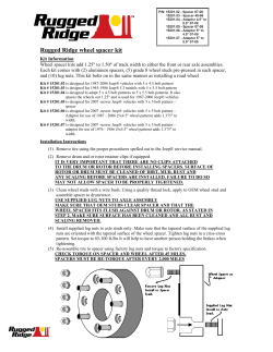


© Copyright 2026