
Health Supervision Guidelines for Children With Achondroplasia
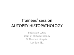
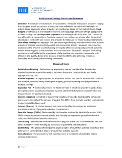
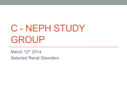
Health
Supervision
for Children
Committee
This set of guidelines
is designed to assist the
pediatrician
in caring for children with achondroplasia confirmed by radiographs
and physical features.
Although
pediatricians
usually
first see
children with achondroplasia
during infancy, occasionally they are called on to advise the pregnant
woman
who has been informed
of the prenatal
diagnosis
of achondroplasia
or asked to examine
the newborn to help establish the diagnosis. Therefore, these guidelines
offer advice for these situations as well.
Achondroplasia
is the most common form of disproportionate
short stature.’ The diagnosis is based
on very specific features on the radiographs,
which
include a contracted base of the skull, a square shape
to the pelvis with a small sacrosciatic
notch, short
pedicles of the vertebrae,
rhizomelic
(proximal)
shortening of the long bones, trident hands, a normal
length trunk, proximal femoral radiolucency, and, by
mid-childhood,
a characteristic
chevron shape of the
distal femoral epiphysis.
Hypochondroplasia
and
thanatophoric
dysplasia are part of the differential
diagnosis, but achondroplasia
can be distinguished
from these because the changes in hypochondroplasia are miider and the changes in thanatophoric
dysplasia are much more severe and invariably lethal.
Achondroplasia
is an autosomal dominant disorder,
but approximately
75% of cases represent new dominant mutations.
The gene for achondroplasia
has
recently been found. Achondroplasia
is due to a
change in the genetic information
for fibroblast
growth factor receptor 3. 2,3 Almost all of the mutations have been found to occur in exactly the same
spot. Now that the gene has been found and the
mutation known, potential therapies and diagnostic
methodologies
are likely to be developed. A great
deal is known about the natural history of the disorder that can be shared with the family. The average
adult height in achondroplasia
is about 4 ft for both
men and women (Figs 1 through 6).4 Other features
include disproportionate
short stature, with shortening of the proximal segment of the limbs, a promiThe recommendations
in this policy statement do not indicate an exclusive
course of treatment for children with genetic disorders, but are meant to
supplement anticipatory
guidelines available for treating the normal child
Provided in the AAP publication, Guidelines for Health Supervision. They
are intended to assist the pediatrician
in helping children with genetic
conditions to participate folly in life. Diagnosis and treatment of genetic
disorders are changing rapidly. Therefore, pediatricians
are encouraged
to view these guidelines
in light of evolving
scientific information.
Clinical geneticists
may be a valuable resource for the pediatrician
seeking additional
information
or consultation.
PEDIATRICS (ISSN 0031 4005). Copyright 0 1995 by the American Academy of Pediatrics.
With
Achondroplasia
on Genetics
nent forehead, a flattened midface, and an averagesized trunk. The head usually appears relatively
large compared with the body. The most common
complication,
occurring
in adulthood,
is related to
lumbosacral
spinal stenosis with compression
of the
spinal cord or nerve roots.5,6 This complication
is
usually treatable by surgical decompression,
if diagnosed at an early stage.
Children
affected
with
achondroplasia
frequently have delayed motor milestones,
otitis media, and bowing of the knees (Fig 7).7 Occasionally
in infancy or early childhood there is symptomatic
airway
obstruction,
development
of thoracolumbar kyphosis,
symptomatic
hydrocephalus,
or
symptomatic
upper cord compression.
Most individuals with achondroplasia
are of normal intelligence and are able to lead independent
and productive lives.6 Because of their disproportionate
short stature, however,
a number of psychosocial
problems
can arise. Families can benefit from anticipatory
guidance and the opportunity
to learn
from other families with children of disproportionate short stature.
The following
guidelines are designed to help the
pediatrician
care for children with achondroplasia
and their families. Issues that need to be addressed at
various age groups are discussed
(Table). These
guidelines are not appropriate for other chondrodysplasias, because each type has its own natural history, complications,
and specific guidelines. It is important that parents also consult a physician with
experience and expertise concerning achondroplasia
early in their child’s development,
because these
guidelines are intended for the general pediatrician
without such experience.
THE PRENATAL
VISIT
Pediatricians may be called upon to counsel a family in which a fetus has achondroplasia or is suspected to have achondroplasia. In some settings, the
pediatrician will be the primary resource for counseling a family. At other times, counseling may already have been provided to the family by a clinical
geneticist and/or the obstetrician. Because of a previous relationship with the family, however, the pediatrician may be called on to review this information and to assist the family in the decision-making
process.
The diagnosis of achondroplasia in the fetus is
most often only made with certainty when one or
both parents have this condition. In this circumstance the parents are usually knowledgeable about
PEDIATRICS
Vol. 95 No. 3 March
1995
443
TABLE.
Achondroplasia
Guidelines
for Health
Supervision*
Infancy, 1 mo-1 y
Age
Prenatal Neonatal
2
mo
4
mo
6
mo
Early Childhood,
9
mo
12
mo
15
mo
18
mo
24
1-5 y
3y
4y
mo
Late Childhood
(5-13 y),
Annual
Adolescence
W-21 y),
Annual
Diagnosis
X-ray film
Whenever the diagnosis is suspected.
Review phenotype
Review proportions
Whenever the diagnosis is suspected.
Whenever the diagnosis is suspected.
Genetic
Counseling
Early intervention
Recurrence risks
Reproductive
options
Family support
Support groups
Long-term
planning
Medical
Evaluation
Growth/weight/
OFC
Orthopedic-if
complication
Neurologic-if
complication
Hearing
Social readiness
Orthodontics
Medical
0
OR
S
OR
S
OR
S
R
OR
S
R
Evaluation
X-ray films-only
to make
diagnosis or if
complication
Ultrasound-of
brain ventricle
size
Social
’
0
Adjustment
Psychosocial
Behavior and
development
School
Sexualitv
s/o
s/o
s/o
S
s/o
S
s/o
s/o
s/o
S
s/o
0
S
s/o
S
s/o
0
0
0
* Assure compliance with the American Academy of Pediatrics “Recommendations for Preventive Pediatric Health Care.”
0 = to be performed; S = subjective, by history; 0 = objective, by a standard testing method; and R = discuss referral to a specialist.
the disorder, the inheritance, and the prognosis for
the offspring.
In most situations in which the parents have normal stature, the diagnosis may only be suspected
based on the observation of disproportionately short
limbs in the fetus by ultrasound. With the frequent
use of ultrasound, approximately one third of cases
of fetal achondroplasia are suspected prenatally.
However, disproportionately
short limbs are observed in a heterogeneous group of conditions. In the
majority of these cases, the specific diagnosis cannot
be made with certainty except by radiography late in
pregnancy or more usually after birth. In these cases,
caution should be exercised when counseling the
family. In those infrequent cases in which the diagnosis is unequivocally established either because of
the familial nature of the disorder or by prenatal
radiography,
the pediatrician may discuss the
following issues as appropriate.
444
1. Review, confirm, and demonstrate laboratory or
imaging studies leading to the diagnosis.
2. Explain the mechanisms for occurrence or recurrence of achondroplasia in the fetus and the recurrence risk for the family.
3. At least 75% of cases of achondroplasia occur in
families in which both parents have average stature and achondroplasia in the offspring occurs
due to sporadic mutation in the gene.
4. Review the natural history and manifestations of
achondroplasia, including variability.’
5. Discuss further studies that should be done,
particularly
those to confirm the diagnosis in
the newborn period. If miscarriage, stillbirth, or
termination occurs, confirmation of diagnosis is
important for counseling family members about
recurrence.
6. Review the currently available treatments and
interventions. This discussion needs to include the
HEALTH SUPERVISION FOR CHILDREN WITH ACHONDROPLASIA
AGE - Years
40
’
B
’
I
2
’
3
’
4
’
!I
’
’
6
’
7
’
8
_,,a
I
IO
9
AGE
I
II
_._
I
I
I2 I3
---
1.
I
I4
I
l5
I
I6
I
I7
I6
-Yews
Fig 1. Height for females with achondroplasia (mean f SD) compared to normal standard curves. Graph is derived from 214
females. (From Horton et a1.4)
,
I 60--
ACHONDROPLASIA
’
160s
’
HEIGHT
MO,. H.189
/
/
/
1
1
/
/ /’ ./
/
1 A’/
JI
I
2
3
4
5
6
7
6
9
IO
II
I2
I3
‘4
‘6
I6
AGE - Years
Fig 3. Mean growth velocities (solid line) for males (top) and
females (bottom) with achondroplasia compared to normal
growth velocity curves (dashed lines, 3rd percentile, mean, 97th
percentile). Data are derived from 26 males and 35 females. (From
Horton et a1.4)
60
i
i
lb
1!l ,h
,!3 ,b
lb
16 ,7
Ii
’
AGE -Years
Fig 2. Height for males with achondroplasia (mean + 2SD) compared to normal standard curves. Graph is derived from 189
males. (From Horton et aL4)
efficacy, complications,
side effects, costs, and
other burdens of these treatments. Discuss possible future treatments and interventions.
7. Explore the options available to the family for the
management and rearing of the child using a nondirective approach. In cases of early prenatal diagnosis, these may include discussion of pregnancy termination,
as well as continuation
of
pregnancy and rearing of the affected child at
home, foster care, or adoption. When both parents
are of disproportionate
short stature, the possibility of double heterozygosity or homozygosity
for
achondroplasia must be assessed. Infants with homozygous achondroplasia usually are either stillborn or die shortly after birth. Homozygous
achondroplasia
can usually be diagnosed prenatally.
8. If the mother is affected with achondroplasia,
a
cesarean section must be performed because of a
small pelvis.Q This surgical procedure usually involves general anesthesia because of the mother’s
spinal stenosis and the consequent risk associated
with conduction (spinal/epidural)
anesthesia. A
mother affected with achondroplasia
may develop respiratory compromise in the third trimester of pregnancy, so baseline pulmonary function
studies should be done. A pregnancy at risk for
homozygosity
should be followed with ultrasound measurements
at 14, 16, 18, 22, and 32
weeks of gestation in order to distinguish
homozygosity
or heterozygosity
from normal
growth patterns in the the fetus. New DNA diagnostic studies are likely to become available.
FROM
HEALTH
SUPERVISION
BIRTH TO 1 MONTH-NEWBORNS
Examination
1. Confirm the diagnosis by radiographic
studies
(the diagnosis of approximately
20% of patients
with achondroplasia has been missed in the past,
because it was not suspected on physical examination in the newborn period and consequently
no radiographs were obtained).
2. Document measurements, including arm span, occipital frontal circumference (OFC), body length,
and upper to lower body segment ratio; note these
measurements
on the achondroplasia
special
growth charts at the end of this document. Review
AMERICAN
ACADEMY
OF PEDIATRICS
445
ACHONDROPLASlA
HEAD CIRCUMFERENCE
Fmd. N .I45
NONTHS
YEARS
AGE
Fig 5. Head circumference for females with achondroplasia compared to normal curves (dashed lines). Data are derived from 145
females. (From Horton et a1.4)
AGE -Years
ACHONDROPLASIA
HEAD
CIRCUMFERENCE
60
YEARS-
.-
AGE
Fig 6. Head circumference for males with achondroplasia compared to normal curves (dashed lines). Data are derived from 114
females. (From Horton et aL4)
AGE -Years
Fig 4. Upper and lower segment lengths for males (top) and
(bottom) with achondroplasia (mean 0 SD). Data are derived from
75 males and 95 females. (From Horton et al?)
the phenotype with the parents and discuss the
specific findings with both parents whenever possible.
3. The OFC should be measured monthly during the
first year. Ultrasound studies of the brain to determine ventricular size should be considered if
the fontanelle size is unusually large, OFC increases disproportionately, or symptoms of hydrocephalus develop.
Anticipatory
Guidance
1. Discuss the specific findings of achondroplasia
with the parents, including:
l
Autosomal dominant inheritance. About 75% of
cases are new mutations. Germline mosaicism
(in which some germ cells are derived from a
normal cell line and some are from a cell line
with a mutation) has been reported, but clearly
the risk of recurrence in sporadic cases is far
below 1%.
l
Most individuals with achondroplasia have
normal intelligence and normal life expectancy.
446
Growth hormone and other drug therapies are
not effective in increasing stature. Experimental
work is being done on leg-lengthening procedures at an older age.‘OJ1
l
Special achondroplasia growth curves and infant development charts have been developed,
and the final expected adult height for persons
with achondroplasia is in the range of about 4
ft.4
2. Discuss the following possible severe medical
complications:
l
Unexpected infant death in less than 3% of
those affected, usually only in the most severe
cases.12Severe upper airway obstruction in less
than 5% of those affected, but consider sleep
studies if there appears to be a problem with
breathing at rest or during sleep, especially if
developmental landmarks lag.13
l
Restrictive pulmonary disease with or without
reactive airway disease occurs in less than 5%
of children with achondroplasia who are
younger than 3 years13;consider pulse oximetry
or evaluation for car pulmonale if there are
signs of breathing problems.
l
Development of thoracolumbar kyphosis is associated with unsupported sitting before there
l
HEALTH SUPERVISION FOR CHILDREN WITH ACHONDROPLASIA
DEVELOPMENTA1 SCREENINGTESTS IN AcHONDROPlASlA
T
n
.
8
Smile
ill
Head Control
113
Pdl cwr
ll6
Sat wtth pwpping
IY
;;;y
z
hll
tq to a stand
Standwith
!I
w
support
l26
Wk
with supprt
1~2
Wk
alone
134
sadMannla/oadda
I
ion
Stand alax
Wingscurds
I I I I-
117
,“Y
,.,,’
I I I I I I I 1 * E...
II
95
l66
Said 2 WOKI phrase
83
s3ii short sentence
80
II III
I III
?,.,t..,^^
I-
I
,I
1
I I I I I 1
3
4
5
6
7
3
9lO11l213WPl22022243333
ci children passing
DENVER dew!qmenta
screening rests
‘%X
Fig 7. Develoumental screening tests in achondroulasia.
I
(FFom Todoro; et al.?.
”
X
Fercent d
achondro@stic
chddren
passing the Item
The 25th, 50th, 75th, and 90th centiles are determined by linear interpolation.
is adequate trunk muscle strength.14
All infants with achondroplasia
have a relatively small foramen magnum, but few become
symptomatic from cord compression at the cervicomedullary
junction.15 This complication
may be manifested by signs and symptoms of a
high cervical myelopathy,
central apnea, or
bothal Rarely, foramen magnum decompression may be recommended.
l
Hydrocephalus
may develop during the first 2
years, i7 so OFC size should be monitored carefully during this time. If a problem is suspected,
refer the infant to a pediatric neurologist or
pediatric neurosurgeon.
l
The common complication
of spinal stenosis
rarely occurs in childhood but manifests in
older individuals
with numbness, weakness,
and altered deep tendon reflexes.18 Children
with severe thoracolumbar
kyphosis are at
greater risk for this problem. It is for this reason
that unsupported
sitting before there is adequate trunk muscle strength is discouraged.
3. Discuss the psychosocial issues related to disproportionate short stature. Refer the affected individual, or the parent of an affected individual,
to
a support group such as Little People of America
or Human Growth Foundation
(see “Resources
for New Parents”). If parents do not wish to join a
group, they may want to meet with or talk to
other affected individuals or parents. Remind parents that most individuals
with achondroplasia
lead productive, independent lives.
4. Discuss with the parents how to tell their family
and friends about their child’s growth problem.
5. Supply the parents with educational books and
pamphlets (see “Resources for New Parents”).
l
t
J
6. Discuss the realistic functional problems for affected individuals.
7. Discuss individual
resources for support, such as
family, clergy, social workers, and friends.
8. Review the prenatal diagnosis and recurrence
risks for subsequent pregnancies.
FROM
HEALTH
1 MONTH
SUPERVISION
TO 1 YEAR-INFANCY
Examination
1. Assess growth and development in comparison
only to children with achondroplasia.
2. Perform physical examination
and appropriate
laboratory studies.
3. Review head growth.
4. Consider performing
a central nervous system
ultrasound at 2, 4, or 6 months if the infant’s
head size increases rapidly in order to evaluate
ventricular
size. If the size of the OFC or
ventricles is increasing rapidly, refer the infant to a pediatric neurologist or pediatric neurosurgeon. At 6 to 12 months, consider performing additional neuroimaging
studies, if appropriate.
5. Check motor development and discuss development; note on the milestone charts for achondroplasia. Expect motor delay but not social or cognitive delay.’
6. Watch for low thoracic or high lumbar gibbus
(posterior angulation
or kyphosis) associated
with truncal weakness. It is recommended
that
parents avoid carrying a child with achondroplasia in curled-up positions. Certain types of child
carriers, swingomatics, jolly jumpers, and “umAMERICAN
ACADEMY
OF PEDIATRICS
447
7.
8.
9.
10.
11.
12.
brella” strollers tend to increase risk for gibbus.
Unsupported
sitting should be avoided.r2 Parents should be instructed
to provide back support during the first year of life.
External rotation of the hips is frequently present
and usually spontaneously
disappears when the
child begins to bear weight. This finding does not
require bracing.19
Check for serous otitis media. Review risk at 6 to
12 months.
Arrange sleep studies if any sign of respiratory
compromise
or delay in developmental
milestones is present.
Refer the infant to a pediatric neurologist
or pediatric neurosurgeon
for reflex asymmetry,
extreme hypotonia,
early hand preference, or excessive head growth.5J7
Consider magnetic resonance imaging or computed tomography
of the foramen magnum region for a severely hypotonic infant or one who
has signs of cord compression.
Magnetic resonance imaging should include the base of the
skull as well as the ventricles
and spinal cord
(Fig S).*O,*l
Discuss filing for Supplemental Security Income
benefits as appropriate.
Anticipatory
Guidance
Review
the personal support
available to the
family.
Review contact with support groups.
Observe the emotional status of parents and intrafamily relationships.
Discuss early intervention
services and the importance of normal socializing experiences with other
children.
5. Ask the parents if they have educated their family
members about achondroplasia;
discuss sibling
adjustment.
6. Review the increased risk of serous otitis media
because of short eustachian tubes. Indicate that an
ear examination is needed with any upper respiratory tract infection.
7. Avoid infant carriers that curl up the infant. This
does not apply to car safety seats, which should
always be used during automobile travel.
HEALTH SUPERVISION
FROM 1 TO 5 YEARS-EARLY
CHILDHOOD
Examination
1. Assess the child’s growth
and development
as
charted on the achondroplasia
growth charts. Obtain lower segment measurements
once weight
bearing is established.
2. Continue to follow head growth.
3. Continue to watch for thoracolumbar
gibbus and
development of lumbar lordosis. Discuss avoiding
the use of walkers, jumpers, or backpack carriers.
Any kyphosis
present should disappear as the
child begins to bear weight. Weight-bearing
and
walking
may occur late; however,
they are expected by 2 years of age. When weight-bearing
448
HEALTH
SUPERVISION
FOR CHILDREN
WITH
4.
5.
6.
7.
begins, the external rotation of the hips should
self-correct
to a normal orientation
within
6
months.
Anticipate
some bowing
of the legs because of
fibular overgrowth
at the knees and ankles. If
bowing leads to an inability to walk, consult a
pediatric orthopedist.
Check the child’s hips for hip flexion contractures.
Prescribe exercises that may decrease lumbar lordosis and hip flexion contractures.*9
Check the
hips for external rotation. Refer the child to a
pediatric orthopedist,
if necessary.
Speech evaluation should be done no later than 2
years of age. If speech is abnormally delayed conductive hearing loss due to chronic serous otitis
media should be excluded.
Watch for obstructive sleep apnea. Children with
achondroplasia often sweat and snore in association
with sleep. If upper airway obstruction is suspected
(increased retraction, choking, intermittent breathing,
apnea, deep compensatory sighs), further pulmonary
evaluation and neurologic examination including so
matic sensory-evoked
responses, sleep studies, and
magnetic resonance imaging are needed.
Anticipatory Guidance
1. Consider
adapting the home so the child can
become independent
(lower the light switches,
faucets, and supply step stools).
2. Occupational
therapy
consultation
may be
needed.
3. Discuss adapting age-appropriate
clothing with
snapless, easy-opening
fasteners
and tuckable
loops.
4. Discuss adaptation of toys, especially tricycles, to
accommodate short limbs.
5. Discuss adaptation of toilets to allow comfortable independent
use, with an extended wand
for wiping.
6. Discuss the use of a stool during sitting so that
the child’s feet are not hanging. Feet need support while the child is sitting at a desk or in a
chair. A cushion behind the child’s back may be
required for good posture.
7. Review weight control and eating habits to avoid
obesity, which becomes a common problem in
mid to late childhood.22
8. Discuss orthodontic bracing in the future and the
possible need for braces after 5 years of age.
9. Encourage
the family to develop activities in
which the affected child can take part; avoid
gymnastics, high diving, acrobatics, and collision
sports.
30. Discuss how to talk with the child and other
friends or family members about short stature.
11. Encourage preschool attendance so that the child
can learn to socialize in an age-appropriate
way,
and work with parents to prepare the teacher
and the other children so the child is not given
unnecessary
special privileges.
12. Discuss toileting at school and special preparations needed by the school because of the child’s
short stature.
ACHONDROPLASIA
,
3
5
7
1,
g
13
15
9.4
5.9
‘.‘A
4.3
7-9 9.1011.,213-1415*-Normal
Years
Months
B
SAQIlTAL
4.1
3.9
3.7
3.5
3.3
i
.._
I.1
.o
.7 1
0,
5
5
7
9
11
1s
10
17
19 n
h#onlho
Yeuo
Fig 8. CT measurements of the foramen magnum. A, transverse; B, sagittal. Normals are plotted as mean ? ED. Achondroplasia as
individual measurements: the solid circles represent patients with and the open circles patients without evidence of neurologic function,
13. Discourage the child from jumping to decrease
unnecessary stress on joints, particularly
the
joints of the spine.
FROM
HEALTH
SUPERVISION
5 TO 13 YEARS-LATE
CHILDHOOD
Examination
Assess and review the child’s growth and development and social adaptation.
Anticipatory Guidance
1. Determine school readiness.
2. Discuss preparation of the school and teacher for
a child with short stature.
3. Prepare the child for psychosocial situations and
discussing issues. Be sure the child can explain
why he or she is short and can ask for help in an
appropriate way. Children with achondroplasia
AMERICAN ACADEMY OF PEDIATRICS
449
4.
5.
6.
7.
8.
9.
10.
11.
12.
13.
14.
usually are included in the regular education
program.
Suggest adaptive aids for the school to cope with
heavy doors, high doorknobs, reaching for the
blackboard,
foot support, and a regular-sized
desk. Also be sure that the child can use the
restroom independently.
Test hearing regularly each year, checking for
possible recurrent serous otitis media.
Check deep tendon reflexes yearly for asymmetry or increased reflexes suggesting spinal stenosis.
Continue to assess history for possible obstructive sleep apnea.
Review socialization and foster independence.
Review weight control. The child may need to
restrict food intake and eat as little as half as
much as an average-sized child eats.
Discuss contact with support groups. It is especially valuable at this age.
Obtain an orthopedic evaluation when the child
is approximately
5 years of age in order to make
appropriate treatment plans, if necessary.
Emphasize correct posture and encourage the
child to consciously decrease lumbar lordosis by
“tucking the buttocks under.”
Develop an activity program with acceptable activities such as swimming and biking. The child
should avoid gymnastics and contact sports because of the potential for neck or back damage
due to existing spinal stenosis.
Review orthodontic and speech status.
9. Continue to encourage participation
in social activities and support groups. It is particularly useful during this age period.
10. Assist in transition to adult care, with emphasis
on continued monitoring
of the spine.
COMMITTEE
LIAISON
Anticipatory
Guidance
450
HEALTH
SUPERVISION
FOR CHILDREN
WITH
TO 1995
REPRESENTATIVES
of
CONSULTANT
Judith G. Hall, MD
RESOURCES
FOR NEW PARENTS
Human Growth Foundation
7777 Leesburg Pike
Falls Church, VA 22043
703/883-1773
or 800/451-6434
Little People of America
PO Box 9897
Washington,
DC 20016
214/388-9576
or 800/24-DWARF
REFERENCES
1.
2.
3.
1. Check on social adaptation.
2. Discuss the diagnosis with the adolescent to be
sure that the adolescent has the vocabulary and
the understanding of the genetic nature of achondroplasia.
3. Discuss sexuality and reproduction,
as well as
the necessity for a cesarean section in women for
childbirth.7
4. Continue orthodontic evaluation.
5. Continue weight counseling.22
6. Encourage the family and affected person to set
career and life goals high and appropriate, as for
other members of the family. Assist in adapting
to an independent life and in obtaining a driver’s
license. (Vocational rehabilitation
may pay.)
7. Discuss college, vocational planning and training, and other plans following high school.
8. Foster independence.
1994
Felix de la Cruz, MD,
National Institutes of Health
James W. Hanson, MD,
American College of Medical Genetics
Jane Lin-Fu, MD, Health Resources
and Services Administration,
DHHS
Paul McDonough,
MD, American College
Obstetricians
& Gynecologists
Godfrey Oakley, MD, Centers for
Disease Control & Prevention
AAP SECTION LIAISON
Beth A. Pletcher, MD,
Section on Genetics & Birth Defects
HEALTH
SUPERVISION
FROM 13 TO 21 YEARS OR OLDERADOLESCENCE
TO EARLY ADULTHOOD
Examination
1. Continue to record parameters.
2. Discuss any signs or symptoms of nerve compression and check deep tendon reflexes, tone,
and sensory findings, if indicated.
3. Review weight and diet.
ON GENETICS,
Margretta R. Seashore, MD, Chairperson
Sechin Cho, MD
Franklin Desposito, MD
Jack Sherman, MD
Rebecca S. Wappner, MD
Miriam G. Wilson, MD
4.
5.
6.
7.
8.
9.
10.
11.
12.
luman achondroplasia.
A multidisciplinary
approach. Proceedings of
he first international
symposium. November
19-21, 1986, Rome, Italy.
hsic Life Sci. 1988;48:1-491
<ousseau F, Bonaventure J, Legeai-Mallet
L, et al. Mutations in the gene
mcoding fibroblast growth factor receptor-3 in achondroplasia.
Nature.
.994;371:252-254
jhiang R, Thompson LM, Zhu YZ, et al. Mutations in the transmemxane domain of FGFR3 cause the most common genetic form of dwarfsm, achondroplasia.
Cell. 1994;78:335-342
lorton WA, Rotter JI, Rimoin DL, Scott CI, Hall JG. Standard growth
:urves for achondroplasia.
J Pediadiatr.1978;93:435-438
lecht JT, Butler IJ. Neurologic morbidity associated with achondropla;ia. J Child Neuuol. 1990;5:84-97
‘yeritz RE, Sack GH, Udvarhelyi
GB. Thoracolumbosacral
laminectomy
n achondroplasia:
long-term results in 22 patients. Am J Med Genet.
1987;28:433-444
rodorov AB, Scott CI, Warren AE, Leeper JD. Developmental
screening
,ests
in
achondroplastic
children.
Am
J Med
Genet.
1981;9:19-23
Rogers JG, Perry MA, Rosenberg LA. IQ measurement in children with
skeletal dysplasia. Pediatrics. 1979;63:894-897
411anson JE, Hall JG. Obstetrics and gynecologic problems in women
Nith chondrodystrophies.
Obstet Gynecol. 1986;67:74-78
‘aley D. Current techniques of limb lengthening.
I Pediatr Orthop.
1988;8:73-92
7imoin DL. Limb lengthening: past, present, and future. Growth Genet
rnd Hormones. 1991;7:4-6
‘auli RM, Scott CI, Wassman ER, et al. Apnea and sudden unexpected
ieath in infants with achondroplasia.
J Pediatr. 1984;104:342-348
ACHONDROPLASIA
13. Stokes DC, Phillips JA, Leonard CO, et al. Respiratory complications of
achondroplasia.
[ Pediutr. 1983;102:534-541
14. Hall JG. Kyphosis in achondroplasia:
probably preventable.
J Pediatr.
1988;112:166-167
15. Reid CS, Pyeritz RE, Kopits SE, et al. Cervicomedullary
compression in
young patients with achondroplasia:
value of comprehensive
neurologic and respiratory evaluation. J Pediatr. 1987;110:522-530
16. Nelson FW, Goldie WD, Hecht JT, Butler IJ, Scott CI. Short-latency
somatosensory
evoked potentials in the management of patients with
achondroplasia.
Neurology. 1984;34:1053-1058
17. Steinbok P, Hall JG, Flodmark 0. Hydrocephalus
in achondroplasia:
the
possible role of intracranial venous hypertension.
7 Neurosurg. 1989;71:
42-48
18. Hecht JT, Butler IJ, Scott CI. Long-term neurological sequelae in achondroplasia. Eur J Pediatr. 1984;143:58-60
19. Siebens AA, Hungerford
DS, Kirby NA. Achondroplasia:
effectiveness
of an orthosis in reducing deformity of the spine. Arch Phys Med Rehnbil.
1987;68:384-388
20. Hecht JT, Nelson FW, Butler IJ, et al. Computerized
tomography
of the
foramen magnum: achondroplastic
values compared to normal standards. Am ] Mcd Gent?. 1985;20:355-360
21. Hecht JT, Horton WA, Reid CS, Pyeritz RE, Chakraborty
R. Growth of
the foramen magnum in achondroplasia.
Am ] Med Genet. 1989;32:
528-535
22. Hecht JT, Hood OJ, Schwartz
J Med Genet. 1988;31:597-602
AMERICAN
RJ, et al. Obesity
ACADEMY
in achondroplasia.
OF PEDIATRICS
Am
451


© Copyright 2026