
C R AMERICAN ACADEMY OF PEDIATRICS Health Supervision for Children With Achondroplasia
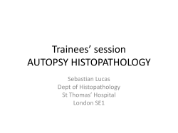
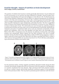
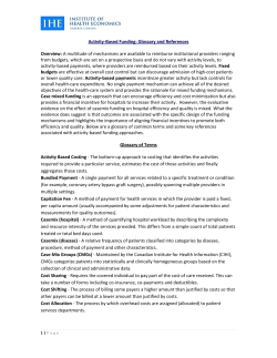
AMERICAN ACADEMY OF PEDIATRICS
CLINICAL REPORT
Guidance for the Clinician in Rendering Pediatric Care
Tracy L. Trotter, MD; Judith G. Hall, OC, MD; and the Committee on Genetics
Health Supervision for Children With Achondroplasia
ABSTRACT. Achondroplasia is the most common condition associated with disproportionate short stature.
Substantial information is available concerning the natural history and anticipatory health supervision needs in
children with this dwarfing disorder. Most children with
achondroplasia have delayed motor milestones, problems with persistent or recurrent middle-ear dysfunction,
and bowing of the lower legs. Less often, infants and
children may have serious health consequences related to
hydrocephalus, craniocervical junction compression, upper-airway obstruction, or thoracolumbar kyphosis. Anticipatory care should be directed at identifying children
who are at high risk and intervening to prevent serious
sequelae. This report is designed to help the pediatrician
care for children with achondroplasia and their families.
Pediatrics 2005;116:771–783; achondroplasia, short stature, children, health supervision.
ABBREVIATIONS. OFC, occipital-frontal circumference; CT, computed tomography.
INTRODUCTION
T
his clinical report is designed to assist the pediatrician in caring for children with achondroplasia confirmed by radiographs and physical
features. Although pediatricians usually first see
children with achondroplasia during infancy, occasionally they are called on to advise a pregnant
woman who has been informed of the prenatal diagnosis of achondroplasia or asked to examine a
newborn to help establish the diagnosis. Therefore,
this report offers advice for these situations as well.
Substantial new information has appeared since
publication of the first policy statement on health
supervision of children with achondroplasia.1 In particular, a great deal has been learned about the molecular genetics of the disorder.2 In addition, a more
complete understanding of how certain serious complications can be minimized or avoided has accrued.3
The new information is incorporated into this report,
which is a revision of the original policy statement.
Achondroplasia is the most common condition associated with severe disproportionate short stature.4
The diagnosis can usually be made on the basis of
clinical characteristics and very specific features on
The guidance in this report does not indicate an exclusive course of treatment or serve as a standard of medical care. Variations, taking into account
individual circumstances, may be appropriate.
doi:10.1542/peds.2005-1440
PEDIATRICS (ISSN 0031 4005). Copyright © 2005 by the American Academy of Pediatrics.
radiographs, which include contracted base of the
skull, square shape of the pelvis with a small sacrosciatic notch, short pedicles of the vertebrae, rhizomelic (proximal) shortening of the long bones, trident hands, a normal-length trunk, proximal femoral
radiolucency, and (by midchildhood) a characteristic
chevron shape of the distal femoral epiphysis. Other
rhizomelic dwarfing disorders such as hypochondroplasia and thanatophoric dysplasia are part of the
differential diagnosis, but achondroplasia usually
can be distinguished from them because the changes
in hypochondroplasia are milder and the changes in
thanatophoric dysplasia are much more severe and
invariably lethal. Achondroplasia is an autosomal
dominant disorder, but approximately 75% of cases
represent new dominant mutations. Achondroplasia
is caused by mutation in the gene that codes for the
fibroblast growth factor receptor type 3 (FGFR3).5–7
Because virtually all of the causal mutations occur at
exactly the same place within the gene,7 molecular
testing is straightforward. It is not necessary to perform molecular testing in every child with a clinical
diagnosis of achondroplasia. However, FGFR3 testing should be performed in children who are in any
way atypical or in circumstances in which differentiation from similar disorders, such as hypochondroplasia, is not certain. Such children also should be
referred for clinical genetic evaluation.
A great deal is known about the natural history of
achondroplasia that can be shared with the family.3,8
The average adult height in achondroplasia is approximately 4 ft for men and women (Figs 1 and 2).9
The most common complication, occurring in adulthood, is related to lumbosacral spinal stenosis with
compression of the spinal cord or nerve roots.10,11
This complication is usually treatable by surgical
decompression if it is diagnosed at an early stage.
Most children with achondroplasia do well. However, children affected with achondroplasia commonly have delayed motor milestones (Fig 3),12,13
otitis media, and bowing of the lower legs.14 Less
commonly, infants and children may have serious
health consequences related to hydrocephalus,
craniocervical junction compression, upper-airway
obstruction, or thoracolumbar kyphosis. Although
they are less common, anticipatory care should be
directed at identifying children who are at high risk
and intervening to prevent serious sequelae. Most
individuals with achondroplasia are of normal intelligence and are able to lead independent and proPEDIATRICS Vol. 116 No. 3 September 2005
771
Fig 1. Height for males with achondroplasia
(mean ⫾ 2.8 standard deviation) compared
with normal standard curves. The graph was
derived from 189 males. (Reproduced with
permission from J Pediatr. 1978;93:435-438.)
ductive lives.15 Because of their disproportionate
short stature, however, a number of psychosocial
problems can arise. Families can benefit from anticipatory guidance and the opportunity to learn from
other families with children of disproportionate
short stature.
The consensus-based guidance in this report is
designed to help the pediatrician care for children
with achondroplasia and their families. Issues that
need to be addressed at various ages are discussed
(Table 1). These suggestions are not appropriate for
other chondrodysplasias, because each type has its
own natural history, complications, and specific
guidelines. Irrespective of the availability of the
guidance in this report, it is important that pediatricians and parents also consult a physician with special experience and expertise concerning achondroplasia early in the child’s life, because this report
only provides generally applicable suggestions that
772
must be tailored to a particular child’s condition and
needs.
THE PRENATAL VISIT
Pediatricians may be called on to counsel expectant parents whose fetus has achondroplasia or is
suspected to have achondroplasia because of recognition on ultrasonography of disproportionate small
stature. In some settings, the pediatrician will be the
primary resource for counseling a family. At other
times, counseling may already have been provided
to the family by a clinical geneticist and/or the obstetrician. Because of a previous relationship with the
family, however, the pediatrician may be called on to
review this information and assist the family in the
decision-making process.
The diagnosis of achondroplasia in the fetus is
made most often with certainty when 1 or both parents have this condition. In this circumstance, the
HEALTH SUPERVISION FOR CHILDREN WITH ACHONDROPLASIA
Fig 2. Height for females with achondroplasia
(mean ⫾ 2.8 standard deviation) compared
with normal standard curves. The graph was
derived from 214 females. (Reproduced with
permission from J Pediatr. 1978;93:435-438.)
parents are usually knowledgeable about the disorder, the inheritance, and the prognosis for the offspring. More often, diagnosis of achondroplasia is
first suspected late in gestation on the basis of longbone foreshortening incidentally discovered by ultrasonography. With the frequent use of ultrasonography, many cases of achondroplasia are first
identified prenatally (after 26 weeks of gestational
age). However, disproportionately short limbs are
observed in a heterogeneous group of conditions.
Misdiagnosis and inaccurate prenatal counseling of
families is common.16 Confirmation of diagnosis
based on ultrasonographic features characteristic of
achondroplasia can be provided by molecular testing
(FGFR3 mutational testing) of prenatal specimens. If
no such confirmation has yet been completed, caution should be exercised when counseling the family.
In this circumstance, the pediatrician should discuss
the tentative nature of the diagnosis and alternatives
that may explain the identified features. The pedia-
trician should also discuss the natural history of
achondroplasia, because it is the most likely explanation for the findings. In cases in which the diagnosis is unequivocally established either because of
the familial nature of the disorder or by prenatal
molecular diagnosis (chorionic villus sampling at
11–13 weeks’ gestation or amniocentesis after 15
weeks’ gestation), the pediatrician may consider the
following steps as appropriate.
1. Review, confirm, and demonstrate laboratory or
imaging studies leading to the diagnosis.
2. Explain the mechanisms for occurrence of achondroplasia in the fetus and the recurrence risk for
the family.
3. Remember that at least 75% of cases of achondroplasia occur in families in which both parents
have average stature. In those cases, achondroplasia in the offspring occurs because of a mutation
in the gene.
AMERICAN ACADEMY OF PEDIATRICS
773
Fig 3. Developmental screening tests in achondroplasia. The bar scale shows the percentage of achondroplastic children passing the item;
the black triangle on top of the bar shows the age at which 90% of normal children pass the same item. The graphs were derived from
197 affected individuals, obtained by questionnaire. (Reproduced with permission from Am J Med Genet. 1981;9:19-23.)
4. Review the natural history and manifestations of
achondroplasia, including variability.3
5. Discuss additional studies that should be performed, particularly those to confirm the diagnosis in the newborn period. If miscarriage, stillbirth, or termination occurs, confirmation of
diagnosis is important for counseling family
members about recurrence.
6. Review currently available treatments and interventions. This discussion needs to include the efficacy, complications, adverse effects, costs, and
other burdens of these treatments. Discuss possible future treatments and interventions.
7. Explore the options available to the family for the
management and rearing of the child using a nondirective approach. In cases of early prenatal diagnosis, these options may include discussion of
pregnancy termination, continuation of pregnancy and rearing of the child at home, foster care,
or adoption. If adoption is planned to another
family, contact may be made with the Little People of America adoption service.17
8. If the mother is affected with achondroplasia, inform her that a cesarean delivery must be performed because of the characteristic small pelvis.18 A mother affected with achondroplasia may
develop respiratory compromise during the third
trimester of pregnancy, so baseline pulmonary
function studies should be performed. Homozygous achondroplasia can be diagnosed prenatally
with molecular testing of the fetus, by either chorionic villus sampling or amniocentesis. A pregnancy at risk of homozygosity should be followed
774
with ultrasonographic measurements at 14, 16, 18,
22, and 32 weeks of gestation to distinguish homozygosity or heterozygosity from normal growth
patterns in the fetus.
9. When both parents are of disproportionate short
stature, assess the possibility of double heterozygosity19 or homozygosity for achondroplasia. Some forms of double heterozygosity lead to
life-threatening problems19; infants with homozygous achondroplasia usually are stillborn or die
shortly after birth.20
HEALTH SUPERVISION FROM BIRTH TO
1 MONTH OF AGE: NEWBORNS
Examination
1. Confirm the diagnosis by radiographic studies
(the diagnosis of approximately 20% of patients
with achondroplasia has been delayed in the past
because it was not suspected on physical examination in the newborn period, and consequently,
no radiographs were obtained).
2. Document measurements, including occipitalfrontal circumference (OFC), body length, and
body weight; plot these measurements on achondroplasia-specific growth charts (Figs 1, 2, and
4 –7). Review the phenotype with the parents and
discuss the specific findings with both parents
whenever possible.
3. The OFC should be measured at every pediatric
contact during the first year (Figs 4 and 5).
HEALTH SUPERVISION FOR CHILDREN WITH ACHONDROPLASIA
AMERICAN ACADEMY OF PEDIATRICS
775
As indicated
S/O
S/O
X
X
X
X
X
X
X
X
2 mo
4
mo
Infancy, 1 mo to 1 y of Age
6
mo
S/O
3
X
S
S/O
3
X
Whenever the diagnosis is suspected
Whenever the diagnosis is suspected
Whenever the diagnosis is suspected
When diagnosis is not certain
Neonatal
3
X
9
mo
S
S/O
3
XR
S
X
X
X
X
X
X
12
mo
S/O
3
S/O
X
15
mo
S/O
3
S/O
18
mo
S
S/O
3
S/O
XR
S
X
X
X
X
24
mo
O
3
S/O
3
y
Early Childhood, 1 to 5 y of Age
3
XR
S
R
O
X
4y
Late Childhood
S
S/O
O
3
XR
S
R
O
X
X
X
X
X
5 to 13 y, Annual
Adolescence
S
S/O
O
X
3
R
X
X
X
X
X
X
13 to 21 y, Annual
These guidelines ensure compliance with AAP recommendations for preventive pediatric health care. FGFR3 indicates fibroblast growth factor receptor type 3; X, to be performed; S, subjective, by
history; O, objective, by a standard testing method; R, discuss referral to a specialist; 3, continue to monitor.
See text
Prenatal
Achondroplasia Guidelines for Health Supervision
Diagnosis
Radiography
Review phenotype
Review proportions
Molecular testing 关FGFR3兴
Genetic counseling
Early intervention
Recurrence risks
Reproductive options
Family support
Support groups
Long-term planning
Medical evaluation
Growth/weight/OFC
Orthopedic consult
Neurology consult
Hearing
Social readiness
Orthodontics
Speech
Medical evaluation
Radiography, only to make
diagnosis or if
complication
CT/MRI brain/cervical
spine
Polysomnography
Social adjustment
Psychosocial
Behavior and development
School
Sexuality
TABLE 1.
Fig 4. Head circumference for males with achondroplasia compared with normal curves (dashed lines).
The graph was derived from 189 males. (Reproduced
with permission from J Pediatr. 1978;93:435-438.)
Anticipatory Guidance
1. Discuss the specific findings of achondroplasia
with the parents, including the following:
• Autosomal dominant inheritance: approximately 75% of cases are new mutations. Germline mosaicism (in which some germ cells are
derived from a normal cell line and some are
from a cell line with a mutation) has been reported, but the risk of recurrence in sporadic
cases is less than 1%.21–23
• Most individuals with achondroplasia have normal intelligence and normal life expectancy.
• Although serious problems may arise during
infancy, such problems affect only 5% to 10% of
infants with achondroplasia.
• Growth hormones, other drug therapies, and
food or vitamin supplements are not effective in
776
significantly increasing stature. Growth-hormone therapy may result in a transient increase
in growth rate. However, the salutary effects
diminish with continued treatment. No study
has clearly demonstrated a significant benefit
with respect to ultimate adult stature.24,25 If
elected, such treatment should be considered
only within a research setting. Extended limb
lengthening using a variety of techniques has
been used far more elsewhere than in North
America. It can result in substantial increases in
ultimate height.26,27 However, it is arduous, not
without risk, and costly. Most families alternatively choose to modify the environment to accommodate the child rather than the converse.
• Special achondroplasia growth curves and infant development charts have been developed
(Figs 1–7); the final expected adult height for
HEALTH SUPERVISION FOR CHILDREN WITH ACHONDROPLASIA
Fig 5. Head circumference for females with achondroplasia compared with normal curves (dashed lines).
Data were derived from 145 females. (Reproduced with
permission from J Pediatr. 1978;93:435-438.)
persons with achondroplasia is approximately 4
ft.9
2. Discuss the following possible severe medical
complications and methods of prevention:
• Unexpected infant death occurs, in the absence
of aggressive evaluation, in approximately 2% to
5% of all infants with achondroplasia.28,29 This
seems to result from central apnea arising secondary to compression of arteries at the level of
the foramen magnum.28 In addition, the universally small foramen magnum may result in a
high cervical myelopathy.30,31 However, with
appropriate assessment and intervention, both
risks can be minimized.31 Parents should be advised to use an infant seat or infant carrier that
has a firm back that supports the neck and to use
a rear-facing car safety seat for as long as possible. They should be counseled to avoid use of
products like mechanical swings and carrying
slings to limit uncontrolled head movement
around the small foramen magnum. There are
instances in which infants with achondroplasia
who showed no clinical abnormality by examination and who were asymptomatic have died
from this complication. Given this, and because
it can be life saving, care of every infant with
achondroplasia should include assessment for
craniocervical junction risks, which includes
careful neurologic history and examination,
neuroimaging, and polysomnography.31 Neuroimaging can be by computed tomography (CT)
with thin cuts and bone windows31 or magnetic
resonance imaging (MRI),32,33 each of which has
benefits and disadvantages: CT allows direct
comparison of foramen magnum size with published achondroplasia standards and often can
be accomplished without sedation or anesthesia
but does not allow direct visualization of the
AMERICAN ACADEMY OF PEDIATRICS
777
Fig 6. Height-by-weight standards in achondroplasia: males. (Reproduced with permission from Am J Med Genet. 1996;62:255-261.)
Fig 7. Height-by-weight standards in achondroplasia: females. (Reproduced with permission from Am J Med Genet. 1996;62:255-261.)
neural elements of interest; MRI provides such a
direct assessment of the brainstem and upper
cervical spinal cord, but no standards for estimation of foraminal size by MRI are available,
and currently it cannot be performed routinely
without sedation. Rapid development of imaging technology suggests that alternative methods may become appropriate in the future.
778
• If severe problems are found (eg, marked abnor-
mality by neurologic examination, such as profound hypotonia or sustained ankle clonus;
markedly diminished foramen magnum size
compared with achondroplasia standard; substantial deformation of the upper cervical spinal
cord; hypoxemic episodes with minimal oxygen
saturations below 85%31), referral to a neurosur-
HEALTH SUPERVISION FOR CHILDREN WITH ACHONDROPLASIA
•
•
•
•
•
geon or other physician skilled and experienced
in the care and treatment of neurologic problems in children with achondroplasia should be
initiated.34
Hydrocephalus is a lifelong risk but is most
likely to develop during the first 2 years.35 OFC
should be monitored carefully during this time.
If the OFC is large or crosses percentiles on the
achondroplasia-specific head-circumference chart,
it is appropriate to refer the infant to a pediatric
neurologist or pediatric neurosurgeon. Baseline
CT or MRI (performed in conjunction with imaging of the craniocervical junction) is valuable
if there is concern about possible hydrocephalus. Repeating neuroimaging to assess change in
ventricular size should be considered if there is
acceleration of head growth compared with
achondroplasia standards or if other signs like
bulging, hard fontanelle, or symptoms of unusual lethargy or intractable irritability develop.
Both ventriculomegaly and excessive extra-axial
fluid are common benign accompaniments of
achondroplasia35 and should not be misinterpreted as indicative of need for shunt placement. Neural ultrasonography may be used to
follow these clinical findings.
Restrictive pulmonary disease occurs in less
than 5% of children with achondroplasia who
are younger than 3 years.36 Living at high elevation markedly increases the risk that restrictive problems will develop. If there are signs of
respiratory distress or evidence of poor weight
gain despite adequate caloric intake, pulse
oximetry (during feeding, when crying, and at
rest) should be considered to monitor oxygenation.
Most infants with achondroplasia develop a thoracolumbar kyphosis. More severe kyphosis is
associated with unsupported sitting before there
is adequate trunk muscle strength.37,38 Parents
should be counseled to avoid unsupported sitting and to avoid devices that cause curved sitting or “C sitting,” such as “umbrella-style”
strollers and soft canvas seats during the first
year of life. Use of feeder seats for upright positioning should be recommended. If severe kyphosis appears to be developing, consider a pediatric orthopedic surgical assessment to
determine if bracing is needed.38
The common complication of spinal stenosis
rarely occurs in childhood but manifests in older
individuals with numbness, weakness, and altered deep tendon reflexes.30 Severe thoracolumbar kyphosis is one mechanism that can give
rise to spinal stenosis. It is for this reason that
unsupported sitting before there is adequate
trunk muscle strength is discouraged.
Anesthesia risk:39 if an individual with achondroplasia needs to have anesthesia and surgery,
the following should be considered:
(a) Care must be taken in manipulation of the
neck, because uncontrolled neck movement
(as may occur with intubation) could lead to
unintentional spinal cord compression sec-
3.
4.
5.
6.
7.
8.
ondary to constriction of the foramen magnum.
(b) Care should be taken to ensure that medication dosages are appropriate for size.
(c) Access to veins is sometimes difficult because of lack of full extension at the elbow.
(d) Generally, spinal anesthesia should be
avoided, particularly when there is kyphosis
or severe lumbar lordosis, because of limited
space within the spinal canal.
(e) General anesthesia should be strongly considered for cesarean delivery for pregnant
women with achondroplasia (they will all
require caesarian delivery because of contracted pelves), because use of epidural anesthesia in these women requires special skill
and expertise.40/P
Discuss the potential psychosocial implications
for both parent and child related to disproportionate short stature. Refer the affected individual or
the parent of an affected individual to a support
group such as Little People of America (also see
“Resources for New Parents”). If parents do not
wish to join a group, they may want to meet with
or talk to other affected individuals or parents.
Remind parents that most individuals with achondroplasia lead productive, independent lives.
Discuss with the parents how to tell their family
and friends about their child’s growth problem.
Supply the parents with educational books and
pamphlets (see “Resources for New Parents”).
Discuss the realistic functional problems for affected individuals.
Discuss individual resources for support, such as
family, clergy, social workers, psychologists, and
friends.
Review the prenatal diagnosis and recurrence
risks for subsequent pregnancies.
HEALTH SUPERVISION FROM 1 MONTH TO
1 YEAR OF AGE: INFANCY
Examination
1. For infants not diagnosed in the newborn period,
arrange for neuroimaging and polysomnography
at the time of diagnosis.
2. Assess growth and development in comparison
only with children with achondroplasia (Figs 1–7).
3. Perform physical examination.
4. Review head growth on achondroplasia-specific
head-circumference charts.
5. Refer the infant to a pediatric neurologist or pediatric neurosurgeon if head size is disproportionately large or crosses percentiles, if there are signs
or symptoms of hydrocephalus, or if there are
indicators of possible craniocervical junction compression, including excessively brisk reflexes,
asymmetric reflexes, ankle clonus, extreme hypotonia, or early hand preference.10,35
6. Consider repeating neuroimaging studies if there
is acceleration of head growth, severe persisting
hypotonia, or any signs of craniocervical junction
compression.31–34 Growth of the foramen magnum may be compared with achondroplasia-specific standards.41
AMERICAN ACADEMY OF PEDIATRICS
779
7. Check motor development and discuss development; note on the milestone charts for achondroplasia. Expect motor delay but not social or cognitive delay.12,13 Check for serous otitis media.
Review risk at 6 to 12 months of age. Formal
behavioral audiometric assessment should be
completed at 9 to 12 months of age. Language
delay may be present secondary to conductive
hearing loss.
8. Continue to monitor for progression of kyphosis
at the thoracolumbar junction. It is recommended
that parents avoid carrying a child with achondroplasia in curled-up (C-sitting) positions. Certain
types of child carriers, mechanical swings, jumpers, and umbrella-style strollers tend to increase
risk for gibbus. Unsupported sitting should be
avoided.37,38 Parents and therapists should be instructed to provide back support during the first
year of life. If severe kyphosis appears to be developing, consider pediatric orthopedic surgical
assessment to determine if bracing is needed.38
9. Be aware that external rotation of the hips is commonly present and usually disappears spontaneously when the child begins to bear weight. This
finding does not require bracing for the infant.
4.
5.
6.
7.
Anticipatory Guidance
1. Review the personal support available to the family.
2. Review contact with support groups.
3. Observe the emotional status of parents and intrafamily relationships.
4. Discuss early-intervention services and the importance of normal socializing experiences with other
children.
5. Ask the parents whether they have educated their
family members about achondroplasia; discuss
sibling adjustment.
6. Review the increased risk of serous otitis media
because of short eustachian tubes. Indicate that an
ear examination is appropriate with any persistent
or severe upper respiratory tract infection or when
parents suspect that ear pain may be present.
7. Advise parents to avoid infant carriers that curl
up the infant. This does not apply to car safety
seats, which should always be used during automobile travel. A rear-facing car safety seat should
be used to the highest weight allowed by a convertible seat (25–30 lb).
8. Discuss filing for Supplemental Security Income
benefits as appropriate.
HEALTH SUPERVISION FROM 1 TO 5 YEARS OF
AGE: EARLY CHILDHOOD
8.
9.
10.
11.
bear weight. Lumbar lordosis usually develops
but rarely requires specific intervention. Weight
bearing and walking may occur late; however,
they are expected by 2 to 2.5 years of age. When
weight bearing begins, the external rotation of
the hips should self-correct to a normal orientation within 6 months.
Anticipate some bowing of the legs. Many children will also have instability of the soft tissues
surrounding the knee and internal tibial torsion.
If positional deformity and instability leads to
difficulty walking, a thrust at the knee (uncontrolled lateral or medial movement with weight
bearing), or chronic pain, consult a pediatric orthopedist.14
Check the child’s hips for hip-flexion contractures. Prescribe exercises that may decrease lumbar lordosis and hip-flexion contractures if indicated. Check the hips for external rotation. Refer
the child to a pediatric orthopedist if necessary.
Screen hearing each year. If otologic history,
hearing screening, or speech development raise
concerns about hearing, formal audiologic assessment should be obtained.
Perform speech evaluation at no later than 2
years of age. If speech is delayed, conductive
hearing loss attributable to chronic serous otitis
media should be excluded.
Watch for obstructive sleep apnea secondary to
smaller-than-average airway size plus physiologic adenoidal hypertrophy.42–44 Most children
with achondroplasia snore. However, if obstructive apnea or disordered breathing in sleep is
suspected (increased retraction, glottal stops,
choking, intermittent breathing, apnea, deep
compensatory sighs, secondary enuresis, recurrent night-time awakening or emesis), then additional pulmonary evaluation and polysomnography are indicated.
Be aware that gastroesophageal reflux may be
more common in children with achondroplasia
and may be more common in those with neurorespiratory complications.45 If reflux is severe, in
addition to usual treatments, consider referral to
a pediatric specialist with experience in treating
gastroesophageal reflux in infants and children.
Do not misinterpret greater-than-average sweating as indicative of serious medical problems; it
is normal in children with achondroplasia.
In rare instances in which diagnosis of achondroplasia is delayed beyond 1 year of age, determine
if neuroimaging is needed on the basis of clinical
signs and symptoms.
Examination
1. Assess the child’s growth and development as
plotted on the achondroplasia growth charts.
2. Continue to follow rate of head growth on the
achondroplasia-specific head-circumference charts.
3. Continue to watch for thoracolumbar gibbus (kyphosis). Discuss avoiding the use of walkers,
jumpers, or backpack carriers. Any kyphosis
present should disappear as the child begins to
780
Anticipatory Guidance
1. Consider adapting the home so that the child can
become independent (eg, lower the light switches,
use lever door handles and lever sink faucets, make
the toilet accessible, and supply step stools) (see
“Resources for Parents”).
2. Determine if an occupational therapy consultation is needed.
HEALTH SUPERVISION FOR CHILDREN WITH ACHONDROPLASIA
3. Discuss adapting age-appropriate clothing with
snapless, easy-opening fasteners and tuckable
loops.
4. Discuss adaptation of toys, especially tricycles, to
accommodate short limbs.
5. Discuss adaptation of toilets to allow comfortable, independent use, with an extended wand
for wiping if needed.
6. Discuss the use of a stool during sitting so that
the child’s feet are not hanging. Feet need support while the child is sitting at a desk, in a chair,
or on the toilet. A cushion behind the child’s back
may be required for good posture and to prevent
chronic back pain. Counsel parents for optimal
protection to use a convertible rear-facing car
safety seat to the highest weight and height allowed by the manufacturer of the seat.46 A rearfacing seat provides the best support protection
and positioning angle for a child with macrocephaly and skeletal dysplasia. Parents may benefit from suggestions for behavioral intervention
to promote continuing the rear-facing position as
long as possible.47
7. Review weight control and eating habits to avoid
obesity, which often becomes a problem in midto late childhood.48
8. Discuss orthodontic bracing in the future and the
possible need for early orthodontic assessment to
consider palatal expansion.
9. Encourage the family to develop activities in
which the child can take part; avoid gymnastics,
diving, trampolines, and collision sports.
10. Discuss how to talk with the child and friends or
family members about short stature.
11. Encourage preschool attendance so that the child
can learn to socialize in an age-appropriate way,
and work with parents to prepare the teacher
and the other children so that the child is not
given unnecessary special privileges.
12. Discuss toileting at school and special preparations needed by the school because of the child’s
short stature (see “Resources for Parents”).
HEALTH SUPERVISION FROM 5 TO 13 YEARS
OF AGE: LATE CHILDHOOD
Examination
1. Assess and review the child’s growth, development, and social adaptation.
2. Plot measures on achondroplasia weight-byheight grids (Figs 6 and 7).
3. Review weight control.48 The child may need to
restrict food intake and eat less than an averagesized child eats.
4. Complete a general and neurologically oriented
physical examination.
5. Check deep tendon reflexes yearly for asymmetry
or increased reflexes that suggest spinal stenosis.
6. Continue to assess history for possible obstructive
sleep apnea.
7. Test hearing each year.
Anticipatory Guidance
1. Determine school readiness.
2. Discuss preparation of the school and teacher for
a child with short stature (see “Resources for
Parents”).
3. Prepare the child for others’ questions and curiosity. Be sure the child can explain why he or she
is short and can ask for help in an appropriate
way. Children with achondroplasia usually are
included in the regular education program.
4. Suggest adaptive aids for the school to cope with
heavy doors, high doorknobs, reaching for the
blackboard, foot support, and a regular-sized
desk. Also, be sure that the child can use the
restroom independently (see “Resources for Parents”).
5. Counsel parents to use a child safety seat with a
full harness to the highest weight allowed by the
manufacturer of the seat and then to transition to
the belt-positioning booster seat for optimal seatbelt positioning.46
6. Review socialization and foster independence.
7. Discuss contact with support groups. They are
especially valuable at this age.
8. Consider obtaining an orthopedic evaluation
when the child is approximately 5 years of age to
make appropriate treatment plans if necessary.
9. Emphasize correct posture and encourage the
child to consciously decrease lumbar lordosis by
“tucking the buttocks under.” If lordosis is severe, consider physical therapy referral to teach
lower abdominal muscle strengthening and pelvic rotation.
10. Develop an activity program with acceptable activities such as swimming and biking. The child
should avoid gymnastics and collision sports because of the potential for neurologic complications secondary to cervical spinal stenosis. If soccer is played, heading should be prohibited.
11. Review orthodontic and speech status.
HEALTH SUPERVISION FROM 13 TO 21 YEARS OR
OLDER: ADOLESCENCE TO EARLY ADULTHOOD
Examination
1. Continue to record growth parameters.
2. Review weight control and diet.
3. Monitor for any signs or symptoms of nerve compression and check deep tendon reflexes, tone,
and sensory findings, if indicated.
4. Continue to assess history for possible obstructive
sleep apnea.
Anticipatory Guidance
1. Check on social adaptation.
2. Discuss the diagnosis with the adolescent to be
sure that he or she has the vocabulary and the
understanding of the genetic nature of achondroplasia.
3. Discuss contraception. The importance and use
of contraception should be discussed with both
males and females. Women with achondroplasia
usually are fertile. Oral contraception should not
be used long-term, because women with achondroplasia may have an increased risk of uterine
fibroids, which may be aggravated by oral conAMERICAN ACADEMY OF PEDIATRICS
781
4.
5.
6.
7.
8.
9.
10.
traceptives. However, using a diaphragm is difficult because of the short arms in achondroplasia. Finding the appropriate long-term contraception
may require consultation with a knowledgeable
gynecologist. Women with achondroplasia often
develop respiratory compromise late in pregnancy; thus, baseline respiratory function studies are
recommended early in pregnancy. All pregnant
women with achondroplasia will require caesarian delivery because of their small pelvic outlet.
Because of spinal stenosis, spinal anesthesia is
not recommended, and most women with achondroplasia should have general anesthesia for
their cesarean delivery. If the partner of the
woman with achondroplasia is of average stature, there will be a 50% risk of the infant having
achondroplasia. If the partner also has short stature, the specific recurrence risk and the possibility of a severely affected infant must be determined so that the pregnancy and neonate can be
managed properly. Prenatal diagnosis should be
discussed to facilitate the management of the
pregnancy for optimal outcome.18
Review orthodontic status.
Continue weight counseling.48,49
Encourage the family and affected person to set
career and life goals high and appropriate, as for
other members of the family. Assist in adapting
to an independent life and in obtaining a driver’s
license. Drivers usually require a vehicle that is
adapted with pedal extenders; extenders that can
be easily mounted and removed as needed are
available. Families may wish to work with a
driver-rehabilitation specialist who is qualified
to assess the driver’s transportation needs and
who can provide them with a list of appropriate
vehicle modifications. Names of qualified evaluators can be obtained by contacting a local rehabilitation center or the Association for Driver
Rehabilitation Specialists (609-844-4433).50 For
most it will be necessary to provide a letter of
justification for disabling of the air bag, because
even with pedal extenders, marked arm foreshortening will preclude positioning at an appropriate distance from the air bag (10 –12 in).47
Individuals who want to have an air bag on-off
switch must read an informational brochure and
submit an official request to the National Highway Traffic Safety Administration (888-DASH-2DOT [www.nhtsa.dot.gov]). Approval does not
guarantee, however, that the request will be honored by a vehicle dealer.51
Discuss college, vocational planning and training, and other plans after high school.
Foster independence.
Continue to encourage participation in social activities and support groups. It is particularly useful during this age period.
Assist in transition to adult care.
Committee on Genetics, 2003–2004
G. Bradley Schaefer, MD, Chairperson
Marilyn J. Bull, MD
Joseph H. Hersh, MD
782
Celia I. Kaye, MD, PhD
Nancy J. Mendelsohn, MD
John B. Moeschler, MD
Howard M. Saal, MD
Chris Cunniff, MD
Past Committee Chairperson
Tracy L. Trotter, MD
Past Committee Member
Contributors
Judith G. Hall, OC, MD
Richard M. Pauli, MD, PhD
Liaisons
James D. Goldberg, MD
American College of Obstetricians
and Gynecologists
James W. Hanson, MD
National Institute of Child Health
and Human Development
American College of Medical
Genetics
Michele Ann Lloyd-Puryear, MD,
PhD
Health Resources and Services
Administration
Sonja A. Rasmussen, MD
Centers for Disease Control and
Prevention
Staff
Paul Spire
RESOURCES FOR PARENTS
Little People of America, Inc.
PO Box 65030
Lubbock, TX 79464-5030
www.lpaonline.org
Parent Coordinators: Grady and Mary Quick
4240 Oak Grove Dr
Carrollton, TX 75010
Books and Pamphlets
Kuklin S. Thinking Big: The Story of a Young Dwarf.
New York, NY: Lothrop, Lee & Shepard Books; 1986
Campbell J, Dorren N. It’s a Whole New View: A
Beginner’s Guide for New Parents of a Child With Dwarfism. Lubbock, TX: Little People America Inc; 1998
(available without charge from Little People of
America at the address listed above)
To Celebrate: Understanding Developmental Differences in Young Children With Achondroplasia and Little
People, Big Schools—Preparing the School for Your
Young Child With Short Stature. Madison, WI: Midwest Regional Bone Dysplasia Clinic; 1997 (both
available at cost from Midwest Regional Bone Dysplasia Clinic, University of Wisconsin, 1500 Highland Ave, Madison, WI 53705-2280)
Summary Chapters
Francomano CA. GeneReviews: achondroplasia.
Available at: www.genetests.org. Accessed July 16,
2003
Pauli RM. Achondroplasia. In: Cassidy SB, Allanson JE, eds. Management of Genetic Syndromes. New
York, NY: Wiley-Liss; 2001:9 –32
HEALTH SUPERVISION FOR CHILDREN WITH ACHONDROPLASIA
REFERENCES
1. American Academy of Pediatrics, Committee on Genetics. Health supervision for children with achondroplasia. Pediatrics. 1995;95:443– 451
2. Horton WA. Fibroblast growth factor receptor 3 and the human chondrodysplasias. Curr Opin Pediatr. 1997;9:437– 442
3. Pauli RM. Achondroplasia. In: Cassidy SB, Allanson JE, eds. Management of Genetic Syndromes. New York, NY: Wiley-Liss; 2001:9 –32
4. Orioli IM, Castilla EE, Scarano G, Mastroiacovo P. Effect of paternal age
in achondroplasia, thanatophoric dysplasia, and osteogenesis imperfecta. Am J Med Genet. 1995;59:209 –217
5. Shiang R, Thompson LM, Zhu YZ, et al. Mutations in the transmembrane domain of FGFR3 cause the most common genetic form of dwarfism, achondroplasia. Cell. 1994;78:335–342
6. Rousseau F, Bonaventure J, Legeai-Mallet L, et al. Mutations in the gene
encoding fibroblast growth factor receptor-3 in achondroplasia. Nature.
1994;371:252–254
7. Bellus GA, Hefferon TW, Ortiz de Luna RI, et al. Achondroplasia is
defined by recurrent G380R mutations in FGFR3. Am J Hum Genet.
1995;56:368 –373
8. Francomano CA. GeneReviews: achondroplasia. Available at: www.
genetests.org. Accessed July 16, 2003
9. Horton WA, Rotter JI, Rimoin DL, Scott CI, Hall JG. Standard growth
curves for achondroplasia. J Pediatr. 1978;93:435– 438
10. Hecht JT, Butler IJ. Neurologic morbidity associated with achondroplasia. J Child Neurol. 1990;5:84 –97
11. Pyeritz RE, Sack GH Jr, Udvarhelyi GB. Thoracolumbosacral laminectomy in achondroplasia: long-term results in 22 patients. Am J Med
Genet. 1987;28:433– 444
12. Todorov AB, Scott CI Jr, Warren AE, Leeper JD. Developmental screening tests in achondroplastic children. Am J Med Genet. 1981;9:19 –23
13. Fowler ES, Glinski LP, Reiser CA, Horton VK, Pauli RM. Biophysical
basis for delayed and aberrant motor development in young children
with achondroplasia. J Dev Behav Pediatr. 1997;18:143–150
14. Kopits SE. Surgical intervention in achondroplasia. Correction of bowleg deformity in achondroplasia. Johns Hopkins Med J. 1980;146:206 –209
15. Thompson NM, Hecht JT, Bohan TP, et al. Neuroanatomic and neuropsychological outcome in school-age children with achondroplasia.
Am J Med Genet. 1999;88:145–153
16. Modaff P, Horton VK, Pauli RM. Errors in the prenatal diagnosis of
children with achondroplasia. Prenat Diagn. 1996;16:525–530
17. Little People of America. Adopting a dwarf child. Available at: http://
lpaonline.org/lpa㛭adoptions.html. Accessed July 16, 2003
18. Allanson JE, Hall JG. Obstetrics and gynecologic problems in women
with chondrodystrophies. Obstet Gynecol. 1986;67:74 –78
19. Flynn M, Pauli RM. Double heterozygosity and compound heterozygosity for disorders affecting bone growth. Am J Med Genet. 2003;121A:
193–208
20. Pauli RM, Conroy MM, Langer LO Jr, et al. Homozygous achondroplasia with survival beyond infancy. Am J Med Genet. 1983;16:459 – 473
21. Mettler G, Fraser FC. Recurrence risk for sibs of children with “sporadic” achondroplasia. Am J Med Genet. 2000;90:250 –251
22. Henderson S, Sillence D, Loughlin J, Bennetts B, Sykes B. Germline and
somatic mosaicism in achondroplasia. J Med Genet. 2000;37:956 –958
23. Sobetzko D, Braga S, Rudeberg A, Superti-Furga A. Achondroplasia
with the FGFR3 1138g–⬎a (G380R) mutation in two sibs sharing a 4p
haplotype derived from their unaffected father. J Med Genet. 2000;37:
958 –959
24. Kanaka-Gantenbein C. Present status of the use of growth hormone in
short children with bone diseases (diseases of the skeleton). J Pediatr
Endocrinol Metab. 2001;14:17–26
25. Seino Y, Yamanaka Y, Shinohara M, et al. Growth hormone therapy in
achondroplasia. Horm Res. 2000;53(suppl 3):53–56
26. Vilarrubias JM, Ginebreda I, Jimeno E. Lengthening of the lower limbs
and correction of lumbar hyperlordosis in achondroplasia. Clin Orthop.
1990;250:143–149
27. Aldegheri R, Dall’Oca C. Limb lengthening in short stature patients.
J Pediatr Orthop B. 2001;10:238 –247
28. Pauli RM, Scott CI, Wassman ER Jr, et al. Apnea and sudden unexpected death in infants with achondroplasia. J Pediatr. 1984;104:342–348
29. Reid CS, Pyeritz RE, Kopits SE, et al. Cervicomedullary compression in
young patients with achondroplasia: value of comprehensive neurologic and respiratory evaluation. J Pediatr. 1987;110:522–530
30. Hecht JT, Butler IJ, Scott CI Jr. Long-term neurological sequelae in
achondroplasia. Eur J Pediatr. 1984;143:58 – 60
31. Pauli RM, Horton VK, Glinski LP, Reiser CA. Prospective assessment of
risks for cervicomedullary-junction compression in infants with achondroplasia. Am J Hum Genet. 1995;56:732–744
32. Bruhl K, Stoeter P, Wietek B, et al. Cerebral spinal fluid flow, venous
drainage and spinal cord compression in achondroplastic children:
impact of magnetic resonance findings for decompressive surgery at the
cranio-cervical junction. Eur J Pediatr. 2001;160:10 –20
33. Rollins N, Booth T, Shapiro K. The use of gated cine phase contrast and
MR venography in achondroplasia. Childs Nerv Syst. 2000;16:569 –575;
discussion 575–577
34. Keiper GL Jr, Koch B, Crone KR. Achondroplasia and cervicomedullary
compression: prospective evaluation and surgical treatment. Pediatr
Neurosurg. 1999;31:78 – 83
35. Steinbok P, Hall J, Flodmark O. Hydrocephalus in achondroplasia: the
possible role of intracranial venous hypertension. J Neurosurg. 1989;71:
42– 48
36. Stokes DC, Phillips JA, Leonard CO, et al. Respiratory complications of
achondroplasia. J Pediatr. 1983;102:534 –541
37. Hall JG. Kyphosis in achondroplasia: probably preventable. J Pediatr.
1988;112:166 –167
38. Pauli RM, Breed A, Horton VK, Glinski LP, Reiser CA. Prevention of
fixed, angular kyphosis in achondroplasia. J Pediatr Orthop. 1997;17:
726 –733
39. Berkowitz ID, Raja SN, Bender KS, Kopits SE. Dwarfs: pathophysiology
and anesthetic implications. Anesthesiology. 1990;73:739 –759
40. Morrow MJ, Black IH. Epidural anaesthesia for caesarean section in an
achondroplastic dwarf. Br J Anaesth. 1998;81:619 – 621
41. Hecht JT, Horton WA, Reid CS, Pyeritz RE, Chakraborty R. Growth of
the foramen magnum in achondroplasia. Am J Med Genet. 1989;32:
528 –535
42. Waters KA, Everett F, Sillence DO, Fagan ER, Sullivan CE. Treatment of
obstructive sleep apnea in achondroplasia: Evaluation of sleep, breathing, and somatosensory-evoked potentials. Am J Med Genet. 1995;59:
460 – 466
43. Zucconi M, Weber G, Castronovo V, et al. Sleep and upper airway
obstruction in children with achondroplasia. J Pediatr. 1996;129:743–749
44. Mogayzel PJ Jr, Carroll JL, Loughlin GM, Hurko O, Francomano CA,
Marcus CL. Sleep-disordered breathing in children with achondroplasia. J Pediatr. 1998;132:667– 671
45. Tasker RC, Dundas I, Laverty A, Fletcher M, Lane R, Stocks J. Distinct
patterns of respiratory difficulty in young children with
achondroplasia: a clinical, sleep, and lung function study. Arch Dis
Child. 1998;79:99 –108
46. American Academy of Pediatrics, Committee on Injury, Violence, and
Poison Prevention. Selecting and using the most appropriate car safety
seats for growing children: guidelines for counseling parents. Pediatrics.
2002;109:550 –553
47. Automotive Safety Program. Transporting Children With Special Health
Care Needs: Training and Resource Manual. Indianapolis, IN: Automotive
Safety Program; 2000:13–14
48. Hecht JT, Hood OJ, Schwartz RJ, Hennessey JC, Bernhardt BA, Horton
WA. Obesity in achondroplasia. Am J Med Genet. 1988;31:597– 602
49. Hunter AG, Hecht JT, Scott CI Jr. Standard weight for height curves in
achondroplasia. Am J Med Genet. 1996;62:255–261
50. National Highway Traffic Safety Administration. Adapting Motor Vehicles for People With Disabilities. Washington, DC: National Highway
Traffic Safety Administration; 2000. Available at: www.nhtsa.dot.gov/
cars/rules/adaptive/brochure/brochure.html. Accessed February 3,
2004
51. National Highway Traffic Safety Administration. Standardized Child Passenger Safety Training Program: Instructor Guide. Washington, DC: National Highway Traffic Safety Administration; 2000:F-22–F-23
All clinical reports from the American Academy of Pediatrics
automatically expire 5 years after publication unless
reaffirmed, revised, or retired at or before that time.
AMERICAN ACADEMY OF PEDIATRICS
783


© Copyright 2026