
Get - Wiley Online Library
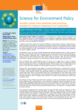
Article
DOI: 10.1002/bkcs.10251
N. Heo et al.
BULLETIN OF THE
KOREAN CHEMICAL SOCIETY
Glucocorticoid Enhances Viability of Human Respiratory Epithelial Cells
Inflicted by Ambient Particulate Matter
Nam Heo,† Ahruem Baek,† Yu Mi Baek,† Sunjoo Byeon,‡ Kum Chan Choi,‡ Joo Yeon Kim,§
Jae Hwan Kwon,§,* and Dong-Eun Kim†,*
†
Department of Bioscience and Biotechnology, Konkuk University, Seoul 143-701, Korea.
*E-mail: [email protected]
‡
Department of Environmental Engineering, Dong-A University, Busan 604-714, Korea
§
Department of Otolaryngolgy-Head and Neck Surgery, Kosin University College of Medicine, Busan
602-702, Korea. *E-mail: [email protected]
Received November 27, 2014, Accepted January 2, 2015, Published online April 21, 2015
Particulate matter (PM) is one of the most common ambient air pollutants, which is derived from diesel
engines, wildfires, yellow dust, and other combustion sources. Especially, PM with an aerodynamic diameter
of <2.5 μm (PM2.5) enhances risks of damaging respiratory organs. Previous studies have reported that PMinduced autophagy and apoptosis can be considered as a phenomenal molecular mechanism of PM-mediated
cytotoxicity in lung cancer epithelial cells. In this study, we investigate the effects of PM2.5 on cellular
responses in human nasal epithelial primary cells and lung cancer cells. The respiratory epithelial cells showed
both time- and concentration-dependent decrease of cell viability when the cells were exposed to PM, which
can be attributed to increased levels of reactive oxygen species (ROS) in the cells. In addition to inducing autophagy via increase of microtubule-associated protein light chain-3 (LC3) puncta, PM exposure also induced the
expression of inflammatory cytokines such as IL-6 and 8 in human respiratory epithelial cells. Thus, inflammation response, as well as autophagosome formation due to increased ROS, is responsible for apoptotic death
of respiratory epithelial cells under challenge of PM. The glucocorticoid ciclesonide used to treat asthma and
allergic rhinitis was shown to be effective in reducing cellular mortality, probably by reducing inflammatory
cytokines. These results suggest that PM2.5-induced inflammation plays a key role in apoptotic cell death in
respiratory epithelial cells, which may be treated by an anti-inflammatory glucocorticoid.
Keywords: Particulate matter, Respiratory epithelial cells, Reactive oxygen Species, Glucocorticoid,
Inflammatory cytokines
Introduction
Particulate matter (PM) is one of the most common urban
ambient air pollutants, which can be produced by industrialization and various other combustions such as fossil fuels, vesicle smoke, and so on.1 In particular, PM with an aerodynamic
diameter of <2.5 μm (PM2.5) has been reported to cause a variety of respiratory and cardiovascular diseases.1 Recent studies
have reported various PM2.5-induced cellular responses such
as autophagy and apoptosis response in lung cancer cells.2–4
Autophagy is known to be the catabolic mechanism for cell
survival that helps to maintain cellular homeostasis, which
involves subcellular degradation of unnecessary or dysfunctional cellular components.5 Through autophagy, decomposed proteins and organelles can be used as the energy
source. Expression of various autophagy-related proteins
involving microtubule-associated proteins light chain 3
(LC3), and degradation of cytosolic components can lead to
the formation of autophagosome, which is a double membrane
structure and which subsequently fuses with lysosomes to
form autolysosomes.6,7 Thus, autophagy has been recognized
Bull. Korean Chem. Soc. 2015, Vol. 36, 1322–1327
as a cellular pathway to maintain homeostasis and adaptation
to environmental stress.8
Recently, it has been reported that PM can induce oxidative
stress as an important molecular mechanism of cell toxicity.2,9
Oxidative stress is caused by reactive oxygen species (ROS),
which are signaling molecules in both cell survival and
cell death.10 Several studies have suggested that inorganic
and organic components from PM exert toxic effects on cells
by increasing oxidative stress in lung epithelial cells.9,11,12
Deng et al. recently observed that autophagy is triggered by
PM2.5-induced oxidative stress in human lung epithelial
A549 cells.2 To date, however, the presence of the PM2.5induced autophagy has not been investigated in nasal epithelial cells, which is important to assess front-line effects
incurred by PMs in the human respiratory system.
Inflammatory response is a nonspecific host defense mechanism, which is generated by external pathogens such as
viruses, bacteria, and parasites. Proinflammatory cytokines
are soluble signaling peptides molecules, which include interlukin-6 (IL-6), IL-8, and tumor necrosis factor-α (TNF-α).
These molecules assist immune response, which could induce
© 2015 Korean Chemical Society, Seoul & Wiley-VCH Verlag GmbH & Co. KGaA, Weinheim
Wiley Online Library
1322
BULLETIN
Article
OF THE
Effect of Glucocorticoid on the Respiratory Cells Incurred by Particulate Matter KOREAN CHEMICAL SOCIETY
or reduce inflammatory responses.13 Very recently, one study
reported that exposure of PM2.5 to mouse-bone-derived macrophage cells enhanced inflammatory potential by increased
secretion of cytokines such as TNF-α and IL-1β in vitro.14 This
study, however, did not use human primary cells derived from
respiratory tissues, which are more suitable to investigate
combined cellular responses such as inflammatory cytokine
release and the accompanying morbidity of neighboring cells.
In pharmacology, steroids or glucocorticoids are used to suppress the adverse effects of inflammatory response by inhibiting the induction of inflammatory cytokines. Inhalation of a
glucocorticoid such as ciclesonide to treat asthma and allergic
rhinitis can inhibit the induction of cytokines implicated by
inflammatory response in the nasal mucous membrane and
bronchial mucosa cells.15
In this study, we investigated the cellular effects by exposure to PM2.5 regarding apoptotic cell death, autophagy, and
inflammatory responses by employing human lung epithelial
A549 cells and human nasal epithelial primary (HNEp) cells.
Compared to the previous studies using the lung cancer cell
line such as A549 cells,2,3,9 our study used human respiratory
primary cells such as HNEp cells cultured from human tissues,
which provide a better in vitro system to assess the physiological responses against PMs than immortalized cancer cell
lines. Furthermore, we demonstrated that ciclesonide is an
effective reagent to attenuate inflammation-related death of
respiratory epithelial cells inflicted by PM.
Experimental
Human Respiratory Epithelial Cells and Glucocorticoid.
The human lung cancer epithelial cell line, A549, was
obtained from the American Type Culture Collection
(ATCC) (Manassas, VA, USA). HNEp cell line HNEpC
was provided by the Kosin University College of Medicine
(Busan, Korea). Cell line A549 and HNEpC were cultured
in Dulbecco's modified minimal essential medium (DMEM)
and RPMI-1640 media supplemented with 100 U of penicillin,
100 μg/mL of streptomycin, and 10% fetal bovine serum
(FBS) and fetal calf serum (FCS). The cells were maintained
at 37 C in a 5% CO2 incubator. The anti-inflammatory glucocorticoid, ciclesonide (Omnaris®), micronized as nasal spray
type, was obtained from Takeda Pharmaceutical Co.
(Osaka, Japan).
PM2.5 Sampling and Preparation. PM2.5 samples were collected in the Dong-A University (Busan, Korea) in April 2014.
PM2.5 samples were collected on Whatman® NucleporeTM filters (diameter 47 mm, pore size 0.2 μm, purchased from GE
healthcare life sciences, Piscataway, NJ, USA) using a cyclone
sampler (about 16.7 L/min, URG Corporation, Chapel Hill,
NC, USA). The filters were equilibrated in 40% relative
humidity and 25 C for 48 h, and then weighed on a highprecision microbalance. PM2.5 particles were extracted from
nitrocellulose filters by immersing them in PBS buffer and
sonicating for 30 min. The extracted samples were then stored
at −80 C until cell exposure. Blank control filters were also
Bull. Korean Chem. Soc. 2015, Vol. 36, 1322–1327
prepared using the same method as above except for the sampling. The amount of PM samples was estimated by the difference in weight between the sampled filter and the blank filter.
Cell Viability Assay. Cell viability was measured by the
methylene blue colorimetric microassay. A549 and HNEp
cells (3 × 10⁴ cells/well) were treated with 50, 100, and
200 μg/mL of PM2.5 for 24 and 48 h. Cells were fixed with
4% paraformaldehyde for 1 h and stained with methylene blue
(1% w/v in borate buffer, Sigma-Aldrich, St. Louis, MO,
USA) for 10 min. After dying at room temperature, the dye
was solubilized using 0.1 M HCl. The absorbance was read
at 595 nm using a VICTOR X3 multilabel plate reader
(PerkinElmer, Waltham, MA, USA).
ROS Assay. A549 and HNEp cells were seeded into 96-well
black plates at a density 3 × 10⁴ cells/well and then incubated
at 37 C. After 24 h incubation, cells were treated with 50, 100,
and 200 μg/mL of PM2.5 or paraquat (Sigma-Aldrich) for 6 h
and then washed with PBS and added to 10 μM 2ʹ,7ʹdichlorofluorescin diacetate (DCF-DA; Sigma-Aldrich). The
fluorescence of DCF was measured by the VICTOR X3 multilabel plate reader (PerkinElmer, Waltham, MA, USA) with
excitation and emission wavelengths of 485 and 535 nm,
respectively.
Confocal Microscopy. A549 and HNEp cells were
cultured on cover slips in a 12-well plate for 24 h. The GFPLC3 plasmid was transfected with LipofectaminTM 2000
(Invitrogen, Carlsbad, CA, USA) for 4 h. Transfected cells
were treated with 100 and 200 μg/mL of PM2.5 or paraquat
(50 μM) for 24 h. Cells were fixed with 4% paraformaldehyde
for further 10 min and washed three times with PBS.
After washing, the treated cells were visualized using a confocal laser scanning microscope (FV-1000, Olympus,
Tokyo, Japan).
Flow Cytometric Analysis of Apoptosis. Apoptosis was
monitored by flow cytometric analysis. A549 and HNEp cells
were seeded into the 24-well plates at a density 6 × 10⁴ cells/
well and then incubated at 37 C. After 24 h incubation, cells
were treated with 50, 100, and 200 μg/mL of PM2.5 or staurosporine (2 μM as positive control, Sigma-Aldrich) for 48 h.
Cells were then harvested and washed twice with cold PBS
and resuspended in a staining buffer containing fluorescein5-isothiocyanate, annexin V, and propidium iodide, provided
in the apoptosis detection kit (BD Biosciences, Franklin
Lakes, NJ, USA), according to the manufacturer's instructions. Flow cytometric data were obtained using a FACSCalibur flow cytometer equipped with BD CellQuest Pro software
(BD Biosciences, San Jose, CA, USA).
Enzyme-linked Immunosorbent Assay (ELISA). A549
and HNEp cells were seeded into 24-well plates at a density
6 × 10⁴ cells/well and then incubated at 37 C. After 24 h incubation, cells were treated with 50, 100, and 200 μg/mL of PM2.5
or lipopolysaccharides at 50 ng/mL (LPS; Sigma-Aldrich) for
24 h. The amounts of proinflammatory cytokines IL-6
and IL-8 were measured with ELISA kits (Quansys Biosciences, Logan, UT, USA) according to the manufacturer's
instructions.
© 2015 Korean Chemical Society, Seoul & Wiley-VCH Verlag GmbH & Co. KGaA, Weinheim
www.bkcs.wiley-vch.de
1323
BULLETIN
Article
OF THE
Effect of Glucocorticoid on the Respiratory Cells Incurred by Particulate Matter KOREAN CHEMICAL SOCIETY
Results and Discussion
Cytotoxicity of PM2.5 in Human Respiratory Epithelial
Cells. First, cell viability assay was performed to assess cellular toxicity after exposure of respiratory epithelial cells to
PM2.5. All previous studies have used lung cancer epithelial
cells for monitoring PM-incurred effects. In addition to lung
epithelial cells, we used the HNEp cells which were isolated
from human nasal mucosa tissues. The use of primary cells
is better to assess physiological effects of PM because the lung
epithelial cells such as A549 cells are immortalized as cancer
cells. A549 and HNEp cells were treated with 50, 100, and
200 μg/mL of PM2.5 for 24 and 48 h. The cell viability measured by methylene blue colorimetric microassay showed that
PM2.5 exposure had significant cytotoxicity in respiratory epithelial cells in a dose- and time-dependent manner (Figure 1).
These results indicated that exposure of PM2.5 causes cytotoxicity, leading to cell death in respiratory epithelial cells.
PM2.5 Generates Oxidative Stress in Respiratory Epithelial Cells. ROS as signaling molecules are involved in autophagy and/or cell death.16 Previous studies have indicated that
PM2.5 can increase the ROS levels in cells.16,17 To monitor the
ROS level in the respiratory epithelial cells upon exposure to
Figure 1. Effect of PM2.5 exposure on cell viability in respiratory epithelial cells. A549 and HNEp cells were treated with 50, 100, and
200 μg/mL of PM2.5 for 24 and 48 h. Cell viability was determined
by methylene blue assay. ∗p < 0.05; ∗∗p < 0.01 vs. control group.
Bull. Korean Chem. Soc. 2015, Vol. 36, 1322–1327
PM2.5, intracellular fluorescence level of ROS was determined
using an ROS-sensing reagent (DCF-DA). After treating with
various concentrations of PM2.5 for 6 h, the respiratory epithelial cells showed increased DCF fluorescence level in a dosedependent manner, indicating that a significantly higher
amount of ROS was generated compared to unexposed control
cells (Figure 2). Exposure of PM2.5 at 100 μg/mL induced
ROS accumulation to a similar extent as with the treatment
of 50 μM ROS-generating reagent (paraquat) in the cells.
PM2.5 Induces Autophagy and Apoptosis. Previous studies
have indicated that PM2.5 as well as nanoparticles induced
autophagy by oxidative stress.2,3,18,19 As we observed that
exposure to PM2.5 of respiratory epithelial cells increased
the ROS levels, we investigated whether PM2.5 induces autophagy and/or apoptosis via oxidative stress. To assess autophagosome formation in cells, expression and puncta of the GFPLC3 protein was monitored under confocal fluorescence
microscopy (Figure 3(a)). Respiratory epithelial cells
increased autophagosome protein GFP-LC3 vacuoles (i.e.,
LC3 puncta) in the cytoplasm after treatment with PM2.5. This
result indicates that increased levels of ROS in the cells
(shown in Figure 2) triggered autophagy in the cells inflicted
by PM2.5. Next, the progress of apoptotic cell death was investigated using flow cytometric analysis of cells exposed to
PM2.5. As the cells were treated with PM2.5, the cells underwent apoptosis in a dose-dependent manner (Figure 3(b)).
Thus, human respiratory epithelial cells exposed to PM2.5
undergo cellular autophagy and subsequent apoptotic cell
death due to enhanced ROS in cells.
Induction of Inflammatory Cytokines by PM2.5 in Respiratory Epithelial Cells. Previous studies have reported that
PM2.5 induced inflammatory cytokines such as IL-8 and
TNF-α in lung cancer cells.20 To observe the inflammatory
response by the PM2.5, we measured the expression of cytokines
Figure 2. Effect of PM2.5 exposure on ROS generation in respiratory
epithelial cells. A549 and HNEp cells were treated with 50, 100, and
200 μg/mL of PM2.5 or paraquat for 6 h. Intracellular ROS level was
determined using DCF fluorescence measurement. The fluorescence
values are expressed as the ratio (fold) of fluorescence between the
exposed cells and the unexposed control cells. Data are means ±
SD of three independent experiments.
© 2015 Korean Chemical Society, Seoul & Wiley-VCH Verlag GmbH & Co. KGaA, Weinheim
www.bkcs.wiley-vch.de
1324
BULLETIN
Article
OF THE
Effect of Glucocorticoid on the Respiratory Cells Incurred by Particulate Matter KOREAN CHEMICAL SOCIETY
Figure 3. Induction of autophagy and apoptotic cell death by PM2.5 exposure in respiratory epithelial cells. (a) Confocal fluorescence microscopy
images of A549 and HNEp cells treated with PM2.5 or paraquat (50 μM) for 24 h. (b) Flow cytometric estimation of apoptotic cell numbers of
A549 and HNEp cells. Cells were treated with PM2.5 or staurosporine (2 μM) for 48 h.
including IL-6 and IL-8 by using ELISA assay in respiratory
epithelial cells. A549 and HNEp cells were treated with various
concentrations (50, 100, and 200 μg/mL) of PM2.5 for 24 h.
In both cell lines, inflammatory cytokines released after the
exposure were significantly higher than those in the control
(Figure 4). Both cell lines showed similar sensitivity to
PM2.5 in producing IL-6 and IL-8 upon exposure to PM2.5,
which was comparable to that when treated with the
Bull. Korean Chem. Soc. 2015, Vol. 36, 1322–1327
cytokine-inducing reagent (i.e., LPS). It has been previously
reported that PM2.5 exposure synergistically increased the
levels of TNF-α and IL-1β in mouse macrophages
in vitro.14 Unlike that in the previous report, our result indicated that IL-6 and IL-8 were mainly secreted in the respiratory
HNEp cells.
Anti-Inflammatory Glucocorticoid Attenuates Cell Toxicity Incurred by PM2.5. Corticosteroids are known to play a
© 2015 Korean Chemical Society, Seoul & Wiley-VCH Verlag GmbH & Co. KGaA, Weinheim
www.bkcs.wiley-vch.de
1325
BULLETIN
Article
OF THE
Effect of Glucocorticoid on the Respiratory Cells Incurred by Particulate Matter KOREAN CHEMICAL SOCIETY
Figure 4. Cytokines secretion after treatment of PM2.5 in human respiratory epithelial cells. Concentrations of inflammatory cytokine
were measured using ELISA after incubation with 50, 100, and
200 μg/mL of PM2.5 or lipopolysaccharide (50 ng/mL) for 24 h.
role in reducing the inflammatory response in cells.21 We
investigated the cell viability of respiratory epithelial cells
exposed to PM2.5 with or without treatment with ciclenoside
(Figure 5(a)), which is an FDA-approved glucocorticoid drug
to treat asthma and allergic rhinitis. A549 and HNEp cells were
treated with various concentrations of PM2.5 in the presence or
absence of ciclenoside for 24 h.
For both cell lines, cell viabilities decreased significantly
less compared to the ciclenoside treatment than without
the drug treatment (Figure 5(b)). The PM2.5-induced cell toxicity was more significantly attenuated by treatment with
ciclenoside (at higher concentration of PM2.5 exposure (i.e.,
200 μg/mL)). Thus, glucocorticoid ciclesonide at optimal concentration (i.e., 5 μg/mL) was shown to be effective in reducing cellular toxicity incurred by PM2.5, probably by
reducing inflammatory cytokines.
Conclusion
We have observed that PM2.5 can induce cellular toxicity by
generating oxidative stress, which can cause the onset of
autophagy and eventual apoptotic cell death in human
Bull. Korean Chem. Soc. 2015, Vol. 36, 1322–1327
Figure 5. Effect of anti-inflammatory glucocorticoid in human respiratory epithelial cells inflicted by PM2.5. (a) Chemical structure of
ciclenoside. (b) A549 and HNEp cells treated with 50, 100, and
200 μg/mL of PM2.5 with or without 5 μg/mL of ciclesonide for
24 h. Cell viability was determined by methylene blue assay. Data
are means ± SD of three independent experiments, and ∗p < 0.05.
respiratory epithelial lung cells and nasal epithelial primary
cell lines. Moreover, PM2.5 induced secretion of inflammatory
cytokines such as IL-6 and IL-8 in both cell lines. The results
suggest that PM2.5-induced oxidative stress and inflammatory
cytokines probably play a key role in causing cell toxicity in
human respiratory epithelial cells. The adverse effect of
PM2.5 on the cell viability was ameliorated by treatment with
the anti-inflammatory glucocorticoid, which might be contributing to the treatment of PM2.5-induced impairment of
© 2015 Korean Chemical Society, Seoul & Wiley-VCH Verlag GmbH & Co. KGaA, Weinheim
www.bkcs.wiley-vch.de
1326
BULLETIN
Article
OF THE
Effect of Glucocorticoid on the Respiratory Cells Incurred by Particulate Matter KOREAN CHEMICAL SOCIETY
respiratory function. Our study is the first in which the glucocorticoid ciclesonide is shown to be effective in treating
asthma and allergic rhinitis by reducing cellular mortality in
human respiratory epithelial cells inflicted by ambient PM
by probably reducing inflammatory cytokines.
Acknowledgments. This work was supported by a National
Research Foundation grant funded by the Korean Government
MSIP (2012M3A9B2028336, to D.-E. Kim) and a grant from
the Kosin University College of Medicine (to J.-H. Kwon).
References
1. J. O. Anderson, J. G. Thundiyil, A. Stolbach, J. Med. Toxicol.
2012, 8, 166.
2. X. Deng, F. Zhang, W. Rui, F. Long, L. Wang, Z. Feng, D. Chen,
W. Ding, Toxicol. in Vitro 2013, 27, 1762.
3. X. Deng, F. Zhang, L. Wang, W. Rui, F. Long, Y. Zhao, D. Chen,
W. Ding, Apoptosis 2014, 19, 1099.
4. M. M. Ulrich, G. M. Alink, P. Kumarathasan, R. Vincent,
A. J. Boere, F. R. Cassee, J. Toxicol. Environ. Health A 2002,
65, 1571.
5. J. D. Rabinowitz, E. White, Science 2010, 330, 1344.
6. A. J. Meijer, P. Codogno, Int. J. Biochem. Cell Biol. 2004,
36, 2445.
7. A. Longatti, S. A. Tooze, Cell Death Differ. 2009, 16, 956.
8. E. White, R. S. DiPaola, Clin. Cancer Res. 2009, 15, 5308.
Bull. Korean Chem. Soc. 2015, Vol. 36, 1322–1327
9. S. Yi, F. Zhang, F. Qu, W. Ding, Environ. Toxicol. 2014, 29, 226.
10. M. B. Azad, Y. Chen, S. B. Gibson, Antioxid. Redox Signal.
2009, 11, 777.
11. S. Billet, G. Garcon, Z. Dagher, A. Verdin, F. Ledoux, F.
Cazier, D. Courcot, A. Aboukais, P. Shirali, Environ. Res.
2007, 105, 212.
12. K. S. Kouassi, S. Billet, G. Garcon, A. Verdin, A. Diouf,
F. Cazier, J. Djaman, D. Courcot, P. Shirali, J. Appl. Toxicol.
2010, 30, 310.
13. B. Ryffel, Toxicol. Lett. 1995, 82–83, 477.
14. M. Ferguson, C. Migliaccio, T. Ward, Inhal. Toxicol. 2013,
25, 766.
15. P. J. Barnes, N. Engl. J. Med. 1995, 332, 868.
16. M. Dewaele, H. Maes, P. Agostinis, Autophagy 2014, 6, 838.
17. Y. Mo, R. Wan, S. Chien, D. J. Tollerud, Q. Zhang, Toxicol.
Appl. Pharmacol. 2009, 236, 183.
18. T. Wang, E. T. Chiang, L. Moreno-Vinasco, G. D. Lang, S. Pendyala, J. M. Samet, A. S. Geyh, P. N. Breysse, S. N. Chillrud,
V. Natarajan, J. G. Garcia, Am. J. Respir. Cell Mol. Biol.
2010, 42, 442.
19. A. Montiel-Davalos, J. Ibarra-Sanchez Mde, J. L. Ventura-Gallegos, E. Alfaro-Moreno, R. Lopez-Marure, Toxicol. in Vitro
2010, 24, 135.
20. K. Peynshaert, B. B. Manshian, F. Joris, K. Braeckmans, S. C. De
Smedt, J. Demeester, S. J. Soenen, Chem. Rev. 2014, 114, 7581.
21. B. Wang, K. Li, W. Jin, Y. Lu, Y. Zhang, G. Shen, R. Wang,
H. Shen, W. Li, Y. Huang, X. Wang, X. Li, W. Liu, H. Cao,
S. Tao, Environ. Sci. Technol. 2013, 47, 10583.
© 2015 Korean Chemical Society, Seoul & Wiley-VCH Verlag GmbH & Co. KGaA, Weinheim
www.bkcs.wiley-vch.de
1327


© Copyright 2026