
Marine and aerophytic Cyanosarcina, Stanieria and
Algological Studies 64
141-157
Stuttgart, Dezember 1991
Marine and aerophytic Cyanosarcina, Stanieria
and Pseudocapsa (Chroococcales) species
from Hellas (Greece)
By K o n s t a n t in o s A n a g n o s t id is and A n d r ia n a P a n t a z id o u
University of Athens, Institute of Systematic Botany, Athens, Hellas (Greece)
With 24 figures in the text
Abstract: Epilithic field and cultured materials of Cyanosarcina ΚονΑδ., Stanieria K o m . et
and Pseudocapsa E r c e g . (Chroococcales) were studied from Hellenic marine and aero
phytic calcareous substrates. A new marine species, Cyanosarcina thalassia A n a g n . et P a n t , and
a new aerophytic, chasmoendolithic species, Cyanosarcina parthenonensis A n a g n . are described.
The marine species Dermocarpa sublitoralis L in d s t . and Cyanocystis sphaerica ( S r t c h . et
G a r d n .) K o m . et A n a g n . are transferred to Stanieria. Morphological features and the life cycle of
the debatable, aerophytic, epilithic type species of Pseudocapsa, P. dubia E r c e g . are elucidated,
confirming the validity of this genus.
Key words: Cyanophytes, cyanobacteria, Chroococcales, Chroococcaceae, Dermocarpellaceae,
Cyanosarcina n. sp., Dermocarpa, Myxosarcina, Cyanocystis, taxonomy,
Stanieria, Pseudocapsa, intergeneric features, taxonomy, morphology, life cycle,
cultures, epilithic, epiphytic, aerophytic (caves), chasmoendolithic, marine (littoral
zone), Hellas (Greece).
A nagn.
PDC*:
SS 02, 021; ST 052, 06, 08, 10; BN 01, 04, 05; MO 01, 09; EC 02, 063, 093; EB
01, 03, 252; ME 06, 10; EP 032, 19; UC 011, 012.
Introduction
The taxonomic position of the non-filamentous cyanophytes has been the subject of
many dicussions by several authors. Until recently they were classified in three (or
four) orders, Chroococcales, Chamaesiphonales (Dermocarpales) and Pleurocapsales,
according to different types of reproduction (G e it l e r 1925, 1932, 1942, F r £ m y 1930,
1934, E l e n k in 1938, F r it s c h 1945, H o l l e r b a c h et al. 1953, D e s ik a c h a r y 1959,
S t a r m a c h 1966, B o u r r e l l y 1970, 1985, K o n d r a t e v a 1975, W a t e r b u r y & S t a n ie r
1978, R ip p k a et al. 1979, 1981, K o n d r a t e v a et al. 1984, R ip pk a & H e r d m a n 1985,
R ip p k a 1988, C a s t e n h o l z & W a t e r b u r y 1989, W a t e r b u r y & R ip p k a 1989, W a t e r
b u r y 1989). Since cell division is basically the same in all cyanophytes (simple binary
fission, multiple fission, presence/absence of nanocytes-baeocytes with many transi
* Phycological Documentation Code - see; Algological Studies 9: 450-481, 1973.
0342-1120/91/0092-0141 $ 4.25
© 1991 E. Schweizerbart’sche Verlagsbuchhandlung, D-7000 Stuttgart 1
142
Konstantinos Anagnostidis and Andriana Pantazidou
tions) and sharp boundaries between the reproductive processes of different unicellular
species are lacking, additional features (polarity, apolarity of cells, etc.) were used in
the recently revised classification system by K o m Ar e k & A n a g n o s t id is (1986). They
classify all unicellular cyanophytes in a single order Chroococcales, consisting of
seven definable and clearly distinguishable families. In that revised system the family
Chroococcaceae contains the genera Chroococcus NAg., 1849, Cyanocybus S c h il l .
1956, Cyanosarcina K o v A c . 1988, Gloeocapsopsis G e it l . 1925 and Pseudocapsa
E r c e g . 1925 that possess spherical cells, and that divide successively in three or more
different planes; nanocytes are lacking. The family Dermocarpellaceae (sensu K o
m A r e k & A n a g n o s t id is 1986) contains genera characterized by cells that may or may
not possess polarity, and that divide exclusively by multiple fission into many nano
cytes; included are the genus Cyanocystis B o r z i 1878 (synonym, Dermocarpa
C r o u a n sensu auctt. post.; sensu W a t e r b u r y & S t a n ie r 1978; sensu W a t e r b u r y
1989) that displays apical-basal polarity, the genus Dermocarpella L e m m . 1907 that
possesses polarity and cleaves entirely into nanocytes, and the genus Stanieria K o m . et
A n a g n . 1986 (synonym, “Dermocarpa” sensu W a t e r b u r y & S t a n ie r 1978; sensu
W a t e r b u r y 1989) that possesses spherical non polarized cells and also cleaves en
tirely into nanocytes.
The present study deals with the systematics of some rare and interesting marine
(epilithic and epiphytic) and aerophytic (epilithic, chasmoendolithic) chroococcacean
and dermocarpellacean (sensu K o m Ar e k & A n a g n o s t id is 1986) cyanophytes. A new
marine species, Cyanosarcina thalassia, and a new aerophytic, chasmoendolithic spe
cies, Cyanosarcina parthenonensis, are described. The species Dermocarpa sublitoralis L in d s t . and Cyanocystis sphaerica (S e t c h . et G a r d n .) K o m . et A n a g n . are
assigned to the recently established genus Stanieria. The diagnostic criteria of Pseudo
capsa dubia E r c e g . are elucidated and the validity of the genus Pseudocapsa is dis
cussed.
Materials and methods
Aerophytic, epilithic material with Pseudocapsa dubia has been collected from the
cave Melidoni, located at the Province Rethymnon of Crete Island, as well as from the
cave of Nympholipton and some other anonymous small caves located at the SE part
on the mountain Hymettos at the Attiki Peninsula. Marine, epilithic and epiphytic
materials with Cyanosarcina thalassia, Stanieria sublitoralis and S. sphaerica were
collected from the littoral zone of calcareous coasts near the villages Vouliagmeni,
Varkiza, Kavouri, Lavrion and Rafina, and the area of Cape Sounion at the Attiki
Peninsula, Aegaeon Pelagos (Aegean Sea), Hellas (Greece). The initial field and culti
vated material derived from the lower part of an exposed marble column of the eastern
part of the Parthenon (ancient Athenian Acropolis) studied by A n a g n o s t id is et al.
(1983) was taxonomically re-evaluated.
Enrichment cultures of C. thalassia, S. sublitoralis and P. dubia were obtained
with BG, MN, and AS ΠΙ media (W a t e r b u r y & S t a n ie r 1978). Cultures were grown
in 18-22 °C and illuminated with white daylight fluorescent tubes (light intensities
about 3.102 cal cm-2 min-1 at 16:8 LD cycle). Photodocumentation and morphometric
evaluation have been carried out by light microscopy (Photomicroscope ΙΠ, Zeiss) on
field and cultured materials.
Marine and aerophytic Chroococcales from Hellas
143
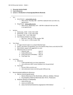
Figs 1-3. Cyanosarcina thalassia A n a g n . et P a n t a z ., LM micrographs; mass culture, liquid MN
medium. 1 - Young aggregates showing the cell size uniformity and the regularity of the planes of
successive divisions. 2-3 - Older aggregates showing the maintenance of fairly regular cubical
packets of cells. [Scale bar: 15 μπι.]
Results
Genus Cyanosarcina Κ ο ν λ έ . 1988
C yanosarcina thalassia spec, nova
(Figs 1-3)
D i a g n o s i s : Thallus epilithicus, pallidus coeruleo-viridis ad substratum carbonaceum
aquamaris adhaerens. Coloniae 2-32 cellulares vel compositae, multicellulares, sarcinoides, ad
12x18 μπι in dimensione, cum tegumentis mucilaginosis tenuis, homogeneis, incoloribus et sine
structura circumdatae. Cellulae sphaericae, post divisionem subglobosae vel hemisphaericae,
144
Konstantinos Anagnostidis and Andriana Pantazidou
2-3 μηι in diametro. Protoplastum aerugineum, homogeneum, raro griseo-olivaceum. Propagatio
cellularum divisione transversali in directiones tres; nanocyta carrentes.
E t y m o l o g y : Thalassia (Greek) = θαλασσια: Epitheton related to the ancient and modem
Hellenic words θαλαττα, θαλασσα = sea, and αλς, α λα ς = salt; living in thalassic (marine)
biotopes.
I c o n o t y p e : Figs 1-3.
T y p e s p e c i m e n : Microscope slides (A t h u - C y 7 5 8 8 3 , A-B) with field populations and
cultured material have been deposited in the Athenian Botanical Museum (Herbarium), Athens
University.
T y p e l o c a l i t y : South western coasts of Attild Peninsula, near the village Varkiza,
Saronikos Gulf, Aegaeon Pelagos (Aegean Sea), Hellas (Greece).
H a b i t a t : Epilithic on calcareous coastal rocks at the marine littoral zone.
D i s t r i b u t i o n : Epilithic on calcareous rocks at the marine littoral zone of the village
Varkiza, Vouliagmeni, Kavouri, and the area of Cape Sounion, Attiki Peninsula, Saronikos Gulf,
Aegaeon Pelagos (Aegean Sea), Hellas (Greece).
The pale blue-green thallus of Cyanosarcina thalassia is epilithic, growing on
marine calcareous substrates, consisting of 2-32-celled or compound, multicelled
colonies arranged in more or less cubical packet-like aggregates, 12 x 18 μιη in
dimension, with thin colourless and structureless mucilaginous envelope (Figs 1-3).
The cells are spherical and after division subspherical or hemispherical, 2-3 μπι in
diameter, with a blue-green, rarely olive-green, finely granular protoplast. The cells
divide successively in three perpendicular planes, not growing before the next division
in the original form, later forming dense packets; nanocytes are lacking.
Stages in the life cycle of C. thalassia were observed by periodic examination of
cultures over several months. The morphology was not affected by the different cul
ture media used; only slight differences in growth rate and the morphometric charac
ters between strains were ascertained.
C. thalassia is the only marine species of the recently established genus and
clearly differs in morphology and ecology from the type species C. fontana and the
related species C. litoralis as well as from the other species of the genus.
Cyanosarcina parthenonensis
A n a g n o s t id is s p e c , n o v a
(Fig.
24 )
S y n o n y m : Myxosarcina concinna P r in t z , p.p., sensu A n a g n o s t id is et al. Nova Hedwigia
38: 246, Fig. 6 (upper part), 22-26, 1983.
D i a g n o s i s : Thallus aerophyticus, chasmoendolithicus, olivaceo-viridis ad substratum
carbonaceum adhaerens. Coloniae 2-16 cellulares vel compositae, multicellulares, sarcinoides,
ad 37 μπι in diametro, cum tegumentis mucilaginosis tenuis, homogeneis incoloribus et sine
structura circumdatae. Cellulae sphaericae, post divisionem subglobosae vel subsphaericae, 2.54.5-(5) μm in diametro. Protoplastum olivaceo-viridum, luteo-viridum, raro luteo-brunneum,
homogeneum, plerumque subtOiter granulatum. Propagatio cellularum divisione transversali in
directiones tres; nanocyta carrentes.
E t y m o l o g y : Parthenonensis (Greek): Epitheton derived from the name Parthenon =
Παρθένων, the unique and most important templum monument of the ancient Athenian
Acropolis, erected in favor of Pallas Athene.
I c o n o t y p e : Fig. 24.
T y p e l o c a l i t y : Eastern part of Parthenon, ancient Athenian Acropolis, Athens, Hellas
(Greece).
H a b i t a t : Aerophytic, chasmoendolithic into fissures and cracks of Pentelic marble (calcite,
microcrystalline of low porosity with veins and chlorite and muscovite) at an exposed weathered
column of Parthenon.
The olive-green thallus of Cyanosarcina parthenonensis is aerophytic, chasmoen
dolithic on calcareous substrate, consisting of 2-16-celled or compound, multicelled
colonies arranged in more or less cubical packet-like aggregates, 10-37 μm in diame
Marine and aerophytic Chroococcales from Hellas
145
ter, with thin colourless and structureless mucilaginous envelope. The cells are spheri
cal and after division subglobose or subspherical, 2.5-4.5-(5) μm in diameter, with
olive-green, yellow-green, rarely olive-brown, finely granulated protoplasts. The cells
divide successively in three perpendicular planes, not growing before the next division
in the original form, later forming dense packets; nanocytes are lacking.
C. parthenonensis was initially placed in the genus Myxosarcina P r in t z 1921,
designated as M. concinna (A n a g n o s t id is et al. 1983). In spite of the fact that Cyano
sarcina is morphologically similar to Myxosarcina and the cell division proceeds in
three or more planes, it never forms nanocytes. Therefore, the cyanophyte from Par
thenon (field and cultured materials) is re-evaluated and assigned to Cyanosarcina.
C. parthenonensis is the only chasmoendolithic, aerophytic species of the genus and
clearly differs in morphometric characters and ecology from the new species C. thalas
sia and the other species of the genus.
Genus Cyanosarcina comprises now nine species known from thermal springs,
freshwater, marine and aerophytic habitats (G e it l e r 1927, 1932, 1942, G e it l e r &
R u t t n e r 1935, E m o t o & Y o n e d a 1941, S c h w a b e 1944, S k u ja 1949, H in d Ak 1975,
H ir a n o & H ir o s e 1977, A n a g n o s t id is et al. 1983, K o v A& k 1988, A n a g n o s t id is ,
unpubl. results; the authors): C. burmensis (S k u ja ) K o v Ac ., C. chroococcoides
(G e it l .) K o v AC., C.fontana KovAc. (type species), C. gelatinosa (E m o t o et Y o n e d a )
KovAc., C. litoralis (S c h w a b e ) K o v Ac ., C. spectabilis (G e it l .) ΚονΑά, C. thermalis
(H in d .) KovAd, and the new species C. thalassia A n a g n . et P a n t , and C. parthenon
ensis A n a g n . Another taxon found in caves and described as Cyanosarcina sp. (A b d e l a h a d 1989) was not taxonomically evaluated. The Cyanosarcina species are char
acterized by sarcinoid-like aggregates and the cell division proceeds in one, two, three
or more, more or less perpendicular directions in the successive generations; nanocytes
are lacking. Cyanosarcina exhibits similarities to the genus Myxosarcina in the way of
cell division but differs in the mode of reproduction as the latter reproduces with
combined binary fission and by nanocytes. It is noted that all the species, except the
type species and C. thalassia, were initially classified under the genus Myxosarcina
(see review in A n a g n o s t id is et al. 1983; Table 1). Cyanosarcina additionally shows
some relationship to Pseudocapsa in the mode of reproduction and the pattern of cell
division (see p. 149). Both genera are classified into the family Chroococcaceae (sensu
K o m Ar e k & A n a g n o s t id is 1986), whereas Myxosarcina into the family Xenococcaceae (sensu K o m Ar e k & A n a g n o s t id is 1986).
Genus Stanieria Kom. et A nagn. 1986
Stanieria sublitoralis
( L in d s t .) c o m b , n o v a
B a s i o n y m : Dermocarpa sublitoralis
p. 30, Fig. 203, 1943.
L in d s t .,
(Figs 4-7)
FI. Mar. Cyanophyc. Schwed. Westkiiste
Stanieria sublitoralis is characterized by solitary spherical cells, 7-12-(18) μm in
diameter, just before multiple fission reaching up to 25 μm. The cell content is bluish
to reddish and more or less homogeneous. Reproduction is taking place by up to 64
spherical motile nanocytes (planocytes) 1.5-2.5-{4) μm in diameter, developing by a
simultaneous division of the protoplast; the nanocytes are released by a rupture of the
mother cell wall and increase in size until the onset of multiple fission.
It was found epilithic widely distributed in the littoral zone of calcareous marine
coasts near the villages Vouliagmeni, Varkiza, Kavouri, Lavrion and Raima, and the
area of Cape Sounion at the Attiki Peninsula, Saronikos Gulf, Aegaeon Pelagos
(Aegean Sea), Hellas (Greece).
Konstantinos Anagnostidis and Andriana Pantazidou
Figs 4 - 7 . Stanieria sublitoralis ( L tndst .) A n a g n . etPA N TA Z., LM micrographs; mass culture, liquid
MN medium. Spherical cells of varying size with or without nanocytes. Liberated and developing
nanocyte groups as well as empty cell-walls are indicated by arrows. [Scale bar: 20 μm.]
Marine and aerophytic Chroococcales from Hellas
147
S. sublitoralis was initially described as Dermocarpa sublitoralis by L in d st e d t
(1943); it was found widely distributed in the sublittoral zone (1-2 m up to 15-20 m
deep) forming red spots attached on various sea animals, algae and mussel valves on
the west coasts of Sweden.
The morphometric characters and the life cycle of the epilithic cyanophyte studied
do not differ significantly from those originally described. L in d s t e d t (1943) measured
cells 10-18 μm in diameter with red or violet protoplasts, and nanocytes (to 128) 2-3
μm in diameter. S. sublitoralis grows well in culture (for more than seven months) on
the solid substrate, exhibiting the typical features of the species, whereas the cells in
the stirred solutions agglomerated and formed anomalies. Obviously, the sessile mode
of life of that marine cyanophyte needs even in culture a substrate to attach. Excep
tionally, in cultured material, the simultaneous cell division does not extend to the
entire cytoplasm and a small portion is left undivided. Cells with such incomplete
simultaneous divisions were also observed in Cyanocystis violacea (C r o u a n ) K o m . et
A n a g n . (H u a et al. 1989) and in Dermocarpella (sensu W a t e r b u r y & S t a n ie r 1978;
W a t e r b u r y 1989); they occur obligatorily in the genus Cyanocystis. After the simul
taneous multiple fission of the cells growing on agar, the resulting nanocytes are
capable of gliding motility for a short period immediately following their release and
react positive phototactic in a light gradient. Planocytes (motile nanocytes) showing a
photoactive response were also observed in the strains PCC 7302 (ATCC 29368), PCC
7303 (ATCC 29369), PCC 7304 (ATCC 29270) and PCC 7437 (ATCC 29371) that
were assigned to the genus Dermocarpa (W a t e r b u r y & S t a n ie r 1978, R ip pk a et al.
1979, W a t e r b u r y 1989).
The strain PCC 7437 originally described as Chroococcidiopsis cyanosphaera
(K o m Ar e k & H in d Ak 1975) and later as Dermocarpa cyanosphaera (W a t e r b u r y &
S t a n ie r 1978, R ip p k a et al. 1979, W a t e r b u r y 1989) represents the type species of the
recently established genus Stanieria by K o m Ar e k & A n a g n o s t id is (1986). The strains
PCC 7302 and PCC 7303 and (probably) the strain PCC 7304 all of marine origin
(W a t e r b u r y & S t a n ie r 1978, R ip p k a et al. 1979, W a t e r b u r y 1989) exhibit many
similarities in ecology and morphology (maximum diameter of vegetative cells 30 μm,
nanocyte diameter 1.5-2 μm) to S. sublitoralis and therefore could be classified under
the latter taxon. Worth noticing is that the strains PCC 7302 and PCC 7303, closely
resemble one another, should probably be assigned to a new species of “Dermocarpa”
according to W a t e r b u r y (1989, p. 1758).
Stanieria sphaerica (S e t c h . e t G a r d n .) c o m b , n o v a
B a s i o n y m : Dermocarpa sphaerica S e t c h . e t G a r d n . in
(Figs
G ardn.
8 -1 0 )
Univ. Cal. Publ., p. 457,
Table 39, Fig. 14, 1918.
S y n o n y m :
biol./Suppl. 73,
Cyanocystis sphaerica
A lg o l.
Stud.
43:
(S e t c h . e t G a r d n .) K o m . e t A n a g n . A r c h . H y d r o -
203, 1986.
The cells are spherical, 6-10 μm in diameter, solitary with bluish more or less
homogeneous content. Reproduction proceeds by a simultaneous, multiple fission of
the protoplast in up to 32 motile spherical nanocytes (planocytes), 1.5-2-(3) μm in
diameter, that are released by a dissolution of the mother cell wall and increase in size
until the onset of multiple fission.
The cell and nanocyte dimensions of the form studied in field material are smaller
than that of the typical species (cells 8-16 μm; nanocytes 2.5-3 μm). S. sphaerica was
found epiphytic on Lyngbya aestuarii L ie b m . attached on calcareous rocks of the
littoral zone at the Attiki Peninsula.
148
Konstantinos Anagnostidis and Andriana Pantazidou
%. r" I
.'v'\
•
*
w
<:
·"*
sSP
'
«
_
8
^
Figs 8-10. Stanieria sphaerica ( S e t c h . et G a r d .) A n a g n . et P a n t a z ., LM micrographs; field
material. 8 - Spherical solitary cells, epiphytic on the marine cyanophyte Lyngbya aestuarii L ie b m .
9 - Cells divided by multiple fission into nanocytes. 10 - Released free, motile nanocytes
(planocytes) or attached on a filament of Lyngbya aestuarii. [Scale bar: 25 μ m ]
This species was initially described as Dermocarpa sphaerica S e t c h . et G a r d n .
1918; recently it was revised and renamed Cyanocystis sphaerica (S e t c h . et G a r d n .)
K o m . et A n a g n . 1986. Nevertheless, the non heteropolar cells (Figs 8-10, 23) corre
spond rather to the definition of the genus Stanieria K o m . et A n a g n . than to the genus
Cyanocystis B o r z i . Therefore, this species is re-evaluated, reclassified and renamed
Stanieria sphaerica.
S. sphaerica e x h ib its a c lo s e re la tio n s h ip in m o r p h o lo g y a n d e c o lo g y to S. subli
toralis a n d th e o n ly d if fe r e n c e s a r e th e c e ll a n d m o tile n a n o c y te (p la n o c y te ) s iz e s ( s e e
a ls o L in d s t e d 1943).
G e n u s Stanieria c o m p r is e s n o w th r e e s p e c ie s , S. cyanosphaera (ty p e s p e c ie s)
k n o w n f r o m m in e r a l s p rin g s o f C u b a (K o m Ar e k & H in d Ak 1975, K o m Ar e k & A n a g
n o s t id is 1986) a n d th e a p p a r e n tly w id e d is tr ib u te d m a r in e s p e c ie s S. sphaerica a n d S.
sublitoralis (S e t c h e l l & G a r d n e r 1919, G eit l e r 1932, F r 6 m y 1934, L in d s t e d t 1943,
K o s in s k a ja 1948, G o n z a l e z & P a r r a 1981, N ev e s 1988, th e a u th o rs ). I t s e e m s
p o s s ib le th a t S ta n ie r ia e x h ib its a m o r e w id e d is tr ib u tio n ; d is c o v e rin g a n d d e s c rip tio n
o f m o r e s p e c ie s is e x p e c te d .
Dermocarpa sphaerica S e t c h . e t G a r d n . (s e n s u R a o 1940, in D e s ik a c h a r y 1959,
174) c o u ld b e p o s s ib ly c o n s id e re d a s a n o th e r ta x o n o f th e g e n u s Stanieria b e c a u s e o f
Marine and aerophytic Chroococcales from Hellas
149
its difference in ecology and morphology. It has larger cells (up to 16.5-(23.1) μm
long!) and was found epiphytic on the chlorophyte Pithophora sp. in a freshwater
habitat of Delhi, India.
Genus Pseudocapsa E r c e g . 1925
Pseudocapsa dubia E rceg. 1925
(Figs 1 1-17)
The organism found aerophytic, epilithic on calcareous rocks (walls) of Hellenic caves
is characterized by more or less spherical 2-16-celled, solitary colonies or by multi
celled packet-like aggregates, up to 170 μm in diameter, with yellowish or colourless,
structured (layered), gelatinous envelopes. The cells are irregularly spherical, during
the division hemispherical, usually arranged radially or fan-like in the colony, 2.5-34.5 μm in diameter; ensheathed cells are 4—6.5 μm in diameter, with a blue-green
homogeneous protoplast. Cells divide successively in three or more different planes
with radially or fan-like oriented cells; cells do not grow before the next division into
the original more or less spherical form. Nanocytes are lacking.
The life cycle of P. dubia proceeds as following: After the liberation from the
colony the ensheathed cells (Figs 12, 14, arrow) start to enlarge and to divide (Figs 11,
13). Initially the cell division takes place transverse in one plane, later in two perpen
dicular planes, thus the resulting colonies resemble those of the genus Chroococcus
(“Chroococcus-lype”; Figs 11, 14—15). Later the cells divide radially in three or more
different, more or less perpendicular planes resulting in the formation of spherical or
ellipsoid colonies up to packet-like aggregates with spherical, subspherical or fan-like
oriented cells (Figs 11-17). The same life cycle were recently described for P.
venkataramanii by KovAcnc (1988) and for P. dubia by A b d e l a h a d (1989).
The morphometric features do not differ significantly from those of the original
(Fig. 20) description (E r c e g o v ic 1925: cells polyhedral, 3-10 μm in diameter, without
special envelopes, yellow-green or blue-green, arranged in one-layered or multi
layered, spherical or irregularly shaped colonies). The developmental stages and the
mode of reproduction observed in our field and cultured materials from Hellenic caves
correspond more or less to those illustrated (Fig. 21a-n) by S t a r m a c h (1966) for
P. dubia·, multiple fission into many nanocytes was never observed. This reproductive
process (absence of nanocyte formation) and the pattern of cell division in radial
direction (without growing up to the original spherical form) with the cell arrangement
in radial or fan-like directions in the colonies (Fig. 19) justifies the classification of
Pseudocapsa in the family Chroococcaceae N a g . (sensu K o m Ar e k & A n a g n o s t id is
1986; see also G e it l e r 1942).
On the other hand, A b d e l a h a d (1989) interpreting some of the developmental
stages of P. dubia and P. venkataramanii observed by S t a r m a c h (1936, 1966,
Fig. 170) and K o v ACik (1988, Figs 24e, 25i, 26) respectively, in relation to her find
ings of “tunicate cells” and clusters of emptied “sporocysts” as early and advanced
stages of nanocyte formation respectively (Figs 15-16), considers that the genus Pseu
docapsa should belong to the family Xenococcaceae (sensu K o m Ar e k et A n a g n o s
t id is 1986), the members of which are defined by cell division in three or more planes
and also by nanocytes (endospores, baeocytes). The term “tunicate cell” is used by
A b d e l a h a d (1989) to indicate “cells of varying size surrounded by a firm sheath” (i.e.
the ensheathed cells) that are considered “the development stage of the nanocytes” and
the term “sporocyst” to indicate “the firm sheath envelope which encloses the cluster
of nanocytes produced by a parental cell”. The term n a n o c y t e (“endospore”,
“baeocyte”) has been used by botanists and/or microbiologists to describe very small
cyanophyte daughter cells (in comparison to the vegetative ones) produced succes11
A rc h iv f. H y d ro b io lo g ie , S u p p l.-B d . 92
150
Konstantinos Anagnostidis and Andriana Pantazidou
Figs 11-14. Pseudocapsa dubia E r c e g ., LM micrographs; field material. 1 1 - 1 2 -Sm all, 1-8-celled
and large multicelled densely packed colonies within yellowish or colourless mucilaginous,
structured (layered) envelopes. 1 3 - 1 4 - Various developmental stages; characteristic colonies with
radially or fan-like oriented cells (arrow); ensheathed solitary cells liberated from the colony (arrow)
and 2-8-celled colonies. [Scale bar: 20 μm.]
Marine and aerophytic Chroococcales from Hellas
151
15
Figs 15-17. Pseudocapsa dubia E r c e g ., LM micrographs; field material. Developmental stages;
1-32 celled spherical colonies. [Scale bar: 20 μm.]
s iv e ly o r s p o n ta n e o u s ly b y m u ltip le f is s io n ( d e ta ils s e e in K o m Ar e k & A n a g n o s t id is
1986, 165-170, s e e a ls o G e it l e r 1932, 1942, 1960, 1979, F r it s c h 1945, B o u r r e l l y
1970, 1985, W a t e r b u r y & S t a n ie r 1978, A n a g n o s t id is e t a l. 1983, W a t e r b u r y &
R ip p k a 1989, A n a g n o s t id is & K o m Ar e k 1990).
G e n u s Pseudocapsa E r c e g . c o m p r is e s f o u r s p e c ie s : P. dubia ( ty p e s p e c ie s ) , P.
maritima K o m . 1956, P. sphaerica (P r o Sk .-L a v r .) K o v Ac . 1988 (sy n . Myxosarcina
sphaerica P r o Sk .-L a v r . 1951) a n d P. venkataramanii KovAc. 1988. P. dubia r e p r e
s e n ts a n a e r o p h y tic , e p ilith ic c y a n o p h y te w ith r e s tr ic te d d is tr ib u tio n o n m o is t c a l
c a r e o u s r o c k s , a n d s ta la g m ite s a n d s ta la c tite s o f c a v e s f o u n d in Y u g o s la v ia , P o la n d ,
I ta ly a n d H e lla s (E r c e g o v i £ 1925, G e it l e r 1932, S t a r m a c h 1966, A n a g n o s t id is e t al.
1983; A b d e l a h a d 1989, A n a g n o s t id is , u n p u b l. r e s u lts ; th e a u th o rs ); its s y s te m a tic
p o s itio n w a s c o n s id e r e d to b e u n c le a r (G e it l e r 1932, S t a r m a c h 1936, 1966, B o u r
relly
1970;
s e e a ls o G e it l e r
1942, ΚονΑάκ 1988,
A bdelahad
1989). P. maritima
w a s f o u n d in B u lg a r ia o n m a r in e c o a s ta l c a lc a r e o u s r o c k s o f th e B la c k S e a (K o m Ar e k
Konstantinos Anagnostidis and Andriana Pantazidou
152
Figs 18-21. Illustrations of the genera Cyanosarcina KovAc., Pseudocapsa E r c e g . 1 8 - 1 9 Schemes of different types of cell division (in 1,2,3 or more planes in successive generations (after
KomArek & A n a g n o s t id is 1986). 2 0 - 2 1 - Pseudocapsa dubia E r c e g . 2 0 - Original illustration
(after E r c e g o v ic 1925). 2 1 - (a-n): Developmental stages (after S t a r m a c h 1966).
1956).
sphaerica is known from mineralized salt waters of USSR (P r o Sk in a -L a v 1951, K r a s a v in a 1968, H o l l e r b a c h & K r a s a v in a 1971, K o n d r a t e v a et al.
1984). P. venkataramanii was recently isolated from soils of India (details see in
P a d m a ja 1972, ΚονΑάκ 1988).
Other Pseudocapsa species not yet taxonomically evaluated could be considered:
1) Pseudocapsa sp. (sensu A b d e l a h a d 1989, Fig. 23) with rounded colonies up to 25 μ m in
P.
renko
diameter, polygonal, bright green or violet, radially oriented cells, 1.5—2-{3) μ m in diameter,
without nanocytes and “tunicate cells”.
2) Pseudocapsa sp. (sensu A n a g n o s t id is , unpubl. results). The studied field and cultured,
aerophytic, chasmoendolithic material from the Parthenon column and identified as Cyanosarcina
parthenonensis (see p. 144) comprises also populations of another chroococcacean cyanophyte.
Marine and aerophytic Chroococcales from Hellas
Fig. 22. Distinguish
ing characters be
tween the genera
Stanieria K o m . et
A n a g n ., Cyanocystis
B o r z i and Dermocarpella L e m m ., family
Dermocarpellaceae
(after K o m Ar e k &
Scheme of cell division
153
Generic mmei
Chroococddiopsis
(p. p., excL typo)
Dermocarpa
(excL typo)
Stanieria gen. n.
A n a g n o s t id is 1 9 8 6 ).
Dermocarpa
(p. p , exd. typo)
Q
Cyanocystis B o r z i
Q
Dermocarpdta
L em m .
22
Fig. 23. Stanieria sphaerica (S e t c h . et G a r d n .) A n a g n . et
P a n t a z . Original illustration (after S e t c h e l l & G a r d n e r
1 9 1 9 ).
154
Konstantinos Anagnostidis and Andriana Pantazidou
Fig. 2 4 . Cyanosarcina parthenonensis
1983). [Scale bar: 10 μm.]
A nagn.
Original illustrations (after
A n a g n o s t id is
et al.
Marine and aerophytic Chroococcales from Hellas
155
The morphometric features of that populations initially assigned to Myxosarcina concinna (sensu
et al. 1 9 8 3 , 246, Fig. 6: lower part) are the following: The cells are more or less
spherical, olive-green, 2.5-3 μm in diameter, initially forming 2-3—4-celled, “Chroococcus-type”
colonies and later dividing in three or more perpendicular directions resulting to radial densely
packed, spherical or irregular multicelled aggregates; ensheathed cells 3.5-5.5 μm in diameter
with yellowish or colourless structured envelopes; nanocytes are lacking. These features are
attributed rather to the genus Pseudocapsa than to Myxosarcina or Cyanosarcina·, therefore, that
cyanophyte is taxonomically re-evaluated and placed to Pseudocapsa, provisionally assigned as
Pseudocapsa sp.
A n a g n o s t id is
In spite of the fact that the type material of P. dubia does not exist and despite all
the mentioned doubts, our observations from field and cultured materials in relation to
those of G e it l e r (1942), A n a g n o s t id is et al. (1983), K o m Ar e k & A n a g n o s t id is
(1986), K o v Ac ik (1988), A b d e l a h a d (1989) and A n a g n o s t id is (unpubl. results) prove
its taxonomic position and support the validity of the genus Pseudocapsa. Obviously,
Pseudocapsa, a “small” genus (sensu A n a g n o s t id is & K o m Ar e k 1985) rarely and
occasionally reported, mainly with the abundantly widespread aerophytic P. dubia,
exhibits a more wide distribution; discovering and description of other species is
expected.
A c k n o w le d g e m e n ts
This research was supported by the Hellenic Ministry of Agriculture (grant 70/2/4) Athinai, by
the Max-Planck-Gesellschaft (Max-Planck-Institut fur Limnologie, PH5n), and by the
Volkswagenwerk-Stiftung, Hannover, Federal Republic of Germany.
R e fe re n c e s
N. (1989): O n f o u r Myxosarcina-hke s p e c i e s ( C y a n o p h y t a ) l i v i n g i n t h e I n f e r n i - A r c h . H y d r o b i o l ./ S u p p l . 82, A lg o l o g i c a l S tu d ie s 54: 3-19.
A n a g n o s t id is , Κ.; E c o n o m o u - A m i l l i , A . & R o u s s o m o u s t a k a k i , M. (1983): E p i l i t h i c a n d
A
bdelahad,
g lio c a v e (Ita ly ).
c h a s m o l i t h i c m i c r o f l o r a ( C y a n o p h y t a , B a c i l l a r i o p h y t a ) f r o m m a r b le s o f t h e P a r t h e n o n
- N o v a H e d w i g i a 38: 227-287.
& K o m Ar e k , J. (1985): Modern approach to the classification system of
cyanophytes 1 - Introduction. - Arch. Hydrobiol./Suppl. 71, Algological Studies 38/39:
291-302.
----(1990): Modem approach to the classification system of cyanophytes 5 - Stigonematales.
- Arch. Hydrobiol./Suppl. 86, Algological Studies 59: 1-73.
B o u r r e l l y , P. (1970): Les algues d ’eau douce. III. - 512 pp., N. Boubee & Cie., Paris.
- (1985): Les algues d ’eau douces, III. - 2. ed., 606 pp., N. Boubee & Cie, Paris.
C a s t e n h o l z , R. W . & W a t e r b u r y , J. B . (1989): Cyanobacteria. - In: S t a l e y , J. T.; B r y a n t ,
M. P .; P f e n n ig , N. & H o l t , J. G. (eds.): B e r g e y ’s manual of Systematic Bacteriology 3:
1710-1728, Williams & Wilkins, Baltimore - Hong Kong - London - Sydney.
D e s ik a c h a r y , T. V. (1959): Cyanophyta. - ICAR Monographs of Algae, 686 pp., New Delhi.
E l e n k in , A. A. (1938): Monographia algarum cyanophycearum aquis dulcium et terrestrium in
finibus URSS inventarum. - 1: 1-684, Moskva - Leningrad.
E m o t o , Y . & Y o n e d a , Y . (1941): Bacteria and algae of the thermal springs in Simane Prefec
ture. - II. - 1. Jap. Bot. 17: 704-720.
E r c e g o v ic , A. (1925): Litofitska vegetacija vapnenaca i dolomita u Hrvatskoj. [La vegetation
des lithophytes sur les calcaires et les dolomites en Croatie]. - Acta Bot. Zagreb 1:
64-114.
F r S m y , P. (1930): Les Myxophycees de l ’Afrique equatoriale franfaise. - Arch. Bot. 2: 1-508.
- (1934): Les Cyanophycees des Cotes d’Europe. - Mem. Soc. n a L Sci. Nat. Mat. Cherbourg
41: 1-236.
F r it s c h , F . E. (1945): The structure and reproduction of algae, II. - 939 pp., The University
Press, Cambridge.
(A c ro p o lis -A th e n s , G re e c e ).
A
n a g n o s t id is ,
K.
156
Konstantinos Anagnostidis and Andriana Pantazidou
L. (1925): Cyanophyceae. - In: P a s c h e r , A. (ed.): Die SUBwasserflora Deutschlands,
Gsterreichs und der Schweiz 12: 1-450, G . Fischer, Jena.
- (1927): (Jber VegetationsPdrbungen in Bachen. - Biol, gener. 3: 791-814.
- (1932): Cyanophyceae. - In: K o l k w it z , R . (ed.): Dr. R a b e n h o r s t ’s Kryptogamenflora von
Deutschland, Osterreich und der Schweiz 14: 1-1196. Akad. Verlagsges., Leipzig.
- (1942): Schizophyta (Klasse Schizophyceae). - In: E n g l e r , A. & P r a n t l , K. (eds.): Die
natiirlichen Pflanzenfamilien lb: 1-232, Duncker & Humblot, Berlin.
- (1960): Schizophyceen. - In: Z im m e r m a n n , W. & O z e n d a , P. (eds.): Handbuch der Pflanzenanatomie, 6: 1 Spez. Teil., 2. Aufl., 131 pp., Gebr. Bomtraeger, Berlin.
- (1979): Einige kritische Bemerkungen zu neuen zusammenfassenden Darstellungen der Morphologie und Systematik der Cyanophyceen. - PI. Syst Evol. 132: 153-160.
G e it l e r , L. & R u t t n e r , F. (1935/1936): Die Cyanophyceen der deutschen limnologischen
Sundaexpedition, ihre Morphologie, Systematik und Okologie. I—II. - Arch. Hydro
biol./Suppl. 14, Trop. Binnengew. 6: 308-369, 371-483.
G o n z a l e z , M. & P a r r a , O. (1981): Cianofitas marinas de Chile. 2. Cianofitas del ambiente
intermareal de algunas localidades de la region del Bio-Bio. - Phycol. lat.-amer. 1: 73110.
H in d Ak , F. (1975): Einige neue und interessante Planktonblaualgen aus der Westslowakei. Arch. Hydrobiol./Suppl. 46, Algological Studies 13: 330-353.
H ir a n o , M. & H ir o s e , H . (1977): Cyanophyceae. - In: H ir o s e , H . & Y a m a g is h i , T. (eds.):
Illustrations of the Japanese fresh-water algae, p. 1-151, Uchidarokakuho Publ. Co.,
Tokyo.
H o l l e r b a c h , Μ. M.; K o s in s k a j a , E. & P o u a n s k u , V. I. (1953): Sinezelenye vodorosli [Bluegreen algae.] - Opred. presnovod. vodoroslej SSSR 2: 1-652, Izd. “Sovetskaja nauka”,
Moskva.
H o l l e r b a c h , Μ. M. & K r a s a v in a , L. K . (1971): Vodorosli. Svodnij ukazatel k otecestvennim
bibliografijam po vodorsljam za 1737-1960 gg. [Algae. Cumulatrive index to the national
bibliography on algae for 1737-1960 incl.]. - 623 pp., Izd. AN SSSR, Bot. Inst., Lening
rad.
H u a , M.; F r ie d m a n n , E. I.; O c a m p o - F r ie d m a n n , R. & C a m p b e l l , S. (1989): Heteropolarity in
unicellular cyanobacteria: Structure and development of Cyanocystis violacea. - PI. Syst.
Evol. 164: 17-26.
K o m Ar e k , J. (1956): Neskol’ko interesnych sinezelenych vodoroslej Cemomorskogo
poberez’ja v okrestnostjach Burgasskoj bucht (Bolgaria). [Some interesting blue-green
algae from Bulgarian coast of Black Sea near Burgas.] - Acta Univ. Carol, biol. (Praha)
2(1): 91-123.
K o m Ar e k , J. & A n a g n o s t id is , K . (1986): Modern approach to the classification system of
cyanophytes. - Arch. Hydrobiol./Suppl. 73. Algological Studies 43: 157-226.
K o m Ar e k , J. & H in d A k , F. (1975): Taxonomy of the new isolated strains of Chroococcidiopsis
(Cyanophyceae). - Arch. Hydrobiol./Suppl. 46, Algological Studies 13: 311-329.
K o n d r a t e v a , N. V. (1975): Morfogenez i osnovnye puti evollljucii gormogonieych
vodoreslej. [Morphoigenesis and the main evolutionary trends in hormogonal algae.] - 302
pp., Izd. “Naukova dumka”, Kiev.
K o n d r a t e v a , N. V.; K o v a l e n k o , Ο. V. & P r ic h o d k o v a , L. P . (1984): Sinozeleni vodorosti Cyanophyta. 1. Zagalna charakteristika sinozelenych vodorostej - Cyanophyta klas
Chrookovi - Chroococcophyceae klas Chamesifonovi - Chamaesiphonophyceae. [Bluegreen algae - Cyanophyta. General characteristics of blue-green algae - Cyanophyta,
Chroococcophyceae, Chamaesiphonophyceae.] - Vizn. prisnovod. Vodorost. Ukr. RSR., I.
1: 1-388, Izd. “Naukova dumka”, Kiev.
K o s in s k a j a , E. K . (1948): Opredelitel’ morskich sinezelenych vodoroslej. [Determination key
for marine blue-green algae.] - 278 pp., Izd. AN SSSR, Moskva - Leningrad.
K o v A c ik , L. (1988): Cell division in simple coccal Cyanophytes. - Arch. Hydrobiol./Suppl.
80, Algological Studies 50-53: 149-190.
K r a s a v in a , L. K. (1968): Bibliografija sovetskoj literatur’ po vodorosljam za 1941-1960 gg.
[Soviet algological literature from 1941-1960 incl.]. - 342 pp., Izd. AN SSSR, Bot. Inst.,
Leningrad.
L in d s t e d t , A. (1943): Die Flora der marinen Cyanophyceen der schwedischen Westkttste. 121 pp., PhD Thesis, Lund.
G
e it l e r ,
Marine and aerophytic Chroococcales from Hellas
157
Μ. M. C. B. (1988): Etude des cyanophycees marines de l a region de Cabo Frio (Rio
de Janeiro, Bresil). - 155 pp., Dr. Thesis, Univ. Paris.
P a d m a j a , T. D . (1972): Studies on coccoid blue-green algae. II. - In: D e s ik a c h a r y , T. V. (ed.):
Taxonomy and biology of blue-green algae, p. 75-127, Univ. Madras.
P r o Sk in a - L a v r e n k o , A. I. (1951): Novie vidi vodoroslej is solenich vodoemov S s s r . 2. [New
algal species from salt waters of USSR. 2] - Bot. Mater. Otd. spor. RasL Bot. Inst. AN
SSSR 7: 69-74.
R ip p k a , R . (1988): Recognition and identification of cyanobacteria. - In: P a c k e r , L . &
G l a z e r , A. N. (eds.): Cyanobacteria. - Methods Enzymol. 167: 28-67, Acad. Press, San
Diego, Cal.
R ip p k a , R .; D e r u e l l e s , J.; W a t e r b u r y , J. B.; H e r d m a n , M. & S t a n ie r , R . Y. (1979): Generic
assignments, strain histories and properties o f pure cultures of cyanobacteria. - J. gen.
Microbiol. I l l : 1-61.
R ip p k a , R . & H e r d m a n , M. (1985): Division patterns and cellular differentiation in cyanobac
teria. - Ann. Inst. Pasteur/Microbiol. 136B: 33-39.
R ip p k a , R .; W a t e r b u r y , J. B. & S t a n ie r , R . Y. (1981): Isolation and purification of cyanobac
teria; some general principles. - In: S t a r r , M. P.; S t o l p , H.; T r u f e r , H. C.; B a l o w s , A. &
S c h l e g e l , H. G. (eds.): The Prokaryotes, 1: 212-220, Springer-Verlag Berlin - Heidelberg
- New York.
S c h w a b e , G. H. (1944): Umraumfremde Quellen. - Mitt. DL Gesell. Nat.-V81kerkde.
Ostasiens (Shanghai), Suppl. 21: 1-300.
S e t c h e l l , W. A. & G a r d n e r , N. L. (1919): The marine algae of the Pacific coast of North
America, I. Myxophyceae. - Univ. Calif. Publ. Bot. 8: 1-139.
S k u j a , H . (1949): SUBwasseralgenflora Burmas. - Nova Acta Reg. Soc. Sci. Uppsal., Ser. 4,
14(5): 1-188.
S t a r m a c h , K. (1936): Zapiski algologiczne I.-II. [Algologische Notizen I.—
II.] - Acta Soc.
Bot. Polon. 13: 23-37.
- (1966): Cyanophyta - sinice, Glaucophyta - glaukofity. - Flora Sodkow. Polski 2: 1-808.
W a t e r b u r y , J. B . (1989): Order Pleurocapsales G e it l e r 1925, e m e n d . W a t e r b u r y a n d
S t a n ie r 1978. - I n : S t a n l e y , J. T.; B r y a n t , M. P .; P f e n n ig , N. & H o l t , J. G . ( e d s .) :
B e r g e y ’ s m a n u a l of S y s t e m a t i c Bacteriology 3: 1746-1770, W i lli a m s & W i l k i n s , Bal
N eves,
ti m o r e - H o n g K o n g - L o n d o n - S y d n e y .
W
aterbury,
J. B. & R ip p k a ,
R.
(1989):
O r d e r C h r o o c o c c a l e s W e t t s t e in
1924,
e m e n d . R ip p k a
e t a l ., 1979. - In : S t a n l e y , J. T.; B r y a n t , M. P .; P f e n n ig , N. & H o l t , J. G . ( e d s .) :
B e r g e y ’ s m a n u a l o f S y s t e m a t i c B a c t e r i o l o g y 3: 1728-1746, W i lli a m s & W i l k i n s , B a l
W
tim o re - H o n g K o n g - L o n d o n - S y d n e y .
J. B. & S t a n ie r , R. Y. (1978): Patterns
of growth and development in pleurocapsalean cyanobacteria. - Microbiol. Rev. 42: 2-44.
aterbury,
The authors’ address:
Prof. Dr. K o n s t a n t in o s A n a g n o s t id is ,
Dr. A n d r ia n a P a n t a z id o u ,
University of Athens,
Faculty of Biology,
Section Ecology and Systematics,
Institute of Systematic Botany,
Panepistimiopolis,
GR-157 84 Athens, Hellas (Greece).


© Copyright 2026