
Long-term ammonia toxicity to the pink-shrimp - INCT
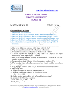
Comparative Biochemistry and Physiology, Part C 150 (2009) 377–382
Contents lists available at ScienceDirect
Comparative Biochemistry and Physiology, Part C
j o u r n a l h o m e p a g e : w w w. e l s ev i e r. c o m / l o c a t e / c b p c
Long-term ammonia toxicity to the pink-shrimp Farfantepenaeus paulensis
Kleber Campos Miranda-Filho a, Grasiela Lopes Leães Pinho b, Wilson Wasielesky Jr. b, Adalto Bianchini c,⁎
a
b
c
Programa de Pós-Graduação em Aqüicultura, Universidade Federal do Rio Grande, 96201-900, Rio Grande, RS, Brazil
Instituto de Oceanografia, Universidade Federal do Rio Grande, 96201-900, Campus Carreiros, Rio Grande, RS, Brazil
Instituto de Ciências Biológicas, Universidade Federal do Rio Grande, 96201-900, Campus Carreiros, Rio Grande, RS, Brazil
a r t i c l e
i n f o
Article history:
Received 15 March 2006
Received in revised form 29 May 2009
Accepted 5 June 2009
Available online 11 June 2009
Keywords:
Ammonia
Chronic toxicity
Farfantepenaeus paulensis
Feeding response
Growth
Metabolism
Shrimp
Predatory activity
a b s t r a c t
Juvenile pink-shrimp Farfantepenaeus paulensis were exposed (75 days) to NH3 (0.016–0.287 mg L− 1) under
static condition with water renewal every 24 h. Experiments were performed at 20 °C, at a water salinity of
15 ppt, and at pH 7.8. Endpoints analyzed were survival, growth and predation rates. After 75 days of
exposure, survival was ≥90% in all concentrations tested. However, growth (carapace length and wet body
mass) was reduced after exposure to NH3 concentrations as low as 0.033 mg L− 1, while the relative
growth (dry body mass and ash content) was reduced after exposure to the highest NH3 concentration
(0.287 mg L− 1). Predatory activity was inhibited after exposure to 0.144 or 0.287 mg L− 1 NH3. Post-larvae
exposed (75 days) to 0.301 mg L− 1 NH3 under the same experimental conditions also showed a reduced
growth (wet body mass) and relative growth (dry body mass). In addition, they showed decreased body
lipids content and increased body glycogen and glucose contents. However, no changes in body protein,
chitin and uric acid contents were observed. Also, NH3 did not affect post-larvae feeding response. Altogether,
findings suggest that F. paulensis reduces its food intake to limit the internal accumulation of nitrogenous
waste products when exposed for long time to high levels of ambient ammonia. As a consequence, shrimp
show a marked change in energy metabolism, characterized by a decreased content of body lipids paralleled
by an increased content of body carbohydrates, resulting in a significant reduction in growth.
© 2009 Published by Elsevier Inc.
1. Introduction
Ammonia is naturally excreted by most aquatic animals and arises
from the degradation of both natural and anthropogenic organic
matter entering the aquatic system. If their production exceeds the
capacity of absorption, recycling and purification of the aquatic
system, accumulation and effects on biota can be observed.
In aqueous solution, total ammonia (Tamm) comprises the highly
toxic un-ionized form (ammonia; NH3) and the generally less-toxic
ionized form (ammonium; NH+
4 ), which are in equilibrium. The
proportions of each chemical species depend mainly on pH, but also
on temperature, salinity and pressure (Whitfield, 1974; Russo, 1985).
The higher NH3 toxicity is mainly because of its high diffusion capacity
throughout biological membranes (Cooper and Plum, 1987; Barimo
and Walsh, 2005). Therefore, the environmental concentration of this
nitrogenous compound is of greatest concern because it easily diffuses
across the body surface and is accumulated. Internally, the ionized
form likely poses the greatest problem because at physiological pH
more than 95% of ammonia exists as NH+
4 .
⁎ Corresponding author. Universidade Federal do Rio Grande, Instituto de Ciências
Biológicas, Campus Carreiros, Av. Itália km 8, 96.201-900, Rio Grande, RS, Brazil.
Tel.: +55 53 3233 6853, +55 53 3233 6848; fax: +55 53 3233 6848.
E-mail address: [email protected] (A. Bianchini).
1532-0456/$ – see front matter © 2009 Published by Elsevier Inc.
doi:10.1016/j.cbpc.2009.06.001
At high environmental concentrations of ammonia, disparity exists
between the response of fish and crustaceans. Up to 300 µM, ammonia
(NH4Cl) stimulates fish growth (Wood, 2004; Barimo and Walsh,
2005). However, such levels of ammonia can be harmful to aquatic
crustaceans, including the pink-shrimp Farfantepenaeus paulensis,
causing several adverse effects such as reduced growth and survival
(Chen and Kou, 1992; Wasielesky et al., 1994; Cavalli et al., 1998; Lin
and Chen, 2001). Adverse effects are observed in both cultured
crustaceans when kept under inappropriate conditions of water
treatment or renewal (Montoya et al., 1999) and wild populations in
natural environments heavily contaminated with organic matter
(Dyer et al., 2003). Despite the bulk of data available on ammonia
effects upon growth and survival rates in crustaceans, information on
the mechanisms of ammonia toxicity remain poorly understood
(Bermudes and Ritar, 2008). Also, evidences on how crustaceans deal
with the excess of ambient or internal ammonia are scarce. Colt and
Armstrong (1981) hypothesized that animals may reduce or even stop
feeding to limit the internal accumulation of nitrogenous waste
products when exposed to high concentrations of ambient ammonia.
Regnault (1996) proposed that aquatic crabs could reduce their
internal level of ammonia storing this compound as glucosamine (or
acetylglucosamine) in tissues involved in the chitin synthesis during
air exposure. In turn, Durand et al. (1999) proposed that aquatic crabs
exposed to air could show an increased synthesis of aminoacids and
uric acid as a strategy to reduce the body levels of ammonia.
378
K.C. Miranda-Filho et al. / Comparative Biochemistry and Physiology, Part C 150 (2009) 377–382
In light of the above, the present study was performed to generate
more data for a better understanding of the long-term ammonia
toxicity, as well as of the strategies adopted by aquatic crustaceans to
deal with long-term exposure to ambient ammonia. To achieve our
goal, the long-term effects of ammonia on survival, growth, predatory
activity, feeding response, and body composition were evaluated in
F. paulensis, a native shrimp from Brazilian coastal and marine waters.
2. Material and methods
2.1. Shrimp collection, cultivation and acclimation
Wild mature F. paulensis (Pérez-Farfante, 1967) were captured
along the Atlantic coast in Southern Brazil, transferred to the
laboratory and maintained at the Aquaculture Marine Station of the
“Universidade Federal do Rio Grande” (Rio Grande, RS, Southern
Brazil). Larvae obtained in the laboratory were reared in seawater at a
salinity of 31 ppt and a temperature of 26 °C until the post-larvae and
the juvenile stages, following previously described techniques
(Marchiori and Boff, 1983; Marchiori, 1996). Juvenile shrimp were
used to evaluate the effect of different concentrations of ammonia on
survival, growth and predation rates. Based on results obtained with
juveniles, a second experiment was performed using post-larvae to
evaluate feeding response and body composition of juvenile shrimp
(4.2 ± 0.15 mm body length) after exposure to a single effective
ammonia concentration under the same experimental conditions
used in the experiments with juveniles.
Before testing, post-larvae and juvenile shrimp were gradually
acclimated to seawater at a salinity of 15 ppt and a temperature of
20 °C in plastic tanks (200 L) for at least two weeks. These conditions
approximate the mean values registered in summer in the Patos
Lagoon estuary (Baumgarten et al., 1995, 2001), where post-larvae
and juveniles of F. paulensis reside until the sub-adult stage
(Marchiori, 1996). A salinity of 15 ppt was obtained by diluting
seawater with dechlorinated tap water.
2.2. Experiment with juveniles
2.2.1. Survival and growth tests
For the respective survival and growth tests, juvenile shrimp were
maintained in 15 L and 70 L plastic tanks at a density of two shrimp
per liter. No sediment was provided to prevent the nitrification
process (Allan et al., 1990). The total number of juvenile shrimp used
was 1440. Tests were run using standard-static procedures with
experimental media being renewed every 24 h. Shrimp were fed once
a day until satiation with metanauplii and adults of Artemia sp.
Experimental media were gently and continuously aerated. Feces,
carapaces and uneaten food were daily removed by siphon and
approximately 100% of the experimental medium was gently changed.
Photoperiod was fixed at 12L:12D.
Five NH3 concentrations and a control (no ammonia addition)
were tested. Nominal concentrations tested were 0.025, 0.050, 0.100,
0.200 and 0.400 mg L− 1 NH3. They were selected to bracket the safe
level of NH3 (0.110 mg L− 1 NH3) to prevent adverse effect on growth
of F. paulensis juveniles (Ostrensky and Wasielesky, 1995). The tested
concentrations were obtained from a stock solution (10 g Tamm L− 1 as
NH4Cl, Merck, USA). Each treatment was carried out in duplicate.
In each experimental unit, water temperature and salinity
(refractometer, Digimed, São Paulo, SP, Brazil) were measured at the
beginning and at the end of the daily routine. Also, one water sample
(200 mL) from each experimental unit was collected at the beginning
and at the end of the daily routine. For each sample, water pH was
measured (pH meter, Digimed DMPH-2, São Paulo, SP, Brazil) and
raised to 10 by addition of NaOH to convert the total ammonia (Tamm)
into NH3. Measurement of the NH3 concentration was then performed
using an NH3-selective electrode (Orion, 95-12, Sergipe, SE, Brazil)
connected to a multimeter (Minipa EP-2002, São Paulo, SP, Brazil). The
NH3 concentration for each water sample was calculated according to
the methodology described by Whitfield (1974) and modified by
Ostrensky et al. (1992), considering water pH, temperature, salinity
and Tamm concentration data for the respective water sample.
The number of live and dead shrimp was recorded every 24 h.
Shrimp were considered dead when they failed to move even when
gently stimulated with a glass pipette. Dead shrimp were removed to
prevent fouling. Survival was recorded over 75 days. In fact, shorter
periods of exposure to levels of ammonia similar to those employed in
the present study were shown to induce significant effects on growth
of both post-larvae (Wasielesky et al., 1994) and adults (Cavalli et al.,
1998) of F. paulensis. Furthermore, the exposure period (75 days)
adopted in the present study corresponds to approximately 50% of the
time expended by this shrimp species in the Patos Lagoon estuary (Rio
Grande, RS, Southern Brazil) until it reaches the sub-adult stage.
Ammonia effects on growth were assessed in 10 juvenile shrimp
randomly sampled from each concentration tested at the beginning of
the experiment and every 15 days up to 75 days of exposure. The
following endpoints were considered: carapace length, wet and dry
body mass, and whole body ash, organic matter, inorganic matter, and
water content. Carapace length (mm) and wet body mass (mg) were
measured according to Neiva and Mistakidis (1966), using a caliper
and an electronic scale (Sartorius, Santo André, SP, Brazil), respectively. Shrimp were dried (48 h) in an oven (60 °C), weighed (dry
body mass), placed (2 h) in a muffle furnace (550 °C) and then
weighed (ash body content) (AOAC, 1965). Using simple proportion
calculation, it was possible to determine the percentage of organic and
inorganic contents with respect to the wet body mass using dry and
ash body content data, respectively. Whole body water content was
determined based on wet and dry body mass data.
2.2.2. Predation test
Five juvenile shrimp were randomly sampled from each experimental unit at the end of the exposure period (75 days) and kept
under the respective experimental conditions for further 24 h. During
this period, they were not fed.
To access the predatory activity, each juvenile shrimp was
individually kept in a 250-mL plastic flask containing 200 mL of the
respective experimental medium and 25 nauplii of Artemia sp. All
nauplii used in the tests were randomly collected from the same
culture batch reared in our laboratory. Juvenile shrimp were allowed
to predate for 1 h and then removed from the test flask. The number of
nauplii remaining in the test flask was counted and the predatory
activity expressed as the number of Artemia consumed per shrimp in
1 h. As five juvenile shrimp were collected from each of the two
replicates of the survival and growth tests, the predatory activity was
individually measured in ten shrimp for each experimental condition.
2.3. Experiment with post-larvae
2.3.1. Feeding response test
Based on the results obtained in the experiment with juveniles,
fifteen post-larvae were kept under control conditions (without
addition of ammonia to the water) or exposed to a single ammonia
concentration (nominal = 0.400 mg L− 1 NH3) for 75 days, as described for the experiment with juveniles. After the exposure period,
surviving shrimp were individually tested for the feeding response
induced by L-isoleucine, following the same procedures previously
described for F. paulensis (dos Santos Fo, 1983; Santos et al., 2000).
Feeding response data was expressed in percentage of positive
responses to the inducer. Tests were run in duplicate.
After the feeding response test, five shrimp from each replicate
(n = 10 per experimental condition) were randomly collected and
individually blotted dried in paper filter, weighed (dry and wet mass;
mg) and frozen (− 20 °C) until analysis of body composition.
K.C. Miranda-Filho et al. / Comparative Biochemistry and Physiology, Part C 150 (2009) 377–382
2.3.2. Body composition analysis
Whole shrimp were thawed, homogeneized in distilled water and
centrifuged (10,000 g; 10 min; 4 °C). For each sample, both supernatant and pellet were collected and separately stored in 1.5 mL
microcentrifuge tubes. Supernatant was used for determination of
whole body total proteins, uric acid, total lipids, glycogen, glucose, and
chitin concentrations. Pellet was used only for chitin determination.
All measurements were done using spectrophotometric techniques.
Total proteins were measured using a commercial reagent kit
based on the Biuret method (Proteínas Totais®, Doles Reagentes,
Goiânia, GO, Brazil). Uric acid concentration was determined using a
commercial reagent kit (Ácido úrico®, Labtest Diagnóstica, Lagoa
Santa, MG, Brazil) based on the uricase and peroxidase enzymatic
assay. Total lipids were measured using a commercial reagent kit
(Bioclim, Quibasa Quimica Básica, Belo Horizonte, MG, Brazil) and
following the procedures described by Schmitt and Santos (1993).
Glycogen and glucose concentrations were determined using a
commercial reagent kit (Glicox®, Doles Reagentes, Goiânia, GO,
Brazil) based on the glucose oxidase method and following the
procedures described by Nery and Santos (1993). Chitin concentration
in both pellet and supernatant was measured following the procedures described by Tsuji et al. (1969) and using D-glucosamine
hydrochloride (Sigma-Aldrich, St. Louis, MO, USA) as standard. Results
from both pellet and supernatant were combined. All results were
expressed in mg g− 1 wet mass.
379
Table 2
Biological endpoints measured in Farfantepenaeus paulensis juveniles kept under
control conditions for up to 60 days.
Time
(days)
Biological endpoints
Carapace length
(mm)
Wet body mass
(mg)
Dry body mass
(mg)
Ash body content
(mg)
0
15
30
45
60
6.4 ± 0.13a
7.1 ± 0.13b
8.1 ± 0.22c
9.3 ± 0.22d
10.0 ± 0.10e
158.0 ± 7.51a
218.3 ± 12.35b
342.2 ± 28.92c
536.0 ± 40.63d
638.2 ± 39.12e
39.5 ± 2.19a
53.5 ± 3.22b
85.4 ± 7.27c
140.7 ± 11.52d
166.9 ± 11.83e
6.3 ± 0.57a
8.3 ± 0.41b
13.3 ± 1.09c
20.4 ± 1.64d
23.7 ± 1.67e
Data are mean values ± SE (N = 10). Different letters indicate significant (P b 0.05)
different mean values.
ments at the same exposure time (data not shown). Therefore, only
one mean value of water temperature (19.8 ± 0.08 °C; N = 450) and
water pH (7.84 ± 0.006; N = 450) was calculated for the whole period
of exposure.
For NH3 concentration, a daily mean value was calculated based on
the data obtained for the two water samples collected for each
replicate, i.e., one at the beginning and one at the end of the daily
routine. Based on these values, no difference was observed between
replicates (data not shown). Therefore, only one mean value was
calculated for each exposure time and experimental condition over
the time of exposure. Since no difference was observed between
daily mean values, an overall mean value was calculated for each
2.4. Data analysis
Data for water chemistry and biological endpoints were expressed
as mean ± SE, except for the feeding response data that were expressed
in percentage. For data from the experiment with juveniles, one-way
analysis of variance (ANOVA) followed by the Duncan's test was used
to detect possible significant differences among treatments for each
time of exposure. Predatory activity data were log-transformed to
meet the ANOVA assumptions for data normality and homogeneity
of variances (Zar, 1999). For data from the experiment with postlarvae, mean values were compared using the Student t test for
independent samples, except for the feeding response data that were
analyzed using the Chi-square test. All statistical analyses were performed using Statistica software (StatSoft, Inc., Tulsa, OK, USA). In all
cases, significance level adopted was 95% (α = 0.05).
3. Results
3.1. Experiment with juveniles
For the same time of exposure, no variation in water temperature
and pH was observed in each experimental unit at the beginning and
the end of the daily routine. Also, no difference was observed between
the two replicates for each experimental condition. Therefore, only
one daily mean value was calculated for each experimental condition.
These mean values were compared over the exposure period
(75 days). No difference was observed between the different times
of exposure for the same experimental condition or between treat-
Table 1
Nominal and measured NH3 concentrations for the different treatments tested.
Nominal concentration
Mean measured concentration
0 = control
0.025
0.050
0.100
0.200
0.400
0.016 ± 0.0005a
0.033 ± 0.0010b
0.047 ± 0.0015c
0.078 ± 0.0028d
0.144 ± 0.0045e
0.287 ± 0.0088f
Data are expressed as mean ± SE. Values are expressed in mg L− 1 NH3. Different letters
indicate significant (P b 0.05) different mean values.
Fig. 1. Biological endpoints measured in Farfantepenaeus paulensis juveniles after
75 days of exposure to different NH3 concentrations. (A) Carapace length (●; left axis)
and wet body mass (○; right axis). (B) Dry body mass (●; left axis) and ash body
content (○; right axis). Data are expressed as mean ± SE (N = 10). Different letters
indicate significant different (P b 0.05) mean values between treatments for each
endpoint analyzed.
380
K.C. Miranda-Filho et al. / Comparative Biochemistry and Physiology, Part C 150 (2009) 377–382
Fig. 2. Predatory activity of Farfantepenaeus paulensis juveniles after 75 days of
exposure to different NH3 concentrations. Data are expressed as mean ± SE (N = 10).
Different letters indicate significant different (P b 0.05) mean values between
treatments.
experimental condition considering the whole period of exposure
(N = 75). The actual mean measured concentrations were deviated by
6 to 32% of the nominal concentrations. The mean NH3 concentration
in the control treatment was 0.016 mg L− 1 NH3 (Table 1).
In all treatments, mortality was negligible (≤10%) after 75 days of
exposure. Growth of control shrimp over the experiment corresponded to 82.8% and 585.4% when measured as carapace length and
wet body mass, respectively.
There were no differences between treatments for any biological
endpoint measured prior to day 75, including at day 0, i.e., at the
beginning of the experiment (data not shown). The mean value for
control shrimp at each exposure time is shown in Table 2. However, a
growth inhibition (carapace length and wet body mass) was observed
in shrimp exposed to NH3 for 75 days, with respect to those kept
under control conditions. Mean carapace length was shorter by 6.8,
6.0, 6.8 and 12.0% at 0.047, 0.078, 0.144 and 0.287 mg L− 1 NH3, respectively. Mean wet body mass was lower by 15.8, 15.4, 11.0, 19.0 and
29.3% at 0.033, 0.047, 0.078, 0.144 and 0.287 mg L− 1 NH3, respectively
(Fig. 1A).
Regarding relative growth, measured as dry body mass or ash body
content, a difference was detected only for shrimp exposed for 75 days
to the highest ammonia concentration tested (0.287 mg L− 1 NH3).
Mean dry body mass and ash content were lower by 31.3% and 27.9%,
respectively (Fig. 1B). In contrast, no NH3 effects on whole body water,
organic and inorganic contents were observed, even after 75 days of
Fig. 4. Body content of lipids, glycogen and glucose in Farfantepenaeus paulensis postlarvae exposed to NH3 (0.301 mg L− 1 NH3) for 75 days. Data are expressed as mean ±
SE (N = 10). ⁎ indicates significant different (P b 0.05) mean values between treatments
for each parameter analyzed.
exposure to NH3 concentrations as high as 0.287 mg L− 1 (data not
shown). The overall mean (±SE) percentage values for control shrimp
were 85.1% (±0.12), 75.6% (±0.12) and 14.9% (±0.11), respectively.
The predatory activity of F. paulensis juveniles was not affected
until after 60 days of NH3 exposure. For control shrimp, the mean
number (±SE) of Artemia predated per hour after 15, 30, 45, and
60 days of NH3 exposure was 0.78 (±0.35), 2.36 (±0.61), 3.74
(±0.67), and 3.99 (±0.89), respectively. However, an inhibition of the
predatory activity was observed after 75 days of exposure to 0.144 or
0.287 mg L− 1 NH3 (Fig. 2).
3.2. Experiment with post-larvae
Based on the results obtained with juveniles, post-larvae were kept
under control conditions or exposed to the highest ammonia
concentration tested in the first experiment (0.4 mg L− 1 NH3). As
described for the experiment with juveniles, only one mean value of
water temperature (20.1 ± 0.07 °C; N = 150) and water pH (7.80 ±
0.01; N = 100) was calculated for the whole period of exposure. NH3
concentration was determined as described for the experiment
with juveniles. Also, an overall mean value was calculated for
the control (0.012 ± 0.002 mg L− 1 NH3) and experimental (0.301 ±
0.0077 mg L− 1 NH3) conditions considering the whole period of exposure (N = 75). The actual mean measured concentration deviated
by 25% of the nominal concentration tested.
After 75 days of test, mortality corresponded to 13.3 and 26.7%
in control and ammonia-exposed shrimp, respectively. Wet and dry body
mass of ammonia-exposed shrimp (456±36 and 120±24 mg, respectively) was lower than that of control shrimp (613±44 and 157±22 mg,
respectively). A 25.6 and 23.6%-reduced growth was observed,
respectively.
Positive feeding response induced by L-isoleucine was not different
in control (65.4 ± 11.6%) and ammonia-exposed shrimp (63.6 ± 9.1%).
Also, no difference was observed in the body content of proteins,
chitin and uric acid between control and ammonia-exposed shrimp
(Fig. 3). However, a lower body content of lipids (decrease of 46.1%)
and higher body contents of glycogen (increase of 53.0%) and glucose
(increase of 70.5%) were observed in ammonia-exposed shrimp when
compared to the levels found in control shrimp (Fig. 4).
4. Discussion
Fig. 3. Body content of proteins, chitin and uric acid in Farfantepenaeus paulensis postlarvae exposed to NH3 (0.301 mg L− 1 NH3) for 75 days. Data are expressed as mean ±
SE (N = 10). ⁎ indicates significant different (P b 0.05) mean values between treatments
for each parameter analyzed.
Data on acute toxicity and effects of ammonia on shrimp kept
under laboratory conditions are largely available in the literature.
However, long-term ammonia toxicity is not so well documented. As
K.C. Miranda-Filho et al. / Comparative Biochemistry and Physiology, Part C 150 (2009) 377–382
discussed below, findings from the present study help to better
understand the mechanisms involved in long-term ammonia toxicity,
as well as the possible strategies adopted by shrimp to deal with the
high levels of ambient ammonia.
The mean NH3 concentrations observed in the control treatments
in the present study (0.016 and 0.012 mg L− 1 NH3 with juveniles
and post-larvae, respectively) is similar to that found in summer
(0.012 mg L− 1 NH3 = 0.61 mg L− 1 NH+
4 at the water temperature,
salinity and pH employed in the present study) in areas non impacted
by organic matter in the Patos Lagoon estuary (Baumgarten et al.,
1995; Niencheski et al., 2004). This finding indicates that control
conditions adopted in laboratory are quite similar to those found in
summer at the Patos Lagoon estuary.
Mean NH3 concentrations measured in the different treatments
ranged from 0.033 to 0.301 mg L− 1 NH3, deviating 6 to 32% from the
nominal concentrations tested. These deviations could be related to
water pH variation (Whitfield, 1974; Erickson, 1985; Ostrensky et al.,
1992), ammonia stability in aqueous environment (Ostrensky, 1991),
and changes in the excretory rate of shrimps exposed to higher
ammonia concentrations (Wickins, 1976; Chen et al., 1993).
The low mortality observed in F. paulensis juveniles in the present
study appears to be a normal response of shrimps from the Penaeidae
family to ammonia exposure (Wasielesky et al., 1994; Cavalli et al.,
1998; Lin and Chen, 2001). In turn, the higher mortality rate observed
in post-larvae exposed to ammonia is in agreement with previous data
showing a higher ammonia sensitivity of post-larvae compared to
juvenile F. paulensis (Ostrensky et al., 1992; Ostrensky and Wasielesky,
1995).
Regarding growth, data from the present study demonstrated that
the long-term ammonia exposure inhibited the shrimp growth
expressed as either wet weight gain or carapace length increase in
both post-larvae and juveniles. It is interesting to note that the effect
on wet weight was more marked than on carapace length in juveniles.
A similar response was observed in F. japonicus juveniles exposed to
ammonia (Chen and Kou, 1992). These findings suggest that ammonia
has a higher inhibition effect on physiological processes involved in
body mass increase than on those related to molting. This could be
related to the infrequent molting of the exoskeleton versus the
continual accumulation of wet body mass.
The lack of change in water, organic and inorganic content in
juvenile shrimp exposed to NH3 for 75 days indicates that shrimp
growth inhibition would be related to a general effect of ammonia on
shrimp energy metabolism. In fact, NH3 has shown to have a negative
effect on the oxidative metabolism (Lehninger, 1990). The observed
change in body composition of F. paulensis post-larvae after long-term
exposure to a high concentration of ambient ammonia (0.301 mg L− 1
NH3) corroborates this idea. This change was characterized by a
marked reduction in body lipid content paralleled by a significant
increase in body carbohydrate (glycogen and glucose) content. This
finding suggests a higher use of carbohydrates as energy source to
cope with the stress induced by the long-term NH3 exposure. In this
case, the increased carbohydrate levels would be explained by a
higher gluconeogenesis rate, having glycerol from lipid oxidation as
substrate for glucose synthesis.
In crustaceans, it is reported that exposure to high levels of
ambient NH3 affects osmoregulation (Lin et al., 1993), inducing to an
increased energy expenditure associated with processes involved in
ionic and osmotic regulations (Spaargaren, 1982; Young-Lai et al.,
1991; Chen and Cheng, 1993b,c; Chen et al., 1993). However, NH3
exposure did not seem to affect osmoregulation in F. paulensis. This
statement is based on the fact that NH3 did not significantly affect
water and inorganic contents in juvenile shrimp. This lack of NH3
effect on osmoregulation could be related to the fact that experiments
were performed on shrimp acclimated to water at salinity 15 ppt, a
condition where F. paulensis juvenile is osmoconforming its body
fluids (Wasielesky et al., 1995).
381
Also, the total amount of energy available for shrimp growth
exposed to ammonia could be decreased due to an increased
expenditure associated with a possible switch from ammoniotelic to
uricotelic pattern as a mechanism of ammonia detoxification (Chen
and Cheng, 1993a,b,c; Chen et al., 1994; Chen and Lin, 1995). However, the strategy to convert ammonia into uric acid or aminoacids
to reduce the internal levels of ammonia, as proposed by Durand
et al. (1999), seems not to be used by F. paulensis. This statement
is based on the fact that body contents of uric acid and proteins were
not significantly altered in post-larvae exposed to a high concentration of NH3 ammonia for 75 days. The strategy to reduce internal
levels of ammonia by storing this nitrogen compound as glucosamine or acetylglucosamine (Regnault, 1996), the substrate for chitin
synthesis, also seems not to be used by F. paulensis. In fact, no
change in the body content of chitin was observed in post-larvae
exposed to NH3.
In addition to a possible increased amount of energy required to
cope with the stress induced by chronic exposure to ambient NH3, the
total amount of energy available for shrimp growth could be also
decreased as a result of the negative effect of NH3 on food intake.
Short-term exposure (15 days) to up ∼0.15 mg L− 1 NH3 was reported
to have no significant effect on food intake in F. paulensis (Wasielesky
et al., 2003). However, data on predatory activity of juveniles in
the present study showed that long-term exposure (75 days) to
0.144 mg L− 1 NH3 or a higher concentration induced a significant
decrease in food intake, leading to a likely reduced amount of energy
available for the whole shrimp metabolism. At this point, one should
assume that the reduced predatory activity could be an indirect effect
of NH3 associated with its negative impact on juvenile shrimp growth.
In fact, a significant positive relationship between carapace length
(y = 2.21x − 15.93; R2 = 0.86) or wet body weight (y = 0.0123x −
2.4375; R2 = 0.95) and predatory activity was observed in control
juvenile shrimp. However, the negative effect of NH3 on growth of
juveniles only explains ∼ 50% of the reduction in predatory activity of
shrimp exposed for 75 days to the highest NH3 concentration tested
(0.287 mg L− 1 NH3). Therefore, other causes must be implicated in
the observed response.
Data from the experiment with post-larvae indicate a lack of a
direct and negative effect of NH3 on shrimp ability to sense and locate
the presence of food in the water. This statement is based on the fact
that the feeding response of post-larvae to the major inducer of
feeding in F. paulensis, i.e. the aminoacid L-isoleucine (dos Santos Fo,
1983), was not modified by long-term exposure to a high ambient NH3
concentration (0.301 mg L− 1). Unfortunately, data generated does not
allow us to evaluate a possible negative NH3 effect on the ability of
shrimps to move and capture the prey.
In light of the above, the observed long-term effects of NH3 on F.
paulensis growth could be explained only by considering the reduced
food intake, a response of shrimp to limit the internal accumulation of
nitrogenous waste products when exposed to high levels of ambient
ammonia, as early proposed by Colt and Armstrong (1981). This
hypothesis would be in line with the possible occurrence of an
alkaline tide followed by a higher rate of ammonia excretion in
crustaceans, as described for fish after voluntary feeding (Bucking and
Wood, 2008). Unfortunately, the occurrence of this phenomenon has
never been evaluated in crustaceans. Therefore, future experiments
need to be performed to confirm the proposed hypothesis.
Acknowledgments
The authors would like to thank Dr. Ronaldo O. Cavalli, Dr. Márcio
R. Milani and Nérile Troca da Cunha from Universidade Federal do Rio
Grande (Rio Grande, RS, Brazil) for their assistance during this work.
The authors gratefully acknowledge the financial support from the
Brazilian National Research Council (CNPq). Adalto Bianchini is a
research fellow from the Brazilian CNPq (# 300906/2006-4).
382
K.C. Miranda-Filho et al. / Comparative Biochemistry and Physiology, Part C 150 (2009) 377–382
References
Allan, G.L., Maguire, G.B., Hopkins, S.J., 1990. Acute and chronic toxicity of ammonia to
juvenile Metapenaeus macleayi and Penaeus monodon and the influence of low
dissolved-oxygen levels. Aquaculture 91, 265–280.
AOAC, 1965. Official Methods of Analysis of the Association of Official Agricultural
Chemists. AOAC, Washington, DC.
Barimo, J.F., Walsh, P.J., 2005. The effects of acute and chronic ammonia exposure during
early life stages of the gulf toadfish, Opsanus beta. Aquat. Toxicol. 75, 225–237.
Baumgarten, M.G.Z., Niencheski, L.F.H., Kuroshima, K.N., 1995. Qualidade das águas
estuarinas que margeiam o município do Rio Grande (RS, Brasil): nutrientes e
detergentes dissolvidos. Atlântica 17, 17–34.
Baumgarten, M.G.Z., Nienchesky, L.F.H., Veek, L., 2001. Nutrientes na coluna da água e na
água intersticial de sedimentos de uma enseada rasa estuarina com aportes de
origem antrópica (RS—Brasil). Atlântica 23, 101–116.
Bermudes, M., Ritar, A.J., 2008. Tolerance for ammonia in early stage spiny lobster (Jasus
edwardsii) phyllosoma larvae. J. Crustac. Biol. 28, 695–699.
Bucking, C., Wood, C.M., 2008. The alkaline tide and ammonia excretion after voluntary
feeding in freshwater rainbow trout. J. Exp. Biol. 211, 2533–2541.
Cavalli, R.O., Peixoto, S., Wasielesky, W.J., 1998. Performance of Farfantepenaeus
paulensis (Pérez-Farfante) broodstock under long-term exposure to ammonia.
Aquac. Res. 29, 815–822.
Chen, J.C., Cheng, S.Y., 1993a. Urea excretion by Penaeus japonicus Bate exposed to
different concentrations of ambient ammonia. J. Exp. Mar. Biol. Ecol. 173, 1–9.
Chen, J.C., Cheng, S.Y., 1993b. Hemolymph PCO2, hemocianin, protein levels and urea
excretions of Penaeus monodon exposed to ambient ammonia. Aquat. Toxicol. 27,
281–292.
Chen, J.C., Cheng, S.Y., 1993c. Hemolymph osmolality, acid–base balance and shift of
ammoniotelic to ureotelic excretory pattern of Penaeus japonicus exposed to
ambient ammonia. Comp. Biochem. Physiol. 106, 733–737.
Chen, J.C., Kou, Y.Z., 1992. Effects of ammonia on growth and molting of Penaeus
japonicus juveniles. Aquaculture 104, 249–260.
Chen, J.C., Lin, C.Y., 1995. Responses of oxygen consumption, ammonia-N excretion and
urea-N excretion of Penaeus chinensis exposed to ambient ammonia at different
salinity and pH levels. Aquaculture 136, 243–255.
Chen, J.C., Nan, F.H., Cheng, S.Y., Sheen, S.S., 1993. Effects of ambient ammonia on
ammonia-N and protein concentration of haemolymph and ammonia-N excretion
of Penaeus chinensis. Mar. Ecol. Prog. Ser. 98, 203–208.
Chen, J.C., Chen, C.T., Cheng, S.Y., 1994. Nitrogen excretion and changes of hemocyanin,
protein and free amino acid levels in the hemolymph of Penaeus monodon exposed
to different concentrations of ambient ammonia-N at different salinity levels. Mar.
Ecol. Prog. Ser. 110, 85–94.
Colt, J.E., Armstrong, D.A., 1981. Nitrogen toxicity to crustaceans, fish, and molluscs. In:
Allen, L.J., Kinney, E.C. (Eds.), Proceedings of the Bio-Engineering Symposium for
Fish Culture. American Fisheries Society, Bethesda, pp. 34–47.
Cooper, A.J.L., Plum, F., 1987. Biochemistry and physiology of brain ammonia. Physiol.
Rev. 67, 440–519.
dos Santos Fo, E.A., 1983. The inducer of the feeding response in Penaeus paulensis
(Crustacea–Decapoda). Physiol. Behav. 31, 733–734.
Durand, F., Chausson, F., Regnault, M., 1999. Increases in tissue free amino acid levels in
response to prolonged emersion in marine crabs: an ammonia-detoxifying process
efficient in the intertidal Carcinus maenas but not in the subtidal Necora puber.
J. Exp. Biol. 202, 2191–2202.
Dyer, S.D., Peng, C., McAvoy, D.C., Fendinger, N.J., Masscheleyn, P., Castillo, L.V., Lim, J.M.,
2003. The influence of untreated wastewater to aquatic communities in the
Balatuin River, The Philippines. Chemosphere 52, 43–53.
Erickson, R.J., 1985. An evaluation of mathematical models for the effects of pH and
temperature on ammonia toxicity to aquatic organisms. Water Res. 19, 1047–1058.
Lehninger, A.L., 1990. Princípios de Bioquímica. Sarvier, São Paulo, SP, Brasil.
Lin, Y.C., Chen, J.C., 2001. Acute toxicity of ammonia on Litopenaeus vannamei Boone
juveniles at different salinity levels. J. Exp. Mar. Biol. Ecol. 259, 109–119.
Lin, H.-P., Thuet, P., Trilles, J.-P., Mounet-Guillaume, R., Charmantier, G., 1993. Effects of
ammonia on survival and osmoregulation of various development stages of the
shrimp Penaeus japonicus. Mar. Biol. 117, 591–598.
Marchiori, M.A., 1996. Guia ilustrado de maturação e larvicultura do camarão-rosa Penaeus paulensis, Pérez-Farfante, 1967. Editora da FURG, Rio Grande, RS, Brasil.
Marchiori, M.A., Boff, M.H., 1983. Induced maturation, spawning and larvae culture of
the pink shrimp Penaeus paulensis, Pérez-Farfante, 1967. Mems. Assoc. Latinoam.
Aquicult. 5, 331–337.
Montoya, R.A., Lawrence, A.L., Grant, W.E., Velaso, M., 1999. Simulation of nitrogen
dynamics and shrimp growth in an intensive shrimp culture system: effects of feed
and feeding parameters. Ecol. Model. 122, 81–95.
Neiva, G.S., Mistakidis, M., 1966. Identificación de algunos camarones marinos del litoral
centro-sur del Brasil. Doc. Técn. (CARPAS, Rio de Janeiro 4, 1–6.
Nery, L.E.M., Santos, E.A., 1993. Carbohydrate metabolism during osmoregulation in
Chasmagnatus granulata Dana, 1851 (Crustacea, Decapoda). Comp. Biochem.
Physiol. 106B, 747–753.
Niencheski, L.F.H., Baumgarten, M.G.Z., Cabrera, L., Juliano, S.K., 2004. Patos Lagoon:
indicators of organic pollution. J. Coast. Res. SI 39, 1356–1359.
Ostrensky, A., 1991. Toxicidade da amônia e do nitrito no processo produtivo de póslarvas do camarão-rosa, Penaeus paulensis Pérez-Farfante, 1967. MSc Thesis,
Universidade Federal do Paraná, Curitiba, PR, Brasil.
Ostrensky, A., Wasielesky, W.J., 1995. Acute toxicity of ammonia to various life stages of
the São Paulo shrimp, Penaeus paulensis Pérez-Farfante, 1967. Aquaculture 132,
339–347.
Ostrensky, A., Marchiori, M.A., Poersh, L.H., 1992. Toxicidade aguda da amônia no
processo produtivo de pós-larvas de Penaeus paulensis Pérez-Farfante, 1967. An.
Acad. Bras. Cienc. 64, 383–389.
Regnault, M., 1996. Air exposure-increase in acetylglucosamine content of some soft
tissues and haemolympf of a crab (Cancer pagurus). J. Exp. Zool. 275, 421–430.
Russo, R.C., 1985. Ammonia, nitrite and nitrate. In: Rand, G.M., Petrocelli, S.R. (Eds.),
Fundamentals of Aquatic Toxicology. Taylor & Francis, USA.
Santos, M.H.S., da Cunha, N.T., Bianchini, A., 2000. Effects of copper and zinc on growth,
feeding and oxygen consumption of Farfantepenaeus paulensis postlarvae (Decapoda: Penaeidae). J. Exp. Mar. Biol. Ecol. 247, 233–242.
Schmitt, A.S.C., Santos, E.A., 1993. Lipid and carbohydrate metabolism of the intertidal
crab Chasmagnathus granulata Dana, 1851 (Crustacea–Decapoda) during emersion.
Comp. Biochem. Physiol. 106A, 329–336.
Spaargaren, D.H., 1982. The ammonium excretion of the shore crab, Carcinus maenas, in
relation to environmental osmotic conditions. Netherlands J. Sea Res. 15, 273–283.
Tsuji, A., Kinoshita, T., Hoshino, M., 1969. Analytical chemical studies on amino sugars. II.
Determination of hexosamines using 3-methyl-2-benzothiazolone hydrazone
hydrochloride. Chem. Pharm. Bull. 17, 1505–1510.
Wasielesky, W.J., Marchiori, M.A., Santos, M.H.S., 1994. Efeito da amônia no crescimento
de pós-larvas do camarão-rosa Penaeus paulensis, Pérez-Farfante, 1967 (Decapoda:
Penaeidea). Nauplius 2, 99–105.
Wasielesky, W.J., Cavalli, R.O., Luvizotto-Santos, R., Bianchini, A., 1995. Determinação do
ponto isosmótico do camarão rosa Penaeus paulensis (Crustacea – Decapoda. XXIV
Encontro Anual de Ciências Fisiológicas, Resumos. Rio Grande (RS, Brazil, p. 65.
Wasielesky, W.J., Bianchini, A., Sanchez, C.C., Poersch, L.H., 2003. The effect of
temperature, salinity and nitrogen products on food consumption of pink shrimp
Farfantepenaeus paulensis. Braz. Arch. Biol. Technol. 46, 135–141.
Whitfield, M., 1974. The hydrolysis of ammonium ions in sea water — a theoretical study.
J. Mar. Biol. Assoc. U.K. 54, 565–580.
Wickins, J.F., 1976. Prawn biology and culture. Oceanogr. Mar. Biol. Ann. Rev. 14, 435–507.
Wood, C.M., 2004. Dogmas and controversies in the handling of nitrogenous wastes: is
exogenous ammonia a growth stimulant in fish? J. Exp. Biol. 207, 2043–2054.
Young-Lai, W.W., Charmantier-Daures, M., Charmantier, G., 1991. Effect of ammonia on
survival and osmoregulation in different life stages of the lobster Homarus
americanus. Mar. Biol. 110, 293–300.
Zar, J.H., 1999. Biostatistical Analysis, 4th Ed. Prentice Hall, New Jersey, USA.


© Copyright 2026