
- Wiley Online Library
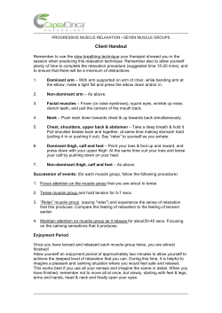
ORIGINAL ARTICLE
Journal of Cachexia, Sarcopenia and Muscle (2015)
Published online in Wiley Online Library (wileyonlinelibrary.com) DOI: 10.1002/jcsm.12045
p62/SQSTM1 but not LC3 is accumulated in sarcopenic
muscle of mice
Kunihiro Sakuma1*, Masakazu Kinoshita1, Yoshinori Ito1, Miki Aizawa1, Wataru Aoi2 & Akihiko Yamaguchi3
1
Research Center for Physical Fitness, Sports and Health, Toyohashi University of Technology, Toyohashi, 1-1 Hibarigaoka, Tenpaku-cho 441-8580, Japan; 2Laboratory of
Health Science, Graduate School of Life and Environmental Sciences, Kyoto Prefectural University, Kyoto, Japan; 3Department of Physical Therapy, Health Sciences University
of Hokkaido, Kanazawa, Ishikari-Tobetsu, Hokkaido, Japan
Abstract
Aim
We investigated the pathway of autophagy signaling linked to sarcopenia of mice.
Methods Young adult (3-month) and aged (24- month) C57BL/6J mice were used. Using real-time PCR, Western blotting, and
immunohistochemical microscopy, we evaluated the amounts of p62/SQSTM1, LC3, and Beclin-1 in the quadriceps muscle
change with aging in mice.
Results Marked fiber atrophy (30%) and many fibers with central nuclei were observed in the aged mice. Western blotting
using homogenate of the cytosolic fraction clearly showed that the amounts of p62/SQSTM1 and Beclin-1 proteins were
significantly increased in the aged skeletal muscle. The amounts of these proteins in both nuclear and membrane fractions
did not change significantly with age. Immunofluorescence labeling indicated that aged mice more frequently possessed
p62/SQSTM1-positive fibers in the cytosol in quadriceps muscle than young ones (aged: 14% vs. young: 1%). In aged muscle,
p62/SQSTM1-positive fibers were significantly smaller than the surrounding p62/SQSTM1-negative fibers. Aging did not elicit
significant changes in the mRNA levels of p62/SQSTM1 and Beclin-1, but decreased LC3 mRNA level. In aged muscle, the
location of p62/SQSTM1 immunoreactivity was similar to that of Beclin-1 protein, but not LC3.
Conclusion
Keywords
Sarcopenia in mice appears to include a marked defect of autophagy signaling.
Sarcopenia; p62/SQSTM1; LC3, Beclin-1; Skeletal muscle; Atrophy
Received: 4 November 2014; Revised: 30 March 2015; Accepted: 23 April 2015
*Correspondence to: Kunihiro Sakuma, Research Center for Physical Fitness, Sports and Health, Toyohashi University of Technology, 1-1 Hibarigaoka, Tenpaku-cho, Toyohashi
441-8580: Tel: 81-532-44-6630, Fax: 81-532-44-6947 Japan, Email: [email protected]
Introduction
Sarcopenia is a term that refers to a loss of skeletal muscle mass
associated with a decrease in muscle strength and increased fatigability. Sarcopenia is the common denominator of the ageing
process, responsible for a general and substantial decline in
physical performance, which leads ultimately to physical disability. The aetiology of sarcopenia is complex and characterized by
the contribution of multiple factors, such as physical activity,
nutritional intake, oxidative stress, and hormonal changes.1–3
Age-related muscle loss is a result of reductions in the size
and number of muscle fibres possibly due to a prominent role
of accelerated apoptosis.4 A most important factor in the
development of muscle loss is the imbalance between protein
synthesis and protein degradation.5 Skeletal muscle contains
various proteolytic systems regulated by lysosome, calpain,
and proteasome. Although evidence suggests that lysosomal
degradation contributes to accelerated muscle proteolysis,
such as in muscle disuse and denervation, the major pathway
responsible for the degradation of contractile protein in skeletal muscle is orchestrated through the ubiquitin-proteasome
system (UPS).5,6 In a variety of conditions, such as cancer, diabetes, denervation, uremia, sepsis, disuse, and fasting, skeletal
muscle undergoes atrophy through the degradation of myofibrillar proteins via the UPS.7–9 Recent advances assert that
muscle atrophy in these conditions shares a common mechanism in the induction of the muscle-specific E3 ubiquitin ligase
atrophy gene-1 (atrogin-1)/muscle atrophy F-box and muscle
ring-finger protein 1 (MuRF1).6,8,10,11 Therefore, it is believed
that these atrogenes also contribute to the fibre degradation
© 2015 The Authors. Journal of Cachexia, Sarcopenia and Muscle published by John Wiley & Sons Ltd on behalf of the Society of Sarcopenia, Cachexia and Wasting Disorders
This is an open access article under the terms of the Creative Commons Attribution-NonCommercial-NoDerivs License, which permits use and distribution in any medium,
provided the original work is properly cited, the use is non-commercial and no modifications or adaptations are made.
2
under normal ageing, in spite of the lack of direct evidence
demonstrating this.12–14
There are contradictory data and varying interpretations in
the literature on the effect of ageing on proteasome-mediated
protein degradation in skeletal muscle. Attaix, Combaret, and
colleagues concluded in their reviews of the literature that it currently remains unclear where proteasome activity and proteasome function are increased or decreased in aged skeletal
muscle.5,7 Interestingly, recent findings indicate that atrogin1-knockout mice are short-lived and experience higher loss of
muscle mass during ageing than control mice,15 indicating that
the activity of E3 ubiquitin ligase is required to preserve muscle
mass during ageing in mice. As indicated by Sandri et al.,15
chronic inhibition of these atrogenes should not be considered
a therapeutic target to counteract sarcopenia because this does
not prevent muscle loss but instead exacerbates weakness.
Moreover, MuRF1 null mice experience higher decay of muscle
strength during ageing than controls, although muscle mass is at
least in part preserved in these mice.16 Collectively, these findings
seem to indicate that the induction of UPS does not occur in
mammalian sarcopenic muscle, and the attenuation of UPS by
several interventions is not effective for preventing this symptom.
Autophagy is a vital cellular process by which intracellular
components are degraded within lysosomes.17,18 Three major
mechanisms of autophagy have been described as follows: (i)
microautophagy, in which lysosomes directly take up cytosol,
inclusions, and organelles for degradation; (ii) chaperonemediated autophagy, in which soluble proteins with a particular
pentapeptide motif are recognized and transported across the
lysosomal membrane for degradation; and (iii) macroautophagy,
in which a portion of cytoplasm, including subcellular organelles,
is sequestered within a double membrane-bound vacuole that
ultimately fuses with a lysosome.19
A decline in autophagy during normal ageing has been
described for invertebrates and higher organisms.20 Inefficient
autophagy has been attributed a major role in the apparent
age-related accumulation of damaged cellular components,
such as undegradable lysosome-bound lipofuscin, protein
aggregates, and damaged mitochondria.21 Interestingly, the degree of age-related changes in autophagy appears to be organspecific. Some researchers demonstrated the upregulation of
LC3-II (active form) and p62/SQSTM1 in the sarcopenic muscles
of mice.22 Demontis and Perrimon23 showed that the function
of the autophagy/lysosome system of protein degradation declined during ageing in the skeletal muscle of Drosophila. This
results in the progressive accumulation of polyubiquitin and
p62/SQSTM1 protein aggregates in senescent Drosophila muscle. Importantly, to the best of our knowledge, changes of autophagy with age have been examined in mammalian skeletal
muscle only by biochemical approaches (western blot using
crude homogenates) but not morphological (e.g. immunofluorescence) ones. Therefore, it has remained to be elucidated
where autophagy-linked molecules are upregulated (i.e. cytosol, nucleus, or cell membrane) in sarcopenic muscle.
K. Sakuma et al.
The purpose of this study was to determine whether
autophagy-linked molecules are upregulated in sarcopenic
mouse muscle and where they are localized in muscle fibres.
Materials and methods
Experimental animals
Young adult (3 months; n = 10) and aged (24 months; n = 10)
C57BL/6J mice were used in this study. The animals were
housed in a temperature (22 ± 2°C) and humidity (60 ± 5%)controlled room regulated to provide alternating 12-h
periods of light and darkness and were allowed to feed (commercial chow) and drink ad libitum. Using excess pentobarbital, mice were killed and the quadriceps and triceps brachii
muscles dissected. The muscles were quickly weighed, frozen
in liquid nitrogen, and then stored at 80°C. This experimental procedure was approved by the Committee for Animal
Research in Toyohashi University of Technology.
Primary antibodies
The antibodies employed in the present study were as
follows: affinity-purified mouse monoclonal antibody to
pan-cadherin (1:1500, Cat. No. ab6528-100, Abcam Ltd, UK)
and to HSP70 (1:2400, Cat. No., ab2787, Abcam Ltd, UK);
affinity-purified rabbit polyclonal antibody to p62/SQSTM1
(1:100–1:1000, Cat. No. M-225, MBL, Nagoya, Japan), to
Beclin-1 (1:1000, Cat. No. #9315, Cell Signaling, Beverly,
USA), to LC3 (1:100–1:1000, Cat. No. PM036, MBL, Nagoya,
Japan), to ubiquitin (linkage-specific K63, 1:2000, Cat. No.,
ab179434, Abcam Ltd, UK), and to lamin A/C (1:1000, Cat.
No., #2032, Cell Signaling, Beverly, USA); and affinity-purified
goat polyclonal antibody to MAP-LC3 (1:50, Cat. No. SC-16756,
Santa Cruz Biotechnology, Santa Cruz, CA, USA).
Tissue preparation, gel electrophoresis, and
immunoblotting
The quadriceps muscle of both mice was homogenized in
10–20 volumes of 50 mM of Tris-HCl pH 7.4, 5 mM of EDTA,
10 μg/mL of phenylmethylsulfonyl fluoride, 0.5 μg/mL of
leupeptin, 0.2 μg/mL of aprotinin, 0.2% NP-40, 0.1% Triton
X-100, and 1 mM of Na3VO4 in a polytron (DIAX 900,
Heidolph-Instruments, Schwabach, Germany) for 30 s. The
homogenized tissues were then centrifuged at 15 000 × g for
25 min at 4°C, and the protein concentration of the supernatant was colorimetrically determined (Bio-Rad protein determination kit, Bio-Rad Laboratories, Richmond, CA, USA).
Sodium dodecylsulfate-polyacrylamide gel electrophoresis
(10–15% acrylamide for detection of p62/SQSTM1, Beclin-1,
Journal of Cachexia, Sarcopenia and Muscle (2015)
DOI: 10.1002/jcsm.12045
3
p62/SQSTM1 accumulated in sarcopenic muscle of mice
and LC3) and western blotting were performed next as
described previously.24 Proteins separated by Sodium
dodecylsulfate-polyacrylamide gel electrophoresis were electrophoretically transferred onto nitrocellulose membranes
(Hybond-ECL Western, Amersham, Arlington Heights, IL, USA).
The blots were then incubated with blocking buffer composed of 0.1% Tween-20 and 1% gelatin in 10 mM of Trisbuffered saline (10 mM of Tris, 135 mM of NaCl, 1 mM of KCl,
and 0.02% NaN3, pH 7.4) for 10 min. The blots were next incubated with primary antibodies overnight, and with goat antirabbit IgG -conjugated AP (1:10 000, Promega Co., Madison,
WI, USA) for l h, and were visualized with Western blue, a stabilized substrate for alkaline phosphatase (Promega Co.). A
densitometric analysis of each blot was performed with a
computerized image processing system (NIH Image 1.63).
Subcellular fractionation
To compare the amount of LC3, p62/SQSTM1, and Beclin-1 protein between young and aged quadriceps muscles in the cytosol,
nucleus, and membrane, rapidly frozen muscle tissue (25–50 mg)
was fractionated into cytosol, membrane, nucleus, and cytoskeleton using a Subcellular ProteoExtract proteome kit from
Calbiochem (EMD Biosciences, La Jolla, CA, USA) according to
the manufacturer’s directions. We identified pure subcellular
fractions using antibodies for heat shock protein 70 (HSP70),
lamin A, and cadherin, each of which was selectively detected
in cytosol, nucleus, and membrane fractions, respectively.
Immunofluorescence
Mouse quadriceps muscle was isolated and frozen in
isopentane. Serial 7-μm transverse sections made with a cryostat (Microm Coldtome, HM-520, Germany) were mounted on
silanized slides (Dako Japan, Tokyo). Cryosections were fixed
with methanol (3 min) and incubated in blocking solution
(2.5–10 % normal horse serum in phosphate buffered saline
(PBS)) for 20 min at room temperature. Sections were incubated with polyclonal anti-p62 and anti-LC3 antibodies at 4°C
overnight. After being washed in PBS, sections were incubated
with anti-rabbit fluorescein isothiocyanate (FITC)-conjugated
(1:100 final dilution; Rockland Immunochemicals, Inc., USA)
and anti-goat Rhodamine-conjugated (1:100 final dilution;
Chemicon International Inc., USA) secondary antibodies. The
muscle sections were mounted in a slowfade antifade kit with
4′-6-diamidino-2-phenylindole (Vector Laboratories, Inc.,
Burlingame, CA, USA). Images were acquired on an Olympus
BX50 inverted microscope with a fluorescent attachment
(Olympus Japan, Inc.) and Photonic Science CCD camera
(DP70, Olympus Japan, Inc.). As described previously,25 we calculated in percentages of myofibres possessing p62/SQSTM1
immunoreactivity in cytosol by analysing 500–700 fibres from
10–15 different parts of cross-sections using six animals. We
conducted the analysis of cross-sectional area (CSA)-showing
p62-positive and p62-negative fibres in aged quadriceps muscles of mice by analyzing all measurable p62-positive fibres
and the surrounding p62-negative fibres (100–120 numbers).
Real-time PCR
Reverse transcription-PCR was performed using total RNA samples obtained from muscle tissues as described previously.26
Real-time PCR using the DNA-binding dye SYBR Green was
employed for the detection of PCR products for RNA sample obtained from cells after synthesized cDNA. The following PCR
primers (Sigma-Aldrich Japan, Hokkaido, Japan) were used:
p62/SQSTM1, 5′-TGTGGAACATGGAGGGAAGAG (forward) and
5′-TGTGCCTGTGCTGGAACTTTC (reverse); MAP1lc3b (LC3B),
5′-GACGGCTTCCTGTACATGGTTT (forward) and 5′-TGGAGTCTTACACAGCCATTGC (reverse); Beclin-1, 5′-GTGCGCTACGCCCAGATC (forward) and 5′-GATGTGGAAGGTGGCATTGAA (reverse);
and GAPDH, 5′-TGTGTCCGTCGTGGATCTGA (forward) and 5′CCTGCTTCACCACCTTCTTGA (reverse). The ratio of the other
signals to that of GAPDH was calculated for every sample.
Statistical analysis
All values were expressed as means ± standard deviation. A oneway analysis of variance was used to evaluate the significance
of differences in morphometric parameter (the percentage of
p62/SQSTM1-positive fibres) and in the mRNA and protein levels
of autophagy-linking molecules in muscle. A Scheffés post hoc test
was conducted if the analysis of variance indicated a significant
difference. P < 0.05 was considered to be statistically significant.
Results
The body weight of the aged mice (31.9 ± 0.6 g) was 18.6% higher
than that of the young adult mice (26.9 ± 0.3 g). The levels of
ageing-associated sarcopenia were similar in each of the skeletal
muscles from aged mice compared with that of the young adults,
as shown by the ~ 27% reduction in muscle wet weight with age.
The degree of muscle loss was even more striking when muscle
mass was normalized to body weight (milligram per gram body
wt), which was 33.2–38.4% lower in aged mice than in young
adult ones. As previously reported,27 the muscle of aged mice
contained several atrophic, centrally nucleated fibres. The percentage (19.1 ± 1.5%) of muscle fibres with central nuclei was significantly increased in the aged quadriceps muscle. Compared
with that of young mice, the mean fibre cross-sectional area
was significantly lower in the quadriceps muscle of aged mice.27
The expression of autophagy-related genes was assessed
in young (3 months old) and old (24 months old) mice
(Figure 1). No significant differences were observed for
Journal of Cachexia, Sarcopenia and Muscle (2015)
DOI: 10.1002/jcsm.12045
4
p62/SQSTM1 or LC3B expression between young and old
mice. We observed a significant age-dependent decrease
in Beclin-1 mRNA in quadriceps muscle (P < 0.05).
We conducted control experiments with subcellular pure
fractions using selective markers (HSP70, lamin A, and
cadherin). We observed selective immunoblots of HSP70 in
cytosol, lamin A in the nucleus, and cadherin in the membrane fraction in spite of minor HSP70 bands also being present in the membrane fraction (Figure 2A). We observed a
prominent band of p62/SQSTM1 at 62 kDa using all of the
fractions in mouse quadriceps muscles (Figure 2B). The cytosol fraction of aged mice possessed more abundant
p62/SQSTM1 protein than that of young mice (Figure 2B). In
contrast, we recognized similar p62/SQSTM1 bands in nuclear
and membrane fractions between young and aged mouse muscles. Densitometric analysis showed a significant increase in the
cytosol fraction in quadriceps muscle of aged mice, but not in
the other two fractions, compared with young mice (Figure 2C).
To obtain the reliability of using antibodies to LC3, we conducted the western blot to detect the LC3-II bands. We
succeeded to observe the apparent LC3-I and LC3-II bands
of quadriceps muscle crude extracts (homogenates) from a
24-h fasted mice (Figure 3A), in which autophagy-dependent
signalling is highly activated. In both young and aged mouse
muscles, western blot analysis showed a prominent band of
LC3 at 22 kDa (LC3-I) but not 18 kDa (LC3-II) in both cytosol
and membrane fractions (Figure 3B). We did not observe
any bands of LC3 using nuclear fractions of both muscle samples. The densitometric analysis showed no significant changes
in the amount of LC3-I protein with age (Figure 3C). Our
western blot detects apparent bands of Beclin-1. Similar to
p62/SQSTM1, we observed an age-related increase in
Beclin-1 protein in the cytosol fraction, but not in the two other
fractions (Figure 3D and 3E).
K. Sakuma et al.
Figure 2 Control experiments with subcellular pure fractions using selective markers indicate selective immunoblots of HSP70 in cytosol,
lamin A in the nucleus, and cadherin in the membrane fraction (A).
HSP70 was also detected at a low level in the membrane fraction.
Western blot analysis showed that p62/SQSTM1 protein was more
abundantly expressed in the cytosolic fraction of the quadriceps muscle
of aged mice than in that of young mice (B and C). No significant difference in the amount of p62/SQSTM1 in the nuclear and membrane fractions was observed in the quadriceps muscle between 3-month-old and
24-month-old mice (B and C). The integrated optical density of p62/
SQSTM1 protein was normalized to the integrated optical density of
each internal control (GAPDH for cytosol fraction, pan-cadherin for
membrane fraction, or lamin A protein for nucleus fraction) (arbitrary
units). Values are means ± standard deviation (n = 6/group).
Figure 1 The mRNA expression levels of autophagy-related genes in
the quadriceps muscle of 3-month-old and 24-month-old mice. Significant age-related decrease was observed in the amount of Beclin-1
mRNA, but not in the other mRNAs. Values are means ± standard
deviation (n = 8/group). The values are expressed as fold change
relative to that in the 3-month-old mice, which was taken as 1.0.
In both young and aged mouse muscles, western blot
analysis showed several prominent bands of Lys63
polyubiquitinated proteins in both cytosol and membrane
fractions (Figure 4A). We did not observe any bands of these
proteins using nuclear fractions of both muscle samples.
The densitometric analysis showed no significant changes
in the amount of Lys63 polyubiquitinated proteins with
age (Figure 4B).
Journal of Cachexia, Sarcopenia and Muscle (2015)
DOI: 10.1002/jcsm.12045
p62/SQSTM1 accumulated in sarcopenic muscle of mice
Figure 3 The LC3-I and LC3-II bands were detected in the quadriceps
muscle crude extracts (homogenates) from a 24-h fasted mice (A).
Compared with 3-month-old mice, aged mice did not show a significant increase in LC3-I protein content (B and C). We could not detect
the active form (LC3-II) in quadriceps in both 3-month-old and 24month-old mice. Similar to p62/SQSTM1, a selective increase of
Beclin-1 protein was observed in the cytosol fraction of aged mice
(C and D). The integrated optical density of LC3-I and Beclin-1 protein
was normalized to the integrated optical density of each internal
control (arbitrary units) (E). Values are means ± standard deviation
(n = 6/group).
5
Figure 4 In both young and aged muscles of mice, several prominent bands of Lys63 polyubiquitinated proteins were detected in
both cytosol and membrane fractions (A). The densitometric
analysis showed no significant changes in the amount of Lys63
polyubiquitinated proteins in quadriceps muscle with age (B). The
integrated optical density of Lys63 polyubiquitinated protein was
normalized to the integrated optical density of each internal
control (arbitrary units). Values are means ± standard deviation
(n = 6/group).
To obtain a clear understanding of the cellular localization
of p62/SQSTM1 and LC3 proteins in young and aged quadriceps muscles of mice, we performed immunofluorescence
staining of single cryosections. p62/SQSTM1 and LC3
immunoreactivity was visualized using FITC-conjugated or
Rhodamine-conjugated antibodies. In young quadriceps muscle, immunolabelling of p62/SQSTM1 was detected in the
membrane of several muscle fibres, but not in the cytosol
(Figure 5A). Apparent p62/SQSTM1 immunoreactivity was
also observed in the membrane and cytosol of aged muscle
fibres (Figure 5D). In contrast, the immunolabelling pattern
of LC3 did not significantly differ between young and aged
muscles (Figure 5B and 5E). We did not apparent
p62/SQSTM1 and LC3 immunoreactivity in both young and
aged muscles in the case of without primary antibodies. We
conducted the analysis of CSA showing p62-positive and
p62-negative fibres in aged quadriceps muscle of mice. We
observed a significant smaller CSA of p62-positive muscle fibres in aged muscle (Figure 5H).
Journal of Cachexia, Sarcopenia and Muscle (2015)
DOI: 10.1002/jcsm.12045
6
K. Sakuma et al.
Figure 5 Serial cryosections of the quadriceps muscle of 3-month-old and 24-month-old mice. p62/SQSTM1 and LC3 immunoreactivity was
visualized using FITC-conjugated or Rhodamine-conjugated antibodies. In young quadriceps muscle, immunofluorescence labelling showed that
p62/SQSTM1 was present in the membrane and at a low level in the cytosol of several muscle fibres (A and C). Marked increases of p62/SQSTM1
immunoreactivity were observed in the membrane and the cytosol of aged muscle fibres (D). No apparent difference in LC3 immunoreactivity was
observed in the muscle between 3-month-old and 24-month-old mice (B and E). p62/SQSTM1-positive muscle fibres exhibit significant smaller
cross-sectional area than p62/SQSTM1-negative fibres in the muscle of aged mice (H). White circles and squares indicate the same fibres on
different immuno images (A–F). White arrows denote the muscle fibres possessing p62/SQSTM1. Bar = 50 μm.
Discussion
Two main conclusions can be derived from the findings of
the present study. First, there was a marked increase in
the amount of several autophagy-regulated molecules
(p62/SQSTM1 and Beclin-1) in the quadriceps muscles of
mice with ageing. Second, our immunofluorescence and
quantitative analyses showed that many muscle fibres
Journal of Cachexia, Sarcopenia and Muscle (2015)
DOI: 10.1002/jcsm.12045
p62/SQSTM1 accumulated in sarcopenic muscle of mice
possess p62/SQSTM1 immunoreactivity in the cytosol of
aged mice.
It is proposed that the autophagy pathway is of potential
importance for protein degradation by lysosome.28,29 Autophagy is a highly regulated process for the bulk degradation
of cytoplasmic components including protein aggregates and
organelles. Autophagy has been implicated in cell death
simultaneously or independently of apoptosis. Skeletal muscle is one of the tissues where autophagy has been detected
in specific disorders such as Danon and Pompe disease as
well as X-linked congenital autophagic vacuolar myopathy.30
More recently, autophagy-linked molecules such as LC3 and
Beclin-1 have been shown to be upregulated in skeletal
muscle under denervation,31 starvation,32 and cachexia.33 In
addition, one recent review has proposed a possible linkage
of autophagy signalling to the fibre atrophy of sarcopenia.34
Although some researchers found the upregulation of LC3
protein22 in aged muscle by western blot using crude homogenates, no descriptive evidence using several analyses has
shown the enhanced expression of autophagy-linked molecules with age. To our knowledge, this is the first study to
involve a systematic investigation of autophagy signalling of
skeletal muscle in aged animals using several different
approaches (real-time PCR, western blot for fractionated
homogenates, and immunofluorescence). Our present study
demonstrated that the localization pattern of p62/SQSTM1
markedly differs from that of LC3 in aged quadriceps muscle.
One interpretation of the selective increase of p62/SQSTM1
protein in fibre cytosol is an autophagic defect in muscle
during ageing. Indeed, cytosol accumulation of p62/SQSTM1
is observed in muscle fibres of sporadic inclusion-body
myositis,35 in knockout mice of Vps15, class III phosphatidylinositol 3-kinase,36 and in mdx mice,37 a Duchenne
muscular dystrophy (DMD) model. All of these mice possess
apparent autophagic defect.
We report here that the protein expression of Beclin-1 was
increased in the aged animals, which is in agreement with a
previous study conducted in rat heart.38 The roles of Beclin1 are manifold,39 and it was first described as a tumour suppressor. With regard to autophagy in muscle adaptation,
O’Leary and Hood31,40 demonstrated that the amounts of
Beclin-1 and LC3-II proteins were markedly elevated in rat
hindlimb muscle following 7 days of denervation. Mouse
tibialis anterior muscle has also been shown to have elevated
mRNA expression of autophagy-related genes including LC3,
Gabarapl1, PI3KIII, Ulk2, Atg12I, Atg4b, and Beclin-1 after 3
or 7 days of denervation.41 Ogata et al.32 have reported increases in LC3-II and p62/SQSTM1 in soleus and plantaris
muscles of rats following 1–3 days of fasting. Some controversy exists as to the adaptive changes in mRNA expression
of autophagy-related molecules with ageing. The upregulation of p62/SQSTM1 and Beclin-1 mRNA could not be detected by our real-time PCR analysis. Although rapid
induction of these molecules has been observed in skeletal
7
muscle subjected to denervation,31 and starvation,32 protein
degradation under sarcopenia occurs more slowly. In addition,
our analysis using subcellular-fractionated homogenates clearly
demonstrated that the abundance of these autophagy-related
molecules occurred only in the cytosol fraction, not in the
membrane and nuclear fractions, of sarcopenic muscle.
We failed to detect marked increases in LC3-I and LC3-II (active form) proteins in aged quadriceps muscle. This result clearly
differs from the finding of Wenz et al., who demonstrated
marked upregulation of LC3-I and LC3-II proteins in biceps
femoris muscle.22 They utilized C57BL6 mice similar to our
group, but these mice were a little younger (22 months old)
than those of utilized in our study (24 months old). Although
we cannot provide an accurate mechanism explaining these results, there may be several reasons for them. One possibility is
the difference of the analysed muscle (biceps femoris vs. quadriceps). Recently, Iida et al.42 demonstrated different manners
of adaptation for LC3-I and LC3-II proteins among several muscles using klotho mutant mice, a type of short-lived mouse
models displaying several ageing-related phenotypes. They indicated marked increases of the ratios of LC3-II to LC3-I in masseter and tongue muscles but not hindlimb gastrocnemius muscle
in klotho mice. A second possibility is the difference in the manner of analysis. We used fractionated homogenate, whereas
Wenz et al.22 used crude homogenate of total muscle. Because
we did not determine the amount of autophagy-related proteins in the cytoskeletal fraction, LC3-I and LC3-II proteins may
exist in this fraction in sarcopenic muscle.
Other candidates may regulate the localization of
p62/SQSTM1 and Beclin-1 in the quadriceps muscle of aged
mice. At the M-band, the mechanically modulated kinase of
titin interacts with a complex of NBR1, p62/SQSTM1, and
MuRFs.43,44 This complex dissociates under mechanical arrest, and MuRF1 and MuRF2 translocate to the nucleus.43,45
p62/SQSTM1 was also shown to translocate together with
MuRF1 and MuRF2 to the nucleus in mechanically arrested
skeletal muscles.45 However, it is not considered that MuRF1
and MuRF2 accumulate in both cell cytosol and/or myonucleus
in sarcopenic muscle, because several researchers15,16 have
recently indicated little possibility of age-related upregulation
of E3-ubiquitin ligases (atrogin-1, MuRF1, etc.) in skeletal
muscle. Intriguingly, both p62/SQSTM1 and NBR1 are similarly
and prominently accumulated in cell cytosol in muscle fibres of
sporadic inclusion-body myositis.35,46 In sarcopenic muscle
fibre, the localization of MuRF may not be regulated by the
MuRF-linked molecules (p62/SQSTM1 and/or NBR1).
It is believed that E3 ubiquitin ligases (atrogin-1 and
MuRF1) contribute to the fibre degradation under normal
ageing, in spite of the lack of direct evidence demonstrating
this.12–14 However, recent findings indicate that atrogin-1knockout mice are short-lived and experience higher loss of
muscle mass during ageing than control mice.15 In addition,
MuRF1 null mice experience higher decay of muscle strength
during ageing than controls.16 As indicated by Sandri et al.,15
Journal of Cachexia, Sarcopenia and Muscle (2015)
DOI: 10.1002/jcsm.12045
8
K. Sakuma et al.
chronic inhibition of these atrogenes should not be considered a therapeutic target to counteract sarcopenia. Demontis
and Perrimon23 showed that the function of the autophagy/
lysosome system of protein degradation declined during ageing in the skeletal muscle of Drosophila. This results in the
progressive accumulation of polyubiquitin protein aggregates
in senescent Drosophila muscle, although our analysis by
western blot did not exhibit significant increase in the Lys63
polyubiquitinated proteins in both cytosol and membrane
fractions in aged mice muscles (Figure 4). Our present study
demonstrated the selective induction of p62/SQSTM1 and
Beclin-1 but not LC3 in the cytosol of sarcopenic muscle
fibres. The selective accumulation of p62/SQSTM1 in skeletal
muscle seems to indicate an autophagic defect. Indeed, more
recent study47 using biopsy samples of young and aged
human volunteers clearly showed the age-dependent autophagic defect such as the decrease in the amount of Atg7
protein and in the ratio of LC3-II/LC3-I proteins. Collectively,
sarcopenia appears to include a partial defect of autophagy
signalling.
The results presented in this paper highlight the adaptive
changes in autophagy-linked molecules in sarcopenic muscle
of mice. Autophagy signalling may regulate protein degradation in aged muscle. More work is required to define the
functional role of upregulated p62/SQSTM1 and Beclin-1
protein in aged muscle.
Acknowledgements
All authors of this manuscript have complied with the guidelines of
ethical authorship and publishing as stated in the Journal of
Cachexia, Sarcopenia, and Muscle 2010; 1:7–8 (von Haehling S,
Morley JE, Coats AJ, and Anker SD).
Funding
This work was supported by a research Grant-in-Aid for Scientific Research C (No. 26350815) from the Ministry of Education, Culture, Sports, Science and Technology of Japan.
Conflict of interest
None declared.
References
1. Aoi W, Sakuma K. Oxidative stress and
skeletal muscle dysfunction with aging.
Curr Aging Sci. 2011; 4: 101–109.
2. Roubenoff R, Hughes VA. Sarcopenia: current concepts. J Gerontol A Biol Sci Med
Sci 2000; 55: M716–M724.
3. Sakuma K, Yamaguchi A. Molecular mechanisms in aging and current strategies to
counteract sarcopenia. Curr Aging Sci
2010; 3: 90–101.
4. Marzetti E, Calvani R, Cesari M, Buford TW,
Lorenzi M, Behnke BJ, et al. Mitochondrial
dysfunction and sarcopenia of aging: from
signaling pathways to clinical trials. Int J
Biochem Cell Biol 2013; 45: 2288–2301.
5. Combaret L, Dardevet D, Béchet D,
Taillandier D, Mosoni L, Attaix D. Skeletal
muscle proteolysis in aging. Curr Opin Clin
Nutr Metab Care 2009; 12: 37–41.
6. Sandri M, Sandri C, Gilbert A, Skurk C,
Calabria E, Picard A, et al. Foxo transcription
factors induce the atrophy-related ubiquitin
ligase atrogin-1 and cause skeletal muscle
atrophy. Cell 2004; 117: 399–412.
7. Attaix D, Aurousseau E, Combaret L, Kee A,
Larbaud D, Rallière C, et al. Ubiquitin-proteasome-dependent proteolysis in skeletal
muscle. Reprod Nut Dev 1998; 38: 153–165.
8. Itakura E, Mizushima N. p62 targeting to
the autophagosome formation site requires self-oligomerization but not LC3
binding. J Cell Biol 2011; 192: 17–27.
9. Mitch WE, Goldberg AL. Mechanisms of
muscle wasting: the role of ubiquitin-
10.
11.
12.
13.
14.
15.
16.
proteasome pathway. N Engl J Med 1996;
335: 1897–1905.
Bodine SC, Latres E, Baumhueter S,
Lai VK, Nunez L, Clarke BA, et al. Identification of ubiquitin ligases required for
skeletal muscle atrophy. Science 2001;
294: 1704–1708.
Cai D, Frantz JD, Tawa NE Jr, Melendez PA,
Oh BC, Lidov HG, et al. IKKbeta/NF-kappaB
activation causes severe muscle wasting in
mice. Cell 2004; 119: 285–298.
Sakuma K, Yamaguchi A. Sarcopenia: molecular mechanisms and current therapeutic strategy. In Cell Aging. NY: Nova
Science Publisher; 2011. p93–152.
Sakuma K, Aoi W, Yamaguchi A. Current
understanding of sarcopenia: possible candidates modulating muscle mass. Pflugers
Arch 2015; 467: 213–229.
Sakuma K, Aoi W, Yamaguchi A. The intriguing regulators of muscle mass in
sarcopenia and muscular dystrophy. Front
Aging Neurosci 2014; 6: 230.
Sandri M, Barberi L, Bijlsma AY, Blaauw B,
Dyar KA, Milan G, et al. Signalling pathways regulating muscle mass in ageing
skeletal muscle: the role of the IGF1-AktmTOR-FoxO pathway. Biogerontology
2013; 14: 303–323.
Hwee DT, Baehr LM, Philp A, Baar K,
Bodine SC. Maintenance of muscle mass
and load-induced growth in Muscle RING
Finger 1 null mice with age. Aging Cell
2014; 13: 92–101.
17. Meijer AJ, Codogno P. Regulation and role
of autophagy in mammalian cell. Int J
Biochem Cell Biol 2004; 36: 2445–2462.
18. Xie Z, Klionsky DJ. Autophagosome formation: core machinery and adaptations. Nat
Cell Biol 2007; 9: 1102–1109.
19. Wang CW, Klionsky DJ. The molecular
mechanism of autophagy. Mol Med 2003;
9: 65–76.
20. Cuervo AM, Bergamini E, Brunk UT, Dröge
W, Ffrench M, Terman A. Autophagy and
aging: the importance of maintaining
“clean” cells. Autophagy 2005; 1: 131–140.
21. Terman A, Brunk UT. Oxidative stress,
accumulation of biological ‘garbage’, and
aging. Antioxid Redox Signal 2006; 8:
197–204.
22. Wenz T, Rossi SG, Rotundo RL, Spiegelman
BM, Moraes CT. Increased muscle PGC-1α
expression protects from sarcopenia and
metabolic disease during aging. Proc Natl
Acad Sci U S A 2009; 106: 20405–20410.
23. Demontis F, Perrimon N. FOXO/4E-BP signaling in Drosophila muscles regulates
organism-wide proteostasis during aging.
Cell 2010; 143: 813–825.
24. Sakuma K, Akiho M, Nakashima H, Nakao R,
Hirata M, Inashima S, et al. Cyclosporin A
modulates cellular localization of MEF2C
protein and blocks fiber hypertrophy in
the overloaded soleus muscle of mice. Acta
Neuropathol (Berl) 2008; 115: 663–674.
25. Akiho M, Nakashima H, Sakata M, Yamasa
Y, Yamaguchi A, Sakuma K. Expression
Journal of Cachexia, Sarcopenia and Muscle (2015)
DOI: 10.1002/jcsm.12045
9
p62/SQSTM1 accumulated in sarcopenic muscle of mice
26.
27.
28.
29.
30.
31.
32.
33.
profile of Notch-1 in mechanically overloaded plantaris muscle of mice. Life Sci
2010; 86: 59–65.
Aoi W, Naito Y, Mizushima K, Takanami Y,
Kawai Y, Ichikawa H, et al. The microRNA
regulates PGC-1α in mouse skeletal muscle
in response to physical activity. Am J Physiol Endocrinol Metab 2010; 298: E799–E806.
Sakuma K, Akiho M, Nakashima H, Akima
H, Yasuhara M. Age-related reductions in
expression of serum response factor and
myocardin-related transcription factor A
in mouse skeletal muscles. Biochim Biophys
Acta 2008; 1782: 453–461.
Sandri M. Autophagy in skeletal muscle.
FEBS Lett 2010; 584: 1411–1416.
Sandri M. Autophagy in health and disease.
3. Involvement of autophagy in muscle atrophy. Am J Physiol Cell Physiol 2010; 298:
C1291–C1297.
Levine B, Kroemer G. Autophagy in the
pathogenesis of disease. Cell 2008; 132:
27–42.
O’Leary MF, Hood DA. Denervation-induced
oxidative stress and autophagy signaling in
muscle. Autophagy 2009; 5: 230–231.
Ogata T, Oishi Y, Higuchi M, Muraoka I.
Fasting-related autophagic response in
slow- and fast-twitch skeletal muscle.
Biochem Biophys Res Commun 2010; 394:
136–140.
McClung JM, Judge AR, Powers SK, Yan Z.
p38 MAPK links oxidative stress to
autophagy-related gene expression in
cachectic muscle wasting. Am J Physiol Cell
Physiol 2010; 298: C542–C549.
34. Rajawat YS, Hilioti Z, Bossis I. Aging: central
role for autophagy and the lysosomal degradative system. Ageing Res Rev 2009; 8: 199–213.
35. Nogalska A, Terracciano C, D’Agostino C,
Engel WK, Askanas V. p62/SQSTM1 is
overexpressed and prominently accumulated in inclusions of sporadic inclusionbody myositis muscle fibers, and can help
differentiating it from polymyositis and
dermatomyositis. Acta Neuropathol 2009;
118: 407–413.
36. Nemazanyy I, Blaauw B, Paolini C, Caillaud
C, Protasi F, Mueller A, et al. Defects of
Vps15 in skeletal muscles lead to autophagic vacuolar myopathy and lysosomal disease. EMBO Mol Med 2013; 5: 870–890.
37. De Palma C, Morisi F, Cheli S, Pambianco S,
Cappello V, Vezzoli M, et al. Autophagy as a
new therapeutic target in Duchenne
muscular dystrophy. Cell Death Dis 2012;
3: e418.
38. Wohlgemuth SE, Seo AY, Marzetti E,
Lees HA, Leeuwenburgh C. Skeletal muscle
autophagy and apoptosis during aging:
effects of calorie restriction and life-long
exercise. Exp Gerontol 2010; 45: 138–148.
39. Cao Y, Klionsky DJ. Physiological functions
of Atg6/Beclin 1: a unique autophagyrelated protein. Cell Res 2007; 17: 839–849.
40. O’Leary MF, Hood DA. Effect of prior
chronic contractile activity on mitochondrial function and apoptotic protein expression in denervated muscle. J Appl
Physiol 2008; 105: 114–120.
41. Zhao J, Brault JJ, Schild A, Cao P, Sandri M,
Schiaffino S, et al. FoxO3 coordinately activates
42.
43.
44.
45.
46.
47.
protein degradation by the autophagic/
lysosomal and proteasomal pathways in
atrophying muscle cells. Cell Metab 2007;
6: 472–483.
Iida R, Kanko S, Suga T, Morito M, Yamane
A. Autophagic-lysosomal pathway functions in the masseter and tongue muscles
in the klotho mouse, a mouse model for
aging. Mol Cell Biochem 2011; 348: 89–98.
Lange S, Xiang F, Yakovenko A, Vihola A,
Hackman P, Rostkova E, et al. The kinase
domain of titin controls muscle gene
expression and protein turnover. Science
2005; 308: 1599–1603.
Puchner EM, Alexandrovich A, Kho AL,
Hensen U, Schäfer LV, Brandmeier B,
et al. Mechanoenzymatics of titin kinase.
Proc Natl Acad Sci U S A 2008; 105:
13385–13390.
Ochala J, Gustafson AM, Diez ML, Renaud
G, Li M, Aare S, et al. Preferential skeletal
muscle myosin loss in response to mechanical silencing in a novel rat intensive care
unit model: underlying mechanisms. J
Physiol 2011; 589: 2007–2026.
D’Agostino C, Nogalska A, Cacciottolo M,
Engel WK, Askanas V. Abnormalities of
NBR1, a novel autophagy-associated protein,
in muscle fibers of sporadic inclusion-body
myositis. Acta Neuropahol (Berl) 2011; 122:
627–636.
Carnio S, LoVerso F, Baraibar MA, Longa E,
Khan MM, Maffei M, et al. Autophagy impairment in muscle induces neuromuscular
junction degeneration and precocious aging.
Cell Reports 2014; 8: 1509–1521.
Journal of Cachexia, Sarcopenia and Muscle (2015)
DOI: 10.1002/jcsm.12045


© Copyright 2026