ABC
docz
Explore
Log in
Create new account
Download
Report
health and fitness
disease
cholesterol
Full Text PDF - Journal of Medical Thesis
Moncton
RIVERINFUSIONS The Benefits of Glutathione
Support for Children Don`t Belong on Tobacco Farms Act
© Simplesa LLC Simplesa Daily 3 Multi Basics: When choosing a
1.800.QUIT.NOW | www.quitlineiowa.org
P.O. Box 2531 Mechanicsville, VA 23116 Phone 804-370
Product Details
Application of the PEN-3 Model on Tobacco Initiation, Use, and
Product Details
Free Tobacco Cessation Programs at Broward College Tobacco
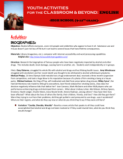
Activities: Biographies: YOUTH
FACTS FOR CONSUMERS ABOUT SETRIA GLUTATHIONE What is glutathione?
© Copyright 2026
About abcdocz
DMCA / GDPR
Report