ABC
docz
Explore
Log in
Create new account
Download
Report
health and fitness
disease
Anemia of Chronic Disease
My Anemia Starter Kit HELLO! My name is C.R. Hume
Laser Charge Tickets 25 (left) Improve communication and reduce coding
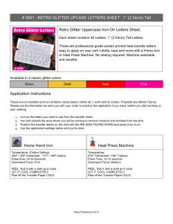
# 0061 : RETRO GLITTER UPCASE LETTERS SHEET : 1”... Retro Glitter Uppercase Iron-On Letters Sheet.
enduraTex Iron On 800-699-5512
Hemoglobin/Hematocrit Child and Teen Checkups (C&TC) For Primary Care Providers
Hemoglobin H Disease What is Hemoglobin H (Hgb H) Disease?
Life Science Journal 2013;10(2)
Haematology Update for General Practice Dr Naim Akhtar Consultant Haematologist
Anemia and adherence to oral iron
T he cardiovascular effects of erythropoietin *
Medicinal Chemistry of Fetal Hemoglobin Inducers for Treatment of -Thalassemia Roberto Gambari
© Copyright 2026
About abcdocz
DMCA / GDPR
Report