
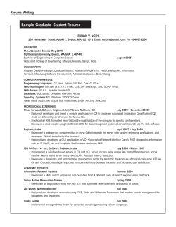
Program & Abstracts KPro Study Group Symposium
Pre-World Cornea Congress
KPRO STUDY GROUP
SYMPOSIUM
Program
April 14, 2015, 7 AM – 5:30 PM
9415 Campus Point Drive, La Jolla, CA
Hosted by:
Natalie Afshari MD
[email protected]
Organized by:
Jose de la Cruz
Jean-Marie Parel
[email protected]
[email protected]
VENUE
7:00am
KPRO STUDY GROUP SYMPOSIUM, April 14 2015, Shiley Eye Institute, UC San Diego
GOLDBERG AUDITORIUM (at Moores Cancer Center behind the Shiley Eye Institute)
Registration & Continental Breakfast + uploading morning talks
8:20am
Welcome/Introduction Natalie Afshari, Jose De La Cruz, Jean-Marie Parel
8:30am
I. Boston KPro International/growth
Moderators: Anthony Aldave, James Chodosh
8:30am
8:40am
8:50am
9:00am
9:10am
Globalization of KPRO – James Chodosh
International Growth – Antony Aldave
Russia KPRO - Boris Malyugin
PanKPRO Initiative – Victor Perez, Jose De La Cruz, Andrea Cruzat, Guillermo Amescua
Discussion
9:20am
II. What have we learned in the last 10 years about complication and management of?
9:20am
9:30am
9:40am
9:50am
10:00am
10:10am
Moderators: Esen Akpek, Victor Perez
Glaucoma – current problems and future fixes – Jeffrey Goldberg
Pre-operative evaluation and tamponade consideration in patients undergoing keratoprosthesis – William Freeman
Extrusion/melts - Esen Akpek
Retroprosthetic membranes – Jose De La Cruz
Infections – Guillermo Amescua
Discussion
10:20am Coffee Break
10:50am III. New Diagnostics/Not yet widely used methods in:
Moderators: Mark Rosenblatt, Kathryn Colby
10:50am Introduction of imaging in KPRO – Jose De La Cruz
11:00am Glaucoma imaging in KPRO – Andrea Cruzat
11:10am New developments in tissue modifications for improved outcomes of the Boston KPro – Joseph Ciolino
11:20am Advantages/Disadvantages in new backplate size/materials – Kathryn Colby
11:30am Presence of silicone oil, no prior corneal surgery and postop contact lens use – Natalie Afshari
11:40am Novel approaches to KPro: an oculoplastics perspective – Don Kikkawa
11:50am Discussion
12:00pm IV. Rapid Fire Free Papers
6 min max
Moderators: Natalie Afshari, Juan Alvarez de Toledo, Jose De La Cruz
1:00pm Discussion
1:10pm
Lunch + uploading afternoon talks
2:10pm
V. MOOKP/Boston Type II
2:10pm
2:20 pm
2:30 pm
2:40 pm
Moderators: Jim Chodosh, Mark Mannis
Improvement MOOKP current procedure & update in complication management – Victor Perez
Tibial KPro - Maria de la Paz
Differences in Type I and Type II – James Chodosh
Discussion
2:50pm
Group Photograph in front of Shiley Bldg and move to Shiley Eye Institute
VI. Novel Devices and True integrating Artificial Corneas
3:20pm
3:30pm
3:40pm
3:50pm
4:00pm
4:10pm
Moderators: Claes Dohlman, Robert Weinreb, Eduardo Alfonso
In search of true integrating artificial corneas - Mark Rosenblatt
KeraKlear - Yichieh Shiuey
Implantation of an electronic pressure sensor with Boston KPro – Thomas Neuhann
Contact lens drug delivery and how it could benefit the Boston KPro – Joseph Ciolino
What do MOOKP and KPro patients see? – Heather Durkee, Victor Hernandez, Jean-Marie Parel
Discussion
3:20pm
VII. Rapid Fire Free Papers
5:20pm
Moderators: Maria de la Paz, Joseph Ciolino, Yichieh Shiuey
Discussion
5:30pm
Final Remarks /Conclusion Natalie Afshari, Jose De La Cruz, Jean-Marie Parel
JMP-JDLC-NA-EA Final Program 4/13/2015
6 min max
Page 1/25
KPRO STUDY GROUP SYMPOSIUM, April 14 2015, Shiley Eye Institute, UC San Diego
Rapid-Fire Free Papers
Morning Session – Moderators: Natalie Afshari, Alvarez de Toledo
1
Incidence of Stevens-Johnson Syndrome and Chemical Burns to the Eye: Relevance to Keratoprosthesis Surgery Michelle White
2
International Outcomes of the Boston Type I Keratoprosthesis in Stevens-Johnson Syndrome - Anthony Aldave
3
Boston Type 1 Kerato-Prosthesis In High Risk Cases – Juan Alvarez de Toledo
4
Long Term Outcomes with Boston Type 1 Keratoprosthesis in Ocular Burns - Lauro Oliveira
5
The advantageous use of Boston KPro in managing combined anterior and posterior segment pathology. A Case study Hatem Kobtan
6
Adjunct CXL of Carrier Cornea in two types of Keratoprosthesis. Technique and Outcomes - Anastasios John
Kanellopoulos
7
Boston type I Keratoprosthesis assisted with intra-prosthetic amniotic membrane (AmniotiKPro sandwich technique) Alejandron Navas
8
Prognostic Value of Peri-Prosthetic Tissue Loss for Corneal Melting - Results of a Longitudinal Anterior Segment OCT
Study in Patients with Boston type 1 Keratoprosthesis - Rodrigo Muller
9
Outcomes of primary Boston Type 1 keratoprosthesis placement - Kimberly Hsu
10 Impact of Pars Plana Vitrectomy on Complications in Aphakic Boston Keratoprosthesis Patients - Allister Gibbons
Afternoon Session – Moderators: Maria de la Paz, Joseph Ciolino
11 Effect of keratoprosthesis on the eye. An animal study - Alja Crnej
12 Scanning Electron Microscopic Analysis of Biofilm Formation in Explanted Human Boston Type I Keratoprostheses.
Sterile vs Infectious Case Comparison - Kavitha Sivaraman
13 KPro: What is the relationship between surface quality and biofilm adherence? - Heather Durkee
14 Optimization of Titanium Surface Characteristic in Boston Keratoprosthesis Backplate - Eleftherios Paschalis
15 Management of end stage corneal disease and chronic uveitic hypotony by combining the Boston type 1
keratoprosthesis with pars plana vitrectomy and silicone oil fill - Tayyeba Ali
16 Five to Ten Year Follow-up of Boston Type 1 Keratoprostheses Demonstrates Stable Visual Acuity and Retention Anthony Aldave
17 Experience with Dohlman-Doane type I keratoprosthesis - Sergio Kwitko
18 Novel Strategies to Improve Keratoprosthesis Outcomes - Juan-Carlos Abad
19 Crosslinking of Donor Corneal Tissue with Glutaraldehyde (GA) for Keratoprosthesis - K.S.Siddharthan
20 Setting up a Keratoprosthesis Service in a Tertiary Care Public Funded Hospital in India - Radhika Tandon
JMP-JDLC-NA-EA Final Program 4/13/2015
Page 2/25
KPRO STUDY GROUP SYMPOSIUM, April 14 2015, Shiley Eye Institute, UC San Diego
Incidence of Stevens-Johnson Syndrome and Chemical Burns to the Eye: Relevance to
Keratoprosthesis Surgery
Michelle White, James Chodosh, Jisun Jang, Claes H Dohlman
Massachusetts Eye and Ear Infirmary, Harvard Medical School, Boston, MA, USA
Purpose: This study was designed to estimate the incidence and distribution of SJS-spectrum (Stevens-Johnson
Syndrome, Toxic Epidermal Necrolysis, Toxic Epidermal Necrolysis/Stevens- Johnson Syndrome overlap), and of
chemical burns (alkali or acid burn of the cornea/conjunctiva) in the United States (US) and project the number of
patients that may benefit from keratoprosthesis surgery worldwide.
Design: Population based observational study.
Subjects: All patients evaluated in 961 hospital-based US emergency departments between July 1, 2010 and June
30, 2012.
Methods: Patients identified retrospectively utilizing two years (2010 and 2011) of Nationwide Emergency
Department Sample (NEDS) data in the US. SJS-spectrum and chemical burn cases were identified using
International Classification of Diseases, Ninth Revision, Clinical Modification diagnostic codes.
Results: A mean of 3,849 new SJS-spectrum cases per year were identified in the US, resulting in an incidence rate
of 12.40 new cases per million per year. Similarly, a mean of 15,798 new chemical burn cases per year were
identified, resulting in an incidence rate of 50.89. The mean age of patients with SJS-spectrum was 48.6 and the
mean age of patients with chemical burns was 34.5. While there was no gender difference for incidence of SJSspectrum, men were twice as likely to suffer from chemical burn compared to women, in the US.
Conclusions: Nearly four thousand people in the US become afflicted with SJS-spectrum every year. How many
eventually end up bilaterally blind and in need of intervention is not known. If the incidence of SJS-spectrum is
approximately uniform the world-over, extrapolation from the US figure would amount to about 87,000 new
cases per year in the world. Similarly, over 15,000 chemical eye burns are reported in the US per year.
Extrapolation to the world is difficult since incidence and severity are anticipated to be higher in the developing
world than in the US. Still, using a US incidence rate, a minimum of 350,000 burn accidents would be expected to
occur worldwide every year. When SJS-spectrum and chemical burns result in severe corneal damage,
keratoprosthesis surgery may be the only option to restore vision in the future.
Contact: [email protected]
JMP-JDLC-NA-EA Final Program 4/13/2015
Page 3/25
KPRO STUDY GROUP SYMPOSIUM, April 14 2015, Shiley Eye Institute, UC San Diego
International Outcomes of the Boston Type I Keratoprosthesis in Stevens-Johnson Syndrome
Anthony J Aldave, Jamie K Alexander, Samar K Basak, Dominga B Padilla, Fei Yu
The Jules Stein Eye Institute, UCLA, Los Angeles, CA, USA
Purpose: To determine the factors influencing outcomes of Boston type I keratoprostheses implantation in
patients with Stevens-Johnson syndrome (SJS) and to compare the results with those implanted in patients
without SJS.
Methods: Data was collected regarding Boston type I keratoprosthesis procedures performed by three surgeons
at three centers: Stein Eye Institute in Los Angeles (UCLA) between May 1, 2004 and July 1, 2013; and Disha Eye
Hospital in Kolkata, India and St. Luke’s Hospital in Manila, Philippines between March 1, 2009, and January 1,
2014.
Data was collected for each procedure regarding the preoperative characteristics, the surgical procedure(s)
performed, interval post-operative visual acuities, keratoprosthesis retention, and complications. Statistical
analysis was performed to identify significant differences between the SJS and non-SJS populations in terms of
visual acuity, retention and complications. Statistical analysis was performed to identify predictors of retention
failure in the SJS population.
Results: During the study periods, 234 keratoprosthesis procedures were performed in 209 eyes of 201 patients
by the three participating surgeons, with 40 of the procedures performed in 27 eyes of 26 patients with SJS.
Compared to the non-SJS series, procedures in SJS patients were more frequently performed as repeat
keratoprostheses (33% vs. 8%, p<0.001) but less frequently in eyes with glaucoma (26% vs. 71%, p<0.001) or
multiple prior penetrating keratoplasties (15% vs. 59%, p<0.001). A significantly greater percentage of patients
with SJS had a CDVA with the keratoprosthesis > 20/200 at last follow-up compared with patients without SJS
(96% vs. 63%, p=0.001). Several post-operative complications were more common in SJS patients, including
corneal stromal necrosis (59% vs. 8%, p<0.001), corneal infiltrate (30% vs. 10%, p<0.001), and persistent corneal
epithelial defect (59% vs. 24% p<0.001). These complications led to a higher rate of retention failure and
secondary surgical procedures in comparison to non-SJS eyes (0.306/eye-year vs. 0.068/eye-year, p< 0.001).
However, following repeat implantations, eyes with SJS were no less likely to ultimately retain a keratoprosthesis
(81% vs. 89%, p=0.335).
Conclusion: The Boston type I keratoprosthesis is an effective means to restore vision in patients with SJS.
Although retention failure and several post-operative complications are more common in SJS patients, sightthreatening complications such as endophthalmitis and retinal detachment are not. Given this, and the lower
incidence of glaucoma in SJS patients, nearly all SJS patients have a final CDVA with the keratoprosthesis > 20/200.
Contact: [email protected]
JMP-JDLC-NA-EA Final Program 4/13/2015
Page 4/25
KPRO STUDY GROUP SYMPOSIUM, April 14 2015, Shiley Eye Institute, UC San Diego
Boston Type 1 Kerato-Prosthesis in High Risk Cases
Alvarez de Toledo J1,2, de la Paz MF1,2, Michael R2, Mantaras J2, Barraquer RI1,2, Seyeddain O3, Grabner G3
1
2
Centro de Oftalmología Barraquer, Barcelona, Spain;
Institut Universitari Barraquer, Universitat Autonoma de
3
Barcelona,Spain; University Eye Clinic, Paracelsus Medical University, Salzburg, Austria
Purpose: To analyze the anatomical and functional results, complications, post-surgical additional procedures of
the Boston K-Pro type 1 implantation in high-risk cases in two Ophthalmology Centers.
Methodology: Retrospective case-series on twenty high-risk cases of chemical burn or muco-cutaneous syndrome
which underwent Boston K-Pro implantation. Standard Boston K-Pro type 1 surgical technique with no additional
surgical measures was used in all cases. Visual results, anatomical retention, postoperative complications and
additional procedures were the main outcome measures.
Results: Eleven patients were operated in Barraquer Eye Institute of Barcelona and 9 patients in University Eye
Clinic in Salzburg. Eighty-five % were male and 15% were female. Mean age was 45.6 years. Primary diagnosis was
alkali burn in 6 eyes, non-specified chemical burn in 3, Lyell’s syndrome in 3, acid burns in 2 (10%) ocular cicatricial
pemphigoid in 2 and non-specified muco-cutaneous syndrome in 1 case. Mean previous number of keratoplasty
procedures was 1.95 (0-10). Mean follow-up was 36.17 months (3-78.5; only 3 cases had less than 6 months of
follow-up). Mean pre-op visual acuity (BCVA; Log-Mar units) was 2.11 (2.6989-1), post-op BECVA (best-ever
corrected VA) was 0.64 (2-0.07918125) and last visit BCVA was 1.34 (3-0.04139269). Retention of the prosthesis
was achieved in 14 cases (70%). In the 6 cases of extrusion, the mean extrusion time was 26.5 months (4-52).
Prosthesis was replaced in 4 cases, 3 being retained at the end of the study and one case had a second extrusion,
which ended in a tectonic keratoplasty. The other 2 cases were also treated with a tectonic keratoplasty.
Complications were frequent and included retro-prosthetic membrane in 9 cases (45%), glaucoma in 7
(35%), extrusion in 6 (30%), melting in 4 (20%), choroidal detachment in 3 (15%) endophthalmitis in 3 (15%),
vitreous hemorrhage in 1 (5%), and descemetocele in 1 (5%). Additional surgical procedures were YAG-laser
membranotomy in 9 cases (45%), new prosthesis replacement in 4 (20%), tectonic keratoplasty in 4 (20%),
enucleation in 1 (5%) conjunctival autograft in 1 (5%) and pars plana vitrectomy in 1 case (5%).
Conclusions: Anatomical and functional results of the Boston K-Pro type 1 are satisfactory in high-risk cases.
Presence of serious complications is common, additional procedures are frequently needed and the final longterm outcome can be compromised.
Contact: [email protected] [email protected]
JMP-JDLC-NA-EA Final Program 4/13/2015
Page 5/25
KPRO STUDY GROUP SYMPOSIUM, April 14 2015, Shiley Eye Institute, UC San Diego
Long Term Outcomes with Boston Type 1 Keratoprosthesis in Ocular Burns
Lauro Augusto de Oliveira, Fernanda Pedreira Magalhães, Flavio E. Hirai, Luciene Barbosa de Sousa
Department of Ophthalmology, Federal University of São Paulo, São Paulo/SP, Brazil
PURPOSE: To report long term outcomes of Boston type I keratoprosthesis (BKPro) in the management of ocular
burn injuries.
METHODS: This was a prospective study including all cases of BKPro implantation for ocular burns at the External
Diseases and Cornea Service of the Federal University of São Paulo, between February 2008 and February 2015.
Twelve patients (12 eyes) were enrolled. Procedures performed to manage ocular injury were identified, and data
were collected regarding patients' ocular history, surgical procedure(s) performed, and postoperative outcomes,
including visual acuity, retention, complications and required surgical procedures.
RESULTS: A total of 13 Type 1 BKPro were implanted in 12 eyes of 12 patients. The mean follow-up period was
61.2 (5 - 83 months). Preoperative best-corrected visual acuity (BCVA) ranged from count fingers to light
perception. Postoperative BCVA was better than 20/200 in 83% of the patients and better than 20/60 in 58.3% of
the patients. The overall BKPro retention rate was 83.3%. The most common complications were retroprosthetic
membrane formation (41.6%) and persistent corneal epithelial defect evolving to corneal melting (33.3%).
Patients who underwent ocular surface procedures such as limbal transplantation prior to BKPRo implantation
had a lower incidence of corneal melting/thinning (p = 0.07), although this was not statistically significant.
CONCLUSION: The anatomical and functional results identified in this study support the use of BKPro in managing
bilateral limbal stem cell deficiency secondary to ocular burns.
Contact: [email protected]
JMP-JDLC-NA-EA Final Program 4/13/2015
Page 6/25
KPRO STUDY GROUP SYMPOSIUM, April 14 2015, Shiley Eye Institute, UC San Diego
The advantageous use of Boston KPro in managing combined anterior and posterior segment
pathology. A Case study
Hatem Kobtan
Department of Ophthalmology, Cairo University, Cairo, Egypt
A 34 year old who was referred to our service with advanced corneal abscess due to a corneal infection of 1
month duration. Examination revealed advanced corneal melting with areas of iris prolapse seen and a vision of
light perception. Culture revealed a coagulase negative staph infection which responded to amniotic membrane
transplantation with topical antibiotic treatment which resulted in healing and extensive vascularization of the
cornea. Ophthalmic ultrasound revealed a total retinal detachment with anterior PVR. At this point a parsplana
vitrectomy through a Landers Wide Field Temporary Keratoprosthesis 8.2mm Lens with extensive dissection of
proliferations at vitreous base and 360 retinotomy-retinectomy with silicone oil injection at the end of the
procedure. The patients graft was re-sutured at the end of the procedure with postoperative follow up by
ultrasound. A total retinal detachment open funnel configuration with epiretinal membranes was seen 3 weeks
later. A second attempt at reattaching the retina using Landers temporary Keratoprosthesis but this time after
flattening the retina with heavy liquid perflorocarbon the Boston KPros was secured after removing the Landers
Keratoprosthesis and the procedure is finalized with the exchange of the PFC silicone oil exchange while clear
visibility is maintained through the Boston Keratoprosthesis.
Results: The patient postoperative visual acuity improved from light perception to 6/36. The case demonstrates
the value of Boston KPro in visual rehabilitation in cases with extensive vascularized recipient beds as well as the
added advantage of working with KPro in complex cases with posterior segment pathology that requires
concomitant intervention.
Contact: [email protected]
JMP-JDLC-NA-EA Final Program 4/13/2015
Page 7/25
KPRO STUDY GROUP SYMPOSIUM, April 14 2015, Shiley Eye Institute, UC San Diego
Adjunct CXL of Carrier Cornea in two types of Keratoprosthesis: Technique and Outcomes
Anastasios John Kanellopoulos
The Laservision Institute, Athens, Greece
Purpose: The purpose of this study was to evaluate safety and efficacy of very high-fluence collagen crosslinking
(CXL) as a means of increasing cornea rigidity and reducing enzymatic digestion in two keratoprosthesis
techniques (a) in the vehicle cornea of Boston keratoprosthesis (Kpro) Type1 and (b) in foldable interlamellar
partial thickness corneal pocket.
Methods: In Boston Keratoprosthesis, eleven consecutive cases (5 with repeat cornea graft failure, 4 ocular
cicatricial pemphigoid, and 2 chemical burn) received donor-vehicle cornea pre-treatment with very high-fluence
prophylactic collagen crosslinking in two sessions. The donor cornea was initially cross-linked with intrastromal
riboflavin instillation through a femtosecond-laser-created pocket; followed by superficial CXL. All cases, at the
completion of the CXL pre-treatment, had the cornea center trephined with femtosecond laser; the vehicle cornea
was trephined with a 9-mm donor punch and the Kpro Type1 was fitted onto the cross-linked donor cornea. We
evaluated visual acuity, corneal surface and donor vehicle cornea stability. Mean follow-up was 7.5(1 to 9) years.
In foldable interlamellar partial thickness corneal pocket (KeraKlear XT Artificial Cornea, Keramed, CA, USA) we
investigated a case with severe ocular surface disease due to limbal stem cell deficiency, resulting in cornea
blindness. The inlay prosthesis was previously implanted as an inlay in a donor vehicle allograft cornea that
underwent collagen cross-linking (CXL) prior to trephination and interlamellar inlay implantation. The inlayvehicle cornea complex was transplanted in a fashion similar to penetrating keratoplasty with a 9.5mm diameter.
Results: In the Boston Kpro cases, mean uncorrected visual acuity improved from light-perception to 20/60. One
case needed follow-up surgery, due to significant melt in the host cornea. None of the eyes developed melts
and/or infection, especially on the vehicle cornea on which the keratoprosthesis was fitted.
In the Keraklear case, the patient was followed for a period of one year. Visual acuity improved from light
perception to the 20/400 within 1 month of surgery and remained stable during the follow-up period. The view
through the central cornea remained clear during the follow-up period and there was no melting of the
surrounding cornea or extrusion of the implant.
Conclusions: Pre-treatment with intrastromal and superficial very high-fluence collagen crosslinking in
conjunction with keratoprosthesis appears to be a safe and effective adjunctive treatment aiming to increase
vehicle donor cornea rigidity and potentially increased resistance to enzymatic degradation. This application may
serve to enhance the biomechanical stability and external disease susceptibility of the donor vehicle cornea in
these advanced external disease cases. The novel CXL technique may reduce the incidence of vehicle cornea melt
in similar keratoprosthesis operations.
Contact: [email protected]
JMP-JDLC-NA-EA Final Program 4/13/2015
Page 8/25
KPRO STUDY GROUP SYMPOSIUM, April 14 2015, Shiley Eye Institute, UC San Diego
Boston type I Keratoprosthesis assisted with intra-prosthetic amniotic membrane (AmniotiKPro
sandwich technique)
Alejandro Navas, Julio C. Hernandez-Camarena, Juan Carlos Serna-Ojeda, Arturo Ramirez-Miranda, Enrique O.
Graue-Hernandez
Department of Cornea and Refractive Surgery, National Autonomous University of Mexico, UNAM, Mexico City, Mexico.
Purpose: To describe a technique of Boston type 1 keratoprosthesis (KPro) implantation assisted with intraprosthetic amniotic membrane.
Methods: Patients with severe ocular surface inflammation and corneal blindness underwent KPro implantation
assisted with intra-prosthetic amniotic membrane. This technique uses a cryopreserved amniotic membrane disc
positioned directly beneath the front plate, with the donor cornea placed after creating a “sandwich” between
the front plate, the amniotic membrane disc and the donor cornea.
Results: KPro remained in place and the amniotic membrane well attached, with improvement in the uncorrected
visual acuity and with anterior segment optical coherence tomography showing the intra-prosthetic amniotic
membrane attached and filling the virtual space between the donor cornea and the anterior plate of the KPro.
Conclusion: This technique could provide an epithelization scaffold and an anti-inflammatory and antimicrobial
platform in patients known to have underlying severe inflammatory ocular surface disease and altered ocular
immunity. A decreased incidence of tissue melting and KPro extrusion could be a potential benefit with this
technique, at the same time the antimicrobial properties of the amniotic membrane and the mechanical closing of
the gap between the optical stem and donor cornea are thought to decrease the rate of the keratitis and
endophthalmitis associated with KPro.
Contact: [email protected]
JMP-JDLC-NA-EA Final Program 4/13/2015
Page 9/25
KPRO STUDY GROUP SYMPOSIUM, April 14 2015, Shiley Eye Institute, UC San Diego
Prognostic Value of Peri-Prosthetic Tissue Loss for Corneal Melting - Results of a Longitudinal Anterior
Segment OCT Study in Patients with Boston type 1 Keratoprosthesis
, Elise Taniguchi, Andrea Cruzat, Bernardo M Cavalcanti, Claes H Dohlman, Pedram Hamrah
Massachusetts Eye and Ear Infirmary, Boston, MA, USA.
Purpose: To prospectively evaluate the presence and alterations of potential spaces and areas of tissue melting
(gaps) in the donor cornea, under the Boston Type 1 Keratoprosthesis (K-Pro1) front plate, as well as corneal
epithelial tissue lipping over the edge of the front plate (epi-lip), using anterior segment-optical coherence
tomography (AS-OCT).
Methods: AS-OCT (RTVue OCT, Optovue Inc., Fremont, CA) was performed at the device-donor corneal interface
in all 4 quadrants around the K-Pro1at two time points (mean follow-up time of 1.3±0.98years). Presence,
alterations, and size of gaps between the front plate and the donor cornea, as well as epi-lips, were evaluated at
baseline and follow-up.
Results: Forty-two eyes of 36 patients were assessed (mean of 4.8±2.9 years after surgery), and 25 eyes of 23
patients were analyzed at follow-up. From 154 OCT quadrants imaged at the first visit, 56 (36.3%) revealed a gap
under the front plate. The gap area was on average 12.7 ± 33.3μm . Among the quadrants with gaps, 19 (34%)
revealed no epi-lip, while among the quadrants without gaps, only 3 (3.0%) had no epi-lip (p<0.0001). In addition,
the epi-lip area was significantly smaller among the quadrants with gaps compared to quadrants without gaps
(22.1 ± 23.8 and 49.5 ± 49.8 μm , p<0.0001). There was a significant correlation between the gap area and epi-lip
area (r=-0.33, p<0.0001). On follow-up examination, 60% of the quadrants remained without gaps (from 20 eyes),
17% remained stable or regressed (from 12 eyes) and 23% progressed (from 12 eyes), with one eye advancing to
extrusion and two eyes having had a prior history of extrusion in the previous K-Pro. Among those quadrants with
gaps that progressed, 30% had no epi-lip during the first visit while, among those quadrants that remained stable
or regressed, 18% had no epi-lip. Finally, among those quadrants that remained without gaps, only 3% had no epilip (p<0.0001). The relative risk for progression of gaps at the follow-up visits was 1.7 (p=0.07); however, it was 3.2
in quadrants with no epi-lips during the first visit, compared to quadrants with epi-lip (p=0.0005).
Conclusions: Patients with K-Pro Type 1 may demonstrate gaps under the front plate. The epi-lip may confer
protection against development of gaps. While corneas in patients without gaps remain stable, presentation of
gaps can progress and could demonstrate a risk factors for corneal melting, requiring closer follow-up.
Contact: [email protected]
JMP-JDLC-NA-EA Final Program 4/13/2015
Page 10/25
KPRO STUDY GROUP SYMPOSIUM, April 14 2015, Shiley Eye Institute, UC San Diego
Outcomes of primary Boston Type 1 keratoprosthesis placement
Kimberly Hsu1, Joann Kang1, Jose de la Cruz1, Maria S Cortina1
Illinois Eye and Ear Infirmary, University of Illinois at Chicago Department of Ophthalmology and Visual Sciences,
Chicago, IL USA
Purpose: To report longer term outcomes on patients who received a Boston Type 1 keratoprosthesis as the
primary penetrating corneal procedure.
Methods: A retrospective study of patients who received a primary Boston Type 1 keratoprosthesis from 2008–
2013 was conducted. Vision, complications, and additional procedures required were collected for patients who
had greater than 1 year follow up.
Results: Twenty-four eyes of 22 patients received a primary Boston Type 1 keratoprosthesis during 2008 – 2013.
Mean follow-up was 47.4 months (range 13.3 to 75.2 months). Preoperative vision ranged from 20/100 to LP. At
last follow up, 70.8% of eyes achieved at least 20/200 vision and 37.5% of eyes achieved greater than or equal to
20/50 vision. Postoperative complications included RPM (37.5%), increased intraocular pressure (58.3%), and
macular edema (25%). There were no cases of endophthalmitis. Retention rate was 79.2%.
Conclusions: In patients who are at high risk for graft failure with traditional penetrating keratoplasty, the Boston
Type 1 keratoprosthesis may be a good alternative. Years after the procedure, many patients retain improved
vision, although close follow up is warranted to monitor for complications.
Contact: [email protected]
JMP-JDLC-NA-EA Final Program 4/13/2015
[email protected]
[email protected]
Page 11/25
KPRO STUDY GROUP SYMPOSIUM, April 14 2015, Shiley Eye Institute, UC San Diego
Impact of Pars Plana Vitrectomy on Complications in Aphakic Boston Keratoprosthesis Patients
Allister Gibbons, Audina Berrocal, Thomas Albini, Daniel Waren, William Feuer, Jean-Marie Parel, Guillermo
Amescua, Victor L Perez
Bascom Palmer Eye Institute, Department of Ophthalmology University of Miami Miller School of Medicine, Miami, FL USA
Purpose: To study the impact of pars plana vitrectomy (PPV) at the time of surgery, versus a limited anterior
vitrectomy on the outcomes and complication rates of the aphakic model of the Boston Keratoprosthesis (BKpro).
Methods: Records of patients with the snap-on aphakic B-KPro implantation during 2007 to 2013 which had over
1 year of follow-up were reviewed. All pre-operative, intra-operative and post-operative data was recorded and
analyzed. Special attention was made to differentiate the group which had received a PPV at the time of surgery
and the both the incidence and timing to appearance of complications over the follow-up period.
Results: Forty-three eyes of forty-one patients were included in the study. Eighteen of these eyes had undergone
a complete PPV at the time of the B-Kpro implantation, the other twenty-five had not. There were no significant
differences in the preoperative, demographic characteristics, clinical background, or indications of B-Kpro in both
groups. The group that had a PPV at the time of surgery had a statistically significant decrease of total
complication rate observed in the Kaplan Meier Survival analysis (p=0.0367). Also, there was a clear trend for
there to be less Retroprosthetic Membrane formation, glaucoma progression, and endophthalmitis (p <0.1) in the
group that received a PPV at the time of surgery.
Conclusions: The results of this retrospective review suggest that a complete PPV, done at the time of surgery,
has a positive impact on decreasing the complication rates of patients undergoing aphakic B-Kpro implantation.
Contact: [email protected]
JMP-JDLC-NA-EA Final Program 4/13/2015
[email protected]
Page 12/25
KPRO STUDY GROUP SYMPOSIUM, April 14 2015, Shiley Eye Institute, UC San Diego
Effect of keratoprothesis on the eye. An animal study
Alja Crnej, Masahiro Omoto, Thomas H Dohlman, Claes H Dohlman, Reza Dana
Massachusetts Eye and Ear Infirmary and Schepens Eye Research Institute, Harvard Medical School, Boston, USA
Purpose: To compare corneal inflammation after syngeneic and allogeneic penetrating keratoplasty (PK) and
miniature Keratoprosthesis (m-KPro) implantation and to assess the extent of damage in the retina and optic
nerve.
Methods: BALB/c (syngeneic) or C57BL/6 (allogeneic) corneas were transplanted onto BALB/c host beds as part of
PK or m-KPro implantation. Corneal inflammation was assessed by determining the frequencies of CD45+
leukocytes, CD4+ T cells, CD11b+ dendritic cells, and Gr-1+ granulocytes/monocytes by flow cytometry and was
analyzed by expression of the pro-inflammatory cytokines TNFα and IL-1β using Real-Time qPCR at 8 weeks posttransplantation. In addition, the loss of retinal ganglion cells and optic nerve fibers was assessed 8 weeks after the
surgery.
Results : Cell frequencies in the syngeneic and allogeneic m-KPro groups were higher compared to the syngeneic
and allogeneic PK groups, respectively. The expression of TNFα was higher in synKPro, alloPK, and alloKPro groups
compared to the naïve and synPK groups at eight weeks. The expression of IL-1β was significantly higher in both
KPro groups as compared to PK groups. The reduction in number of retinal ganglion cells and optic nerve fibers
were higher in both allogeneic compared with syngeneic groups.
Conclusion: Although m-KPro device augments the inflammatory response in cornea after its implantation,
allogenicity is greater contributor to inflammation. We suggest using syngeneic or decellularized corneal tissue as
a Boston-KPro carrier. Results also suggest that retina and optic nerve could be damaged by chronic inflammation
after allogeneic transplantation.
Contact: [email protected]
JMP-JDLC-NA-EA Final Program 4/13/2015
Page 13/25
KPRO STUDY GROUP SYMPOSIUM, April 14 2015, Shiley Eye Institute, UC San Diego
Scanning Electron Microscopic Analysis of Biofilm Formation in Explanted Human Boston Type I
Keratoprostheses. Sterile vs Infectious Case Comparison
Kavitha R Sivaraman1,2, Joshua H Hou1,3, Jin Hong Chang1, Irmgard Behlau4,5, M Soledad Cortina1, Jose de la Cruz1
1
Illinois Eye and Ear Infirmary, University of Illinois at Chicago Department of Ophthalmology and Visual Sciences, Chicago, IL
2
3
USA; Bascom Palmer Eye Institute, University of Miami Miller School of Medicine, Miami, FL USA; University of Minnesota
4
Department of Ophthalmology, Minneapolis, MN USA; Microbiology & Molecular Biology and Ophthalmology, Tufts
5
University School of Medicine, Boston, MA USA; Infectious Diseases, Mount Auburn Hospital, Cambridge, MA USA.
Purpose: To describe the morphological distribution of host tissue and microbial biofilms on the intraocular
surfaces of Boston Type I Keratoprostheses (KPro) explanted due to corneal melt.
Methods: Retrospective scanning electron microscopy (SEM) study of four explanted Boston Type I KPros
composed of polymethylmethacrylate (PMMA) and titanium. SEM images of KPro-associated ocular surfaces
were reviewed for the presence of inflammatory cells, microbes, and/or biofilm formation. One sterile KPro Type
I was also imaged to serve as a device only control.
Results: All four KPros were explanted due to culture-negative, clinically ’sterile’ donor corneal melt with
impending KPro extrusion. In all cases, the rough, irregular surfaces of the device harbored more adherent corneal
epithelia and stromacytes, inflammatory cells and bacteria than the smooth, polished surface of the KPro optic.
One KPro showed not only evidence of prior bacterial colonization, but marked biofilm formation.
Conclusions: SEM images of explanted KPros explanted for ‘sterile’ corneal melt demonstrated evidence of biofilm
formation, despite negative donor cornea cultures and the absence of clinical suspicion for infection. These
results suggest that ‘sterile’ corneal melt may be due to inflammatory host responses to low microbial burdens as
seen in biofilms and/or released antigens after antibiotic-induced lysis. There was increased adherence of host
tissue cells and microbial biofilms on the non-polished surfaces of the KPro. Polishing the intraocular PMMA and
titanium KPro surfaces may decrease microbial adhesion and biofilm formation in KPro patients, but what the
impact this will have on rates of post-op endophthalmitis is unknown.
Contact: [email protected]
JMP-JDLC-NA-EA Final Program 4/13/2015
[email protected]
Page 14/25
KPRO STUDY GROUP SYMPOSIUM, April 14 2015, Shiley Eye Institute, UC San Diego
KPro: What is the relationship between surface quality and biofilm adherence?
Heather Durkee1, Darlene Miller2, Shawn P Kelly1, Patricia L Blackwelder4, Mariela C Aguilar1, Victor L Perez3,
Guillermo Amescua3, Jose de la Cruz5, Jean-Marie Parel1
1
2
3
Ophthalmic Biophysics Center, Ocular Microbiology Laboratory, ABLEH, Bascom Palmer Eye Institute, University of Miami
4
Miller School of Medicine, Miami, FL’ . University of Miami Center for Advanced Microscopy (UMCAM) and Marine
5
Geosciences (RSMAS), University of Miami, Coral Gables, FL, Department of Ophthalmology, University of Illinois Eye & Ear
Infirmary, Chicago, IL, USA.
Purpose: To investigate the relationship between surface quality and biofilm adherence in explanted Boston Type
I, Type II, and modified-osteoodonto keratoprostheses (KPros) using confocal and scanning electron microscopy.
Methods: Failed KPros were collected from patients undergoing KPro explantation at Bascom Palmer Eye
Institute, Miami, FL, USA. In the operating room, the KPro samples were placed in a container with balanced salt
solution immediately after removal. The fresh, un-fixed KPro samples were stained with a live/dead fluorescent
stain and confocal microscopy was performed to visualize the microbial adherence and cellular growth. Images
were taken across the entire anterior and posterior surfaces of the KPro samples to characterize the complete
anterior and posterior faces of the KPro specimen (including the optic, backplate, locking ring). After confocal
microscopy, the KPro sample was fixed in 10% formalin, immersed in PBS buffer, dehydrated in a graded series of
ethanol, dried in HMDS, and sputter-coated with Palladium for scanning electron microscopy. Images of the
anterior and posterior surfaces of the KPro were obtained using SEM at multiple magnifications (30x–5000x).
Results: Confocal microscopy and SEM images revealed rough surfaces in all regions of the KPros. The confocal
microcopy revealed cellular growth in areas of more irregularities. The high magnification SEM images showed
many bacteria and biofilm colonies attached to the KPros. In one case, the patient also had an intraocular lens
(IOL) which was analyzed as was the KPro to relate surface features to microbial adherence. The IOL had super
polished surfaces with almost no microbial adherence.
Conclusion: A dual-imaging approach provided better visualization of the surface quality and biofilm/microbial
growth on the surfaces of the failed KPros. Understanding the relationship between biofilm adherence and rough
surface quality may bring to light the mechanism of KPro failure.
Contact: [email protected]
[email protected]
Support: USAMRMC Department of Defense W81XWH-09-1-0674, Florida Lions Eye Bank, Drs. Karl R Olsen and Martha E
Hildebrandt, NIH P30EY1481 (Center Grant), Research to Prevent Blindness, Henri and Flore Lesieur Foundation (JMP).
Scientific support was provided by: Eduardo Alfonso, Yoh Sawatari, Aleksandra V Rachitskaya, Audina M. Berrocal, Jose
Antonio Bermudez Manger, Kavitha Sivaraman, Florence Cabot, Nidhi Relhan Batra, Carolina de Freitas and Gabe Gaidosh.
JMP-JDLC-NA-EA Final Program 4/13/2015
Page 15/25
KPRO STUDY GROUP SYMPOSIUM, April 14 2015, Shiley Eye Institute, UC San Diego
Figure 1: (Left) Confocal microscopy and (Right) Scanning electron microscopy images of internal KPro surface
shows cellular activity and biofilm.
JMP-JDLC-NA-EA Final Program 4/13/2015
Page 16/25
KPRO STUDY GROUP SYMPOSIUM, April 14 2015, Shiley Eye Institute, UC San Diego
Optimization of Titanium Surface Characteristic in Boston Keratoprosthesis Backplate
Eleftherios I. Paschalis, Chengxin Zhou, James Chodosh, Claes H. Dohlman
Department of Ophthalmology, Boston Keratoprosthesis Laboratory, Massachusetts Eye and Ear Infirmary and Schepens Eye
Research Institute, Harvard Medical School, Boston, MA, USA
Purpose: Even though titanium (Ti) has been extensively used in orthopedic surgery and dentistry, it was only
recently approved by the FDA and Europe for use in the eye as Boston keratoprosthesis (B-KPro) backplates. A
major limitation of Ti is the shiny metallic appearance which makes it cosmetically less favorable compared to
previously used PMMA. To circumvent this limitation, B-KPro backplates have been systematically sandblasted to
generate surface roughness and to reduce metallic reflection. The present work presents a method for changing
the color of the Ti to somewhat match the natural color of the patient’s eye and provides for the first time data of
the impact of Ti surface roughness in human corneal cells.
Methods: Coloring of the Ti was achieved by electrochemically anodization that resulted in a Ti oxide film
formation on the surface of the backplate. The thickness of the film was controlled by the applied voltage. The
technique was evaluated in vitro (human corneal cells) and in vivo (rabbit intra-corneal implantation) for
biocompatibility and safety. Rough Ti surfaces (RMS= 4.413, 2,426, 0.690) and polished (RMS= 0.175) were
assessed in vitro using human corneal/limbal epithelial cells (HCLE), stromal fibroblasts (HCFs), endothelial cells
and HeLa cells. Cell cytotoxicity and proliferation assays were performed. Cells were challenged with ascorbic acid,
w/wt TGFβ1 to simulate the corneal micro-environment after injury and assessed for collagen deposition, cell
morphology and α-smooth muscle actin (αSMA) expression.
Results: Coloration of Ti was achieved by anodization at 33 and 13.4 volts, respectively with a current supply of 3
Amps. In vitro and in vivo assessment showed no significant differences in tolerance compared to non-colored Ti.
However rough Ti surface topography resulted to significant inhibition of HCLE and HCF cell proliferation, as
compared to polished Ti. Polished Ti provided better cell adhesion, remarkable directionality in cell alignment and
had parallel aligned, fibrillar collagen meshwork, while rough Ti had randomly aligned cells and collagen deposits.
Challenge with TGFβ1 caused increased αSMA expression on the rough Ti surface, less collagen deposition and
randomly oriented collagen fibrils as compared to those on polished Ti which had parallel aligned collagen fibrils.
Conclusion: Blue and brown Ti coloring was achieved by electrochemical anodization and was equally
biocompatible and as safe as the standard non-coated Ti. Ti surface roughness significantly affected corneal cell
expression, with epithelial and stromal cells being highly affected by the topography. Contrary to the established
paradigm in orthopedics, polished Ti surface better facilitated corneal cell proliferation, matrix deposition, and
reduced αSMA expression under TGFβ1 stimulation as compared to rough Ti surface. These results suggest that
BK-Pro Ti coloring may improve the cosmesis and acceptance by patients and that controlled Ti surface roughness
manipulation may provide better tissue biointegration (smooth surface) or reduced retroprosthetic membrane
formation (rough surface) in the future.
Contact: [email protected]
Acknowledgments/disclosures: This study was supported by the Boston Keratoprosthesis Fund. The authors have no financial conflicts to disclose. All authors are full-time
employees of Massachusetts Eye and Ear Infirmary, a non-profit organization and manufacturer of the Boston Keratoprosthesis.
JMP-JDLC-NA-EA Final Program 4/13/2015
Page 17/25
KPRO STUDY GROUP SYMPOSIUM, April 14 2015, Shiley Eye Institute, UC San Diego
Management of end stage corneal disease and chronic uveitic hypotony by combining the Boston type
1 keratoprosthesis with pars plana vitrectomy and silicone oil fill
Tayyeba K Ali, Guillermo Amescua, Janet L Davis, Allister Gibbons, Victor L Perez
Bascom Palmer Eye Institute, Department of Ophthalmology, University of Miami Miller School of Medicine, Miami, FL, USA
Purpose: To review the outcomes of Boston type 1 keratoprosthesis (B-KPro) implantation in combination with
pars plana vitrectomy and silicone oil for the treatment of end stage bullous keratopathy and uveitic hypotony.
Methods: This was a retrospective chart review. Six eyes of 5 patients underwent B-KPro implantation, pars plana
vitrectomy, and silicone oil placement. All preoperative characteristics, including visual acuity and intraocular
pressure, intraoperative events, and postoperative outcomes were analyzed.
Results: Mean patient age was 63.9 years (range 40.9-86.4 years). Mean pre-operative BCVA was logMAR 2.73
(range 2.60 to 3.00). Initially, a gain of visual acuity was seen in half the sample with mean BCVA logMAR 1.90
(Range 0.90 to 3.00). At final follow-up (mean 25.8 months and range 12 to 52 months), mean visual acuity was
logMAR 2.23 (range 1.20 to 3.00) and no eye lost vision. Two eyes (33%) had previously undergone corneal
transplantation, one of which was an endothelial keratoplasty (DSAEK). The other 4 eyes had a B-Kpro as their first
corneal procedure. No intraoperative complications occurred, though one patient had a concomitant retinal
detachment (RD) repair for pre-existing RD. B-KPro retention rate was one hundred percent. One patient
developed a retroprosthetic membrane (RPM); no patients experienced complications such as melt,
endophthalmitis, or extrusion. The one patient who had an RPM had a psuedophakic B-Kpro, which may have
contributed to persistence of RPM. There were no uveitic flares and hypotony was controlled, though further
intervention, including repeat SO fill in one patient, was required over the extended follow-up of these patients.
Conclusions: B-KPro implantation in combination with pars plana vitrectomy and intraocular silicone oil fill is a
safe, though end-stage, procedure that maintains control of uveitis, can improve vision in some
chronically hypotonous eyes, and may delay or prevent phthisis.
Contact: [email protected]
JMP-JDLC-NA-EA Final Program 4/13/2015
Page 18/25
KPRO STUDY GROUP SYMPOSIUM, April 14 2015, Shiley Eye Institute, UC San Diego
Five to Ten Year Follow-up of Boston Type 1 Keratoprostheses Demonstrates Stable Visual Acuity and
Retention
Anthony J Aldave, Jaime Alexander, Fei Yu
The Jules Stein Eye Institute, UCLA, Los Angeles, CA, USA
Purpose: To evaluate the long-term outcomes of Boston type I keratoprosthesis implantation
Methods: A retrospective, interventional case series of eyes undergoing keratoprosthesis surgery by a single
surgeon (AJA) prior to September 2009 with 60 months follow-up. Preoperative characteristics and postoperative
outcomes including CDVA, complications and retention were determined.
Results: Sixty eight keratoprostheses were implanted in 54 eyes of 51 patients, with the initial procedure
performed before September 2009 in all eyes. The mean follow-up was 66 months, with a range between 2
months and 10 years. The primary indication for keratoprosthesis implantation was corneal transplant failure
(50% of eyes). Pre-operative CDVA was > 20/200 in 6% of eyes, while annual post-operative CDVA between 5 and
10 years after surgery was > 20/200 in 55%, 63%, 62%, 100%, 100% and 100% of eyes. Failure to obtain and
maintain 20/200 CDVA following keratoprosthesis implantation was most commonly due to glaucoma. The most
common post-operative complications were retroprosthetic membrane formation (48%), persistent epithelial
defect formation (44%), and elevated intraocular pressure (24%). No eye developed endophthalmitis. Twenty six
of the 68 keratoprosthesis (38%) implanted in 18/54 eyes (33%) were not retained (retention failure rate 0.088
eye-year), although only 2 retention failures occurred between 5 and 10 years after keratoprosthesis surgery.
Conclusions: The Boston type I keratoprostheses provide > 20/200 CDVA in the majority of patients 5-10 years
following implantation. While glaucoma is the most common vision-threatening post-operative complication,
endophthalmitis was not observed. The retention rate of the Boston type I keratoprosthesis remains stable more
than 5 years after surgery, and is significantly higher than the repeat corneal transplant survival rate.
Contact: [email protected]
JMP-JDLC-NA-EA Final Program 4/13/2015
Page 19/25
KPRO STUDY GROUP SYMPOSIUM, April 14 2015, Shiley Eye Institute, UC San Diego
Experience with Dohlman-Doane type I keratoprosthesis
Sérgio Kwitko1,2, Tiago Lansini 1, Diane Marinho 1,2
1
Department of Ophthalmology, Hospital de Clinicas de Porto Alegre, Brazil
OftalmoCentro, Porto Alegre, Brazil
2
Objective: To report the outcomes of Dohlman-Doane type 1 keratoprosthesis (Boston KPro) implantation in 36
eyes
Methods: A retrospective case series was performed on all patients undergoing Boston KPro implantation at
Hospital de Clinicas de Porto Alegre, Brazil, performed between September 2005 and August 2014. A minimum
follow-up of 6 months was required. Using a computerized patient database, 36 eyes of 30 patients were
identified that met these criteria. Best-spectacle corrected visual acuity (BSCVA), complications and retention
time of KPro were evaluated.
Results: underlying disease were failure of multiple previous corneal transplants in 19 eyes (keratoconus, bullous
keratopathy, congenital glaucoma, angle-closure glaucoma, Fuchs dystrophy, and Acanthamoeba keratitis),
alkaline burns in 11 eyes, Stevens-Johnson Syndrome in 3 eyes, graft versus host disease in 2 eyes, thermal burn in
one eye, and aniridia in one eye. Preoperative visual acuity was 20/200 or worse in all eyes. Postoperative BSCVA
ambulatory vision (20/200 or better) was achieved in 63% of the eyes after 3 years, 58.3% after 4 years, 47.6%
after 5 years, and 57.1% after 7 years. Main causes for poor postoperative VA were glaucoma, retinal detachment
and endophthalmitis. Postoperative complications included posterior capsule opacity in 27.8%, corneal melting in
25%, cystoid macular edema 22.2%, sterile vitritis in 19.4%, fugal keratitis in 13.9%, retroprosthetic membrane
formation in 11.1%, anterior chamber fibrin membrane in 5.5%, retinal detachment in 5.5%, and fungal
endophthalmitis in 5.5%. Sixteen eyes (50%) had preoperative glaucoma, and 6 other eyes (16.7%) developed
postoperative glaucoma. Retention KPro rate was 100% in the first year, 91.7% in the second year, 88.9% in the
third year, and 86.1% after 5 years of KPro implantation. Mean follow-up period was 64.7 ± 48.8 months (6 to 111
months).
Conclusion: Boston type I is a good option to restore vision in cases of corneal blindness in which penetrating
corneal transplantation does not have a good prognosis. One main advantage is that it does not require systemic
immunosuppression. Despite the potential complications, type I Boston KPro provided visual improvement in our
series of patients with an otherwise poor prognosis. Immune diseases and ocular surface keratinization had the
worst results. Retention rate decreases overtime due to late retroprosthetic membrane formation and corneal
necrosis. Glaucoma is a frequent cause of poor visual rehabilitation in these patients.
Contact: [email protected]
JMP-JDLC-NA-EA Final Program 4/13/2015
Page 20/25
KPRO STUDY GROUP SYMPOSIUM, April 14 2015, Shiley Eye Institute, UC San Diego
Novel Strategies to Improve Keratoprosthesis Outcomes
Juan-Carlos Abad1, Miguel Cuevas2, James Jaramillo3, Maria Clara Jaramillo3, Oscar Villada4
1
2
Cornea and External Disease Service, clínica CLOFAN, Cornea, External Disease and Ocular Immunology Services, clinic
3
4
CLOFAN and University of Antioquia, Ophthalmology Residents, University of Antioquia, Epidemiology Service, Hospital
Universitario San Vicente Foundation, Medelin, Colombia.
Purpose: To report the clinical results of a group of 22 patients (22 eyes) which had a Boston type I
keratoprosthesis (KPro) implanted in which novel surgical and postop modifications were introduced to try to
improve the outcome.
Methods: Such strategies were 1) All patients were rendered aphakic; 2) Complete pars plana vitrectomy at the
time of surgery; 3) Use of oversized titanium back plates; 4) Almost universal use of glaucoma draining devices
(GDD); 5) Use of medroxiprogesterone and oral doxicycline and ascorbic acid; 6) Avoidance of medications that
could activate collagenases such as topical fourth-generation quinolones, latanoprost and prednisolone acetate;
7) Cleaning of the bandage contact lens with iodine povidone at every patient’s visit.
These patients were treated between January 2011 and November 2014 with an average follow-up of 18 months
(range 3-39 months). There were 17 men and 5 women with an average age of 52±18 years (range 17-84
years). Fifty percent of the cases had a disease without active inflammation of the ocular surface, 23% had a
history of chemical or thermal burns, 18% had diseases with active inflammation of the ocular surface and the
remaining 9% had Stevens-Johnson syndrome. The Indications for implanting the Kpro were a failure of one or
more corneal grafts in 59% of cases; 23% had had one or more Kpros with either PMMA or undersized titanium
back plates that had extruded and 18% did had a hostile ocular surface for a primary penetrating
keratoplasty. Eighty-six percent of the cases had a visual acuity (VA) ≤ 20/200 in the eye to intervene (6 cases had
light perception). Visual acuity values better than 20/200 in 37%, 50% and 60% of the cases at the 3, 6 and 12
months visits. There were 2 Kpro extrusions (91% Kpro retention rate).
Results: The most frequent complication was poorly controlled intraocular pressure in 54%, requiring implant of a
first or second GDD in 24% of the overall cases. Other complications were vitreous hemorrhage in 13%; retinal
detachment in 4 patients (1 was successfully repaired); there were two cases of infections (endophthalmitis and
keratitis) related to an inferotemporal GDD exposure and there was 1 case of retro-optic retroprosthetic
membrane (RPM) related to an unoperated vitreous hemorrhage. There seemed to be less deposit in the
backplate holes compared with prior PMMA backplate cases. Two patients had bare light perception at the end
of follow-up.
Conclusion: Although Kpro extrusion and RPM formation (both behind the optic and in the backplate holes) seem
to be reduced by the implementation of these techniques, glaucoma continues to be a problem in these very
difficult patients.
Contact: [email protected]
JMP-JDLC-NA-EA Final Program 4/13/2015
Page 21/25
KPRO STUDY GROUP SYMPOSIUM, April 14 2015, Shiley Eye Institute, UC San Diego
Crosslinking of Donor Corneal Tissue with Glutaraldehyde (GA) for Keratoprosthesis – A preliminary
report of cases
Kuttappalayam S Siddharthan, Jagadeesh K Reddy, Adithya Gowda, Rushita Kamdar
Cornea and Refractive Services, Sankara Eye Center, Coimbatore, Tamil Nadu, India
Purpose: The aim of this study was to investigate the possibility of induction of cross-links in donor
corneal tissue in order to increase the stiffness as a basis for an ingenious technique of keratoprosthesis.
Methods: Collagenous biomaterials can be stabilized by chemical and physical agents and glutaraldehyde (GA) is
the most commonly used crosslinking agent for collagen based biomaterials. It increases the crosslinks both
intrahelical and interhelical and is thus known to increase the tensile strength of the tissue. Studies have shown
that GA exerts the maximum biochemical effect on the cornea when compared to other cross-linking agents like
genipin and UC- CLX.
At our clinic, we pre-treated the cornea with 0.5% GA for 4 minutes (adequate time for Schiff base formation)
followed by thorough washing under running normal saline for 10 minutes to remove any residual glutaraldehyde
which may have toxic effects after transplantation. It may be noted that higher GA concentration and longer
reaction times may lead to formation of pendant GA related molecules rather than crosslinks. We then used this
GA treated cornea as the donor tissue in the keratoprosthesis (Auro K pro manufactured by Aravind eye Hospitals,
India) for five patients.
Results: During the surgical procedure of punching and then suturing the keratoprosthesis to the host bed, we
noted increased stiffness of the graft biomaterial, attributed to the crosslinking effect of GA. On subsequent
follow up, the pre-treated corneas re epithelialized well with no adverse effects on the eye. Two patients have 6
months follow up and three patients have 3 months follow up.
Conclusions: This increased tensile strength would in due course prevent donor tissue melt and leaks, this being
the primary concern with widespread use of keratoprosthesis. Eventually it would lead to an increased life of the
keratoprosthesis. In our presentation we will show the procedure of tissue treatment, surgical procedure and the
post-operative results of these patients.
Contact: [email protected]
JMP-JDLC-NA-EA Final Program 4/13/2015
Page 22/25
KPRO STUDY GROUP SYMPOSIUM, April 14 2015, Shiley Eye Institute, UC San Diego
Setting up a Keratoprosthesis Service in a Tertiary Care Public Funded Hospital in India
Radhika Tandon, Onkila Bhutia, Ajoy Roychowdhary
1
2
Dr Rajendra Prasad Centre for Ophthalmic Sciences, Centre for Dental Education and Research, All India Institute of Medical
Sciences, New Delhi-110029, India
Purpose: To review the long term outcome of keratoprosthesis surgical services delivery.
Methods: File notes, case records, administrative letters, grant applications, ethics submissions, clinical protocols
notes and updates, clinic registers, operating room records, medical social workers records, applications to stores
for purchase of medications and devices, follow up information and information provided by patient contact by
telephone were gathered, catalogued and analyzed. Clinical and socioeconomic profile of patients, resources
utilized, types of devices and surgical procedures, length and duration of surgery, surgical expenses, short term
and long term results in terms of improvement in vision, change in quality of life, impact on family and life
changing events, expenses incurred in visiting hospital and medications used, retention and complications were
determined.
Results: An estimated total of 65 patients were operated at the centre of which 40 patients under care of the
authors were included for detailed analysis. Age ranged from 20 to 72 years with a mean of 46yrs (SD 20) The
underlying end stage corneal disease was attributed to Stevens Johnson Syndrome or Ocular Cicatricial
Pemphigoid (38%), prior failed corneal grafts (40%), end stage limbal stem cell deficiency (5%) high risk
keratoplasty with healed keratitis and neovascularization in 4 quadrants (5%), aphakia with silicon oil keratopathy
(5%), combination of factors or others (7%). Devices used in sequential order of initiation of the introduction of
surgical procedures to the service included: Singh Worst Champagne Cork KPro (13%), OOKP (28%), Boston Type
1 or its prototypes (48%), Boston Type 2 (3%) and temporary KPro assisted combined vitreoretinal and
keratoplasty procedures (8%). The overall rate of improvement in vision was 95% and improvement in quality of
life 80%. Long term clinical outcome in terms of retention rate including those who completed minimum 5 years
of follow up and excluding those who had temporary KPro assisted keratoplasty was estimated to be 77%. Major
ophthalmic complications noted included extrusion, endophthalmitis (10%), irreparable retinal detachment (3%),
exchange due to instability (3%%), surgical removal due to uncontrollable infection (3%) and progressive ciliary
body failure leading to phthisis bulbi (5%).
Conclusion: Keratoprosthesis surgery offers a life changing option to patients with end stage blinding corneal
disease. With passage of time and changing technology a better understanding of case selection, selection of
devices and procedures, clinical protocols, anticipated outcome and estimated prognosis has emerged.
Contact: [email protected]
JMP-JDLC-NA-EA Final Program 4/13/2015
Page 23/25
KPRO STUDY GROUP SYMPOSIUM, April 14 2015, Shiley Eye Institute, UC San Diego
NOTES
JMP-JDLC-NA-EA Final Program 4/13/2015
Page 24/25
KPRO STUDY GROUP SYMPOSIUM, April 14 2015, Shiley Eye Institute, UC San Diego
Compliments of Juan Carlos Abad MD
For the KPro Study Group:
Jose De La Cruz
[email protected]
Jean-Marie Parel
[email protected]
This KPro Study Group Symposium is endorsed by the
JMP-JDLC-NA-EA Final Program 4/13/2015
Page 25/25


© Copyright 2026