
Integrating Biosensors for Air Monitoring and Breath-Based
Integrating Biosensors for Air Monitoring and
Breath-Based Diagnostics
NIKLAS SANDSTRÖM
Doctoral Thesis
Stockholm, Sweden 2015
Front cover picture:
Top: A droplet of water on a silicon diaphragm with 25 µm perforations.
Middle: A 0.9 mm thick microfluidic OSTE+ cartridge fabricated by reaction
injection molding and direct covalent bonding.
Bottom: Two different biosensor cartridges integrated with quartz crystal resonators
using mechanical clamping (left) and covalent bonding (right).
TRITA-EE 2015:020
ISSN 1653-5146
ISBN 978-91-7595-560-5
KTH Royal Institute of Technology
School of Electrical Engineering
Department of Micro and Nanosystems
Akademisk avhandling som med tillstånd av Kungliga Tekniska högskolan framlägges
till offentlig granskning för avläggande av teknologie doktorsexamen i elektrisk
mätteknik fredagen den 22 maj 2015 klockan 10.00 i Q2, Osquldas väg 10,
Stockholm.
Thesis for the degree of Doctor of Philosophy at KTH Royal Institute of Technology,
Stockholm, Sweden.
© Niklas Sandström, May 2015
Tryck: Universitetsservice US AB, Stockholm, 2015
iii
Abstract
The air we breathe is the concern of all of us but nevertheless we only
know very little about airborne particles, and especially which biological
microorganisms they contain. Today, we live in densely populated societies
with a growing number of people, making us particularly vulnerable to air
transmission of pathogens. With the recent appearance of highly pathogenic
types of avian influenza in southeast Asia and the seasonal outbreaks of
gastroenteritis caused by the extremely contagious norovirus, the need for
portable, sensitive and rapid instruments for on-site detection and monitoring
of airborne pathogens is apparent.
Unfortunately, the integration incompatibility between state-of-the-art
air sampling techniques and laboratory based analysis methods makes
instruments for in-the-field rapid detection of airborne particles an unresolved
challenge.
This thesis aims at addressing this challenge by the development of novel
manufacturing, integration and sampling techniques to enable the use of labelfree biosensors for rapid and sensitive analysis of airborne particles at the
point-of-care or in the field.
The first part of the thesis introduces a novel reaction injection molding
technique for the fabrication of high quality microfluidic cartridges. In
addition, electrically controlled liquid aspiration and dispensing is presented,
based on the use of a thermally actuated polymer composite integrated with
microfluidic cartridges.
The second part of the thesis demonstrates three different approaches of
biosensor integration with microfluidic cartridges, with a focus on simplifying
the design and integration to enable disposable use of the cartridges.
The third part to the thesis presents a novel air sampling technique
based on electrophoretic transport of airborne particles directly to microfluidic
cartridges. This technique is enabled by the development of a novel
microstructured component for integrated air-liquid interfacing. In addition,
a method for liquid sample mixing with magnetic microbeads prior to
downstream biosensing is demonstrated.
In the fourth part of the thesis, three different applications for airborne
particle biosensing are introduced and preliminary experimental results are
presented.
Niklas Sandström, niklas. sandstrom@ ee. kth. se
Department of Micro and Nanosystems, School of Electrical Engineering
KTH Royal Institute of Technology, SE-100 44 Stockholm, Sweden
iv
Sammanfattning
Vi är alla måna om en bra luftkvalité men trots det vet vi förvånansvärt
lite om vilka partiklar som luften innehåller och speciellt vilka biologiska
mikroorganismer som finns på dessa partiklar. Vi lever idag i tätt befolkade
samhällen med en stadigt växande befolkning, vilket gör oss extra sårbara
för luftburen smitta. Med de senaste årens uppkomst av högpatogena typer
av fågelinfluensa i sydostasien och de årligen kommande utbrotten av vinterkräksjuka så har behovet av snabba, känsliga och portabla analysinstrument
för detektion och övervakning av luftburna patogener blivit uppenbart.
Tyvärr så är nuvarande tekniker för insamling av luftprover och laboratoriebaserad analys inte kompatibla för att kunna integreras till ett instrument
som möjliggör fältbaserad och snabb detektion av luftburna partiklar. Den
utmaningen återstår att lösas.
Denna avhandling presenterar nya tekniska framsteg som avser att
möjliggöra användningen av biosensorer för snabb och känslig analys av
luftburna partiklar vid ”point-of-care” eller i fält.
Den första delen av avhandlingen introducerar en ny formsprutningsteknik
för tillverkning av högkvalitativa mikrofluidikchip. Dessutom presenteras en
metod för elektriskt styrd vätskehantering i mikrofluidikchip som är baserad
på en termisk expanderbar polymerkomposit.
Den andra delen av avhandlingen demonstrerar tre olika metoder för
integration av biosensorer med mikrofluidikchip, med fokus på att förenkla
designen och möjliggöra engångsanvändning.
Den tredje delen av avhandlingen presenterar en ny elektrostatisk insamlingsteknik för luftburna partiklar direkt till mikrofluidikchip. Denna
teknik möjliggörs genom utvecklandet av en ny integrerad mikrokomponent
som exponerar västkan i ett mikrofluidikchip för omgivande luft. Dessutom
demonstreras en metod för snabb omskakning och blandning av vätskeprover
med magnetiska mikropartiklar i direct anslutning till ett mikrofluidikchip.
I den fjärde delen av avhandlingen presenteras tre olika applikationer för
mätning med biosensorer av luftburna partiklar tillsammans med preliminära
experimentella resultat.
Niklas Sandström, niklas. sandstrom@ ee. kth. se
Avdelningen för Mikro- och nanosystem, Skolan för elektro- och systemteknik
Kungliga Tekniska Högskolan, 100 44 Stockholm
v
Till min älskade familj
Contents
Contents
vi
List of Publications
ix
Abbreviations
xv
Introduction to the Thesis
Scope . . . . . . . . .
Objective . . . . . . .
Contributions . . . . .
Structure . . . . . . .
.
.
.
.
.
.
.
.
.
.
.
.
.
.
.
.
.
.
.
.
.
.
.
.
.
.
.
.
.
.
.
.
.
.
.
.
.
.
.
.
.
.
.
.
.
.
.
.
.
.
.
.
.
.
.
.
xvii
xvii
xvii
xvii
xviii
.
.
.
.
.
.
.
.
.
.
.
.
.
.
.
.
.
.
.
.
.
.
.
.
.
.
.
.
.
.
.
.
.
.
.
.
.
.
.
.
.
.
.
.
1 Introduction to Biosensing of Airborne Particles
1.1 What’s in the air? . . . . . . . . . . . . . . . . . .
1.2 Airborne pathogens . . . . . . . . . . . . . . . . . .
1.3 State-of-the-art techniques for bioaerosol sampling
1.4 Labs-on-chip and biosensors - but only for liquids...
1.5 The interdisciplinary incompatibility problem . . .
.
.
.
.
.
.
.
.
.
.
.
.
.
.
.
.
.
.
.
.
.
.
.
.
.
.
.
.
.
.
.
.
.
.
.
.
.
.
.
.
.
.
.
.
.
.
.
.
.
.
1
1
1
2
3
4
2 Novel Materials and Manufacturing Methods for Labs-on-Chip
2.1 Introduction . . . . . . . . . . . . . . . . . . . . . . . . . . . . . . . .
2.1.1 Prototyping of microfluidic polymer cartridges . . . . . . . .
2.1.2 Industrial manufacturing of microfluidic polymer cartridges .
2.1.3 OSTE and OSTE+ thermosetting polymers for labs-on-chip .
2.1.4 Methods for on-chip liquid actuation in disposable cartridges
2.2 Prototyping of high-quality microfluidic OSTE+ cartridges . . . . .
2.2.1 Reaction injection molding of microstructured OSTE+ parts
2.2.2 OSTE+ parts assembly by direct covalent bonding . . . . . .
2.2.3 Demonstration of OSTE+ microfluidic cartridges . . . . . . .
2.3 Electrically controlled single-use liquid aspiration and dispensing . .
2.4 Discussion . . . . . . . . . . . . . . . . . . . . . . . . . . . . . . . . .
7
7
8
8
8
10
10
10
11
12
14
15
3 Advances in Biosensor Integration with Microfluidic Cartridges
17
vi
.
.
.
.
vii
CONTENTS
3.1
.
.
.
.
.
.
.
.
.
.
.
.
.
.
.
.
.
.
.
.
.
.
.
.
.
.
.
.
.
.
.
.
.
.
.
.
.
.
.
.
.
.
.
.
.
.
.
.
17
17
18
22
22
24
25
26
26
28
29
31
33
36
38
40
4 Direct Airborne Particle Sampling to Microfluidic Cartridges
4.1 Introduction . . . . . . . . . . . . . . . . . . . . . . . . . . . . . .
4.1.1 Electrostatic precipitation . . . . . . . . . . . . . . . . . .
4.1.2 Corona discharge . . . . . . . . . . . . . . . . . . . . . . .
4.1.3 Air-liquid interfacing . . . . . . . . . . . . . . . . . . . . .
4.2 Perforated diaphragms for air-liquid interfacing . . . . . . . . . .
4.3 Air sampling to microfluidic cartridges . . . . . . . . . . . . . . .
4.3.1 Sampling of airborne particles in ambient air . . . . . . .
4.3.2 Sampling of aerosols in breath . . . . . . . . . . . . . . .
4.3.3 Impact of ESP sampling on QCM frequency stability . . .
4.4 Rapid sample mixing in an on-chip sample tube . . . . . . . . . .
4.5 Discussion . . . . . . . . . . . . . . . . . . . . . . . . . . . . . . .
.
.
.
.
.
.
.
.
.
.
.
.
.
.
.
.
.
.
.
.
.
.
41
41
42
42
43
43
45
45
47
50
51
53
5 Applications for Biosensing of Airborne Particles
5.1 Introduction . . . . . . . . . . . . . . . . . . . . . . .
5.2 Detection of narcotics substances in filters . . . . . .
5.3 Monitoring and detection of norovirus in ambient air
5.4 Breath-based diagnostics of influenza . . . . . . . . .
.
.
.
.
.
.
.
.
55
55
55
58
64
3.2
3.3
3.4
Introduction . . . . . . . . . . . . . . . . . . . . . . . . . .
3.1.1 Label-free biotransducer technologies . . . . . . . .
3.1.2 Quartz crystal microbalance . . . . . . . . . . . . .
3.1.3 Rupture event scanning . . . . . . . . . . . . . . .
3.1.4 Anharmonic detection technique . . . . . . . . . .
3.1.5 Silicon photonic ring resonators . . . . . . . . . . .
3.1.6 State-of-the-art biosensor integration approaches .
Design considerations for microfluidic biosensor cartridges
3.2.1 Cartridges for quartz crystal resonators . . . . . .
3.2.2 Cartridges for photonic ring resonators . . . . . . .
Novel approaches to biosensor integration . . . . . . . . .
3.3.1 Mechanical clamping in thermoplastic cartridges .
3.3.2 Adhesive bonding using foils and tapes . . . . . . .
3.3.3 Direct covalent bonding with OSTE cartridges . .
3.3.4 Direct covalent bonding with OSTE+ cartridges .
Discussion . . . . . . . . . . . . . . . . . . . . . . . . . . .
.
.
.
.
.
.
.
.
.
.
.
.
.
.
.
.
.
.
.
.
.
.
.
.
.
.
.
.
.
.
.
.
.
.
.
.
.
.
.
.
.
.
.
.
.
.
.
.
.
.
.
.
.
.
.
.
.
.
.
.
.
.
.
.
.
.
.
.
.
.
.
.
.
.
.
.
6 Discussion and Outlook
67
Summary of Appended Papers
69
Acknowledgments
73
References
75
Paper Reprints
85
List of Publications
The thesis is based on the following peer-reviewed international journal
papers and conference proceedings:
1. ”Reaction injection molding and direct covalent bonding of OSTE+ polymer
microfluidic devices”, N. Sandström, R. Z. Shafagh, A. Vastesson, C. F.
Carlborg, W. van der Wijngaart and T. Haraldsson, accepted for publication
in Journal of Micromechanics and Microengineering
2. ”Liquid Aspiration and Dispensing Based on an Expanding PDMS Composite”, B. Samel, N. Sandström, P. Griss and G. Stemme Journal of
Microelectromechanical Systems, vol. 17, no. 5, pp. 1254-1262, 2008.
3. ”An integrated QCM-based narcotics sensing microsystem”, T. Frisk, N.
Sandström, L. Eng, W. van der Wijngaart, P. Månsson, and G. Stemme,
Lab on a Chip, vol. 8, no. 10, pp. 1648-1657, 2008.
4. ”One step integration of gold coated sensors with OSTE polymer cartridges
by low temperature dry bonding”, N. Sandström, R. Z. Shafagh, C.
F. Carlborg, T. Haraldsson, G. Stemme, and W. van der Wijngaart,
Proceedings of the 16th IEEE International Solid-State Sensors, Actuators
and Microsystems Conference (TRANSDUCERS), pp. 2778-2781, 2011.
5. ”Integration of microfluidics with grating coupled silicon photonic sensors
by one-step combined photopatterning and molding of OSTE”, C. ErrandoHerranz, F. Saharil, A. M. Romero, N. Sandström, R. Z. Shafagh, W. van
der Wijngaart, T. Haraldsson, and K. B. Gylfason, Optical Express, vol. 21,
no. 18, pp. 21293-21298, 2013.
6. ”Electrohydrodynamic enhanced transport and trapping of airborne particles
to a microfluidic air-liquid interface”, N. Sandstrom, T. Frisk, G. Stemme,
and W. van der Wijngaart, Proceedings of the 21st IEEE International
Conference on Micro Electro Mechanical Systems (MEMS), pp. 595-598,
2008.
7. ”Aerosol sampling using an electrostatic precipitator integrated with a
microfluidic interface”, G. Pardon* and L. Ladhani*, N. Sandström, M.
ix
x
LIST OF PUBLICATIONS
Ettori, G. Lobov and W. van der Wijngaart, Sensors and Actuators:B, vol.
212, pp. 344-352, 2015
xi
The contribution of Niklas Sandström to the different publications:
1. all design, major part of fabrication, experiments and writing
2. all design, fabrication and experiments of the lateral aspiration, part of writing
3. all simulations, major part of writing
4. all design, major part of fabrication, experiments and writing
5. part of design, fabrication and writing
6. all design, fabrication and experiments, major part of writing
7. all initial design, simulation and experimental validation of the concept of
electrostatic precipitation of airborne particles to liquid collectors, part of
design, fabrication, experiments and writing
xii
LIST OF PUBLICATIONS
In addition, the author has contributed to the following peer-reviewed
international journal papers:
8. ”Wafer-Scale Manufacturing of Bulk Shape-Memory-Alloy Microactuators
Based on Adhesive Bonding of Titanium-Nickel Sheets to Structured Silicon
Wafers”, S. Braun, N. Sandström, G. Stemme, and W. van der Wijngaart
Journal of Microelectromechanical Systems, vol. 18, no. 6, pp. 1309-1317,
2009.
9. ”Increasing the performance per cost of microsystems by transfer bonding
manufacturing techniques”, W. van der Wijngaart, R.M. Despont, D.
Bhattacharyya, P.B. Kirby, S.A. Wilson, R.P.R. Jourdain, S. Braun, N.
Sandström, J. Barth, T. Grund, M. Kohl, F. Niklaus, M. Lapisa, and F.
Zimmer MST News, vol. 2, pp. 27-28, 2008.
The work has also been presented at the following peer-reviewed
international conferences:
10. ”Integration of polymer microfluidic channels, vias, and connectors with
silicon photonic sensors by one-step combined photopatterning and molding
of OSTE”, C. Errando-Herranz, F. Saharil, A. M. Romero, N. Sandström,
R. Z. Shafagh, W. van der Wijngaart, T. Haraldsson, and K. B. Gylfason Proceedings of the 17th IEEE International Conference on Solid-State Sensors,
Actuators and Microsystems (TRANSDUCERS), pp. 1613-1616, 2013.
11. ”Microfluidic and transducer technologies for lab on a chip applications”,
D. Hill, N. Sandström, K. Gylfason, F. Carlborg, M. Karlsson, T.
Haraldsson, H. Sohlström, A. Russom, G. Stemme, T. Claes, P. Bienstman,
A. Ka?mierczak, F. Dortu, M. J. Banuls Polo, A. Maquieira, G. M. Kresbach,
L. Vivien, J. Popplewell, G. Ronan, C. A. Barrios, and W. van der Wijngaart
Proceedings of the Annual International Conference of the IEEE Engineering
in Medicine and Biology Society, vol. 2010, pp. 305-307, 2010.
12. ”On-chip liquid degassing with low water loss”, J. M. Karlsson, T. Haraldsson,
N. Sandström, G. Stemme, A. Russom, and W. van der Wijngaart
Proceedings of the Micro Total Analysis Systems (?TAS) Conference on
Miniaturized Systems for Chemistry and Life Sciences, pp. 1790-1792, 2010.
13. ”Screening of antibodies for the development of a fast and sensitivie influenza:
A nucleoprotein detection on a nonlinear acoustic sensor”, K. Leirs, N.
Sandström, L. Ladhani, D. Spasic, V. Ostanin, D. Klenerman, W. van
der Wijngaart, S. Ghosh, and J. Lammertyn Proceedings of the European
Conference on Novel Technologies for In Vitro Diagnostics, 2014.
14. ”Rapid ultra-sensitive detection of influenza A nucleoproteins using a microfluidic nonlinear acoustic sensor”, K. Leirs, N. Sandström, L. Ladhani,
xiii
D. Spasic, V. Ostanin, D. Klenerman, W. van der Wijngaart, J. Lammertyn,
and S. Ghosh Proceedings of the Biosensors Conference, edition 24, 2014.
15. ”A laterally operating liquid aspiration and dispensing unit based on an
expanding PDMS composite”, B. Samel, N. Sandström, P. Griss, and G.
Stemme Proceedings of the Micro Total Analysis Systems (?TAS) Conference
on Miniaturized Systems for Chemistry and Life Sciences, vol. 1, pp. 530-532,
2007.
16. ”Wafer-Scale Manufacturing of Robust Trimorph Bulk SMA Microactuators”,
N. Sandström, S. Braun, T. Grund, G. Stemme, M. Kohl, and W. van der
Wijngaart Proceedings of the Actuators 08 Conference, pp. 382-385, 2008.
17. ”Full wafer integration of shape memory alloy microactuators using adhesive
bonding”, N. Sandström, S. Braun, G. Stemme, and W. van der Wijngaart Proceedings of the 15th IEEE International Conference on Solid-State
Sensors, Actuators and Microsystems (TRANSDUCERS), pp. 845-848, 2009.
18. ”Microsystems for Airborne Sample Detection”, N. Sandström, T. Frisk, G.
Stemme, and W. van der Wijngaart Proceedings of the Mini-symposium on
Opto-Microfluidics for Biological Large Scale Integration, 2008.
19. ”Lab-on-a-chip microsystems for point-of-care diagnostics”, N. Sandström,
K. Gylfason, F. Carlborg, M. Karlsson, T. Haraldsson, H. Sohlström, A.
Russom, D. Hill, G. Stemme, and W. van der Wijngaart Proceedings of the
Microfluidics in Bioanalytical Research and Diagnostics Conference, pp. 3435, 2010.
20. ”Robust microdevice manufacturing by direct lithography and adhesivefree bonding of off-stoichiometry thiol-ene-epoxy (OSTE+) polymer”, A.
Vastesson, X. Zhou, N. Sandström, F. Saharil, O. Supekar, G. Stemme,
W. van der Wijngaart, and T. Haraldsson Proceedings of the 17th IEEE
International Conference on Solid-State Sensors, Actuators and Microsystems
(TRANSDUCERS), c, pp. 408-411, 2013.
21. ”Robust microdevice manufacturing by direct lithography and adhesivefree bonding of off-stoichiometry thiol-ene-epoxy (OSTE+) polymer”, A.
Vastesson, X. Zhou, N. Sandström, F. Saharil, O. Supekar, G. Stemme,
W. van der Wijngaart, and T. Haraldsson Proceedings of the 17th IEEE
International Conference on Solid-State Sensors, Actuators and Microsystems
(TRANSDUCERS), c, pp. 408-411, 2013.
22. ”Rapid Fabrication Of OSTE+ Microfluidic Devices With Lithographically
Defined Hydrophobic/ Hydrophilic Patterns And Biocompatible Chip Sealing”, X. Zhou, C.F. Carlborg, N. Sandström, A. Haleem, A. Vastesson,
F. Saharil, W. van der Wijngaart, and T. Haraldsson Proceedings of the
xiv
LIST OF PUBLICATIONS
Micro Total Analysis Systems (?TAS) Conference on Miniaturized Systems
for Chemistry and Life Sciences, pp. 134-136, 2013.
23. ”OSTE+ microfluidic devices with lithographically defined hydrophobic/
hydrophilic patterns and biocompatible chip sealing : OSTEmer Allows
Easy Fabrication of Microfluidic Chips”, X. Zhou, C.F. Carlborg, N.
Sandström, A. Vastesson, F. Saharil, W. van der Wijngaart, and T.
Haraldsson Proceedings of the European Conference on Novel Technologies
for In Vitro Diagnostics, pp. 26-27, 2014.
Abbreviations
Abbreviation
Ab
ADT
BSA
DMA
EHD
ESP
LoC
LoD
NP
NV
OSTE
OSTE+
OSTE+RIM
PDMS
PBS
PCB
PoC
QCM
QCM-D
REVS
RIM
SEM
TEM
TSM
VLP
XB
Explanation
Antibody
Anharmonic Detection Technique
Bovine Serum Albumin
Differential Mobility Analysis
ElectroHydroDynamic
ElectroStatic Precipitation
Lab-on-Chip
Limit-of-Detection
NucleoProtein
NoroVirus
Off-Stoichiometric Thiol-Ene
Off-Stoichiometric Thiol-Ene Epoxy
Off-Stoichiometric Thiol-Ene Epoxy Reaction Injection Molding
PolyDiMethylSiloxane
Phosphate Buffered Saline
Printed Circuit Board
Point-of-Care
Quartz Crystal Microbalance
Quartz Crystal Micorbalance with Dissipation monitoring
Rupture Event Scanning
Reaction Injection Molding
Scanning Electron Microscopy
Transmission Electron Microscopy
Thickness Shear Mode
Virus-Like-Partice
Expancel Beads (microspheres)
xv
Introduction to the Thesis
Scope
The work summarized in this thesis has been conducted within the framework of
three different research projects, RPA, Rapp-Id and Norosensor, which are described
in chapter 5. The thesis is a summary of seven peer-reviewed international journal
papers and conference proceedings. It also includes preliminary results that are yet
unpublished but that have made significant contributions to the projects mentioned
above.
Objective
The objective of this thesis is to bring technological advancements that enable the
use of highly sensitive and disposable microfluidic biosensors for the detection of
airborne particles.
The targeted applications include monitoring and detection of airborne pathogens
and narcotic substances in the field, and breath-based diagnostics at point-of-care.
Contributions
In pursuit of the objective of this thesis, the following technical advancements were
made (figure 1):
• Prototyping of high-quality microfluidic cartridges using an industrial-like
reaction injection molding technique (paper 1)
• Electrically controlled single-use liquid aspiration and dispensing in microfluidic cartridges (paper2 ).
• Biosensor integration with microfluidic cartridges featuring a significant
reduction of design and integration complexity compared to state-of-the-art
(paper 3, 4 and 5).
• Air-liquid interfacing integrated in close proximity to the sensor in microfluidic biosensor cartridges, enabling rapid detection of captured airborne
particles (paper 3 and 6).
xvii
xviii
ABOUT THE THESIS
• Efficient airborne particle sampling, in ambient air and synthetic breath,
directly to air-liquid interfaces in microfluidic cartridges (paper 6 and 7).
• Rapid off-chip sample mixing and seamless sample transfer to microfluidic
cartridges.
Structure
The thesis is structured as follows:
• Chapter 1 gives an introduction to airborne particles and state-of-the-art air
sampling techniques and highlights why rapid and sensitive detection using
biosensors is not applicable to air airborne particles with current technology.
• In chapter 2, the fabrication of microfluidic polymer cartridges is demonstrated by the use of a novel prototyping method and a thermally actuated
composite material for on-chip liquid handling.
• In chapter 3, the development of biosensor integration with microfluidic
cartridges for point-of-care applications is presented using one conventional
and two novel approaches.
• In chapter 4, a novel technique for sampling of airborne particles directly
to microfluidic devices is demonstrated as well as an efficient technique for
sample mixing with magnetic microbeads.
• In chapter 5, applications for the sampling and biosensing of airborne particles
are presented, in which technologies developed in this thesis are used.
• Chapter 6 discusses the outlook of biosensing of airborne particles with respect
to the contributions made in this thesis.
xix
Chapter 2 - Novel Materials and Manufacturing Methods for Labs-on-Chip
Reaction injection molding of mirofluidic cartridges
(paper 1)
On-chip liquid aspiration and dispensing
(paper 2)
Chapter 3 - Advances in Biosensor Integration with Microfluidic Cartridges
Mechanical clamping
Adhesive bonding
(paper 3)
Direct covalent bonding
(paper 4, 5)
Chapter 4 - Direct Airborne Particle Sampling to Microfluidic Cartridges
Air-liquid interfacing in microfluidic
cartridges (paper 3, 6, 7)
Sampling of airborne particles to
microfluidic cartridges (paper 6, 7)
Rapid off-chip liquid sample mixing in
connection to microfluidic cartridges
Chapter 5 - Applications for Biosensing of Airborne Particles
Detection of influensa virus, norovirus
and narcotics in ambient air (paper 3)
biosensor cartridge
Detection of norovirus
in breath (paper 7)
Figure 1: Overview of the main contributions of the thesis.
Chapter 1
Introduction to Biosensing of
Airborne Particles
1.1
What’s in the air?
To me, air is intriguing. We seldom give it much thought, but when we sense that
good food is cooking, that spring is approaching or that someone just broke wind,
we know that there is more in air than just oxygen. It is an invisible and, to some
extent, unexplored world that is full of airborne particles, which are too small to
be seen but nevertheless affecting our daily lives.
So, what are these airborne particles? Well, there are both non-biological and
biological particulate matter in air, commonly categorized by size: particles with a
hydrodynamic diameter smaller than 10 microns and a diameter smaller than 2.5
microns. Specifically, the particles are considered potentially dangerous due to that
they are able to travel deep into our lungs, causing more severe effects compared
to larger particles that get caught in the upper respiratory tract.
The non-biological particulate matter, such as dust and pollutants, consists
of solid particles and aerosol droplets, generated either by naturally occurring
processes or by human activities. The biological particulate matter, such as bacteria
and virus, is commonly referred to as the bioaerosol and consist of both viable
and nonviable particles, which might just be fragments of a complete bioparticle.
Whereas both types of particulate matter can cause human diseases, particles in
bioaerosols can cause acute and severe infections that can be further transmitted
form person-to-person and are therefore of particular interest in the work of this
thesis since.
1.2
Airborne pathogens
Much effort is put into understanding the effects of the particulate matter in our
atmosphere on human health, particularly of the non-biological species [1]. Less
1
2
CHAPTER 1. INTRODUCTION
is known about bioaerosols and specifically about the transmission of airborne
pathogens.
There are several pathogens that are known to be transmitted via air from
person-to-person, including virus such as influenza (flu), varicella (chicken pox) and
rubeola (measles), and bacteria such as mycobacterium tuberculosis (tuberculosis),
bordetella pertussis (whooping cough) and methicillin-resistant staphylococcus
aureus (e.g. pneumonia). These airborne pathogens can travel on solid particles,
such as dust and skin flakes, or in aerosol droplets generated by breathing, talking,
coughing, sneezing, vomiting, etc.
A recent investigation revealed a tight correspondence with the concentration
of bioaerosols to the activities and number of people in lecture halls [2]. Another
investigation showed a high presence of certain pathogens in the ambient air in
hospitals [3]. Larger aerosol droplets rapidly settle but smaller droplets quickly
evaporate to form droplet nuclei that are suspended in air for long periods of time
[4]. Consequently, the pathogens spread between people in the same room but also
over long distances, from one ward to another, potentially causing major outbreaks
with significant impact on both healthcare and economy.
There are also airborne pathogens that are transmitted from other sources than
infected persons, such as bacterial and fungal spores, bacteria in bird droppings or
in liquid-based systems such as in air-conditioning. Also flushing toilets have been
found to generate pathogen-containing aerosols [5]. When humans are exposed
to the environment in which such pathogens exist, there is a risk of airborne
transmitted infection.
1.3
State-of-the-art techniques for bioaerosol sampling
There are several available techniques for sampling and detecting airborne particles.
Microscale airborne particles, such as dust, can be detected and quantified but not
identified using optical particle counters. Finer airborne particles in the nanometer
range, such as gas compounds, can be analyzed using mass spectrometry. However,
to analyze bioaerosols, other methods are required.
A passive method for bioaerosol sampling is to use a settle plate, to which
the airborne particles sediment. A petri dish settle plate enables subsequent
culturing, counting and identification of species. However, passive methods are
only recommended in settings where active methods are not applicable [6].
Three of the most common active sampling methods that are used for bioaerosols
are filtering, impactors and impingers. In filtering, the air is pumped through a
device, such as a cyclone (SKC Inc., USA), in which the particles are trapped
in a mechanical filter with a precisely defined pore size. Airborne pathogens risk
dehydration after capture but subsequent analysis, for example by microscopy, is
possible by transferring the captured species to culture media or water dissolvable
gelatin.
1.4. LABS-ON-CHIP AND BIOSENSORS - BUT ONLY FOR LIQUIDS...
3
Impactors are based on devices in which high air flows make airborne particles
impact on a solid collector surface or alternatively an adhesive or a petri dish. Multistage impactors, such as the Andersen cascade impactor (Thermo Fisher Scientific
Inc, USA), consist of several sections designed for the capture of increasingly smaller
particles. Long sampling times introduce the risk of dehydration and the particles
can rupture due to mechanical stress upon impact.
In impingers, such as the commonly used Biosampler (SKC Inc., USA), the
sampled particles of a bioaerosol collide with, and become trapped in, a liquid
instead of a solid surface as in impactors. This makes the particles available for
immediate liquid-based analysis, such as polymerase chain reaction (PCR) [7].
Whereas there is no risk of dehydration of the particles, mechanical stress at
impingement can cause rupture of the particles. Impingers require relatively large
liquid volumes and may therefore require subsequent up-concentration to enable
detection of low concentration samples.
The commonly used sampling methods for bioaerosols that are presented
above rely on laboratory-based analysis of the capture particles. No established
methods offer a sensitive detection, identification and quantification at the point of
sampling. However, a sensitive and direct detection of airborne pathogens at pointof-care (PoC) close to the patient, or for air monitoring in healthcare and other
facilities, would potentially enable rapid diagnostics, disease spreading and outbreak
prevention, and immediate bioaerosol controls after cleansing of contaminated
facilities.
The recent outbreak of Ebola in western Africa posed a great challenge for the
world community, which lacked sufficient resources, organization and tools to stop
the outbreak at an early stage (ref). When a similar outbreak of an airborne disease
happens, such as an avian flu (H5N1) pandemic [8], we better be well prepared
to handle the situation immediately and efficiently. Despite the lack of rapid and
sensitive bioaerosol detection by currently available methods, there are emerging
techniques with great potential.
1.4
Labs-on-chip and biosensors - but only for liquids...
The last couple of decades, there has been a tremendous technology development
the last couple of decades for the analysis of pathogens and other bioparticles in
liquid samples, such as blood, urine and nasal swabs. This is particularly true for
labs-on-chip (LoC) devices, which feature microfluidic channels that enable rapid
analysis due to short diffusion times, well controlled sample handling due to laminar
flow and low consumption of liquids and reagents due to miniaturization.
The development LoC devices has been accompanied by a strong development
of biosensors, which ideally offer rapid and sensitive detection with high specificity
of particles in liquid. Integrating biosensors with LoC devices potentially makes up
for very powerful analysis systems, which we are likely to encounter more frequently
in future healthcare.
4
CHAPTER 1. INTRODUCTION
Before that happens, there are issues that need to be addressed. Less technically
advanced LoC systems are already established and used extensively worldwide at
PoC, such as laminar flow pregnancy tests, but devices with more sophisticated
features, such as integrated biosensors, are sparse. Advanced desktop biosensor
instruments have been successfully commercialized and used for bioparticle analysis
in laboratory settings. The reasons for why such instruments have not yet
been successfully applied to PoC applications in healthcare are complex, but
might depend on factors such as technical hurdles, low user friendliness, market
unacceptance, high RnD and prototyping costs and high cost-per-test.
Considering our growing and aging population, there is an urgent need to bring
in the latest technological advancements into the healthcare. Therefore, further
research is required to address some of the above mentioned and remaining issues.
The potential market is largely fragmentized, with applications that differ
significantly in needs and requirements. Therefore, access to straightforward
methods for prototyping is crucial during the development of new LoC and
biosensor devices. As of today, the de facto standard material and fabrication
method of microfluidic polymer devices in academic research is not applicable to
industrial manufacturing, thus requiring complete redesigning of the devices to
enable commercialization [9].
One focus of this thesis, is to develop new manufacturing methods that bridge the
fabrication technology gap between research prototyping and industrial manufacturing of microfluidic devices.
When diagnostic tests are performed at the PoC or when measurements are
conducted in fieldwork, the test cartridges are typically disposed after use. This is
due to the risk of pathogen transmission and contamination. Therefore, the cost
per test must be low, in the range of a few euros. This poses a great challenge for
microfluidic biosensor cartridges, partly due to the cost of all subcomponents, but
primarily due to the fabrication cost in the back-end processing when the biosensor
is integrated with the microfluidic cartridge [10].
Another focus of this thesis, is to reduce the design and integration complexity of
biosensor cartridges by the development of novel integration approaches
1.5
The interdisciplinary incompatibility problem
Unfortunately, the multifaceted tool box that now exists for LoCs and biosensors
is exclusive for liquid based samples. To open up the same possibilities for the
detection and analysis of airborne particles, such as bioaerosols, compatible air
sampling and air-to-liquid interfacing techniques must be developed. Additionally,
concatenation of airborne sampling and on-chip detection can be used in purification
steps for other biosensing applications, as exemplified by narcotic sensing in 5.
1.5. THE INTERDISCIPLINARY INCOMPATIBILITY PROBLEM
5
To solve the incompatibility problem of the air sampling and liquid-based sensing
technologies, an interdisciplinary approach is required.
A third focus of this thesis, is to develop technology that enables us to use existing
high performance LoC and biosensor devices for the direct detection of airborne
particles, and in particular airborne pathogens.
Enjoy!
Chapter 2
Novel Materials and
Manufacturing Methods for
Labs-on-Chip
This chapter presents a new prototyping method for manufacturing of microfluidic
devices, aiming to address the fabrication technology gap between academic
research and industrial manufacturing as discussed in chapter 1.4.
First, a background to existing fabrication technologies of microfluidic polymer
cartridges is given in section 2.1. Thereafter, a novel high-fidelity replication
technique based on reaction injection molding is demonstrated in section 2.2 for
the fabrication of microfluidic polymer cartridges with exceptionally low residual
stress. In section 2.3, a thermally responsive composite is demonstrated for singleuse liquid aspiration and pumping in disposable microfluidic devices.
2.1
Introduction
Microfluidic cartridges are predominantly made in glass, silicon and polymers using
various fabrication techniques, including photolithography, etching, machining
and replication. Micro structuring of glass and silicon is typically based on
semiconductor processing methods and is expensive for relatively large chips, such
as centimeter-scale microfluidic devices. In contrast, polymer microstructuring
features relatively low material and fabrication costs and is compatible with a wide
range of polymers with different material characteristics. Therefore, microfluidic
polymer cartridges are favorable for use in PoC and fieldwork applications that
require disposable cartridges.
7
8
2.1.1
CHAPTER 2. FABRICATION
Prototyping of microfluidic polymer cartridges
Since Whitesides et al. introduced the thermosetting polymer polydimethylsiloxane
(PDMS) for the fabrication of microfluidic cartridges [11], it has become the work
horse in academic research and is by far the most commonly used material for
microfluidic cartridges. Microstructured parts are replicated through casting using
microstructured molds, such as photolithographically defined SU-8 structures on
silicon wafers. The parts are typically assembled by oxygen plasma bonding to
generate complete microfluidic cartridges [12] but other techniques also exist [13].
Recently, new promising thermosetting polymers were introduced, including
polyurethane [14] and thiol-ene based resins [15], featuring significantly shorter
processing times compared to PDMS.
2.1.2
Industrial manufacturing of microfluidic polymer
cartridges
For industrial manufacturing of microfluidic polymer cartridges, thermoplastic
polymers are exclusively used, including cyclic olefin polymer (COP), cyclic olefin
copolymer (COC), polymethylmethacrylate (PMMA), polycarbonate (PC) and
polystyrene (PS) [16]. In contrast to PDMS, thermoplastic parts can be rapidly
manufactured in large quantities using industrial replication techniques, of which
micro injection molding is the most established method [10].
However, the thermoplastic polymers and fabrication techniques suffer from
an inherent and significant drawback: the fabricated parts have surfaces that
are chemically non-reactive to themselves or to common biosensor materials, thus
precluding seamless and facile surface modifications and heterogenous integration.
To build a complete and functional microfluidic device from two or more
thermoplastic parts, laborious and/or difficult to control back-end processes are
required that depend on either modification of the polymer surface by techniques
such as plasma bonding, thermal bonding or ultrasonic bonding, or on the addition
of new material by chemical bonding or adhesive bonding [17]. Using such methods
for the heterogenous integration of biosensors with polymer cartridges severely
limits the ability to produce low cost and disposable products, thus eliminating
the potential use in PoC and fieldwork applications.
2.1.3
OSTE and OSTE+ thermosetting polymers for
labs-on-chip
Off-stoichiometric thiol-ene (OSTE) polymers are a new type of thermosetting
polymers for the fabrication of micro structured devices, offering the unique ability
of direct covalent bonding of molded and microstructured parts. An overview of its
capabilities is given by Haraldsson et al. [18].
2.1. INTRODUCTION
9
Single vs. dual polymerization thiolene polymers
The thermoset currently exists in two different versions, OSTE [19] and OSTE+
[20]. OSTE is a UV curable material and is based on thiol and allyl functionalized
monomers, respectively, whereas OSTE+ is a UV/UV or UV/thermally curable
material formed by dual polymerization reactions and is based on thiol, allyl and
epoxy functionalized monomers, respectively.
The first polymerization reaction of OSTE+ is the same as in OSTE and is
based on the UV polymerization of the monomers with thiol and allyl groups.
In OSTE, the resulting fully polymerized and off-stoichiometric material has an
excess of either thiol or allyl and has either soft or stiff mechanical properties
depending on the selected formulation. In OSTE+, the partially polymerized and
off-stoichiometric material has an excess of thiol and has soft mechanical properties.
In the second reaction of OSTE+, the remaining monomers with unreacted
thiol are polymerized with the monomers that have epoxide groups in a second UV
or thermally-initiated curing step. This makes the material fully polymerized so
that it achieves its end chemical and mechanical properties, which is either soft or
stiff depending on the selected formulation. OSTE+ is then stoichiometric and has
hydroxyl groups on its surface and, in contrast to most other common polymers for
microfabrication, it is natively hydrophilic [20].
In back-end processing, the remaining functional surface groups of polymerized
OSTE (thiol or allyl) and partially polymerized OSTE+ (thiol and epoxide) are
available for surface reactions, including surface modifications [21] and/or covalent
bonding [22]. During the back-end processing, the second reaction of OSTE+ is
initiated and the material becomes fully polymerized.
Casting of OSTE and OSTE+
OSTE and OSTE+ can be be molded by casting to form microfluidic parts, see
figure 2.1. Firstly, the polymer precursor is poured into a microstructured mold
that is either left open or closed with a transparent lid. Secondly, the thiol-allyl
polymerization reaction is initiated and completed by exposure to UV-light. Lastly,
the mold is opened and the microstructured and polymerized OSTE or partially
polymerized OSTE+ part is demolded. The molded part is then subjected to
back-end processing or, in the case of OSTE+, directly finished by the second
thiol-epoxide polymerization initiated by a last UV or thermal curing step.
The casting of OSTE+, particularly in PDMS molds, suffers from three significant drawbacks. The first thiol-allyl polymerization is exothermic and generates
excessive heat. This is not an issue in OSTE, however, in OSTE+ the generated
heat during molding can trigger the second thiol-epoxide polymerization reaction,
causing a premature complete polymerization of the material. Consequently, the
molded part risks irreversible bonding with the mold and the unique back-end
processing capabilities based on the direct covalent reactions are lost. Secondly,
casting does not allow for the molding of vertical interconnects without squeeze-
10
CHAPTER 2. FABRICATION
film formation. Thirdly, the material shrinkage during polymerization limits the
replication fidelity and introduces residual stress in the molded part.
2.1.4
Methods for on-chip liquid actuation in disposable
cartridges
In fieldwork applications or in PoC applications where contagious patient samples
are handled, it is preferred to have liquids self-contained in the microfluidic cartridge
for safe and easy disposal and for limiting the risk for contamination. This requires
on-chip liquid handling. Several on-chip liquid dispensing methods in microfluidic
polymer cartridges have been proposed [23–25], but liquid aspiration, which in many
cases is needed to introduce samples and reagents prior to subsequent downstream
dispensing, has often been neglected needs to be addressed.
2.2
2.2.1
Prototyping of high-quality microfluidic OSTE+
cartridges
Reaction injection molding of microstructured OSTE+
parts
Reaction injection molding of OSTE+, termed OSTE+RIM, is a novel fabrication
method developed during the work of this thesis and is presented in paper 1 and
in figure 2.1. OSTE+RIM combines the merits of OSTE+ polymers, by efficiently
separating the dual polymerization reactions, with the molding of microstructured
parts with high replication fidelity. This is enabled by two important features of
OSTE+RIM, temperature stabilization and shrinkage compensation.
Temperature stabilization for separated dual polymerizations
Temperature stabilization of the mold maintains a low polymerization temperature
during molding, despite the exothermic reaction, and thereby separates the dual
polymerization reactions in the first curing step. This is achieved by using a mold
material with high heat conductance, e.g. Al, optionally assisted by active cooling
through e.g. a Peltier element.
Shrinkage compensation for high-fidelity replication
Shrinkage compensation during molding is achieved by using the special shrinkage
behavior of OSTE+, which solidifies at a late stage of the first thiol-allyl
polymerization. During shrinkage, the liquid OSTE+ precursor refills from inlet
and outlet reservoirs into the mold cavity. Consequently, the partially polymerized
molded part features high replication fidelity with low residual stress. The second
thiol-epoxide polymerization, used in the back-end processing, introduces very little
shrinkage and residual stress (ref).
11
2.2. OSTE+ PROTOTYPING
Overview of the OSTE+RIM process
The OSTE+RIM process begins with the assembly of the mold and is followed by
the injection of the OSTE+ precursor into the mold cavity via an injection port
while air escapes through a ventilation port. To initiate the first polymerization
step, the OSTE+ precursor in the mold is exposed to UV-light. While the allylthiol reaction lasts, the generated heat is dissipated via the mold and the liquid
OSTE+ precursor backfills into the mold from the injection and the ventilation
ports to compensate for shrinkage. The mold is then opened and the replicated
OSTE+ part is demolded. Unwanted polymer structures from the mold injection
and ventilation channels are cut off and removed.
Casting/Injection
a)
OSTE or OSTE+
Molding
Fabricated part
Parts assembly
Bonding
Fabricated device
Casting
UV polymerization of allyl-thiol
OSTE+: UV/heat polymerization of thiol-epoxide
OSTE: heat-accelerated reaction of allyl-thiol
PDMS mold
MAGNIFICATIONS
polymerization
shrinkage
exothermic
polymerization
less reactive surface
after casting
the shrinkage leads to
low replication fidelity and
high residual stress
the generated heat risks
initiating the second
thiol-epoxide reaction
of OSTE+
OH OH OH OH
b)
squeeze-film formation
OSTE+
inaccurate replication
gives rounded corners
warpage
premature dual polymerization
high shrinkage
high residual stress
closed through-holes
OSTE+RIM
UV polymerization of allyl-thiol
OSTE+: UV/heat polymerization of thiol-epoxide
OSTE+
Al mold
MAGNIFICATIONS
shrinkage
compensation
temperature
stabilization
precursor backfills the
mold cavity from
inlet/outlet reservoirs
heat dissipation through
the mold to prevent the
initiation of the
thiol-epoxide reaction
highly reactive surface
after OSTE+RIM
O SH
O SH
OSTE+
high replication fidelity
gives sharp corners
no warpage
partial polymerization
low shrinkage
low residual stress
open through-holes
Figure 2.1: Processing schemes for a) casting of OSTE and OSTE+ polymers and b)
OSTE+RIM for OSTE+ polymers.
2.2.2
OSTE+ parts assembly by direct covalent bonding
After OSTE+RIM, the replicated and intermediately polymerized OSTE+ part is
available for back-end processing. In a typical process, the OSTE+ part is aligned
to, and brought in contact with another part for direct covalent bonding. The
stack is exposed to UV-light and/or heated in an oven to initiate and/or accelerate,
depending on which OSTE+ formulation is being used, and complete the second
thiol-epoxide polymerization to achieve a covalent bonding between the parts and
to obtain the final material properties of the polymer.
12
2.2.3
CHAPTER 2. FABRICATION
Demonstration of OSTE+ microfluidic cartridges
Microfluidic OSTE+ cartridges made by combining OSTE+RIM and direct covalent bonding, demonstrated in figure 2.2 and 2.3, are characterized by strong leaktight bonding, native hydrophilic surfaces, high optical transparency, accurately
replicated microstructures, no detectable warpage, homogenous thickness and little
residual stress.
Figure 2.2: Photographs of a microstructured aluminum mold for OSTE+RIM and the resulting
OSTE+ cartridge.
13
2.2. OSTE+ PROTOTYPING
a)
top view
OSTEMER 322 microfluidic device
10 mm
b)
meander channel
c)
chamber
straight channel
0.3 mm
2 mm
0.2 mm
0.4 mm
magnified top view
magnified top view
e)
d)
tube connector
tube
0.9 mm
side view
10 mm
magnified side view
Figure 2.3: Photographs of an OSTE+ microfluidic device fabricated by OSTE+RIM
14
2.3
CHAPTER 2. FABRICATION
Electrically controlled single-use liquid aspiration and
dispensing
In the work of this thesis, an on-chip liquid aspiration and dispensing method based
on a thermally responsive polymer composite was developed, see paper 2. The
composite consists of Expancel microspheres, also referred to as expandable beads
(XB), which have liquid hydrocarbon enclosed in a hard thermoplastic polymer
shell. The shell softens upon heating, allowing the spheres to irreversibly expand.
The expandable microspheres were embedded in a soft polymer matrix consisting of PDMS. The thermally responsive PDMS-XB composite is actuated by
localized heating, which triggers a local volume expansion. Using selective bonding,
the composite was integrated with a microfluidic system on a PCB with resistive
heaters, see figure 2.4 a.
a) Illustration of a microfluidic pump based on a PDMS-XB composite
plastic sheet
PDMS
PDMS-XB
PCB
channel
heaters
b) The inlet channel is filled with liquid
inlet channel
c) The liquid fills the formed microfluidic chamber
first chamber actuation
d) The inlet/outlet valves are closed/opened
actuated inlet valve
selective bonding
actuated outlet valve
e) The liquid is pumped by closing the chamber
last chamber actuation
1 mm
Figure 2.4: a) Illustration and b-e) photographs showing the sequence of liquid aspiration and
dispensing using the thermally actuated PDMS-XB composite.
Upon actuation, the volume change of the composite allow for the formation
and closing of microfluidic cavities and channels. Thereby, a liquid can first be
aspirated into a formed microfluidic cavity, see figure 2.4 b-c, whereafter it can be
dispensed into a downstream microchannel by closing and opening inlet and outlet
valves, respectively, see figure 2.4 d-e. The resistive heater that was used to close
the formed fluidic cavity was designed such that the heat propagation is aligned
with the direction of the liquid flow to avoid trapping of liquid during dispensing.
The on-chip liquid handling system was electronically controlled and was
based on heterogenous integration with a PCB, which could also be used for the
integration of quartz crystal biosensors as shown below in section 3.3.4. This
potentially enables the fabrication of disposable biosensor cartridges with selfcontained on-chip liquid handling for fieldwork and PoC applications.
2.4. DISCUSSION
2.4
15
Discussion
OSTE+RIM brings a strong advancement in the field of prototyping of microstructured parts, enabled by heat stabilization and shrinkage compensation. It is
based on an industrial-like molding technique for thermosetting polymers with
low requirements on technical setup and offers an easy-to-use and straight-forward
process suitable for research and development.
Combined with the facile back-end processing of OSTE+ polymers, OSTE+RIM
provides high-fidelity manufacturing of microfluidic cartridges that feature low
residual stress, high optical transparency and high mechanical stiffness. The
resulting cartridges are very similar to industrially manufactured thermoplastic
cartridges. Thus, OSTE+RIM can potentially bridge the fabrication technology
gap between academic research and industrial manufacturing of microfluidic
cartridges.
Given the additional benefits of OSTE+ processing, such as facile surface modifications, strong heterogeneous integration capabilities and hydrophilic surfaces,
OSTE+RIM could potentially also challenge established manufacturing techniques
in industry for certain commercial applications.
In preliminary tests, we have successfully explored the possibilities to extend
the capabilities of OSTE+RIM even further. Due to that the OSTE+ precursor
has relatively low viscosity and only solidifies upon active curing, it allows for a
straightforward injection into the mold cavity. This feature enables the possibility of
batch-fabrication by having multiple mold cavities in a single mold. In preliminary
tests, we have successfully molded four parts in a single batch. These parts
constituted the top and bottom pairs of two microfluidic cartridges. After molding,
the parts were handled and aligned, with the use of a carrier/release liner, in pairs
of bottom parts and top parts. In the back-end processing, the bottom and top
parts were bonded in a single batch, generating two complete microfluidic cartridge.
Future research will include more parts in a single batch for demonstrating this
process.
Another intriguing possibility of OSTE+RIM processing is the in-mold heterogeneous integration of various materials and components with the OSTE+
polymer during polymerization. In preliminary tests, we patterned a mold with an
electrically conductive spray prior to molding. During OSTE+RIM , the OSTE+
precursor reacted with the electrically conductive and patterned material. Upon
demolding, the material was transferred from the mold to the replicated OSTE+
part. Although the electrical conductivity of the transferred patterns was low
on the molded part and the process needs further optimization, it conceptually
demonstrates the possibilities of heterogenous integration in the molding step of
OSTE+RIM.
A similar approach is to integrate a functional material in the bulk of the
replicated part by first mixing it with the polymer precursor. This was tested for
the integration of Expancel beads with molded OSTE+ parts. To retain the same
functionality of the OSTE+ composite as PDMS-XB, an OSTE+ formulation with
16
CHAPTER 2. FABRICATION
soft mechanical end properties was used. The results demonstrated a successful
expansion of the composite upon thermal actuation. With further optimization of
the composition of the composite, liquid aspiration and dispensing can potentially
be integrated in an OSTE+ microfluidic cartridge.
One possibility with great potential is two-component OSTE+RIM, in which
the precursors of two different polymer formulations are subsequently injected into
the mold, generating a replicated part that consists of multiple materials. This
technique already exists in industrial injection molding but in contrast, OSTE+RIM
for example enables the fabrication of replicated parts that consists of two different
materials with very similar surface chemical properties but different mechanical
properties. This idea is yet to be tested but is potentially attractive for a number
of applications, including the fabrication of integrated gaskets in OSTE+ biosensor
cartridges.
Chapter 3
Advances in Biosensor Integration
with Microfluidic Cartridges
This chapter introduces new methods for the manufacturing of high performance
microfluidic biosensor cartridges for disposable use, aiming for reducing the
design and integration complexity of current biosensor cartridges, as discussed in
chapter 1.4. The work in this thesis is focused on two different types of labelfree biosensors, namely acoustic quartz crystal resonators and optical silicon ring
resonators.
First, an introduction to label-free biotransducer techniques and conventional
biosensor integration approaches is given in section 3.1. Thereafter, important
design considerations are explained for the integration of quartz crystals and
silicon photonic ring resonators in section 3.2. Lastly, new advances for biosensor
integration with microfluidic cartridges based on three different approaches are
demonstrated in section 3.3.
3.1
3.1.1
Introduction
Label-free biotransducer technologies
Biosensing is commonly categorized into label-based and label-free biosensing.
The former is based on the indirect measurement of an analyte particle that
has been labeled with an entity that is easily detected, such as a fluorophore.
A well established label-based method is enzyme-linked immunosorbent assay
(ELISA) [26]. Label-based biosensing can achieve detections of single molecules [27]
but typically only offers end-point readouts.
In contrast, label-free biosensing refers to the direct measurement of an analyte
particle, for example by the binding of the particle to an immobilized receptor on
the surface of a transducer. Together, the receptor and the transducer form what is
commonly referred to as a biotransducer, which converts biological signals or events
17
18
CHAPTER 3. ADVANCES IN BIOSENSOR INTEGRATION
Quartz crystal resonator
Photonic ring resonator
Time
TSM oscillation amplitude
Drive voltage
Increasing TSM oscillation amplitude
No bond breakage
Drive voltage
Increasing TSM oscillation amplitude
Power transmission
Bond breakage
ADT
3f current
Signal of AU
REVS
Dissipation shift
Frequency shift
QCM and QCM-D
Resonance shift
Wavelength
Optical power density distribution
captured antigen
immobilized receptor
AT-cut quartz crystal
Si3N4 waveguides on SOI
Figure 3.1: Illustration of biosensor techniques based on quartz crystal resonators, including
QCM and QCM-D, REVS and ADT, and photonic ring resonators.
into a detectable readout signal.
A recent review of recent developments in the field of label-free electrical,
mechanical and optical biosensors is given in [28]. Label-free biosensing typically
features real-time monitoring of the binding events and therefore allows for
instantaneous detection in the presence of analyte particles. This is particularly
favorable for the applications that are targeted in the work of this thesis, where
particle detection both at PoC and in fieldwork require rapid analysis.
This thesis is focused on two types of resonating label-free biosensors, which are
based on acoustic and optical transducers. One type is piezoelectric quartz crystals,
which are well established biosensors that are commonly referred to as quartz
crystal microbalances (QCMs) [29]. The other type is silicon photonic sensors,
which have a had strong development during the last decade particularly for LoC
applications where miniaturization is critical [30]. Whereas silicon photonic sensors
allow for significant miniaturization and multiplexing, QCM-based biosensors only
require small-scale electronics to operate and are thus suitable for the integration in
compact and portable instruments. These two types of biotransducer technologies
are introduced in more detail below and illustrated figure 3.1.
3.1.2
Quartz crystal microbalance
QCMs are acoustic mass sensing transducers based on piezoelectric thickness shear
mode (TSM) quartz crystal resonators and were first introduced by Sauerbrey in
1959 [31]. They were later demonstrated for mass sensing in liquid conditions in
1980 [32, 33], which paved the way for liquid-based biosensing.
19
3.1. QUARTZ CRYSTAL MICROBALANCE
a)
Q-sense Omega Auto
b)
Attana Cell 200
Figure 3.2: Photographs of commercially available QCM instruments. a) Q-sense Omega Auto
(QCM-D), reproduced with permission by Biolin Scientific. b) Attana Cell 200 (QCM), reproduced
with permission by Attana.
Since then, QCM has been applied to a range of different bioapplications,
including the analysis of cells and of receptor-ligand interactions, and the detection
of pathogens and of biomarkers. QCM-based biosensing systems have been
successfully commercialized through desktop laboratory machines (Attana, Q-sense,
QCMLab, Stanford Research Systems and International Crystal Manufacturing),
designed primarily for industrial and academic research and development, see
figure 3.2.
AT-cut quartz crystal characteristics
In most QCM biosensing applications, AT-cut quartz crystals are used, see
figure 3.3. The quartz sensors are typically shaped into thin circular disks. A
circular electrode is located on each side of the disk. The two electrodes are of
different size with the largest electrode facing the liquid, which is referred to as the
top side. When an electrical high-frequency signal is applied to the electrodes, a
TSM oscillating motion is induced in the crystal at a resonance frequency, which is
typically 5-30 MHz and depends on the thickness of the disk.
The oscillation amplitude is distributed with a Gaussian profile over the
crystal [34], see figure 3.4, and has a maximum in the central region, which by that
features the region of highest sensitivity, whereas the oscillation is negligible towards
the perimeter. The gradient of the shear-wave oscillation amplitude also induces an
out-of-plane motion on AT-cut crystals [35–39] which must be considered for the
integration of quartz crystal resonators with microfluidic cartridges, as discussed in
section 3.2.
20
CHAPTER 3. ADVANCES IN BIOSENSOR INTEGRATION
Figure 3.3: Commercially available 5 MHz quartz crystal sensors for QCM-D applications.
Reproduced with permission by Biolin Scientific.
Biosensing principle
When a particle binds to the surface of the QCM sensor and interacts with the
surface motion, the resonance frequency of the transduced signal is altered. For
the binding of small nanoscale particles, such as nucleic acids, polypeptides or
proteins, the addition of mass approximately corresponds to an increase of the
thickness of the crystal and the resonance frequency decreases. The typical lower
limit-of-detection (LoD) for QCMs in biosensing applications is approximately 5
ng/ml [40]. The high sensitivity of resonating quartz crystals comes from the
enormous acceleration, which is approximately 107 g [41], that analytes that are
rigidly bound to the transducer surface experience. If this thesis were to be
subjected to this acceleration, you would have to lift approximately three hundred
tons the next time you turn the page.
For the binding of large microscale particles, such as bacteria, the frequency
response from the sensor can be more ambiguous. For example, upon binding of
microbeads to a QCM sensor, the transduced signal can show an increase of the
resonance frequency instead of a decrease [42]. This is due to viscoelastic energy
losses of the bound receptor-bead complex on the surface of the sensor, which is
21
3.1. QUARTZ CRYSTAL MICROBALANCE
Shear-wave oscillation amplitude over a 10 MHz AT-cut quartz crystal
y
Particle displacement
Oscillation amplitude
max
Cross section
Non-oscillating region
x
min
5
4
3
2
1
0
1
2
3
4
5
mm
Figure 3.4: . The plot shows a measured oscillation amplitude profile over a 10 MHz ATcut quartz crystal (data reproduced by permission from QCM Labs, Sweden). The illustration
indicates the oscillating and non-oscillating regions of the transducer surface. The cross section
illustrates the shear wave lateral displacement (in the x-direction) of a rigidly bound particle upon
an applied sinusoidal electric field over the thickness of the crystal (in the y-direction). Note, the
shape of the amplitude profile depends on the design of the quartz crystal disk and the electrodes.
particularly prominent in liquid conditions. Therefore, QCM biosensing based on
resonance frequency shift measurements is primarily suitable for the detection of
particles below the micrometer scale.
The viscoelastic losses that can yield inaccurate results in traditional QCM
sensing, can be utilized by measuring the change in quality factor (Q) of the sensor.
QCM with dissipation monitoring (QCM-D) is a technique that simultaneously
analyzes the resonance frequency shifts and energy dissipation (D, inverse of Q)
of the transduced signal as the oscillation decays upon termination of the applied
AC signal [43–45]. The technique enables biosensing of larger particles and soft
materials and has been commercialized (Q-sense, Biolin Scientific Holding AB,
Sweden) and used extensively in biotechnology research [46].
Summary
QCM is a well proven and established technique successfully demonstrated both
in research and in commercial applications. The technique is highly sensitive
and in combination with, e.g. dissipation monitoring, is suitable for the realtime detection of a range of different analytes. However, most of the instruments
that have been developed as of today have been designed for laboratory work
22
CHAPTER 3. ADVANCES IN BIOSENSOR INTEGRATION
and/or have been relatively large and complex. Much of the bulk and complexity
in existing commercial instruments comes from advanced and automated liquid
handling systems. The QCM technique itself only requires small-scale electronics
and has great potential for integration in easy-to-use and compact instruments for
PoC applications [29].
3.1.3
Rupture event scanning
Biosensing principle
Rupture event scanning (REVS) is a quartz crystal resonator technique that
exclusively uses the viscoelastic energy losses on the transducer surface for
sensing [47], which for example has been demonstrated for the detection of single
virus particles [48]. In REVS, an AT-cut quartz crystal is driven close to its
resonance frequency by a sinusoidal oscillation at an increasing amplitude to cause
unbinding by rupture of the bound particles from the sensor surface. When the
dissociation happens, abrupt changes in the transduced electrical signal is measured
at the third harmonic (3f), i.e. at three times the resonance frequency.
Summary
It is argued that weaker interactions dissociate at lower oscillation amplitudes
and hence, specifically bound particles can be distinguished from those that are
specifically bound. However, recent investigations imply that the signal from the
bond rupture could originate from a cooperative process of unbinding, rather than
a rupture, and experiments showed that the selectivity between specifically and
unspecifically bound particles could be questioned [45, 49]. Additionally, since
the onset of any unbinding event is mediated by thermal fluctuations, making the
dissociation random in time, reproducibility is likely challenging with the REVS
technology. The method is also destructive, making repeated measurements of the
analyte impossible.
3.1.4
Anharmonic detection technique
Anharmonic detection technique (ADT) is a newly developed measurement method
based on quartz crystal resonators [49, 50]. It is similar to REVS in that it uses
the viscoelastic energy losses at increasing oscillation amplitudes for sensing. In
contrast to REVS, ADT relies on limited maximum oscillation amplitudes to avoid
the event of rupture of the surface bound particles, making ADT a non-destructive
technique and enabling multiple consecutive amplitude scans.
Biosensing principle
When a measurement is performed, the viscoelastic energy losses of the bound
receptor-particle complex increase non-linearly as the oscillation amplitude is
3.1. ANHARMONIC DETECTION TECHNIQUE
23
increased. This non-linear response is transduced into an anharmonic signal by
the quartz crystal, resulting in a measurable electrical current at the third (or
higher) odd harmonic.
Large particles that are directly captured by receptors on the sensor surface
typically provide stronger anharmonic signals compared to small particles due to
that the viscoelastic energy losses increase with the size and mass of the bound
particle [51]. The anharmonic signal can be further amplified by introducing a linker
for the attachment of the receptor to the sensor surface. Upon binding of a particle
to a receptor, the linker acts as a spring that is stretched during the oscillating
motion of the crystal, thus increasing the viscoelastic losses on the sensor surface.
Thus, for the direct binding and detection of particles, ADT is most suitable for
micrometer scale particles rather than small molecules in the nanometer range and
in contrast to QCM, the use of an elastic linker-receptor complex is advantageous.
ADT has been experimentally used for the direct detection of 6400 bacterial
spores, which corresponds to a mass of 9.6 ng, visually confirmed using optical
microscopy [51]. Based on the measurements of the spores and assuming a detection
limit corresponds to a signal-over-noise ration of 2, the reported lower limit-ofdetection was 430 spores.
Discrimination of surface interactions
Interestingly, the profile of the 3f electrical current response upon amplitude
scanning is different in ADT if the particle is strongly bound (chemisorbed), loosely
bound (physisorbed) or if the sensor surface is clean [51]. Thus, it is possible to
distinguish between specific and unspecific surface interactions. In addition, the
ADT has the unique ability among biosensors to physically remove unwanted and
loosely bound particles from the sensor surface. When the amplitude scanning is
performed, the maximum amplitude can be adjusted to provide enough energy to
break the weak (van der Waals) bonds to the physisorbed particles while preserving
the bonds to the more strongly bound chemisorbed particles. The unspecifically
bound particles can thereby be rinsed from the sensor surface, increasing the
selectivity of the biotransducer in addition to the specificity provided by the
receptors.
Summary
ADT is a promising biotransducer technique but compared to the well proven and
established QCM technique it is still at its infancy and needs further research
in terms of repeatability, quantifiability, etc. For example, it has still not been
investigated how the out-of-plane motion in AT-cut quartz crystals impacts on the
ADT response. In addition, as for all quartz crystal resonators, the signal response
is likely different depending on where on the crystal surface a particle binds due to
the difference in oscillation amplitude. This likely impacts on the reproducibility
of measurements of low concentration samples, where there is less averaging of
24
CHAPTER 3. ADVANCES IN BIOSENSOR INTEGRATION
the surface density of bound particles. Nevertheless, in combination with tailormade bioassays, it could potentially allow for the detection of particles in very low
concentration samples.
The ADT technology has a promising potential as a biotransducer technique
for PoC applications and field work due to four important factors: it features high
sensitivity; it enables the discrimination between unspecific and specific surface
interactions; it allows for the direct detection of large particles, potentially whole
virus and bacteria, and; it potentially allows for miniaturization and integration
of the ADT instrument due to that the technique is based only on small-scale
electronics.
3.1.5
Silicon photonic ring resonators
Resonating biotranducers can also be based on optical sensing. One example of
such technique is silicon photonic ring resonators [30, 52], consisting of a ring
waveguide in close proximity to a neighboring straight waveguide, which is coupled
to an external light emitter and detector. Incoming light in the straight waveguide
couples to the ring resonator, in which the light resonates at effective wavelengths
that fit an integer number of times in the ring.
Biosensing principle
In biosensing applications, the surface of the ring waveguide is coated with
receptors. When a particle binds, the refractive index changes locally in the range of
the evanescent field of the light in the ring waveguide. The change of refractive index
introduces measurable shifts of the resonance wavelengths. During the last decade,
several photonic ring resonators have been successfully demonstrated in a range of
biosensing applications, including the detection of proteins [53], cancer biomarkers
[54] and virus [55]. The technique has recently also been commercialized (Genalyte
Inc., USA). The lower limit-of-detection is commonly reported as the smallest
detectable surface concentration of the analyte on the sensor surface, with state-ofthe-art values of approximately 3 pg/mm2 [56].
Summary
Silicon photonic ring resonators are fabricated by standardized manufacturing
processes available in the semiconductor industry. Their small size allows for
three important advantages compared to quartz crystal resonators: wafer-scale
batch fabrication, potentially lowering the cost per device; multiplexed biosensing,
enabling the analysis of several different particles in a single measurement, and;
higher absolute mass sensitivity, enabling the detection of low concentration
samples.
The advantage of small chip size and wafer-scale batch fabrication is however
hampered by the fact that the footprint of the microfluidic system required to
3.1. STATE-OF-THE-ART APPROACHES
25
provide the sample to the sensor is often much larger in size compare to the sensor
[57]. In addition, photonic waveguides typically require off-chip light emittors
and detectors, increasing the overall size and making the integration and chip-toinstrument tolerances critical. The high sensitivity of Si photonic ring resonators
compared to acoustic transducer techniques is particularly advantageous when
detecting small analytes, such as proteins, in low volumes, but less critical in the
detection of large analytes, such as virus and bacteria. Moreover, the LoD is limited
in practical biosensing applications due to unspecific binding, which can only be
partially circumvented by relying on surface chemistry and advanced bioassays.
3.1.6
State-of-the-art biosensor integration approaches
Quartz crystal packaging
In industrial quartz crystal resonators, such as those used for timing applications,
HC-49 crystal holders or similar models are used. The crystal is suspended by
two metal wires that clamp to electrical contact pads on the edges of each side
of the crystal, where electrically conductive adhesive glue provides mechanically
stability. The crystal is protected from surrounding electrical disturbances by a
grounded metal cap. However, such cartridges are not compatible with biosensing
applications.
Existing QCM biosensor cartridges, such as those from Attana, Q-sense,
QCMLab, Stanford Research Systems and International Crystal Manufacturing,
share a de facto standard design approach with three common denominators: firstly,
the crystal is mounted in the cartridge by mechanical clamping; secondly, the
cartridge consists of multiple parts, typically including O-rings for liquid sealing
and screws for mounting, and; thirdly, the integration relies on manual assembly.
This type of integration and cartridge design allows for disassembly of the
cartridge after measurements for thorough cleaning of the cartridge parts and for
replacing the crystal prior to new measurements. Therefore, such cartridges are
especially suitable for laboratory applications and are extensively used in academic
research.
Electrical contacting is commonly made by: clamping a wire between the bottom
o-ring and the contact pads of the quartz crystal; by clamping the bottom side of the
quartz crystal and its contact pads towards contact pads an a bottom substrate,
such as a PCB, or; by direct contacting to the quartz crystal via spring-loaded
contact pins in the cartridge holder. Specifically, all of these methods apply a force
to the quartz crystal, which risks affecting the performance of the crystal.
Packaging of silicon photonics
Photonic ring resonators developed in academic research are predominantly integrated with microfluidic PDMS cartridges by plasma bonding [57, 58] or clamping
[59], whereas commercially available photonic ring resonator biosensors share a
26
CHAPTER 3. ADVANCES IN BIOSENSOR INTEGRATION
similar clamping-based integration approach using gaskets as described above
(Genalyte Inc., USA).
Packaging of disposable biosensor cartridges - an unresolved challenge
Whereas clamping of the sensor with a microfluidic cartridge that accommodates
gaskets, such as O-rings, is suitable for laboratory applications, it is less attractive
for disposable cartridges in fieldwork and PoC applications due to high component
and integration costs. The inability of all commonly used polymers to directly bond
to other components for heterogeneous integration hinders the development and
commercialization of technically advanced polymeric microdevices [10]. Whereas
sensors, such as quartz crystals and silicon photonic ring resonators, are industrially
manufactured in large quantities at low cost and well-characterized surface biofunctionalization protocols are readily available, new ways to heterogeneously integrate
sensors with polymer cartridges are needed to realize high-performance biosensor
instruments with lower cost disposable cartridges for PoC and fieldwork.
In this thesis, the development of microfluidic biosensor cartridges has been
focused on two fields of applications: laboratory use for the development of
biosensing platforms, and; disposable use in end applications at PoC or fieldwork.
To be able to integrate a biosensor with a microfluidic cartridge, it is first important
to understand existing design limitations and considerations.
3.2
3.2.1
Design considerations for microfluidic biosensor
cartridges
Cartridges for quartz crystal resonators
Physical aspects
Quartz crystals are acoustic resonators with a physical motion, and therefore,
special consideration is required for their physical integration with microfluidic
cartridges. To combine quartz crystal biosensors with liquid samples, a liquid flow
cell above the crystal is required. An open flow cell is not advised due to the
undefined height of the liquid layer, which is discussed below. Hence, a closed flow
cell with a defined height and a fluidic inlet/outlet is preferred.
As shown in figure 3.4, AT-cut quartz crystals have no, or very little, oscillation
towards the perimeter of the crystal. The non-oscillating area of the crystal
is therefore suitable for mounting the crystal into a cartridge, for example by
clamping. In this way, dampening of the crystal oscillation as a result of the
mounting is limited or even absent.
However, when strong clamping forces are applied to the crystal, even at
its perimeter, the crystal risks being mechanically stressed which risks affecting
the oscillation performance. Therefore, the clamping force should not be more
3.2. DESIGN CONSIDERATIONS
27
than what is required to ensure leak tightness during normal fluidic operation.
Alternatively, other mounting and liquid sealing approaches can be explored.
AT-cut quartz crystals are susceptible to interference from compressional
acoustic waves in liquid that are generated out-of-plane by the shear wave oscillation
of the crystal [35–39]. To circumvent the interference of the crystal oscillation from
the standing waves with wavelength λ generated in the liquid flow cell, optimal flow
cell heights, h, must be chosen and are calculated by:
h = (2n + 1) λ4 [m]
λ=
c
f
[m],
where c is the speed of sound in liquid (1500 m/s at RT ) and f is the resonance
frequency of the quartz crystal. For 10 MHz crystals, which are used in the QCM
applications, the optimal flow cell heights are h = 37.5, 112.5, 187.5, . . . µm, whereas
for 14.3 MHz crystals, which are used in the ADT applications, the optimal flow
cell heights are h = 26.2, 78.7, 131.1, . . . µm.
Alternatively, the shape of the flow cell ceiling can be non-parallel to the crystal
surface, for example dome-shaped, to distribute the reflected compressional acoustic
waves towards the side of the flow cell rather then straight back towards the
oscillating crystal surface.
All quartz crystal resonators are susceptible to temperature fluctuations but
AT-cut crystals have, in contrast to many other crystal cuts, a cubic temperature
dependence, making them relatively frequency stable over a certain temperature
range. However, even minor changes in temperature can have impact on highly
sensitive measurements. To ensure that the crystal, reagents and buffers all have
the same temperature during measurements, a temperature controller integrated in
the cartridge holder of the instrument is advised, particularly in field applications.
In laboratory settings, the temperature of all parts are more easily controlled and
equalized. Therefore, we have not employed an active temperature controller for
measurements presented herein.
Electrical aspects
The electrical contacting of the quartz crystal is important. Both the contact
resistance and the contacting force must be simultaneously low for optimal electrical
and mechanical performance of the crystal, respectively.
Conventional electrical contacting methods in biosensor cartridges rely on
applying a mechanical force to the contact pads of the quartz crystal, with the
risk of affecting the mechanical performance of the crystal. Other methods that
avoid direct contacting have been demonstrated, such as wireless and electrodeless
contacting in QCM applications [60], however, no direct comparison with
conventional methods have been made.
28
CHAPTER 3. ADVANCES IN BIOSENSOR INTEGRATION
Quartz crystal resonators are susceptible to electrical disturbances from nearby
objects, which is described as the proximity effect [61]. Therefore, electrical
shielding of the sensor cartridge is critical to avoid random changes of the resonance
frequency in real applications. Polymer sensor cartridges can not provide the
electrical shielding by them selves. Instead, the electrical shielding should be
provided by the cartridge holder in the QCM instrument.
Performance aspects
The quartz crystal cartridges that are presented in this thesis, have been fabricated,
throughout the years, to provide proof-of-concept for a range of applications, as
detailed in 5. The performance quality requirements for these cartridges have in
all cases been low drift and low noise. Thus, prior to any biosensing in-house or by
partners, the cartridges have been verified for noise level below 1 Hz and no drift
within 10 minutes in liquid conditions. Other performance parameters have not
been investigated because the scope of the work has, at any moment in time, been
on delivering functional components in the frame of the respective projects.
3.2.2
Cartridges for photonic ring resonators
Aspects of miniaturization
Silicon photonic ring resonators also require special design considerations for
their integration with microfluidic cartridges, however, not so much in terms of
mechanical clamping.
In the silicon wafer-scale processing of miniaturized photonic ring resonators,
a large number of chips per wafer can be fabricated. When such sensors are
batch-fabricated with integrated microfluidics, the significantly larger microfluidic
cartridge determines the chip size of each photonic sensor on the wafer. To lower
cost and utilize the large number of chips that are possible to produce per wafer,
it is important that also the microfluidics, including fluidic connectors, can be
miniaturized to a footprint that matches the photonics chips.
Due to the small size of the ring resonators, the microfluidic channels over
the waveguide rings need to be scaled down accordingly to be able to focus the
sample to the sensor surface and not let it pass by on the sides of the ring.
Consequently, the miniaturization leads to strict alignment tolerances of the sensor
and the microfluidics, which need to be considered in the design and integration.
Optical aspects
The coupling of light to the sensor from external light emitters and detectors
requires through-hole openings in the microfluidic cartridge. Grating couplers
on the Si surface are typically in the micron size range whereas through-holes
in the cartridge are significantly larger due to limitations in the used fabrication
techniques, such as punching holes in PDMS [57]. If the fabrication method would
3.3. NOVEL APPROACHES TO BIOSENSOR INTEGRATION
29
allow for a decrease in the hole size, less cartridge area but more accurate alignment
would be required.
3.3
Novel approaches to biosensor integration
This section will present advances in biosensor integration with microfluidic
cartridges using quartz crystal and silicon photonic biosensors. The main focus
is on quartz crystal biosensor cartridges, which are overviewed in figure 3.5.
The biosensors have been integrated with microfluidic cartridges using three
different approaches: mechanical clamping, which is the conventional approach;
adhesive bonding, and; direct covalent bonding. The cartridges are presented in
chronological order within each integration approach.
Although these cartridges have been develop at different moments in time,
within different research projects and for different applications, the overall aim
has been to reduce the design and integration complexity to address the objective
of the thesis and enable the manufacturing of disposable biosensor cartridges.
All cartridges were connected to their corresponding instruments in custom
designed cartridge holders prior to use.
CHAPTER 3. ADVANCES IN BIOSENSOR INTEGRATION
cartridge
rapid integration
few parts
only polymers
microscopy compatible
disposable/reusable
easy manual handling
microscopy compatible
reusable
conventional design
reusable
key features and
improvements
lab work
lab work
fieldwork
lab work
lab work
intended
use
injection
molding,
cutting
milling,
DRIE,
cutting
milling
injection
molding,
milling,
cutting
milling
fabrication
methods
medical-grade
biologically inert
tape
vertically
conductive
foil
press-fit
fastening
by friction
fasten with
spring-loaded
clips
fasten with
screws
assembly
methods
4
4
4
11
12
10 MHz
QCM
10 MHz
QCM
14.3 MHz
ADT
14.3 MHz
ADT
10 MHz
QCM
quartz
crystal
external
spring-loaded
contact
vertically
conductive
foil
external
spring-loaded
contact
PCB
HC-49
industrial
holder
electrical
contacting
~ 80 µm
(not optimal)
~ 50 µm
(not optimal)
~ 79 µm
~ 79 µm
~ 112 µm
flow cell
height
low
medium
low
low
low
frequency
noise/drift
Figure 3.5: Overview of the QCM cartridges presented in this thesis.
#parts
air-liquid interfacing
no clamping forces
few parts
partly reusable
fieldwork
(prototype)
injection molded parts
only UPS class VI materials
no clamping forces
few parts
disposable
high/low
Adhesive bonding
low
~ 112 µm
~ 112 µm
HC-49
industrial
holder
external
spring-loaded
contact
(alt. PCB)
10 MHz
QCM
10 MHz
QCM
3
3
(alt. 4)
bonding of
thiol-gold
bonding of
epoxide-quartz
thiol-gold
(epoxide-PCB)
casting
fieldwork
reaction
injection
molding
lab work
(prototype)
direct bonding
no clamping forces
few parts
(-) not enclcosed
disposable
strong direct bonding
hydrophilic surfaces
bonds to other substrates
batch-manufacturing potential
no clamping forces
few parts
disposable
Mechanical clamping
30
Reducued design and integration complexity
Covalent bonding
31
3.3. NOVEL INTEGRATION APPROACHES
3.3.1
Mechanical clamping in thermoplastic cartridges
Three different QCM and ADT cartridges with sensor integration based on
mechanical clamping were designed and fabricated for use in the development of
novel biosensing platforms. Whereas the first two cartridges are aimed solely for
laboratory work, the third type of cartridge is designed also with consideration to
disposable use, hence it features a less complicated design and integration approach
compared to the first two cartridges.
First, the design and integration method of each cartridge are introduced and
thereafter a comparison between them is made.
Cartridge assembly with screws
The first type of cartridge with sensor integration based on mechanical clamping was
used for QCM sensing of norovirus in application ??, see chapter 5. The cartridge
was clamped by screws and consisted of twelve parts: a 10 MHz quartz crystal
and a HC-49 crystal holder (International Crystal Manufacturing Inc., USA), two
UPS class VI EPDM O-rings (Trelleborg, Sweden), two machined microfluidic
thermoplastic parts and six screws, see figure 3.6. The electrical connection to
the quartz crystal was made through the HC-49 crystal holder. The height of the
liquid flow cell was approximately 112 microns.
a)
fluidic port
b)
flow cell
top plastic part
screw
top o-ring
electrical connector
quartz crystal
bottom o-ring
threaded screw hole
1 cm
bottom plastic part
Figure 3.6: A QCM biosensor cartridge based on mechanical clamping using screws.
Cartridge assembly with clips
The second cartridge with sensor integration based on mechanical clamping was
used for ADT sensing of influenza and norovirus in application 5.4 and 5.3, see
chapter 5. The cartridge was clamped by spring-loaded clips and consisted of
seven parts: a 14.3 MHz quartz crystal (LapTech Inc., Canada), an EPDM
O-ring (Trelleborg, Sweden), two commercial microfluidic thermoplastic parts
(Microfluidic ChipShop Gmbh, Germany), a double-sided medical-grade adhesive
tape (Adhesive Research, Ireland), a thermoplastic spacer and a gold-coated and
32
CHAPTER 3. ADVANCES IN BIOSENSOR INTEGRATION
patterned printed circuit board (PCB), see figure 3.6. The electrical connection was
made by direct contacting of the gold electrodes on the quartz crystal to contact
pads on the PCB. The height of the liquid flow cell was approximately 79 microns.
a)
b)
fluidic port
flow cell
top plastic part
adhesive
bottom plastic part
o-ring
quartz crystal
spacer
PCB
1 cm
Figure 3.7: An ADT biosensor cartridge based on mechanical clamping using clips (not shown
in the figure).
Press-fit cartridge assembly
The third cartridge with sensor integration based on mechanical clamping was used
for ADT sensing of influenza and norovirus in application 5.4 and 5.3, see chapter 5.
The cartridge was clamped with a press-fit assembly provided by two specific pillarhole structures and consisted of four parts: a 14.3 MHz quartz crystal (LapTech
Inc., Canada), an EPDM O-ring (Trelleborg, Sweden), two machined microfluidic
thermoplastic parts, see figure 3.6. The electrical connection was made via springloaded contact pins in the cartridge holder to the gold electrodes on the quartz
crystal. The height of the liquid flow cell was approximately 79 microns.
a)
b)
channel
flow cell
top plastic part
press-fit pillar
o-ring
quartz crystal
press-fit hole
bottom plastic part
1 cm
electrical port
Figure 3.8: Photograph of an ADT biosensor cartridge based on mechanical clamping using a
press-fit assembly.
3.3. NOVEL INTEGRATION APPROACHES
33
Comparison of mechanically clamped cartridges
The first cartridge, which is assembled with screws, is based on the conventional
integration approach used in many of the commercial devices. In addition, it uses
an industrial HC-49 crystal holder for electrical contacting of the crystal. Thus,
its design and integration approach relies on well-proven methods and enables
disassembly and reuse of all parts, making it suitable for laboratory work.
The second cartridge is also aimed for laboratory work but enables easier manual
handling compared to the first cartridge due to that is clamped by spring-loaded
clips. The main technical advancement that it brings is its compatibility with
microscopy analysis of the full sensor surface, which is enabled by moving the fluid
connections to the sides of the cartridge.
The third cartridge is aimed for laboratory work but with consideration to the
requirements on disposable cartridges, making it conceptually different from the
first two cartridges. It features a novel and rapid press-fit type of assembly, which
can be adapted by its tightness to allow for single use or reuse, and consists of a
minimal amount of parts required for mechanical clamping.
In the first cartridge, the quartz crystal is clamped between two O-rings, whereas
in the second and the third cartridge the quartz crystal is clamped by an O-ring on
top but against a stiff flat surface at the bottom. Potentially, using O-rings both at
the top and bottom provides a softer contact with less risk of inducing mechanical
stress in the crystal but having a stiff bottom surface below the crystal helps to
more accurately define the flow cell height in the clamped state.
The electrical contacting was made using three different approaches. However,
in the third cartridge the electrical contacting to the quartz crystal was moved
off-chip to the crystal holder. This allowed the third cartridge to only consist of
polymer parts in addition to the quartz crystal. Moreover, the third cartridge
was assembled with a press-fit assembly, further reducing the number of parts and
simplifying the integration.
Thus, for mechanical clamping of the quartz crystal in a microfluidic cartridge,
the press-fitted cartridge provided the most promising approach for a disposable
biosensor cartridge. However, biosensor cartridges based on mechanical clamping
still require multiple parts that are integrated by a serial pick-and-place assembly,
inevitably limiting the reduction of manufacturing costs.
3.3.2
Adhesive bonding using foils and tapes
The number of parts in a biosensor cartridge can be reduced and the integration
simplified by changing from double-sided to single-sided mounting of the sensor.
This can be realized by using adhesive bonding, where the top side of the sensor
is directly attached to the cartridge via the adhesive. The adhesive provides both
the attachment of the sensor and sealing of the liquid flow cell. Specifically, the
bottom side of the quarts crystal resonator is not in contact with other parts and
can therefore oscillate without mechanical interference. No forces are applied to
34
CHAPTER 3. ADVANCES IN BIOSENSOR INTEGRATION
the crystal in such configuration so the risk of introducing mechanical stress is
significantly reduced compared to mechanical clamping.
Below, single-sided bonding of quartz crystals with microfluidic cartridges using
two different types of adhesives is described. One cartridge is for liquid-based
samples and the other is for airborne samples and includes an air-liquid interface at
the top of the flow cell. The cartridges are first presented and thereafter compared.
Assembly with a vertically conductive adhesive foil
Single-sided bonding of quartz crystals with microfluidic cartridges was demonstrated for the QCM detection of narcotic substances, as described in application 5.2, see chapter 5 and paper 3. The cartridge consisted of four parts: a 10
MHz quartz crystal (Biosensor Applications AB, Sweden); a vertically conductive
double-sided adhesive foil (VCAF) (3M, USA); a machined thermoplastic part at
the bottom and; a microfluidic silicon chip at the top, see figure 3.9. The parts were
bonded by the adhesive foil, which also provided liquid confinement and electrical
contact between the electrodes on the quartz crystal and gold contact pads on the
silicon chip. Spring-loaded contact pins provided the contact between silicon chip
and the QCM instrument. The silicon chip included a perforated diaphragm for
air-liquid interfacing and a microfluidic system, as described in section 4.2. The
liquid flow cell height was defined by the adhesive foil which had a thickness of 50
microns.
a)
b)
channel
flow cell
diaphragm
silicon chip
conductive adhesive
quartz crystal
bottom plastic part
1 cm
fluidic port
electrical port
Figure 3.9: Photograph of QCM biosensor cartridges assembled with vertically conductive
adhesive tape.
Assembly with a medical-grade adhesive tape
Prototypes of another approach using single-sided bonding of quartz crystals with
microfluidic cartridges were demonstrated within the frame of the Rapp-Id project.
The prototypes consisted of four parts: a 10 MHz quartz crystal (QCMLab,
Sweden); two commercial microfluidic thermoplastic parts (Microfluidic ChipShop
GmbH., Germany) and; a medical-grade double-sided adhesive tape (Adhesive
35
3.3. NOVEL INTEGRATION APPROACHES
Research Inc., Ireland), see figure 3.10. The cartridge was bonded by the adhesive
tape, which has been developed for diagnostic applications and is claimed by the
manufacturer to be inert to biological samples. The electrical contacting was done
directly to the electrodes on the quartz crystal via spring-loaded contact pins in
the cartridge holder, similarly as in the press-fitted cartridge above. In the current
design, the cartridge prototypes did not include an air-liquid interface but were
intended for liquid samples. The liquid flow cell height was defined by the adhesive
foil which had a thickness of 80 microns.
a)
b)
channel
flow cell
top plastic part
adhesive
quartz crystal
bottom plastic part
1 cm
fluidic port
electrical port
Figure 3.10: Photograph of QCM biosensor cartridges assembled with medical-grade adhesive
tape.
Comparison of adhesive bonded cartridges
The first cartridge based on adhesive bonding brings two significant technical
advancements. The first advancement is the use of the multifunctional vertically
conductive adhesive foil, which enables sensor integration, liquid confinement and
electrical contacting. The second advancement is the integration of a perforated
diaphragm, which enables air-liquid interfacing in close proximity to the biosensor
in the microfluidic cartridge. The cartridge is primarily intended for single-use, but
in laboratory work the cartridge parts can be reused by submerging the cartridge
in isopropanol, which makes the adhesive foil swell and the parts disassembled.
The second cartridge using adhesive bonding is a prototype based on the
integration of a quartz crystal resonator with only commercial products: injection
molded thermoplastic parts, and a double-sided adhesive tape, both of which are
made from medically certified materials. The cartridge has not been used for
any specific biosensing measurement but demonstrates the potential of integrating
quartz crystal resonators in commercial microfluidic products.
Using adhesives in microfluidic biosensing applications introduces several potential risks. Firstly, the sample can become absorbed by the adhesive and the adhesive
can cause contamination due to leaching into the sample liquid. Secondly, the longterm mechanical stability of adhesive tapes in microfluidic biosensing applications
is unknown. Thirdly, using adhesive tape makes it difficult to consider the optimal
36
CHAPTER 3. ADVANCES IN BIOSENSOR INTEGRATION
flow cell height for AT-cut quartz crystals in the design due to that only certain
tape thicknesses are available. To further reduce the number of cartridge parts,
to simplify the sensor integration and to avoid the above mention issues, direct
bonding of the sensor to the cartridge was investigated.
3.3.3
Direct covalent bonding with OSTE cartridges
Thiol-functionalized molecules, such as polyethyleneglycols (PEGs), are commonly
used to form monolayers on gold surfaces, e.g. as linkers in immunoassays [62].
The thiol groups spontaneously form covalent-like bonds to the gold atoms, which
are stronger than gold-gold bonds [63]. The thiol-gold complexes allow for surface
rearrangements of the bound molecules [64], thus enabling efficient formation of
dense monolayers.
Our hypothesis was that quartz crystal sensors could be directly integrated with
OSTE cartridges by covalent-like bond formation between the top gold electrodes
on quartz crystals sensors surfaces and the thiol functional surface groups on OSTE
cartridges. In addition, reorientation of the surface-mobile thiol-gold complex on
the gold surface potentially enables the relaxation of bound polymer chains into a
non-strained position during bonding, generating minimal amounts of stress at the
bond interface. This hypothesis was tested by the fabrication of novel OSTE QCM
cartridges.
OSTE thiol-gold bonding to quartz crystals
An OSTE QCM cartridge based on direct covalent bonding of the quartz crystal for
the integration with a microfluidic cartridge was demonstrated for QCM sensing in
paper 4. The cartridge consisted of three parts: a 10 MHz quartz crystal and a HC49 crystal holder (International Crystal Manufacturing Inc., USA) and; an OSTE
microfluidic part, see figure 3.11. The OSTE part was casted and directly bonded
to the quartz crystal via the formation of thiol-gold bonds on the outer perimeter
of the top electrode of the crystal. The electrical connection to the quartz crystal
was made through the HC-49 crystal holder. The height of the liquid flow cell was
approximately 112 microns.
The OSTE QCM cartridge provided a void-free and leak-free bond interface
that could withstand differential pressures of up to 2 bars. A performance analysis,
reported in paper 4, showed a resonance frequency signal with no detectable
drift while monitoring the signal for ten minutes. This result indicates that the
hypothesis of that the single-sided bonding featured low stress at the bond interface
is plausible, since stress in the crystal originating from the mounting typically
generates drift in the resonance frequency signal. The noise level was approximately
+- 4 Hz, which was undesirably high but expected from the non-encapsulated
and exposed crystal making it susceptible to disturbances from the surrounding
environment.
37
3.3. NOVEL INTEGRATION APPROACHES
fluidic port
flow cell
OSTE part
electrical connector
quartz crystal
1 cm
Figure 3.11: Photograph of a QCM OSTE cartridge assembled by direct covalent bonding.
To form a complete OSTE cartridge that encapsulates the crystal, an additional
OSTE part with an excess of allyl functional groups is needed. The additional
OSTE part would constitute the bottom part of the complete cartridge, whereas
the existing OSTE part, which accommodates the crystal, would constitute the
top part. The allyl groups of the additional OSTE part would react with the thiol
groups of the existing OSTE cartridge to form a bond, thereby enclosing the crystal.
The thiol-gold bonding between the OSTE cartridge and the crystal sets
limitations to the geometrical design of the gold electrodes on the quartz crystal, i.e.
the gold electrode must extend towards the non-oscillating perimeter of the crystal
to be available for bonding. However, the further the electrode is extended towards
the perimeter, the broader the oscillating region of the crystal will be. Thus, there
is a conflict in the design requirements, making the integration of quartz crystals
to OSTE cartridges less favorable.
OSTE thiol-isocyanate bonding to silicon photonic chips
Thiols have the ability to bond to other materials than gold. One example
is isocyanate, which is commonly used as a linker for surface attachment of
proteins [65]. Previous work has demonstrated ”click” wafer bonding of OSTE to
silicon wafers by first coating the silicon surface with isocyanate [66]. By combining
the fabrication technology for making microfluidic OSTE cartridges using casting
with the click bonding approach, we explored the possibility to directly integrate
silicon photonic ring resonators with microfluidic OSTE cartridges, described in
paper 5.
Casting of OSTE is limited to single-sided replication of structures in the mold
and does not allow for the fabrication of open through-holes in the OSTE part
needed for fluidic and optical interconnects. In order to circumvent these issues we
adapted the casting approach to include double-sided replication of structures in
the mold and to be combined with the photolithographic capabilities of OSTE
polymers [67, 68]. This allowed the replication of relatively large fluidic tube
connectors in the bottom side of the mold and the replication of microfluidic
38
CHAPTER 3. ADVANCES IN BIOSENSOR INTEGRATION
channels in the top side of the mold. The open through holes were created
by photolithography that was done in the same step at the UV-light initiated
polymerization during molding. Thus, the novel combination of double-sided
replication during casting and photolithography promoted a straightforward singlestep fabrication of the microfluidic OSTE part.
The molded OSTE cartridge was subsequently bonded with the silicon photonic
chip at an elevated temperature for 10 min, at which the silicon-isocyanate surface
had time to covalently bond with the thiol surface of the OSTE polymer. The
resulting silicon photonic sensor cartridge is shown in figure 3.12.
a)
b)
fluidic port
channel
optical port
OSTE part
silicon photonic chip
optical grating ring wavegudie
1 mm
Figure 3.12: Photograph of a silicon photonic OSTE cartridge assembled by direct covalent
bonding.
The cartridge included a 100 micron channel over the ring resonator, separated
by only 75 micron wide bond areas from the through-holes used for optical probing.
The tight spacing allowed for a grating coupler spacing of only 500 micron, thus
reducing the wafer footprint of the silicon photonics compared to what is required
for PDMS cartridges [69]. Measurements of ethanol and methanol mixed in
DI water yielded a volume refractive index sensitivity of 50.5 nm/RIU, in good
agreement with previously reported similar devices [70].
3.3.4
Direct covalent bonding with OSTE+ cartridges
Partially polymerized OSTE+ cartridges fabricated by OSTE+RIM provide epoxide functional groups on its surface, in addition to thiol groups. The additional
surface functionality gives OSTE+ bonding properties similar to epoxy glue.
OSTE+ has been demonstrated to bond to a range of materials [71], including itself
and directly to silicon dioxide, i.e. quartz. Hence, we investigated the possibility to
use OSTE+ to form a complete cartridge that integrates and encapsulates a quartz
crystal sensor by direct dry-bonding, independent of the gold electrode design. The
preliminary results are presented below.
The cartridge consisted of three parts: a 10 MHz quartz crystal (QCMLab,
Sweden); an OSTE+ microfluidic part and; a bottom substrate, see figure 3.13. The
39
3.3. NOVEL INTEGRATION APPROACHES
bottom substrate was either another OSTE+ part or a PCB (QCMLab, Sweden),
as seen in the figure.
a)
b)
fluidic port
flow cell
OSTE+ part
quartz crystal
conductive adhesive
PCB
1 cm
Figure 3.13: Photograph of a QCM OSTE+ cartridge assembled by direct covalent bonding.
The OSTE+ part was fabricated using OSTE+RIM followed by direct bonding
with the quartz crystal and the bottom substrate in a single step. The quartz
crystal was only bonded to the top OSTE+ part and was separated from the bottom
substrate. Prior to bonding, the PCB was prepared by depositing droplets of EPOTEK conductive epoxy glue (Epoxy Technology Inc., USA) on its contact pads.
Upon assembly, the conductive glue came in contact with the electrodes of the
quartz crystal, thereby providing electrical contact. The bottom OSTE+ part
accommodated through-holes for direct electrical contacting of the quartz crystal
by spring-loaded contact pins in the cartridge holder. The top OSTE+ part, which
was bonded to the top surface of the quartz crystal, included flow cell with an
approximate height of 112 microns and fluidic inlet/outlet.
A preliminary analysis of the cartridge demonstrated leak-tight liquid operation
and no frequency drift during a 10 minutes test period. The noise level was
approximately 0.2 Hz, significantly lower compared to the OSTE QCM cartridge.
By the use of the epoxy functional groups in the OSTE+ material, the OSTE+
part can bond to both the gold and the quartz surfaces of the crystal, thus, no
consideration is needed with regard to the electrode design. The OSTE+ cartridge
also gives freedom in selecting an appropriate bottom substrate, such as another
OSTE+ or a PCB.
40
3.4
CHAPTER 3. ADVANCES IN BIOSENSOR INTEGRATION
Discussion
In this chapter, novel materials and integration approaches have been demonstrated
for the integration of quartz crystal and silicon photonic resonators, which are
commonly used transducers for biosensing applications.
Double-sided mechanical clamping of the biosensor is the conventional integration method, used both in industry and academic research. It is not dependent on
the chemical properties of the cartridge materials, provides mechanical stability and
allows for disassembly of the cartridge. However, mechanically clamped cartridges
are best suited for laboratory applications. For disposable use in PoC and fieldwork
applications, such cartridges introduces unnecessary high costs due to a relatively
high number of cartridge materials, often including metals, and a serial pick-andplace type of assembly. However, it is potentially possible to adapt mechanical
clamping for disposable use by using a press-fit assembly, which simplifies the
integration and allows for a reduction of the number of cartridge parts.
An alternative to double-sided clamping of the sensor with the cartridge is singlesided bonding, either by the use of adhesives or directly by covalent reactions.
The use of adhesives does not allow for a reduction of cartridge parts compared
to the clamped and press-fitted cartridge, but by using single-sided mounting it
eliminates the need for clamping forces which potentially increases the performance
of quartz crystal resonators. It does however require and additional and complicated
alignment step when applying the adhesive, which limits its potential use in
disposable cartridges.
Newly developed materials and fabrication techniques, such as OSTE+RIM,
open up possibilities for a completely new type of integration approach, namely
direct bonding of the sensor with the cartridge. As demonstrated in the OSTE+
QCM cartridge, the number of cartridge parts was reduced to a minimum and the
direct integration of the sensor with the cartridge simplifies the integration process
to a single step. The OSTE+ parts were molded using an industrial-like reaction
injection molding process, which potentially allows for batch manufacturing of
cartridge parts. Thus, OSTE+ biosensor cartridges are potentially very promising
for disposable use in PoC or fieldwork applications but further analysis of the sensor
performance is still required.
Chapter 4
Direct Airborne Particle Sampling
to Microfluidic Cartridges
This chapter introduces novel methods for sampling of airborne particles to
microfluidic biosensor cartridges, aiming to enable the use of existing high
performance LoC and biosensor devices for the direct detection of airborne particles.
First, an introduction is given to corona discharge and electrostatic precipitation
of airborne particles in section 4.1. Also, air-liquid interfacing to microfluidic
devices is introduced. Thereafter, a novel component for robust air-liquid
interfacing in biosensor cartridges is demonstrated in section 4.2. A method of
electrostatic precipitation directly to microfluidic cartridges is demonstrated in
section 4.3. Two different applications are presented, sampling of breath-based
aerosols in section 4.3.1 and sampling of aerosols and particles in ambient air
in section 4.3.2 . Lastly, a novel method for up-concentrating captured airborne
sample in liquid is demonstrated in section 4.4.
4.1
Introduction
Airborne particle sampling is of interest for a range of different applications, such
as air monitoring and detection of pathogenic particles or other harmful substances
in the field but also applications in healthcare, such as breath-based diagnostics at
PoC.
Conventional methods for air sampling and immediate biosensing are not
compatible. This issue is addressed in the work of this thesis by the development
of an electrostatic precipitator integrated with microfluidic cartridges. First, a
background to relevant state-of-the-art technologies is provided.
41
42
4.1.1
CHAPTER 4. AIRBORNE PARTICLE SAMPLING
Electrostatic precipitation
Electrostatic precipitation (ESP) is based on that charged airborne particles are
transported by electrohydrodynamic (EHD) flow and captured to a collector
electrode. The charged airborne particles are transported by three mechanisms:
diffusion; migration; and convection.
Whereas today, ESP is predominantly used for filtering particulate matter,
e.g. in air purifiers, the technology has also been investigated for air sampling
applications. Already in 1955, Kraemer et al. [72] explained, theoretically, the
charging and capture of aerosol droplets in the presence of electrostatic fields and
recently the technology has been applied to the sampling of bioaerosols [73, 74].
ESP has also been studied as a potential technology for the capture of breath
aerosol [75, 76]. However, so far, no ESP device has provided a method that allows
for direct detection of the captured particles using a downstream biosensor.
4.1.2
Corona discharge
To utilize ESP for air sampling, the airborne particles must be charged before being
subjected to an electric field. In this thesis, the corona discharge needles were chose
to both provide charging of the particles and function as one of the electrodes
generating the the electrostatic field. The main advantages of this technique is that
it is based on only small-scale electronics and integrates well with air sampling to
microfluidic devices, as demonstrated below.
When corona discharge is used in ESPs, a strong electric field is generated
between a high-curvature discharge electrode and a low-curvature collector electrode, typically in a point-to-plane or wire-to-plane configuration [77]. The electric
field is generated by setting the collector electrode at electrical ground and by
applying a high voltage to the discharge electrode, which induces the corona
discharge. The corona causes ionization of the surrounding air molecules close
to the discharge electrode. The ionized particles accelerate in the electric field
towards the collector electrode. The polarity of the applied voltage determines
wether a negative or positive corona is formed, generating either negatively or
positively charged particles, respectively.
Surrounding airborne particles are charged either by field charging or diffusion
charging. In the former case, ions generated by the corona discharge actively
collide with the airborne particles when accelerating in the electric field whereas in
the latter case, ions randomly collide with the airborne particles due to Brownian
motion.
Some investigations have shown that airborne pathogens are inactivated by
ionization through corona discharge [78], whereas other reports show the viability
of certain pathogens after capture [79], which allows e.g. subsequent culturing of
bacteria. The impact of the ionization on the bioaerosol particles is much affected
by the field strength, humidity, air composition, etc. How the ionization affects
4.2. PERFORATED DIAPHRAGMS FOR AIR-LIQUID INTERFACING
43
the binding of captured pathogens in subsequent biosensing using immunoassays is
unknown and needs further investigation.
4.1.3
Air-liquid interfacing
Capture and direct detection of airborne particles in bioaerosols have been
demonstrated with quartz crystal resonators in open air [80–82]. However, it has
also been questioned wether or not the receptors on the biosensor surface are able
to function properly in a dehydrated state in air [83]. To address bioreceptor
denaturation, it has been suggested to use hydrogels on the biosensor surface to
maintain the activity of the antibodies in air [84]. However, biosensing based on
immunoassays is typically very sensitive to contamination and unspecific binding
and none of the above methods allow for rinsing the surface prior to measurements.
In contrast to previous research on direct biosensing of airborne particles, this
thesis focuses on the capture of airborne particles directly to the liquid environment
of microfluidic cartridges to ensure the performance and longevity of the receptors
on the surface of label-free biosensors, and to allow for state-of-the-art liquid based
bioassays.
To capture airborne particles directly to a microfluidic cartridge, an air-liquid
interface on the cartridge is needed. An air-liquid interface in a macroscopic well,
provides large capture interface area but does not provide robustness to flooding
against well flooding or drying caused by gravity or pressure variations, e.g., manual
handling or liquid actuation [85].
A microscale air-liquid interface provides better robustness to flooding and
collapsing by featuring a low Bond number, i.e. that the surface tension overcomes
gravitational forces and pressure variations in the liquid. Air-liquid interfaces
that are fixated by surface tension in microfluidic cartridges can be provided by
microstructured surfaces, e.g. pillar forests. Such interface has been demonstrated
for the detection of airborne explosives [86] but suffers from long fluidic transport
paths of the sample from the air-liquid interface to the downstream sensor.
4.2
Perforated diaphragms for air-liquid interfacing
In this work, a perforated silicon diaphragm was designed and fabricated for the
purpose of air-liquid interfacing, see paper 3 and figure 4.1. The diaphragm was
fabricated in a microfluidic chip by photolithography and deep reactive ion etching
(DRIE). The diaphragm has a diameter of 5 mm and was designed in different
variations, including hexagonal-shaped perforations with an apothem of 12.5,
22.5 or 40 microns, respectively. A smaller apothems provide better mechanical
robustness whereas a larger apothems provide a higher fill-factor of the air-liquid,
i.e. air-liquid over air-solid areal ratio. The thickness of the diaphragm, hence also
the depth of the perforations, was 20 microns. In addition to the diaphragm, the
silicon chip also included microfluidic channels for the transport of liquid to, and
from, the air-liquid interface.
44
CHAPTER 4. AIRBORNE PARTICLE SAMPLING
a) Si chip with a perforated diaphragm and micrfluidic channels
b) 25 µm pores
microfluidic channels
b) 45 µm pores
perforated diaphragm
b) 80 µm pores
Figure 4.1: SEM images showing c) an overview of a Si chip with microfluidic channels and a
perforated diaphragm a pore size diameter of b) 25 mu, c) 45 mu, and d) 80 mu.
The silicon chip was integrated in a QCM cartridge, see section 3.3.2, and was
used for development of a novel electrostatic air sampling system, see section 4.3.1,
and QCM sensing of airborne narcotics in application 5.2.
A perforated diaphragm for air-liquid interfacing allows for the integration with
a sensor in close proximity. In the cartridge that is demonstrated in section 3.3.2,
the distance between the diaphragm and the sensor surface is only 50 microns.
The short distance from the air-liquid interface to the sensor surface features
three important advantages. Firstly, the airborne particles that are absorbed at
the air-liquid interface have a very short distance to diffuse before reaching the
sensor surface, enabling rapid sensor response. Secondly, during their diffusive
transportation the particles do not encounter other solid surfaces in the fluidic
system, other than the diaphragm, thereby limiting the risk of sample losses due
to unspecific binding. Lastly, since the particles are not convectively transported
in long microchannels and tubing, the risk of sample dispersion greatly reduces.
Evaporation at air-liquid interfaces
One major challenge of air-liquid interfaces in microfluidic cartridges is evaporation.
A large air-liquid interfacial area is advantageous for improved capture of airborne
particles but suffers from liquid loss due to evaporation. Evaporation causes
increased salinity of the capture liquid and the microfluidic cartridge risks drying
out.
The effects of evaporation can be compensated for by a continuous refilling of
the liquid to the air-liquid interface. This is possible when using a silicon perforated
4.3. AIR SAMPLING TO MICROFLUIDIC CARTRIDGES
45
diaphragm due to that it provides enough mechanical rigidity not to severely deflect
or break during operation and due to that its hydrophilic surfaces provide strong
enough surface tension in water to avoid a collapse of the air-liquid interface.
Each pore functions as a Laplace valve, in which the surface tension keeps the
liquid in a fixed position over a certain liquid pressure range. As described in paper 3
, large negative or positive pressure loads can break the valve and subsequently
cause air introduction or flooding, respectively. However, the perforated silicon
diaphragms presented herein, are able to withstand typical microfluidic flow rates
required to compensate for evaporation at the air-liquid interface.
4.3
Air sampling to microfluidic cartridges
Above, the challenge of air-liquid interfacing in microfluidic devices was address
and in this section, a solution for compatible airborne particle sampling directly to
the air-liquid interface of a microfludic cartridge is demonstrated.
We use a novel electrode configuration in an ESP sampler where the liquid of
a microfluidic cartridge is the collector electrode. The liquid collector electrode
is a key feature that enables tree important functions that occurs simultaneously.
Firstly, the airborne sample is captured directly to the liquid of a microfluidic
cartridge, thus eliminating the need for subsequent sample-to-liquid transfer.
Secondly, the sample is subjected to a tremendous up-concentration as it is collected
from liters of air into a microlitre liquid environment. Lastly, the sample can be
immediately detected by adjacent or downstream liquid-based analysis, such as
integrated biosensing.
In order to use the liquid of a microfluidic cartridge as the collector electrode,
the liquid must be electrically conductive. Phosphate buffered saline, which is a
commonly used buffer in biological applications and has a NaCl concentration of 0.9
percent, was successfully tested to provide enough electrical conductance to work
efficiently as a collector electrode in the corona discharge setup.
The work focused on two different applications: the sampling of ambient air
for the detection, monitoring, and public surveillance of airborne particles, see
application 5.3, and; the sampling of a patient’s breath for non-invasive PoC
diagnostics and companion diagnostics for drug development, see application 5.4.
Ambient air sampling and breath sampling have different requirements in terms
of particle size, humidity, volumes, etc. Therefore, two different ESP devices were
developed and tested but similarly, both devices were based on a negative corona
discharge system.
4.3.1
Sampling of airborne particles in ambient air
A proof-of-concept ESP device was developed and tested for the sampling of
airborne particles in ambient air, see illustration in figure 4.2 and paper 6. The
device included a -16 kV corona discharge needle and a microfluidic cartridge as
46
CHAPTER 4. AIRBORNE PARTICLE SAMPLING
Ionization
corona discharge
electrode
oo-
oo-
DC -16kV
Capture
Absorption
electrid field
microfluidic cartridge with
a Si perforated diaphragm
collector - air-liquid interface
o-
oo-
o-
oo-
o-
o-
ambient air
o-
o-
o-
o-
o-
o-
o- o- oo-
Transport
o-
liquid
ambient air
liquid electrode
0.9% NaCl(aq)
o-
o-
oGND
o-
Figure 4.2: Schematic illustration of ESP capture of airborne particles in ambient air to a
microfluidic device.
collector, vertically aligned and horizontally separated in open air by 15 cm. The
microfluidic cartridge consisted of a thermoplastic part and a silicon chip with
a perforated diaphragm. The thermoplastic part accommodated 150 muL of 0.9
percent NaCl in DI water, which was electrically grounded to generate an electric
field between the liquid and the corona discharge needle. The silicon chip was
bonded to the thermoplastic part by double-sided adhesive tape.
The ESP device was experimentally tested by the capture of airborne smoke
particles in ambient air. Two types of smoke was used: ammonium chloride smoke
used for measurements and stearic acid smoke used for visualization. Both types
contained airborne smoke particles in the size range om 0.1-1 micron (PM2.5). The
capture of stearic acid smoke is shown in figure 4.3 a and b. The photographs
visualize how the smoke is accelerated towards the air-liquid interface of the
microfluidic cartridge when the ESP device is active by the application of a high
voltage to the corona discharge needle.
pH-measurements of the liquid in the microfluidic collector cartridge after
capture of airborne ammonium chloride smoke particles showed significant shifts
in acidity, even after only 30 s of sampling. These particles absorb into the aqueous
liquid as shown in figure 4.3 d, whereas the larger and hydrophobic smoke particles
of stearic acid only adsorb to the surface of the air-liquid interface without absorbing
into the liquid, as shown in figure 4.3 e. Thus, ESP sampling of airborne particles
to microfluidic devices is limited to water dissolvable particles, which is similar to
any type of water-based analysis.
4.3. AIR SAMPLING TO MICROFLUIDIC CARTRIDGES
a) Corona discharge OFF
corona
needle
b) Corona discharge ON
air-liquid
interface
smoke
c) No particles
47
EHD wind
microfluidic
collector
d) NH4Cl
smoke particles
accelerate towards
air-liquid interface
e) CH3(CH2)16COOH
clean Si perforated diaphragm hydrophilic particles absorb hydrophobic particles adsorb
Figure 4.3: ESP capture of airborne smoke particles in ambient air to a microfluidic device.
a) the smoke is moving straight up when ESP is off. b) the smoke is accelerated towards the
air-liquid interface of the microfluidic cartridge when the ESP is on.
4.3.2
Sampling of aerosols in breath
Guiding airborne particle flow in breathing tubes
Another part of the ESP development was focused on the capture of exhaled
air particles used for breath-based diagnostics in application 5.4. In contrast to
the sampling of airborne particles in ambient air, breath aerosols are preferably
captured in closed systems in order to avoid dilution of the analyte with ambient
air. The ESP presented above was therefore adapted for a preliminary investigation
of capture of airborne particles in a breathing pipe/tube used as part of the medical
breathing and respiratory equipment in clinical settings.
A plastic y-junction pipe substituted a medical breathing pipe/tube during the
experimental testing, see figure 4.4. A stearic acid smoke source was positioned at
the bottom opening of the pipe and the -16 kV corona discharge needle was inserted
in a small hole in the side of the pipe in its bottom region. An electrically grounded
microfluidic collector cartridge was positioned outside one of the two top openings
of the pipe. When the ESP sampler was off, the smoke stream rised through the
pipe and split in two at the y-junction, generating two equally sized smoke streams
that each exited through one of the two top openings of the pipe, see figure 4.4 a.
When the ESP sampler was turned on, the smoke particles were ionized and the
stream was accelerated and directed entirely towards the top opening where the
collector was positioned, see figure 4.4 b. No traces of smoke were visible at the
48
CHAPTER 4. AIRBORNE PARTICLE SAMPLING
a) Corona discharge OFF
smoke
smoke particles
exit both ends
of the tube
b) Corona discharge ON
collector
corona
needle
smoke
smoke particles
accelerate towards
the collector
Figure 4.4: ESP capture of airborne smoke particles transported through a plastic y-shaped
pipe. a) the smoke comes out from both outlets when the ESP is off. b) the smoke is accelerated
towards the collector at one of outlets when the ESP is on.
other top opening of the pipe. In this configuration, not all smoke particles were
captured by the collector but some were also escaping pass the collector into the
ambient air, as seen in the figure.
The challenge of ionizing and trying to guide the air flow in a limited space that
is enclosed by plastic walls, as in a plastic pipe, is that the resulting charge-buildup
on the plastic surface can significantly affect the ESP characteristics. In the above
investigation, it was however demonstrated that it is possible to efficiently charge
and steer the airborne particles in a confined plastic environment. This result
shows promising potential for the integration of ESP sampling in existing medical
breathing and respiratory equipment.
Capture of synthetic breath aerosols
A new ESP system was specifically developed for the sampling of exhaled air in
application 5.4 and is described in paper 7. The sampler was integrated in a
handheld thermoplastic device that accommodated an enclosed sampling volume
with inlets and outlets for the air flow.
With consideration to design guidelines that were established by finite element
modeling, a prototype ESP sampler for breath aerosol was fabricated, see figure 4.5
a. The device included a centered inlet for the breath sample and surrounding
inlets for air sheath flow. The device was circular to promote a vortex-free air flow
through the device. Three corona discharge needles were positioned concentrically
below the inner orifices of the sheath flow inlets at optimal locations according to
the FE simulations. A liquid collector with an open air-liquid interface was centrally
positioned at an inter-electrode distance D from the corona discharge needles. The
liquid was grounded whereas the corona discharge voltages applied to the needles
were varied in between the measurements.
49
4.3. AIR SAMPLING TO MICROFLUIDIC CARTRIDGES
Breath aerosol model
85%RH air
Waste Outlet
HEPA
filter
Nebulizer
Inlet 1
Reference Biosampler impinger
85%RH air
Inlet 2
80%
HEPA
filter
20%
Aerosol
droplets flow
HEPA filter
Compressor
ESP dimensions
ø 100
ø Rn
ø 20
Sheath flow
HEPA
filter
Corona needles
10
BioSampler impinger
-1 to -5.5 kV
D
Ion flux
1
Aerosol
particle
trajectories
Gas flow
ø 15
GND
Flow sensor
Pressure
sensor
120˚
Vacuum pump
Liquid collector
HEPA filter
Outlet
Electrostatic
ESPprecipitator(ESP)
sampler
4
25
3 cm
3.5
7 cm
Collection Efficiency (%)
3
Corona Current ( µA)
3cm
5cm
7cm
5 cm
2.5
Approximate
corona onset
for 3cm
2
1.5
1
20
19
Impinger
15
10
10
6
4
5
2
0.5
0
0
0cm
8
3
3.5
4
4.5
Voltage (kV)
5
5.5
0
0
0.5
ESPoff
1
1.5
2
0
2.5
2
Corona Current (μA)
4
6
3
8
3.5
10 12
4
Figure 4.5: a) Illustration of the ESP sampler for capturing synthetic breath aerosol to a
microfluidic device. b) Current-voltage relationship for the ESP, plotted for D = 3 (circle), 5
(square), and 7 (diamond) cm. c) Capture Efficiency of the ESP as a percentage of the estimated
input aerosol, ndye input for D = 0 (triangle, inset), 3 (circle), 5 (square), 7 (diamond) cm. The
shaded ellipses allow for ease of visualization of the data points for each inter-electrode distance.
The error bars indicate measurements made in triplicates. The efficiency of the state-of-the-art
reference impinger was measurement consistent and is also indicated.
50
CHAPTER 4. AIRBORNE PARTICLE SAMPLING
The ESP sampler was connected to an upstream breath aerosol model that
generated a synthetic breath aerosol containing color dye particles as target
particles. The generated aerosol particles were 1.0-2.7 micron in size (PM2.5),
thus slightly larger compared to aerosol particles generated from breath, which is
typically < 1 micron in size. Downstream, the sampler was connected to a reference
Biosampler impinger, which is the gold standard in state-of-the-art air sampling
equipment. More details on the measurement procedures are found in paper 7.
It was seen that no corona was produced at low negative discharge voltages,
whereas a stable self-sustained corona discharge with a quasi-linear relationship to
the corona current was present at higher negative discharge voltages, see figure 4.5
b.
A maximum absolute collection efficiency of 21 percent was achieved for an
inter-electrode distance D = 3 cm and corona discharge voltage of V = -5.5 kV
(I = 4 microA), whereas lower voltages and/or different inter-electrode distances
reduced the collection efficiency. When the ESP was turned off, the collection
efficiency of aerosol droplets was consistently < 1 percent, confirming the success
of electrostatic precipitation as mechanism for aerosol capture.
The ESP sampler performed well compared to the state-of-the-art Biosampler
impinger, which had a collection efficiency of 19 percent in our tests. In addition,
due to the small volume of the liquid collector of the ESP sampler, it achieved
a 10 fold higher up-concentration of the captured dye particles compared to the
Biosampler impinger. The volume of the liquid collector can be further decreased, as
demonstrated in paper 6, enabling even more concentrated samples and potentially
also highly sensitive and rapid detection of analytes when integrated with a
biosensor.
4.3.3
Impact of ESP sampling on QCM frequency stability
Many QCM oscillator circuits apply electrical ground to the top gold electrode of
the quartz crystal, which is in contact with the sample liquid. The crystal is then
less susceptible to external electrical disturbances. Interestingly, such configuration
potentially enables the integration of QCM systems with ESP sampling, in which
the QCM system provides electrical ground to the liquid collector electrode of the
microfluidic collector cartridge.
To test the viability of direct integration of a QCM system with an ESP system,
a commercial QCM oscillator (International Crystal Manufacturing Inc., USA) was
integrated with the ESP sampler in section 4.3.1, see figure 4.6. The electric
ground potential of the QCM oscillator was shared, via the top electrode of the
quartz crystal, with the microfluidic collector cartridge of the ESP sampler. The
corona discharge needle was positioned 5 cm and 15 cm above the microfluidic
cartridge in two subsequent tests, in which the ESP sampler was turned on for 5 s
and 10 s, respectively. The microfluidic collector cartridge included a silicon chip
with a perforated diaphragm for providing the air-liquid interface. The resonance
51
Frequency shift (Hz)
4.4. RAPID SAMPLE MIXING IN AN ON-CHIP SAMPLE TUBE
300
280
260
240
220
200
180
160
140
120
100
80
60
40
20
0
-20
300 Hz after 5 s of ESP
DC -16kV
5 cm
15 cm
190 Hz after 10 s of ESP
biosensor cartridge
stabilized 80 s after ESP onset
0
10
ESP ON
20
30
40
50
60
70
80
90
100
110
120
Time (s)
Figure 4.6: The graph shows the impact of the high-voltage ESP on the QCM resonance
frequency of a microfluidic biosensor cartridge with an air-liquid interface that functioned as
the collector electrode. The ESP sampler and the QCM shared a common electrical ground.
frequency of the QCM system was recorded before, during and after the ESP system
was on.
Unsurprisingly, the results show that the high voltage and strong electrical field
of the ESP sampler greatly affects the QCM system by triggering a rapid increase
of the resonance frequency. It is also seen that the distance between the corona
discharge needle and the microfluidic sensor cartridge affects the magnitude of the
frequency increase; a shorter distance gives a more rapid increase of the frequency.
However, after the ESP sampler is turned off, the resonance frequency decreases
and stabilizes at the same baseline as the one that was established before the ESP
sampling. Thus, the ESP sampler only has a temporary and transient impact on the
QCM system. It is therefore not possible to perform real-time QCM measurements
during ESP sampling, but the integration of the QCM and ESP systems potentially
allows for measuring frequency shifts before and after ESP sampling.
4.4
Rapid sample mixing in an on-chip sample tube
The ESP sampling of airborne particles to liquid for subsequent biosensing does not
necessarily require direct sampling to the microfluidic biosensor cartridge but could
be done to a separate liquid container, e.g. as the one used for breath sampling
52
CHAPTER 4. AIRBORNE PARTICLE SAMPLING
a) Illustration of bead mixing and trapping
outlet
sample tube
b) Before mixing
c) After 4 s mixing
beads aggregated
beads dispersed
d) Before trapping
e) After flushing/trapping
waste tube
mixing b-c)
vibrator
flexible fluidic joint
microfluidic device
flow
direction
trapping d-e)
sensor
magnet under channel
beads
in sample
tube
magnet
under channel
trapped
beads
Figure 4.7: a) Illustration of the magnetic bead mixing and trapping in a microfluidic device.
b) The beads are magnetically aggregated in a sample tube that is connected to the microfluidic
device at the bottom and an electromechanic actuator at the side (side view). c) The beads are
homogeneously dispersed in the liquid sample in the tube after 4 seconds of active mixing (side
view). d) A magnet is positioned underneath a micro channel of the microfluidic device (top
view). e) The sample liquid is flushed through the device and the magnetic beads are trapped in
the channel (top view). Subsequently, fresh liquid can be restored in the channel.
in section 4.3.2. Specifically, for the detection of influenza in application 5.4,
sample pre-treatment is required to be separate from the biosensor flow cell in the
microfluidic cartridge due to the initial use of strong lysis buffers that inactivates the
immobilized receptors on the sensor surface. After exposure to the lysis buffer, the
target particles are mixed with, and bound to, receptor-coated superparamagnetic
microbeads. Thereafter, the lysis buffer is replaced with standard buffer liquid to
enable subsequent biosensing. This procedure is done off-chip in a sample tube,
which potentially could be used as the collector in the ESP sampling.
A solution for seamless integration of sample pre-treatment in the sample tube
with subsequent biosensing in a microfluidic cartridge was developed, see figure 4.7.
A 500 muL sample tube (Microfluidic ChipShop GmbH., Germany) was connected
to a microfluidic chip (Microfluidic ChipShop GmbH., Germany) by a flexible fluidic
joint in the form of silicone tube. The sample tube was positioned in an ellipsformed metal ring, connected to a small electromechanical solenoid actuator.
The tube was filled with phosphate buffered saline (PBS) containing 2.8 micron
superparamagnetic beads (Life Technologies, Thermo Fisher Scientific Inc.,USA)
that were temporarily aggregated at the side of the tube using a permanent magnet
, see figure 4.7 b. The tube was then shaken by applying a 30 Hz sinusoidal AC
signal to the actuator, which moved the upper part of the tube in an ellips-like
trajectory as it followed the motion of the metal ring. The shaking of the tube
started an immediate dispersion of the beads in the liquid, simulating mixing and
binding of target analytes with receptor-coated beads. After 4 s of actuation, the
beads had been dispersed into a homogenous distribution in the liquid, see figure 4.7
c (side-view) and d (top view).
Thereafter, the liquid in the tube was aspirated through the microfluidic
cartridge, see figure 4.7 e. The beads were separated from the sample liquid in a
4.5. DISCUSSION
53
microfluidic channel by an external permanent magnetic underneath the cartridge.
While the beads were immobilized in the channel by the magnet, the sample liquid
was replaced with fresh buffer, thereby enabling downstream biosensing of the
beads.
4.5
Discussion
In this work, sampling of airborne particles directly to microfluidic cartridges was
demonstrated. This was enabled by the development of a novel component for onchip air-liquid interfacing and an ESP technique that integrated with microfluidic
cartridges.
The air-liquid interface was provided by a perforated silicon diaphragm in which
surface tension fixated the liquid meniscus in the pores. Thereby, the air-liquid
interface was able to withstand gravitation and pressure variations, making it robust
for practical use. In addition, it was possible to compensate for evaporation at the
air-liquid interface by flowing liquid under the diaphragm without risking flooding
or a collapse of the interface.
Air-liquid interfacing in microfluidic devices has previously been demonstrated
by the use silicon pillar arrays but relied on downstream analysis of the captured
particles. In contrast, the perforated diaphragm allowed to integrate a biosensor in
close proximity of only 50 microns to the air-liquid interface, enabling immediate
detection of the captured particles with no risk of sample losses to parasitic surface
in the microfluidic system.
However, for biosensing of airborne particles at PoC or in fieldwork, disposable
cartridges are required. The perforated silicon diaphragms demonstrated in this
work take up a relatively large area on a silicon wafer and is fabricated by DRIE
through the wafer. Hence, the resulting cost per silicon diaphragm is too high to
be a viable alternative for disposable use.
Preferably, also the diaphragm should be made of polymer but unfortunately,
polymers do not offer the same mechanical stiffness as silicon, which potentially
would make the diaphragm too weak and flexible to hold the air-liquid interface in
a steady position. A polymer microstructure for air-liquid interfacing would need
to use another type of design. One example is micro pillars, which can be molded
simultaneously as the rest of the cartridge.One drawback of a polymer pillar array
is similar to the silicon pillar array in that it does not allow for placing the biosensor
in close proximity to the air-liquid interface. However, that is a compromise that
might be required in a disposable device.
Combining on-chip air-liquid interfacing with the developed ESP technique
for capturing airborne particles directly to the liquid of a microfluidic biosensor
cartridge opens up a range of new possibilities for biosensing applications. By
eliminating the need for sample handling after capture and prior to subsequent
analysis, the proposed method potentially enables automated and rapid sampling
54
CHAPTER 4. AIRBORNE PARTICLE SAMPLING
and detection of airborne particles. This fits well with the requirements in PoC and
fieldwork.
However, in certain situations sample pre-treatment and amplification is still
required. As discussed in application 5.4, the sample pre-treatment is not
compatible with the immunoassay on the sensor surface. Therefore, an offchip sample pre-treatment technique was developed in which the sample first is
collected to a sample tube and thereafter seamlessly transferred to a flow cell in
a microfluidic cartridge. To enable this process for use in an automated fashion
in a disposable device, liquid aspiration and dispensing offered by the thermally
actuated composite, PDMS-XB, could be integrated in the microfluidic cartridge.
This would allow for a completely electrically controlled device, from ESP air
sampling, to PDMS-XB liquid handling and QCM/ADT biosensing.
One challenge in air sampling is the capture of unwanted dust particles. Such
particles could clog the air-liquid interface and interfere with the biosensing by
unspecific interactions with the sensor surface. However, ionized airborne particles
can be separated based on their size and number of acquired charges with an
industry standard technique called differential mobility analysis (DMA), in which
particles of different size impact at different locations on the collector. Combining
the ESP technique demonstrated in this work with DMA would enable a size
discrimination of particles that are captured to the air-liquid interface, thus further
up-concentrating and purifying the sample.
Chapter 5
Applications for Biosensing of
Airborne Particles
5.1
Introduction
In this chapter, three applications for biosensing of airborne particles are introduced, enabled by technological advancements demonstrated in this thesis, and
initial experimental results are presented.
First, detection of narcotic substances via airborne sample transport from a
sampling filter to the liquid of a biosensor cartridge is presented in section 5.2.
Thereafter, monitoring and detection of norovirus in ambient air is presented in
section 5.3. Lastly, breath-based diagnostics of influenza is presented in section 5.4.
These three applications have been or are currently targeted in research projects
in which framework the work of this thesis has been conducted. As of today,
the primary task of most biosensor cartridges described in this thesis has been to
support bioassay and instrument development, in which liquid-based samples are
predominantly used.
The measurement results presented below are achieved during the development
phase of those larger biosensor projects. Whereas they do not represent the best
possible sensitivities or LoDs of the QCM and ADT biosensors, they demonstrate
the relevance, function and performance of the technical work in this thesis.
5.2
Detection of narcotics substances in filters
This work was based on a collaboration with Biosensor Applications AB, Sweden.
We provided the miniaturization and integration of the biosensor cartridge and
microfluidic air-liquid interfacing whereas they provided reagents and QCM
instrumentation. The collaboration resulted in paper 3.
This project was the start of our work on QCM technology and the integration
of quartz crystals with microfluidic cartridges. It was also in this work that we first
55
56
CHAPTER 5. BIOSENSING APPLICATIONS
developed the perforated silicon diaphragm for air-liquid interfacing.
Background
Biosensor Applications are focused on providing rapid on-site detection of drugs and
explosives, which is critical for the border control and customs, narcotics police,
traffic police, prisons, and rehabilitation centers. Narcotic traces are typically
present on surfaces, fabrics, skins and in oral fluids. The traces are sampled by the
use of filters that are swiped on suspected surfaces and then subsequently processed
and analyzed in a liquid-based system.
In this work, we investigate the possibility of transferring narcotic substances
from a filter, via air and into the liquid of a miniaturized microfluidic biosensor
cartridge. A filter-air-liquid sample transfer potentially enables a rapid up
concentration of the narcotic substances and, in addition, also a rapid detection
due to the close proximity of the biosensor to the air-liquid interface.
Contributions
The work of this thesis has contributed with:
• QCM biosensor cartridge, presented in section 3.3.2,
• microfluidic air-liquid interfacing, presented in section 4.2,
Measurement results
The QCM biosensing system is based on a competitive immunoassay, including a
narcotics substance, Ab, and antigens. The Ab has higher affinity to the narcotics
molecules than to the antigen. The antigens are pre-immobilized on the sensor
surface. Thereafter, the Ab are introduced to the sensor and bind to the antigen.
Subsequently, narcotic molecules that are captured in a sample filter are vaporized
by a heat pulse. The airborne molecules absorb into the liquid of the microfluidic
cartridge via the air-liquid interface. The molecules quickly diffuse 70 microns down
to the sensor surface where the Ab release from the immobilized antigens and bind
to the narcotics molecules in the liquid. This generates a frequency increase of the
QCM due to the unloading of mass on the sensor surface.
Measurement results from the detection of 200 ng ecstasy and 100 ng cocaine
is shown in figure 5.1 and figure 5.2, respectively. The spikes in the resonance
frequency at the beginning of each narcotics deposition originate from the thermal
actuation used to vaporize the sample. However, the blank runs demonstrate that
the resonance frequency quickly stabilizes at the original baseline after each spike.
For this project, a novel biosensor cartridge was developed that only consisted
of four parts, which were integrated using adhesive bonding with a vertically
conducting adhesive foil. The tape provided the integration of the parts, liquid
5.2. DETECTION OF NARCOTICS SUBSTANCES IN FILTERS
57
Detection of ecstacy
Figure 5.1: The graph shows the binding of Ab, unbinding of Ab upon subsequent transfer and
absorption of ecstasy via the air-liquid interface and a control performed as a blank run, i.e. a
thermal vaporization step without narcotics molecules .
Detection of cocaine
Figure 5.2: The graph shows the binding of Ab, unbinding of Ab upon subsequent transfer and
absorption of cocaine via the air-liquid interface and two controls performed as blank runs before
and after the addition of cocaine.
confinement in the microfluidic system and electrical contacting to the quartz
crystal sensor.
In addition, this work demonstrated the integration of an air liquid interface
with a microfluidic biosensor cartridge that was positioned in close proximity, i.e.
70 microns, to the sensor surface. This enabled the rapid transfer of airborne
narcotics molecules into the microfluidic cartridge and subsequent detection by the
biosensor.
58
5.3
CHAPTER 5. BIOSENSING APPLICATIONS
Monitoring and detection of norovirus in ambient air
In this section I describe the work that has been conducted within the framework
of one Swedish research project, RPA - Rapid Pathogen Analyzer (ended), and one
European research project, Norosensor (ongoing).
The RPA project was an interdisciplinary collaboration among Swedish research
groups where the KTH Micro and Nanosystems department provided the technology development. Other project partners contributed with the immunoassay development, including the production of norovirus (NV) virus-like-particles (VLPs)
and antibody (Ab) production.
RPA was our starting point for the development of biosensor and sampling
technologies for the rapid detection of airborne virus. This included the novel
approach of direct sampling of airborne particles to microfluidic cartridges and further development of perforated diaphragms for air-liquid interfacing and biosensor
integration.
Norosensor is an ongoing research project with partners from several European
countries that contribute by the development of assays, biosensing techniques
and instrumentation, whereas the KTH Micro and Nanosystems department
contributes with the development of biosensor cartridges, air sampling technology
and integration.
Much of the technology development that started in RPA has been transferred
to, and is being further developed in, Norosensor. Norosensor also includes
industrial partners and is more focused on the targeted application compared to
RPA, which in comparison was more research oriented. In addition to the QCM
technique, Norosensor also explores the possibility of using ADT as a biosensor
platform for the detection of virus.
Background
Norovirus, also known as norwalk virus, is one of the most common causes of
acute gastroenteritis [87], leading to symptoms such as stomach pain, nausea and
severe diarrhea and vomiting. It is most common in the winter season but exists
throughout the year [88].
A person who is infected typically develops symptoms after 12-48 hours after
being exposed to the virus and the illness lasts 1-3 days. No long-lasting immunity
is developed by persons who have had the illness. It is foremost among young
children and older adults that the illness can be serious, due to the risk of severe
dehydration.
Norovirus infection is associated with approximately 90 percent of epidemic
non-bacterial acute gastroenteritis worldwide [87]. In the U.S., approximately
twenty million people get the illness each year and it causes up to seventy thousand
hospitalizations and eight hundred deaths per year according to the Center for
Disease Control and Prevention, USA.
5.3. AIR MONITORING OF NOROVIRUS
59
The virus transmits directly from person-to-person [89] or indirectly via
contaminated food or water [90]. Several studies have indicated that a common
cause of infection is via aerosolized virus, primarily generated from vomiting [89,
91, 92].
Outbreaks commonly occur in daycare centers, nursing homes, schools and
cruise ships. However, it is in hospitals where the illness is particularly serious,
leading to outbreaks that have a major impact on both the healthcare and economy.
In one documented case, an outbreak caused illness in 69 percent of the staff and
33 percent of the patients in the emergency room, and a total number of 635 infected
persons in the hospital staff [92]. Such outbreaks not only lead to discomfort for
the infected persons, but also causes isolation or temporary shut-down or rooms
and entire wards, postponed and/or cancelled operations and a prolonged illness
and an increased risk of fatality among immunosuppressed patients [93].
State-of-the-art methods methods for detection and diagnostics includes the
collection of whole stool or vomitus and analysis by PCR, ELISA or transmission
electron microscopy (TEM) [94]. It has been demonstrated that PCR can detect
as few as 20 virions [95]. However, such methods rely on laboratory facilities and
take several hours, or up to days, before providing an answer. A recent study
on the binding of norovirus virus-like-particles (NV-VLPs) to glycosphingolipids in
supported lipid bilayers has demonstrated label-free biosensing by using QCM-D,
but with the purpose to evaluate strain-specific binding characteristics [96]. A novel
label-based method demonstrated a limit-of-detection (LoD) of approximately 106
NV-VLPs/ml using a fluorescent TIRF-enabled readout [97], however, the reported
assay time was 2 hours and required an advanced imaging setup for readout.
If the presence of aerosolized norovirus can be rapidly detected on-site, large
outbreaks can potentially be prevented. This work focuses on two primary
applications: air monitoring for early detection and warning of the presence of
norovirus in ambient air, and; rapid detection of norovirus in ambient air after
cleansing and disinfection of rooms in which infected patients have been present.
Potentially, the proposed method can also be adapted for rapid diagnostics of
patients that are suspected to have a norovirus infection.
Contributions
The work of this thesis has contributed with:
• QCM and ADT biosensor cartridges, presented in section 3.3.1,
• ESP sampling of airborne particles in ambient air directly to microfluidic
cartridges, presented in section 4.3.1,
• microfluidic air-liquid interfacing, presented in section 4.2,
• sample-pretreatment and microbead mixing in an on-chip sample tube,
presented in section 4.4.
60
CHAPTER 5. BIOSENSING APPLICATIONS
Measurements with the QCM system have been conducted in our laboratory,
as part of this thesis work, whereas the ADT-based measurements have been
conducted by our collaboration partners.
QCM measurement results
Preliminary measurement results for the detection of NV-VLPs in liquid have been
produced in the work of this thesis and are shown in figure 5.3.
The surface of the quartz crystal was coated with either monoclonal or
polyclonal Ab that were specific to norovirus, see figure 5.3 a. The immobilization
of Ab was done either by direct unspecific binding of the Ab to the gold electrode
or by binding to pre-immobilized protein A. The surface was subsequently blocked
with bovine serum albumin (BSA) to prevent further unspecific binding. Nonpathogenic NV-VLPs were used as model particles to substitute real pathogenic
norovirus sample.
The tests were performed using a commercial QCM instrument (QCMLab,
Sweden) and the cartridge presented in section 3.3.1. First, a stable baseline
frequency was obtained in PBS buffer solution followed by an injection of 5 µg/ml
of NV-VLPs for 10 minutes. Thereafter, the flow cell and sensor surface were rinsed
with PBS buffer and a new baseline frequency was obtained. All injections were
made with a peristaltic pump and a constant liquid flow of 50 µl/min.
The resulting frequency shifts were 18 Hz for the sensor with polyclonal Ab and
30 Hz for the sensor with monoclonal Ab. Given a total injection volume of 0.5 ml,
the resulting mass sensitivities were 139 ng/Hz and 83 ng/Hz, respectively. These
figures are comparable to the results of a similar study where the detection of 5
µg/ml of dengue virus in PBS solution resulted in frequency shifts ranging from
10 to 40 Hz [98]. In that study, the binding of the virus to antibodies immobilized
via protein A resulted in the largest frequency shifts compared to when other
immobilization protocols were used.
A steady-state binding/unbinding condition or saturation of the sensor surface,
i.e. that all binding sites were occupied, were not observed during the measurement,
indicated by the continuos and steady decline in resonant frequency. A control was
conducted with the injection of BSA, resulting in no significant frequency shift. No
drift was observed during the measurement and the noise level was equal to, or
below, the frequency resolution of 1 Hz of the instrument (i.e. 1 sample readout
per second).
The short-term measurement in figure 5.3 a shows a linear decrease of the
resonance frequency upon exposure of NV-VLP to the sensor. A long-term
measurement was conducted to evaluate the binding kinetics and wether a steadystate condition between binding and unbinding or saturation of bound particles on
the surface is reached.
First, a stable baseline frequency was obtained in PBS buffer solution followed
by a continuous injection of 5 µg/ml of NV-VLPs until the sample liquid was
consumed and the liquid flow stopped after 2.5 hours. The resulting frequency
61
5.3. AIR MONITORING OF NOROVIRUS
a) Binding of NV-VLP to monoclonal and polyclonal Ab
injection of 5µg/ml NV-VLP
b) Long-term binding of NV-VLP to monoclonal Ab
injection of 5µg/ml NV-VLP
rinsing with buffer
sample consumed,
liquid flow stopped
monoclonal Ab
Frequeny shift (Hz)
Frequeny shift (Hz)
polyclonal Ab
monoclonal Ab
NV-VLP
NV-VLP
mono-Ab
poly-Ab
mono-Ab
protein A
protein A
Time (s)
Time (s)
Figure 5.3: QCM measurements were conducted in a) a short time period for detection of NVVLP captured by immobilized monoclonal or polyclonal Ab, and in b) an extended period of
time for detection of NV-VLP captured by immobilized monoclonal Ab. The measurements were
conducted using the cartridge presented in section 3.3.1.
shift could not be evaluated due the sudden stop of liquid flow and due to that it
was not possible to distinguish the binding of NV-VLPs from unspecific binding
since no rinsing with PBS buffer was performed.
The response in resonance frequency during the long-term measurement shows a
relatively steady decrease, still with no significant indication of reaching a steadystate condition or saturation on the surface. This indicates that the biosensor
system is either limited by the reaction kinetics or by the mass transport of sample
to the sensor.
Since the performance of the produced antibodies were analyzed using ELISA
during the development stage, which relied on over-night incubation times and
only generated end-point readouts, there is no available information about the
association and dissociation rate constants of the Ab. Even though the Ab showed
excellent binding capabilities in the ELISA tests, there is no guarantee that they
are suitable for rapid detection in a label-free biosensor system.
However, the above results could also indicate a limitation in mass transport
of the relatively large VLPs. Approximating the VLPs as particles with a radius
of 19 nm, their diffusion coefficient in water can be estimated to be 1.1 ∗ 10−11
m2 /s [96]. In no flow conditions the binding at the sensor surface is likely diffusion
limited. A continuous liquid flow, as used in the these experiments, maintains the
concentration of VLPs in the liquid close to the sensor surface and limits the risk
of sample depletion. However, based on the flow cell height, the liquid flow and the
diffusion coefficient of the VLPs, it is estimated that approximately only 10 percent
of the VLPs have time to diffuse down to the sensor surface before being flushed
away out from the flow cell. Thus, optimal flow conditions needs to be investigated
in future work.
62
CHAPTER 5. BIOSENSING APPLICATIONS
Another uncertainty in the above measurements is the availability of binding
sites on the sensor surface. The results of the Ab immobilization on the sensor
surface, which originated from the protocols used for ELISA and TEM analysis of
norovirus, was not thouroughly characterized. Other immobilization methods that
rely on chemisorption rather than physisorption, such as the use of thiolated PEG
molecules and EDC/NHS chemistry [99], have been proven successful and might be
considered in future work.
The above measurements were conducted to evaluate if a rapid detection of
NV-VLP using a QCM biosensor was possible. Although the presented results are
preliminary, they indicate that the sensitivity is too low for the targeted application
with the current biosensor. This is addressed in ongoing research projects by: 1)
shifting to alternative assays combined with novel signal amplification techniques
and improvement of the QCM instrumentation, and by; 2) the use of ADT, which
is described below.
Nevertheless, the QCM measurements provided evidence of the high performance and reliability of the QCM biosensor cartridge. The cartridge remained
leak-tight under a liquid flow for hours of operation. In addition, the sensor featured
no observable drift and low noise levels.
ADT measurement results
Our collaboration partners in the University of Cambridge and Loughborough University have developed the method of ADT and corresponding ADT instruments.
ADT potentially promises better sensitivity and lower limits of detection compared
to QCM and is therefore being explored for the use of rapid virus detection.
In Norosensor, a sandwich assay is used for the ADT detection of norovirus. It
includes primary Ab or aptamers that are immobilized on the sensor surface and
secondary Ab or aptamers that are immobilized on magnetic microbeads. Both the
primary and the secondary Ab or aptamers are specific to norovirus. When the
norovirus particles bind to both the sensor surface and the microbead, they form
an immobilized complex that potentially provides large viscoelastic energy losses
as the quartz crystal oscillates and consequently strong ADT signals.
For the development of the ADT and the immunoassays, ADT biosensor
cartridges for liquid-based samples were designed and fabricated. The cartridges
are presented in section 3.3.1.
Preliminary measurement results provided by our collaboration partners are
showed in figure 5.4. The measurements were conducted using the cartridge
presented in section 3.3.1. The aim of the measurement was to evaluate the response
of the sensor upon binding of the bead-anti-NV complex to the sensor surface, which
is a part of the immunoassay that is being developed. A solution of 106 beads/ml
was used.
The results in figure 5.4 a show a characteristic ADT response where the
specifically bound bead-anti-NV-anti-IgE complex gives a high 3f current at high
drive amplitudes whereas the addition of IgG coated beads gives a lower and similar
5.3. AIR MONITORING OF NOROVIRUS
63
Figure 5.4: An ADT measurement of beads coated with anti-norovirus IgE that are specifically
bound to the sensor surface by immobilized anti-IgE. A control with unspecific binding of beads
that were coated with IgG was performed. a) The raw 3f signal is plotted against the drive voltage.
b) The modulus of the difference in complex 3f signal from the baseline signal is plotted against
the 1f current. The measurements were conducted using the cartridge presented in section 3.3.1.
signal as pure buffer solution. In figure 5.4 b, the same measurement data is
normalized to the signal generated in pure buffer and plotted against the 1f current.
This provides a clearer representation of the ADT output signals upon specific and
unspecific binding.
Consider an ideal case where beads bind to the sensor surface in a sandwich
assay with NV-VLPs as in the immunoassay described above. In addition, consider
that each bead binds only to one VLP, and that all bead-VLP complexes bind
to the sensor surface and generate a similar response as seen in figure 5.4.
Given the number of beads used in the measurement above, the generated signal
would correspond to a detection of 106 VLPs/ml. Noro-VLPs have a molecular
weight of 1.17 ∗ 104 kDa [97], thus the detected NV-VLP concentration would
be approximately 20 pg/ml. This concentration is approximately five orders of
magnitude lower compared to the one used in the QCM measurement above
and comparable to [97]. Thus, the detection of 106 microbeads/ml presented in
the measurement above demonstrates the potentially high sensitivity of the ADT
combined with a microbead-based sandwich assay.
However, the above exercise was based on ideal conditions, which are typically
unrealistic in practice, and does not necessarily represent the expected outcome in
a real measurement. Nevertheless, since the beads are the main contributor to the
3f current signal and the main function of the analyte particles, being intact virus
capsids or only protein fragments, is to attach the beads to the sensor surface, the
above measurement is a strong indication of the potential of ADT.
The ADT cartridge used in the experiment demonstrated great performance and
reliability. The quality factor of the sensor was approximately 2800 in liquid and
strong 3f currents were recorded even in low concentration samples, as demonstrated
64
CHAPTER 5. BIOSENSING APPLICATIONS
above. This type of cartridge has been used by our project partners for three
consecutive years without failure.
5.4
Breath-based diagnostics of influenza
This work has been conducted within the framework of the European multidisciplinary research project Rapp-Id (ongoing), which includes technical competence,
industrial participation and clinical expertise. Several different technology platforms are being developed for rapid diagnostics of infectious diseases. We are
involved in a platform based on ADT where the aim is to develop a point-of-care
biosensor instrument for non-invasive breath-based diagnostics of influensa.
In Rapp-Id, we provide the development of biosensor cartridges, air-liquid
interfacing, aerosol sampling technology and integration, whereas our collaborators provide the development of ADT instrumentation, immunoassays, samplepretreatment protocols, etc. As in Norosensor, much of the technology that was
developed in RPA has been transferred to Rapp-Id for further development.
Background
Over the past few years, the concern of an influenza pandemic has increased since
the emergence of a highly pathogenic avian influenza A (H5N1) in southeast Asia,
causing what is commonly referred to as the bird flu [8]. The strain is maintained
in bird populations but according to investigations made by the world health
organization, WHO, a total number of 630 human cases with a confirmed infection
of H5N1 was reported. The infections resulted in the death of 375 people. These
concerns were further supported by the emergence of a pandemic caused by a new
influenza virus (H1N1).
Aerosol transmission is considered to be major cause for the spreading of
influenza A [100]. Further studies have strengthen this hypothesis and it suggests
that aerosol transmission, in particular, is responsible for the most severe cases of
disease involving viral infection of the lower respiratory tract [101].
Rapid influenza diagnostics is used for screening patients with suspected
influenza and provides timely results to support clinical decision making [102]. It
can help to facilitate antiviral treatment, decrease inappropriate use of antibiotics
and reduce the duration of treatment at hospitals. In addition, rapid influenza tests
are also used for investigating suspected influenza outbreaks, such as those caused
by H5N1 and H1N1, to facilitate prompt implementation of control measures [103].
However, commercial tests for rapid diagnostics of influenza suffer from low
sensitivity [103] and require confirmatory test results provided by conventional
laboratory based methods, such as shell vial culture, tissue cell viral culture,
or RT-PCR [103]. A recent review of emerging biotechnology-based detection
systems for rapid diagnostics of influenza identifies that there has been considerable
progress in the development of new tests, but also highlights the urgent demand for
more sensitive, simple and rapid detection [104]. According to the review, further
5.4. BREATH-BASED DIAGNOSTICS OF INFLUENZA
65
development of diagnostic assays should aim at achieving affordable point of care
detection ability to facilitate diagnostics in the field.
This work focuses on the development of an ADT-based biosensor system for
rapid and highly sensitive PoC influenza diagnostics. In addition, we forego the
conventional sampling methods, which include sputum and swab samples and that
cause discomfort for the patient, with a non-invasive sampling of breath aerosol
based on our developed ESP system.
Contributions
The work of this thesis has contributed with:
• ADT biosensor cartridges, presented in section 3.3.1,
• ESP sampling of breath-based aerosol directly to microfluidic cartridges,
presented in section 4.3.2,
• microfluidic air-liquid interfacing, presented in section 4.2,
• sample-pretreatment and microbead mixing in an on-chip sample tube,
presented in section 4.4.
Measurements with the ADT system has been conducted by our collaboration
partners.
Measurement results
An immunoassay for the biosensor is being developed in Rapp-Id by our project
partners. However, the direct detection of influenza virus using a label-free
biosensor is challenging due to the lack of conservative epitopes that are easily
accessible for surface immobilized Ab or aptamers. In Rapp-Id, this issue is
circumvented by lysing the virus and target nucleoproteins (NPs), which are less
subjected to mutation. A single influenza virus holds about one thousand NPs.
Thus, the release of the NP generates a thousand-fold sample up-concentration,
which is advantageous for the downstream biosensing.
The biosensor is based on a sandwich immunoassay, including magnetic
microbeads. The released NPs are first bound in solution to microbeads that are
coated with primary anti-NV or aptamers. Thereafter, the NV-bead complex binds
to secondary anti-NV or aptamers that are immobilized on the sensor surface.
A disadvantage is that the lysis buffer inactivates the secondary Ab on the
sensor surface. Therefore, the first binding step must take place outside of the
sensor flow cell. The lysis require a relatively large volume of liquid. Therefore, we
developed a method for sample lysis and mixing with microbeads that is performed
in a sample tube, connected to the microfluidic cartridge, see section 4.4. After
lysing, mixing and binding, the lysis solution can be replaced with buffer solution
while the NV-bead complexes are held in position by magnetic forces. Thereafter,
66
CHAPTER 5. BIOSENSING APPLICATIONS
bead with
primary anti-NP
3f current (AU)
influenza nucelprotein (NP)
secondary anti-NP
linker
~1000 influenza nucelproteins (NP)
control
Drive amplitude (V)
Figure 5.5:
An ADT measurement of 1000 influenza nucleoproteins, approximately
corresponding to 1 virus particle, which gives a significant difference in the 3f current compared
to the baseline signal taken before the capture of the nucleoproteins. The measurements were
conducted using the cartridge presented in section 3.3.1.
the NV-bead complexes introduced to the flow cell and captured onto the sensor
surface by the secondary Ab or aptamer.
A preliminary measurement provided by our collaboration partners is shown
in figure 5.5. The measurement was conducted using the cartridge presented in
section 3.3.1. The aim of the measurement was to evaluate the response of the sensor
upon binding of the NV-bead complex. A solution of 1000 purified nucleoproteins
was used.
The measurement shows significantly stronger 3f current upon binding of the
NP-bead complex compared to the baseline signal in buffer solution. The results
indicate that potentially very low concentration samples of influenza virus can be
detected. It also demonstrates that small particles, such as proteins, can be detected
using ADT when combined with a microbead-based sandwich assay. However, these
are preliminary results and further testing needs to be conducted to confirm the
reproducibility, quantifiability, sensitivity, specificity, etc.
Similarly to the cartridges used in the previously presented measurements,
the ADT cartridge used in this experiment was based on mechanical clamping.
However, it is conceptually different in terms of design. It is mounted by a press-fit
assembly and consists of a minimal amount of parts to allow for future disposable
use. The cartridge demonstrates leak-tight operation, rapid assembly/disassembly
and excellent performance during ADT measurements. This type of cartridge has
been used by our project partners for one year without failure.
Chapter 6
Discussion and Outlook
In this thesis, novel techniques were demonstrated for the integration of airborne
particle sampling to microfluidic biosensor cartridges and were designed and
developed for use at PoC or in-the-field applications. Specifically, the presented
techniques for airborne particle sampling, sample pretreatment and mixing, on-chip
liquid actuation and biosensing are all based on small-scale electronic instrumentation. The airborne sampling and subsequent biosensing were demonstrated to
be electrically compatible and the sample transport from air to liquid to sensor
was seamlessly integrated. The results potentially enable the development of an
integrated system in a portable instrument.
We also developed new methods for manufacturing and heterogeneous integration with the aim to close the fabrication technology gap between academic
research and industrial production and thus enable a quick and seamless transfer
of technological advancements from science to society.
In particular, a new industrial-like and easy-to-use reaction injection molding
technique was developed for the fabrication of thermoplastic-like microfluidic
cartridges. This is a major step forward to closing the fabrication technology gap
to industrial manufacturing compared the workhorse in academia today, namely
casting of PDMS.
Additionally, we demonstrated benefits of the new molding technique compared
to industrial injection molding of thermoplastic parts, e.g. by the fabrication
of disposable QCM biosensor cartridges. The integration of the QCM with the
cartridge was based on direct covalent bonding, which enables a much more
straightforward and uncomplicated back-end process compared to conventional
integration approaches and reduces the amount of cartridge parts to a minimum,
i.e. microfluidic polymer parts and a QCM. Furthermore, the reaction injection
molding and direct integration techniques potentially allow for batch processing,
which would make a significant difference in the reduction of fabrication costs
compared to the conventional serial pick-and-place type of integration. Thus, the
novel molding and integration techniques potentially address many of the existing
67
68
CHAPTER 6. OUTLOOK
challenges of making a heterogeneously integrated biosensor cartridge for disposable
use.
However, the new reaction injection molding technique needs further optimization and adaption before proving itself suitable for industrial manufacturing,
e.g. by further decreasing the molding cycle-times. Therefore, additionally,
advancements were made in the integration of biosensors with thermoplastic
microfluidic cartridges. By employing a press-fit type of assembly, the design
and integration complexity of a new biosensor cartridge was substantially reduced
compared to commercially available cartridges. Future development of OSTE+RIM
towards two-component injection molding, i.e. simultaneous molding of both
stiff and rubbery materials, could potentially allow integrating the o-ring as a
structural part of the polymer cartridge. In my view, this is how far conventional
manufacturing technologies could be used for adapting current biosensor cartridges
designed for laboratory settings to the use as disposables at PoC or in the field.
To conclude, I believe that future instruments for biosensing of airborne particles
will open a new world for analysis, where we can learn how to better understand the
presence and transport of airborne particles. This will hopefully lead to improved
healthcare, better prevention of disease outbreaks, and potentially also enable new
exciting applications that are yet undiscovered.
Summary of Appended Papers
Paper 1: Reaction injection molding and direct covalent bonding of OSTE+
polymer microfluidic devices
In this paper, we present OSTE+RIM, a novel reaction injection molding process that combines the merits of off-stoichiometric thiol-ene epoxy
thermosetting polymers with the fabrication of high quality microstructured
parts. OSTE+RIM uniquely incorporates temperature stabilization and
shrinkage compensation of the OSTE+ polymerization during molding,
enabling rapid processing, high replication fidelity and low residual stress.
The replicated parts are directly and covalently bonded in the back-end
processing, generating complete devices. The method is demonstrated with
the fabrication of optically transparent, warp-free and natively hydrophilic
microfluidic cartridges.
Paper 2: Liquid Aspiration and Dispensing Based on an Expanding PDMS
Composite
This paper introduces a method for for on-chip liquid aspiration and dispensing in microfluidic cartridges. The actuation mechanism is based on singleuse thermally expanding polydimethylsiloxane (PDMS) composite, which
allows altering its surface topography by means of individually addressable
integrated heaters. The method is demonstrated by the fabrication of
microfluidic devices that are designed to create an enclosed cavity in the
system, due to locally expanding the initially unstructured composite. This
enables negative volume displacement and leads to the event of liquid
aspiration. Subsequently, the previously formed cavity is closed by a
secondary actuation step, leading to downstream liquid dispensing in the
microfluidic cartridge.
Paper 3: An integrated QCM-based narcotics sensing microsystem
In this paper, we present the design, fabrication and successful testing
of an QCM biosensor for airborne narcotics detection. The biosensor
cartridge consists of only four parts, including a perforated diaphragm for
air-liquid interfacing. The parts are integrated using a vertically conductive
double-sided adhesive foil, which provides both electrical contacting and
69
70
SUMMARY OF APPENDED PAPERS
liquid containment. The biosensor absorbs airborne narcotics molecules and
performs a liquid immunoassay on the sensor surface. The system was
demonstrated with a successful detection of 100 ng of cocaine and 200 ng
of ecstasy.
Paper 4: One step integration of gold coated sensors with OSTE polymer
cartridges by low temperature dry bonding
This paper introduces a novel method for the integration of biosensors
with microfluidic OSTE cartridges by direct covalent bonding. OSTE
thermosetting cartridges are casted and then bonded to gold-coated quartz
crystals by gold-thiol reactions. The resulting bond interface is shown
to be completely homogenous and void free and the cartridge is tested
successfully to a differential pressure of up to 2 bars. This approach delivers a
straightforward and rapid high-quality integration aiming for the production
of robust, low cost and disposable biosensor cartridges.
Paper 5: Integration of microfluidics with grating coupled silicon photonic sensors
by one-step combined photopatterning and molding of OSTE
In this paper, we present a novel integration method for packaging silicon
photonic sensors with polymer microfluidics cartridges, designed to be suitable
for wafer-level production methods. We demonstrate the fabrication, in a
single step, of an OSTE microstructured part and the subsequent unassisted
dry bonding with a grating coupled silicon photonic ring resonator sensor chip.
The microfluidic layer features photopatterned through holes (vias) for optical
fiber probing and fluid connections, as well as molded microchannels and
tube connectors. The method addresses the previously unmet manufacturing
challenges of matching the microfluidic footprint area to that of the photonics,
and of robust bonding of microfluidic cartridges to biofunctionalized surfaces.
Paper 6: Electrohydrodynamic enhanced transport and trapping of airborne
particles to a microfluidic air-liquid interface
This paper introduces a novel approach for efficient sampling of airborne
particles in ambient air to a microfluidic cartridge by integrating an electrohydrodynamic air pump with a microfluidic air-liquid interface. In our
system, a negative corona discharge partially ionizes the air around a sharp
electrode tip while the electrostatic field accelerates airborne particles towards
an electrically grounded air-liquid interface, where they absorb. The airliquid interface is fixated at the microscale pores of a perforated silicon
diaphragm, each pore functioning as a static Laplace valve. Our system was
experimentally tested using airborne smoke particles of ammonium chloride
and aqueous salt solution as the liquid. We measured that EHD enhanced
transport of the particles from the air into the liquid is enhanced over 130
times compared to passive trapping.
71
Paper 7: Aerosol sampling using an electrostatic precipitator integrated with a
microfluidic interface
In this paper, we present development of a point-of-care system aiming for
breath-based sampling of airborne particles directly to a microfluidic cartridge
for subsequent analysis. The system involves an electrostatic precipitator
that uses corona charging and electrophoretic transport to capture aerosol
droplets onto a microfluidic air-to-liquid interface for downstream analysis.
A theoretical study of the governing geo- metric and operational parameters
for optimal electrostatic precipitation is presented. The fabrication of an
electrostatic precipitator prototype and its experimental validation using a
laboratory-generated synthetic breath aerosol is described. Collection efficiencies were comparable to those of a state-of-the-art Biosampler impinger,
with the significant advantage of providing samples that are at least 10 times
more concentrated.
Acknowledgments
Now, a long but worthwhile micro journey has come to an end and without the
great help of others I would not have gotten this far.
First of all, I would like to express my sincere gratitude to, Wouter. Thank you
for supervising me during my doctoral studies with lots of enthusiasm, inspiration
and trust as well as for providing me the opportunity to try my wings on this
multidisciplinary adventure through science.
I would also like to thank and congratulate Göran for creating and preserving
an exceptionally motivating, innovative and multicultural research environment as
well as for an outstanding leadership that allows an open and positive atmosphere
in the group.
Many thanks also to Tommy for his unwearied support in which he always brings
a down-to-earth perspective on things, independent on the subject of matter, and
also for bringing all aspects of doctoral studies to a higher level.
I would like to thank Björn for introducing me to this exciting field of research
and for supervising me during my master thesis project, which was the first leg of
this journey into a miniaturized world.
I would especially like to thank my former master thesis student Reza, who is
now my colleague, office roommate, and close friend. Thank you for all the cheerful
moments, hard work and contributions you have made to this thesis, despite all the
challenges that life has brought you. You are a fighter and there is nothing that
can stop you!
Thank you for the financial support for the work of this thesis, which was partly
provided by: Vinnova, Swedish Foundation for Strategic Research and the Swedish
Research Council in the RPA project; Innovative Medicines Initiative in the RappID project; and the 7th Framework Programme by the European Commission in
the Norosensor project.
I would also like to thank: my old office roommate Kristinn for sharing your
immense knowledge, both while working and eating Italian food; Fredrik C for
your very contagious positive attitude; Gaspard for interesting and thoughtful longinto-the-night-at-some-hotelroom discussions; Andreas for the exciting time-lapse
planning and for diving deep into fun tech-talks; Staffan for giving me the laughs
of my life with your extraordinary nice Hollywood voice; Hans for sharing your
views and knowledge of, not only the technological but also the artistic aspect, of
73
74
ACKNOWLEDGMENTS
photography; Laila for being a great coworker but in the same breath a caring and
sincere friend; Jonas for making everyone smile by bringing irony into any serious
matter; Stefan B for a long and true friendship, despite you being a badass ”mcknutte” ;-) Thomas for all the joyful storytelling on one of my favorite subjects,
adventures in the archipelago; Alexander for letting me share train delay complaints
over a lunchbox; Mikael S for inspiring me to live life more like a scout; Mikael H for
bringing some Småland atmosphere to the capital; and Umer for being the original
role model for beard growing students.
I am also very grateful and would like to express my appreciation to our
technicians, the mechatronic wizards Kjell and Mikael B, and the queen of silicon,
Cecilia. In addition, work would have been much more difficult without the help
from our truly outstanding and hardworking administrators, Erika and Ulrika.
Thank you!
At the department of Micro and Nanosystems, I have met some wonderful
colleagues that have shared their knowledge, friendship and awesomeness with me
and I would therefor like to say thank you to: Frank, Joachim, Niclas, Oleksandr,
Fredrik F, Mikhail, Bernhard, Simon, Valentin, Carlos, Xuge, Henrik F, Henrik
G, Hithesh, Maoxiang, Miku, Gabriel, Jessica, Floria, Mina, Federico, Stephan,
Fritzi, Xiaojing, Xinghai, Xiamo, Nutapong, Martin, Adit, Mikael A, Farizah and
Zargham.
I have also had the chance to work with some great master thesis students and
I would like to thank and wish good luck to Sveinbjörg, Jutta, Gleb, and Javad.
The various projects that I have worked in have given me the opportunity to
collaborate with many different people in Europe, and I would particularly like to
thank: Jeroen, Dragana, Karen, and Francesco in Leuven; David and Victor in
Cambridge; Sourav, Igor, Shilpa and Carlos in Loughborough; Lieven and his team
at JnJ; Johan and Jimmy at Getinge; Lars and Vasile at QCMLab; Hans, Berit,
Rolf and Tony at KI; Lennart, Marie and Johan at LiU; and Mats and Annika at
SU.
On a personal note, I am forever grateful for the unreserved support given by
my mum and dad and parents-in-law and would like to thank you for all your
encouragement, pep talks, advice, and practical support as well as for your love
and caring of my family.
Lastly, I want to thank my family. Tuva and Elliot, you are my greatest source
of inspiration in life and everyday you challenge me to explain complex things more
easily, which I consider is the most important skill of a scientist. Mia, thank you
for all the love, support, strength and understanding that you give to me. With
these last words of the thesis, I want to thank you for always standing by my side,
I love you!
Niklas Sandström,
Stockholm, April 26, 2015
References
[1]
R. M. Harrison and J. Yin, “Particulate matter in the atmosphere: which
particle properties are important for its effects on health?” The Science of
the total environment, vol. 249, no. 1-3, pp. 85–101, Apr. 2000.
[2]
H. Brandl, A. von Däniken, C. Hitz, and W. Krebs, “Short-term
dynamic patterns of bioaerosol generation and displacement in an indoor
environment,” Aerobiologia, vol. 24, no. 4, pp. 203–209, 2008.
[3]
I. Eames, J. W. Tang, Y. Li, and P. Wilson, “Airborne transmission of disease
in hospitals,” Journal of The Royal Society Interface, vol. 6, no. 6, pp. 407–
702, Oct. 2009.
[4]
X. Xie, Y. Li, A. T. Y. Chwang, P. L. Ho, and W. H. Seto, “How far droplets
can move in indoor environments – revisiting the Wells evaporation–falling
curve,” Indoor Air, vol. 17, no. 3, pp. 211–225, Jun. 2007.
[5]
D. Johnson, R. Lynch, C. Marshall, K. Mead, and D. Hirst, “Aerosol
Generation by Modern Flush Toilets,” dx.doi.org, vol. 47, no. 9, pp. 1047–
1057, Jul. 2013.
[6]
B. M. Andon, “Active air vs. passive air (settle plate) monitoring in routine
environmental monitoring programs.” PDA journal of pharmaceutical science
and technology / PDA, vol. 60, no. 6, pp. 350–355, Nov. 2006.
[7]
J. S. West, S. D. Atkins, J. Emberlin, and B. D. L. Fitt, “PCR to predict
risk of airborne disease,” Trends in microbiology, vol. 16, no. 8, pp. 380–387,
Aug. 2008.
[8]
A. Abbott and H. Pearson, “Fear of human pandemic grows as bird flu sweeps
through Asia.” Nature, vol. 427, no. 6974, pp. 472–473, Feb. 2004.
[9]
H. Becker, “Mind the gap!” Lab on a Chip, vol. 10, no. 3, pp. 271–273, 2010.
[10]
U. M. Attia, S. Marson, and J. R. Alcock, “Micro-injection moulding of
polymer microfluidic devices,” Microfluidics and Nanofluidics, vol. 7, no. 1,
pp. 1–28, 2009.
75
76
REFERENCES
[11]
J. C. McDonald and G. M. Whitesides, “Poly(dimethylsiloxane) as a material
for fabricating microfluidic devices.” Accounts of chemical research, vol. 35,
no. 7, pp. 491–499, Jul. 2002.
[12]
D. C. Duffy, J. C. McDonald, O. Schueller, and G. M. Whitesides, “Rapid
prototyping of microfluidic systems in poly(dimethylsiloxane),” Analytical
Chemistry, vol. 70, no. 23, pp. 4974–4984, 1998.
[13]
M. A. Eddings, M. A. Johnson, and B. K. Gale, “Determining the
optimal PDMS–PDMS bonding technique for microfluidic devices,” Journal
of Micromechanics and Microengineering, vol. 18, no. 6, p. 067001, Jun. 2008.
[14]
J. S. Kuo, L. Ng, G. S. Yen, R. M. Lorenz, P. G. Schiro, J. S. Edgar, Y. Zhao,
D. S. W. Lim, P. B. Allen, G. D. M. Jeffries, and D. T. Chiu, “A new
USP Class VI-compliant substrate for manufacturing disposable microfluidic
devices,” Lab on a Chip, vol. 9, no. 7, pp. 870–876, 2009.
[15]
D. Bartolo, G. Degré, P. Nghe, and V. Studer, “Microfluidic stickers,” Lab on
a Chip, vol. 8, no. 2, pp. 274–279, 2008.
[16]
H. Becker and C. Gärtner, “Polymer microfabrication technologies for
microfluidic systems,” Analytical and Bioanalytical Chemistry, vol. 390, no. 1,
pp. 89–111, 2008.
[17]
C.-W. Tsao and D. L. Devoe, “Bonding of thermoplastic polymer
microfluidics,” Microfluidics and Nanofluidics, vol. 6, no. 1, pp. 1–16, 2009.
[18]
T. Haraldsson, C. F. Carlborg, and W. van der Wijngaart, “ OSTE: a novel
polymer system developed for Lab-on-Chip,” SPIE MOEMS- . . . , vol. 8976,
pp. 897 608–897 608–6, Mar. 2014.
[19]
C. F. Carlborg, T. Haraldsson, K. Öberg, M. Malkoch, and W. van der
Wijngaart, “Beyond PDMS: off-stoichiometry thiol–ene (OSTE) based soft
lithography for rapid prototyping of microfluidic devices,” Lab on a Chip,
vol. 11, no. 18, pp. 3136–3147, Sep. 2011.
[20]
C. F. Carlborg, A. Vastesson, Y. Liu, W. van der Wijngaart, M. Johansson,
and T. Haraldsson, “Functional off-stoichiometry thiol-ene-epoxy thermosets
featuring temporally controlled curing stages via an UV/UV dual cure
process,” Journal of Polymer Science Part A: Polymer Chemistry, vol. 52,
no. 18, pp. 2604–2615, Sep. 2014.
[21]
X. Zhou, C. F. Calborg, and N. Sandstrom, “Rapid Fabrication of OSTE+
Microfluidic Devices with Lithographically Defined Hydrophobic/Hydrophilic
Patterns and Biocompatible Chip Sealing,” in Proceedings of the 17th
International Conference on Miniaturized Systems for Chemistry and Life
Sciences, 2013.
REFERENCES
77
[22]
F. Saharil, F. Forsberg, Y. Liu, P. Bettotti, N. Kumar, F. Niklaus,
T. Haraldsson, W. van der Wijngaart, and K. B. Gylfason, “Dry
adhesive bonding of nanoporous inorganic membranes to microfluidic devices
using the OSTE(+) dual-cure polymer,” Journal of Micromechanics and
Microengineering, vol. 23, no. 2, p. 025021, Feb. 2013.
[23]
E. T. Carlen and C. H. Mastrangelo, “Electrothermally activated paraffin
microactuators,” Microelectromechanical Systems, Journal of, vol. 11, no. 3,
pp. 165–174, Jun. 2002.
[24]
C. G. Cooney and B. C. Towe, “A thermopneumatic dispensing micropump,”
Sensors and Actuators a-Physical, vol. 116, no. 3, pp. 519–524, Oct. 2004.
[25]
P. Griss, H. Andersson, and G. Stemme, “Expandable microspheres for the
handling of liquids.” Lab on a Chip, vol. 2, no. 2, pp. 117–120, May 2002.
[26]
R. M. Lequin, “Enzyme immunoassay (EIA)/enzyme-linked immunosorbent
assay (ELISA).” Clinical chemistry, vol. 51, no. 12, pp. 2415–2418, Dec. 2005.
[27]
S. Schultz, D. R. Smith, J. J. Mock, and D. A. Schultz, “Single-target molecule
detection with nonbleaching multicolor optical immunolabels.” Proceedings of
the National Academy of Sciences of the United States of America, vol. 97,
no. 3, pp. 996–1001, Feb. 2000.
[28]
S. U. Senveli and O. Tigli, “Biosensors in the small scale: methods and
technology trends.” IET nanobiotechnology / IET, vol. 7, no. 1, pp. 7–21,
Mar. 2013.
[29]
P. Prakrankamanant, “Quartz crystal microbalance biosensors: prospects for
point-of-care diagnostics.” Journal of the Medical Association of Thailand =
Chotmaihet thangphaet, vol. 97 Suppl 4, pp. S56–64, Apr. 2014.
[30]
A. L. Washburn and R. C. Bailey, “Photonics-on-a-chip: recent advances in
integrated waveguides as enabling detection elements for real-world, lab-ona-chip biosensing applications,” Analyst, vol. 136, no. 2, pp. 227–236, 2011.
[31]
G. Z. Sauerbrey, Sauerbrey: Use of quartz vibration for weighing thin... Google Scholar. J. Physik, 1959.
[32]
T. Nomura and O. Hattori, “Determination of micromolar concentrations of
cyanide in solution with a piezoelectric detector,” Analytica chimica acta, vol.
115, pp. 323–326, Mar. 1980.
[33]
P. L. KONASH and G. J. BASTIAANS, “Piezoelectric-Crystals as Detectors
in Liquid-Chromatography,” Analytical Chemistry, vol. 52, no. 12, pp. 1929–
1931, 1980.
78
REFERENCES
[34]
B. A. Martin and H. E. Hager, “Velocity Profile on Quartz Crystals Oscillating
in Liquids,” Journal of Applied Physics, vol. 65, no. 7, pp. 2630–2635, 1989.
[35]
B. A. Martin, “Flow Profile Above a Quartz Crystal Vibrating in Liquid,”
Journal of Applied Physics, vol. 65, no. 7, pp. 2627–2629, 1989.
[36]
S. M. Reddy, J. P. Jones, and T. J. Lewis, “The coexistence of pressure waves
in the operation of quartz-crystal shear-wave sensors,” Journal of Applied
Physics, vol. 83, no. 5, pp. 2524–2532, Mar. 1998.
[37]
R. Lucklum, S. Schranz, C. Behling, F. Eichelbaum, and P. Hauptmann,
“Analysis of compressional-wave influence on thickness-shear-mode resonators
in liquids,” Sensors and Actuators a-Physical, vol. 60, no. 1-3, pp. 40–48, May
1997.
[38]
V. M. Mecea, J. O. Carlsson, and R. V. Bucur, “Extensions of the quartzcrystal-microbalance technique,” Sensors and Actuators a-Physical, vol. 53,
no. 1-3, pp. 371–378, May 1996.
[39]
V. M. Mecea, “From quartz crystal microbalance to fundamental principles
of mass measurements,” Analytical Letters, vol. 38, no. 5, pp. 753–767, 2005.
[40]
K. Wong-ek, O. Chailapakul, N. Nuntawong, K. Jaruwongrungsee, and
A. Tuantranont, “Cardiac troponin T detection using polymers coated quartz
crystal microbalance as a cost-effective immunosensor,” Biomedizinische
Technik/Biomedical Engineering, vol. 55, no. 5, pp. 279–284, Oct. 2010.
[41]
A. Arnau Vives, Ed., Piezoelectric Transducers and Applications.
Heidelberg: Springer Berlin Heidelberg, 2004.
[42]
A. Pomorska, D. Shchukin, R. Hammond, M. A. Cooper, G. Grundmeier,
and D. Johannsmann, “Positive Frequency Shifts Observed Upon Adsorbing
Micron-Sized Solid Objects to a Quartz Crystal Microbalance from the Liquid
Phase,” Analytical Chemistry, vol. 82, no. 6, pp. 2237–2242, Feb. 2010.
[43]
M. Rodahl and B. Kasemo, “A simple setup to simultaneously measure the
resonant frequency and the absolute dissipation factor of a quartz crystal
microbalance,” Review of Scientific Instruments, vol. 67, no. 9, pp. 3238–
3241, Sep. 1996.
[44]
F. Höök, M. Rodahl, P. Brzezinski, , and B. Kasemo, “Energy Dissipation
Kinetics for Protein and Antibody−Antigen Adsorption under Shear
Oscillation on a Quartz Crystal Microbalance,” Langmuir, vol. 14, no. 4,
pp. 729–734, Jan. 1998.
[45]
M. Edvardsson, M. Rodahl, B. Kasemo, and F. Höök, “A dual-frequency
QCM-D setup operating at elevated oscillation amplitudes.” Analytical
Chemistry, vol. 77, no. 15, pp. 4918–4926, Aug. 2005.
Berlin,
REFERENCES
79
[46]
M. C. Dixon, “Quartz crystal microbalance with dissipation monitoring:
enabling real-time characterization of biological materials and their
interactions.” Journal of biomolecular techniques : JBT, vol. 19, no. 3, pp.
151–158, Jul. 2008.
[47]
M. A. Cooper, “Biosensing using rupture event scanning (REVS)™,”
Measurement Science and Technology, vol. 14, no. 11, pp. 1888–1893, Nov.
2003.
[48]
M. A. Cooper, F. N. Dultsev, T. Minson, V. P. Ostanin, C. Abell, and
D. Klenerman, “Direct and sensitive detection of a human virus by rupture
event scanning.” Nature biotechnology, vol. 19, no. 9, pp. 833–837, Sep. 2001.
[49]
S. K. Ghosh, V. P. Ostanin, and A. A. Seshia, “Anharmonic Interaction
Signals for Acoustic Detection of Analyte,” Analytical Chemistry, vol. 82,
no. 9, pp. 3929–3935, Apr. 2010.
[50]
——, “Anharmonic Surface Interactions for Biomolecular Screening and
Characterization,” Analytical Chemistry, vol. 83, no. 2, pp. 549–554, Dec.
2010.
[51]
S. K. Ghosh, V. P. Ostanin, C. L. Johnson, C. R. Lowe, and A. A.
Seshia, “Probing biomolecular interaction forces using an anharmonic
acoustic technique for selective detection of bacterial spores,” Biosensors &
bioelectronics, vol. 29, no. 1, pp. 145–150, Nov. 2011.
[52]
W. Bogaerts, P. De Heyn, T. Van Vaerenbergh, K. De Vos, S. Kumar Selvaraja, T. Claes, P. Dumon, P. Bienstman, D. Van Thourhout, and R. Baets,
“Silicon microring resonators,” Laser & Photonics Reviews, vol. 6, no. 1, pp.
47–73, Jan. 2012.
[53]
A. L. Washburn, M. S. Luchansky, A. L. Bowman, and R. C.
Bailey, “Quantitative, label-free detection of five protein biomarkers using
multiplexed arrays of silicon photonic microring resonators.” Analytical
Chemistry, vol. 82, no. 1, pp. 69–72, Jan. 2010.
[54]
A. L. Washburn, L. C. Gunn, and R. C. Bailey, “Label-Free Quantitation
of a Cancer Biomarker in Complex Media Using Silicon Photonic Microring
Resonators,” Analytical Chemistry, vol. 81, no. 22, pp. 9499–9506, 2009.
[55]
M. S. McClellan, L. L. Domier, and R. C. Bailey, “Label-free virus detection
using silicon photonic microring resonators,” Biosensors & bioelectronics,
vol. 31, no. 1, pp. 388–392, 2012.
[56]
K. De Vos, J. Girones, T. Claes, Y. De Koninck, S. Popelka, E. Schacht,
R. Baets, and P. Bienstman, “Multiplexed Antibody Detection With an Array
of Silicon-on-Insulator Microring Resonators,” Photonics Journal, IEEE,
vol. 1, no. 4, pp. 225–235, 2009.
80
REFERENCES
[57]
C. F. Carlborg, K. B. Gylfason, A. Kaźmierczak, F. Dortu, M. J. B.
Polo, A. M. Catala, G. M. Kresbach, H. Sohlström, T. Moh, L. Vivien,
J. Popplewell, G. Ronan, C. A. Barrios, G. Stemme, and W. van der
Wijngaart, “A packaged optical slot-waveguide ring resonator sensor array
for multiplex label-free assays in labs-on-chips,” Lab on a Chip, vol. 10, no. 3,
pp. 281–290, Feb. 2010.
[58]
M. S. Luchansky and R. C. Bailey, “High-Q optical sensors for chemical and
biological analysis,” Analytical Chemistry, 2011.
[59]
V. Zamora, P. Lützow, M. Weiland, D. Pergande, and H. Schröder, “ Highly
sensitive integrated optical biosensors,” SPIE . . . , pp. 893 307–893 307–7,
Mar. 2014.
[60]
H. Ogi, “Wireless-electrodeless quartz-crystal-microbalance biosensors for
studying interactions among biomolecules: a review.” Proceedings of the
Japan Academy. Series B, Physical and biological sciences, vol. 89, no. 9,
pp. 401–417, 2013.
[61]
G. Y. Yu and J. Janata, “Proximity effect in quartz crystal microbalance.”
Analytical Chemistry, vol. 80, no. 8, pp. 2751–2755, Apr. 2008.
[62]
N. K. Chaki and K. Vijayamohanan, “Self-assembled monolayers as a tunable
platform for biosensor applications,” Biosensors & bioelectronics, vol. 17, no.
1-2, pp. 1–12, Jan. 2002.
[63]
D. Krüger, H. Fuchs, R. Rousseau, D. Marx, and M. Parrinello, “Pulling
monatomic gold wires with single molecules: an Ab initio simulation.”
Physical review letters, vol. 89, no. 18, p. 186402, Oct. 2002.
[64]
F. P. Cometto, P. Paredes-Olivera, V. A. Macagno, and E. M. Patrito,
“Density functional theory study of the adsorption of alkanethiols on Cu(111),
Ag(111), and Au(111) in the low and high coverage regimes.” The journal of
physical chemistry. B, vol. 109, no. 46, pp. 21 737–21 748, Nov. 2005.
[65]
J. Escorihuela, M. J. Bañuls, J. G. Castelló, V. Toccafondo, J. GarcíaRupérez, R. Puchades, and Á. Maquieira, “Chemical silicon surface
modification and bioreceptor attachment to develop competitive integrated
photonic biosensors,” Analytical and Bioanalytical Chemistry, vol. 404, no. 10,
pp. 2831–2840, 2012.
[66]
F. Saharil, C. F. Carlborg, T. Haraldsson, and W. van der Wijngaart,
“Biocompatible "click" wafer bonding for microfluidic devices.” Lab on a Chip,
vol. 12, no. 17, pp. 3032–3035, Sep. 2012.
[67]
J. F. Ashley, N. B. Cramer, R. H. Davis, and C. N. Bowman, “Soft-lithography
fabrication of microfluidic features using thiol-ene formulations.” Lab on a
Chip, vol. 11, no. 16, pp. 2772–2778, Aug. 2011.
REFERENCES
81
[68]
J. M. Karlsson, F. Carlborg, and F. Saharil, “High-resolution micropatterning
of off-stoichiometric thiol-enes (OSTE) via a novel lithography mechanism,”
. . . for Chemistry and . . . , 2012.
[69]
E. P. Kartalov and S. R. Quake, “Microfluidic device reads up to four
consecutive base pairs in DNA sequencing-by-synthesis,” Nucleic Acids
Research, vol. 32, no. 9, pp. 2873–2879, May 2004.
[70]
K. De Vos, I. Bartolozzi, E. Schacht, P. Bienstman, and R. Baets, “Silicon-onInsulator microring resonator for sensitive and label-free biosensing.” Optics
Express, vol. 15, no. 12, pp. 7610–7615, Jun. 2007.
[71]
F. Saharil, L. El Fissi, Y. Liu, F. Carlborg, D. Vandormael, L. A.
Francis, W. van der Wijngaart, and T. Haraldsson, “SUPERIOR DRY
BONDING OF OFF-STOICHIOMETRY THIOL-ENEEPOXY (OSTE(+))
POLYMERS FOR HETEROGENEOUS MATERIALLABS-ON-CHIP,” . . .
Micro Total Analysis . . . , pp. 1831–1833, 2012.
[72]
H. F. Kraemer and H. F. Johnstone, “Collection of Aerosol Particles in
Presence of Electrostatic Fields,” Industrial & Engineering Chemistry, vol. 47,
no. 12, pp. 2426–2434, Dec. 1955.
[73]
T. Han and G. Mainelis, “Design and development of an electrostatic sampler
for bioaerosols with high concentration rate,” Journal of Aerosol Science,
vol. 39, no. 12, pp. 1066–1078, Dec. 2008.
[74]
M. Tan, F. Shen, M. Yao, and T. Zhu, “Development of an Automated
Electrostatic Sampler (AES) for Bioaerosol Detection,” dx.doi.org, vol. 45,
no. 9, pp. 1154–1160, May 2011.
[75]
A. Miller, G. Frey, G. King, and C. Sunderman, “A Handheld Electrostatic
Precipitator for Sampling Airborne Particles and Nanoparticles,” dx.doi.org,
vol. 44, no. 6, pp. 417–427, Apr. 2010.
[76]
L. S. Christensen, K. E. Brehm, J. Skov, K. W. Harlow, J. Christensen, and
B. Haas, “Detection of foot-and-mouth disease virus in the breath of infected
cattle using a hand-held device to collect aerosols,” Journal of Virological
Methods, vol. 177, no. 1, pp. 44–48, Oct. 2011.
[77]
J.-S. Chang, P. A. Lawless, and T. Yamamoto, “Corona discharge processes,”
Plasma Science, IEEE Transactions on, vol. 19, no. 6, pp. 1152–1166, Dec.
1991.
[78]
D. Dobrynin, G. Friedman, A. Fridman, and A. Starikovskiy, “Inactivation of
bacteria using dc corona discharge: role of ions and humidity,” New Journal
of Physics, vol. 13, no. 10, p. 103033, Oct. 2011.
82
REFERENCES
[79]
J.-M. Roux, O. Kaspari, R. Heinrich, N. Hanschmann, and R. Grunow,
“Investigation of a New Electrostatic Sampler for Concentrating Biological
and Non-Biological Aerosol Particles,” dx.doi.org, vol. 47, no. 5, pp. 463–471,
Jan. 2013.
[80]
T. W. Owen, R. O. Al-Kaysi, C. J. Bardeen, and Q. Cheng, “Microgravimetric
immunosensor for direct detection of aerosolized influenza A virus particles,”
Sensors and actuators. B, Chemical, vol. 126, no. 2, pp. 691–699, 2007.
[81]
J. Lee, J. Jang, D. Akin, C. A. Savran, and R. Bashir, “Real-time detection of
airborne viruses on a mass-sensitive device,” Applied Physics Letters, vol. 93,
no. 1, 2008.
[82]
D. R. P. Morris, J. Fatisson, A. L. J. Olsson, N. Tufenkji, and A. R.
Ferro, “Real-time monitoring of airborne cat allergen using a QCM-based
immunosensor,” Sensors and actuators. B, Chemical, vol. 190, pp. 851–857,
Jan. 2014.
[83]
L. RAJAKOVIC, V. GHAEMMAGHAMI, and M. THOMPSON, “Adsorption on Film-Free and Antibody-Coated Piezoelectric Sensors,” Analytica
chimica acta, vol. 217, no. 1, pp. 111–121, 1989.
[84]
D. D. Stubbs, W. D. Hunt, S. H. Lee, and D. F. Doyle, “Gas phase activity
of anti-FITC antibodies immobilized on a surface acoustic wave resonator
device,” Biosensors & bioelectronics, vol. 17, no. 6-7, pp. 471–477, Jun. 2002.
[85]
M. Michalzik, R. Wilke, and S. Buttgenbach, “Miniaturized QCM-based flow
system for immunosensor application in liquid,” Sensors and actuators. B,
Chemical, vol. 111, pp. 410–415, 2005.
[86]
T. Frisk, N. Sandström, L. Eng, W. van der Wijngaart, P. Månsson, and
G. Stemme, “An integrated QCM-based narcotics sensing microsystem ,”
Lab on a Chip, vol. 8, no. 10, pp. 1648–1657, 2008.
[87]
L. Lindesmith, C. Moe, S. Marionneau, N. Ruvoen, X. Jiang, L. Lindblad,
P. Stewart, J. LePendu, and R. Baric, “Human susceptibility and resistance
to Norwalk virus infection.” Nature medicine, vol. 9, no. 5, pp. 548–553, May
2003.
[88]
A. W. Mounts, T. Ando, M. Koopmans, J. S. Bresee, J. Noel, and R. I.
Glass, “Cold weather seasonality of gastroenteritis associated with Norwalklike viruses.” Journal of Infectious Diseases, vol. 181 Suppl 2, no. Supplement
2, pp. S284–7, May 2000.
[89]
P. J. Marks, I. B. Vipond, D. CARLISLE, D. DEAKIN, R. E. Fey, and E. O.
Caul, “Evidence for airborne transmission of Norwalk-like virus (NLV) in a
hotel restaurant,” Epidemiology and infection, vol. 124, no. 03, pp. 481–487,
Jun. 2000.
REFERENCES
83
[90]
M. Noda, “Statistical analysis of attack rate in norovirus foodborne
outbreaks,” International journal of food microbiology, vol. 122, no. 1-2, pp.
216–220, Feb. 2008.
[91]
P. J. Marks, I. B. Vipond, F. M. Regan, K. Wedgwood, R. E. Fey, and
E. O. Caul, “A school outbreak of Norwalk-like virus: evidence for airborne
transmission.” Epidemiology and infection, vol. 131, no. 1, pp. 727–736, Aug.
2003.
[92]
L. A. SAWYER, J. J. MURPHY, J. E. KAPLAN, P. F. PINSKY,
D. CHACON, S. WALMSLEY, L. B. SCHONBERGER, A. PHILLIPS,
K. FORWARD, C. GOLDMAN, J. BRUNTON, R. A. FRALICK, A. O.
CARTER, J. WILLIAM G GARY, R. I. GLASS, and D. E. LOW, “25- TO 30NM VIRUS PARTICLE ASSOCIATED WITH A HOSPITAL OUTBREAK
OF ACUTE GASTROENTERITIS WITH EVIDENCE FOR AIRBORNE
TRANSMISSION,” American Journal of Epidemiology, vol. 127, no. 6, pp.
1261–1271, Jun. 1988.
[93]
S. Schwartz, M. Vergoulidou, E. Schreier, C. Loddenkemper, M. Reinwald,
M. Schmidt-Hieber, W. A. Flegel, E. Thiel, and T. Schneider, “Norovirus
gastroenteritis causes severe and lethal complications after chemotherapy and
hematopoietic stem cell transplantation,” Blood, vol. 117, no. 22, pp. 5850–
5856, Jun. 2011.
[94]
R. HF, S. M, B. S, W. A, P. W, and D. HW, “Laboratory diagnosis of
norovirus: which method is the best?” Intervirology, vol. 46, no. 4, pp. 232–
238, Dec. 2002.
[95]
M. JA and B. LD, “Laboratory diagnosis of norovirus.” Clinical laboratory,
vol. 52, no. 11-12, pp. 571–581, Dec. 2005.
[96]
G. E. Rydell, A. B. Dahlin, F. Höök, and G. Larson, “QCM-D studies
of human norovirus VLPs binding to glycosphingolipids in supported lipid
bilayers reveal strain-specific characteristics.” Glycobiology, vol. 19, no. 11,
pp. 1176–1184, Nov. 2009.
[97]
M. Bally, M. Graule, F. Parra, G. Larson, and F. Höök, “A virus biosensor
with single virus-particle sensitivity based on fluorescent vesicle labels and
equilibrium fluctuation analysis,” Biointerphases, vol. 8, no. 1, p. 4, Feb.
2013.
[98]
C.-C. Su, T.-Z. Wu, L.-K. Chen, H.-H. Yang, and D.-F. Tai, “Development of
immunochips for the detection of dengue viral antigens,” Analytica chimica
acta, vol. 479, no. 2, pp. 117–123, Mar. 2003.
[99]
F. Frederix, K. Bonroy, W. Laureyn, G. Reekmans, A. Campitelli, W. Dehaen,
, and G. Maes, “Enhanced Performance of an Affinity Biosensor Interface
84
REFERENCES
Based on Mixed Self-Assembled Monolayers of Thiols on Gold,” Langmuir,
vol. 19, no. 10, pp. 4351–4357, Apr. 2003.
[100] R. Tellier, “Review of aerosol transmission of influenza A virus.” Emerging
infectious diseases, vol. 12, no. 11, pp. 1657–1662, Nov. 2006.
[101] Tellier, “Aerosol transmission of influenza A virus: a review of new studies,”
Journal of The Royal Society Interface, vol. 6, no. 6, pp. 302–790, Sep. 2009.
[102] T. M. Uyeki, “Influenza diagnosis and treatment in children: a review of
studies on clinically useful tests and antiviral treatment for influenza.” The
Pediatric infectious disease journal, vol. 22, no. 2, pp. 164–177, Feb. 2003.
[103] T. M. Uyeki, R. Prasad, C. Vukotich, S. Stebbins, C. R. Rinaldo, Y.-h. Ferng,
S. S. Morse, E. L. Larson, A. E. Aiello, B. Davis, and A. S. Monto, “Low
sensitivity of rapid diagnostic test for influenza.” Clinical Infectious Diseases,
vol. 48, no. 9, pp. e89–92, May 2009.
[104] T. R. Shojaei, M. Tabatabaei, S. Shawky, M. A. M. Salleh, and D. Bald,
“A review on emerging diagnostic assay for viral detection: the case of avian
influenza virus.” Molecular biology reports, vol. 42, no. 1, pp. 187–199, Jan.
2015.
Paper Reprints
85


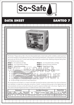


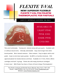


© Copyright 2026