
as a PDF
THE UNIVERSITY OF KANSAS
PALEONTOLOGICAL CONTRIBUTIONS
VE RTE B RATA
ARTICLE
Pages
9
1-32, Plate 1, Figures 1-32
OSTEOLOGY OF MYLAGAULUS LAEVIS, A FOSSORIAL
RODENT FROM THE UPPER MIOCENE OF COLORADO
By SYLVIA ROBINSON PAGAN
(Contribution
from the Museum of Natural History)
THE UNIVERSITY OF KANSAS PUBLICATIONS
November 21, 1960
PRINTED BY
THE UNIVERSITY OF KANSAS PRESS
LAWRENCE
THE UNIVERSITY OF KANSAS PALEONTOLOGICAL CONTRIBUTIONS
VERTEBRATA, ARTICLE
9, PAGES 1-32, PLATE 1, FIGURES 1-32
OSTEOLOGY OF MYLAGAULUS LAEVIS, A FOSSORIAL RODENT FROM
THE UPPER MIOCENE OF COLORADO 1
By SYLVIA ROBINSON PAGAN
Department of Zoology, University of Kansas
CONTENTS
PAGE
PAGE
ABSTRACT INTRODUCTION
Acknowledgments Materials and methods COMPARATIVE OSTEOLOGY Axial skeleton Skull Mandible Dentition Vertebrae Cervical Thoracic Lumbar Sacrum Caudal Ribs Pectoral girdle and forelimb Scapula Clavicle Humerus 5
5
5
5
6
6
6
9
10
11
11
11
11
12
12
12
12
12
13
13
A dissertation submitted in partial fulfillment of the requirements for
the Degree of Master of Arts in Zoology at the University of Kansas in
August, 1956. Modifications as of May, 1959.
Ulna Radius Manus Carpals Metacarpals Phalanges Pelvic girdle and hind limb Pelvis Femur Tibia Fibula Pes Tarsals Astragalus Calcaneum Metatarsals Phalanges DISCUSSION SUMMARY AND CONCLUSIONS REFERENCES 14
16
16
16
16
16
17
17
18
20
20
20
20
20
21
21
21
21
27
29
ILLUSTRATIONS
PLATE
PAGE
1. Skull and mandible of Mylagaulus and of
Aplodontia. Facing page
28
FIGURE
PAGE
1. Lateral view of fifth lumbar vertebra, KU9808
2. Posterior view of anterior free caudal vertebra,
KU9808 3. Lateral view of fifth lumbar vertebra, KU-
e 32020
4. Posterior view of anterior free caudal vertebra,
KU32020 a
5. Lateral view of right scapula, KU9808 6. Lateral view of right scapula, KU32020 7. Medial view of right scapula, KU9808 8. Medial view of right scapula, KU32020 9. Anterior view of right humerus, KU9808 10. Anterior view of right humerus, KU32020 11. Posterior view of right humerus, K1J9808 12. Posterior view of right humerus, KU32020 ....
13. Lateral view of left radius and ulna, KU9808 ....
14. Lateral view of left radius and ulna, KU32020 8
15. Third metacarpal and digit, KU9808 11
11
11
11
13
13
13
13
15
15
15
15
15
15
16
FIGURE
PAGE
16. Third metacarpal and digit, KU32020 a 16
17. Lateral view of right innominate bone,KU32020 e 17
18. Lateral view of right innominate bone, KU9808 17
19. Dorsal view of right innominate bone, KU9808 17
20. Dorsal view of right innominate bone, KU32020e 17
21. Anterior view of right femur, KU9808 19
22. Anterior view of right femur, KU32020 19
23. Posterior view of right femur, KU9808 19
24. Posterior view of right femur, KU32020 19
25. Anterior view of right tibia, KU9808 19
26. Ventral view of tarsal articulating surface of
tibia, KU9808 19
27. Anterior view of right tibia, KU32020 a 28. Ventral view of tarsal articulating surface of
tibia, KU32020
29. Dorsal view of left astragalus and calcaneum,
KU9808 30. Dorsal view of left astragalus and calcaneum,
KU32020 8 31. Lateral view of third metatarsal, KU9808 32. Lateral view of third metatarsal, KU32020
19
19
21
21
21
21
5
MIOCENE FOSSORIAL RODENT FROM COLORADO
ABSTRACT
MATERIALS AND METHODS
A skeleton of Mylagaulus (Mylagaulidae), a fossorial
rodent from upper Miocene beds in Colorado is compared with Aplodontia, the only living member of the
Aplodontidae. These comparisons suggest that certain
common characters are due to phyletic relationship and
others to a similar fossorial habit. Mylagaulus is more
fossorial than Aplodontia. The fossorial trends seen in
Mylagaulus foreshadow the fossorial structures seen in
Epigaulus, last of the Mylagaulidae.
The material used, in the University of Kansas Museum of Natural History unless otherwise noted, was
as follows.
Specimens
KU9808
KU9807
INTRODUCTION
The rodent family Mylagaulidae, ranging from
early Miocene to middle Pliocene, is regarded as an
offshoot of the family Aplodontidae. Both groups
became fossorial, the Mylagaulidae notably more so
than the Aplodontidae. The discovery of an excellent
skeleton of Mylagaulus, from upper Miocene beds in
northeast Colorado, affords an opportunity to compare
a member of the Mylagaulidae (suborder Sciuromorpha) with Aplodontia, the only living member of
the Aplodontidae.
Comparisons aim to reveal : (1) which common
characters are due to phyletic relationship and which
arc due to a similar habitat, (2) what differences are
caused by a difference in degree of adaptation, (3)
something of the life habits of Mylagaulus, and (4)
structural relationship of Mylagaulus to Epigaulus,
the last of the Mylagaulidae. Determination of the difference in degree of fossorial adaptation may help
clarify the effect of fossorial adaptation on basic
rodent morphology.
ACKNOWLEDGMENTS
Grateful acknowledgment is made to Dr. ROBERT W.
WILSON for suggesting the problem and for advice and
for criticism of the work, to Drs. E. RAYMOND HALL and
PETER P. VAUGHN for their constructive criticisms of the
manuscript and to Miss LUCY REMPLE for advice on the
illustrations which were prepared by the author.
After the main part of my study was completed, the
pertinent material of the genus Mylagaulus in the collection of the American Museum of Natural History, in New
York City, was examined. For the privilege of studying
the last-mentioned material, I thank Dr. George Gaylord
Simpson and Mrs. Rachel Nichols.
KU9969
FOSSIL SPECIMENS
Locality
SW 1/4 sec. 26, T. 12 N.,
R. 55 W., Logan County,
Colorado
WP/2 sec. 27, T. 12 N.,
R. 55 W., Logan County,
Colorado
E 'A sec. 27, T. 12 N.,
R. 55 W., Logan County,
Colorado
AMNH9043 Cedar Creek, Logan
County, Colorado
AMNH17576 Sinclair Draw, Sioux
County, Nebraska
Nature of material
Damaged skull, lower jaws
and postcranial skeleton
Anterior part of skull without cheek teeth
Complete humerus, fragments of cranial and postcranial skeleton, several
cheek teeth
Anterior part of skull with
teeth, damaged right mandible with teeth, right and
left ilia with attached
sacrum
Well-preserved skull lacking
nasal bones, part of the
basicranial region and the
right zygomatie arch.
KU—University of Kansas
AMNH—American Museum of Natural History
KU9808 and KU9807 were collected by E. C. GALBREATH in 1951 and reported on by him in the year 1953
(p. 96).
KU9969 was found by R. R. CAMP in 1956 and has
not previously been reported.
AMNH9043, the type of Mylagaulus laevis, was described and figured by MATTHEW in the year 1901 (p. 378379).
AMNH17576 was described and figured by MATTHEW
in the year 1924 (p. 75-82).
GEOLOGIC AGE OF FOSSILS
The geologic age of KU9808, KU9807 and KU9969
is late Miocene (late Barstovian); Pawnee Creek Formation (Kennesaw local fauna).
AMNH9043 also is from the late Miocene (Barstovian), Pawnee Creek Formation. AMNH17576 is from
the latest middle Miocene (late Hemingfordian), lower
Snake Creek beds.
RECENT SPECIMENS
Specimens of Recent fossorial rodents used in the
study here reported are recorded as follows.
THE UNIVERSITY OF KANSAS PALEONTOLOGICAL CONTRIBUTIONS
6
Materials
Locality
Specimens( a)
Date Collected
Aplodontia rufa rufa (Rafnesque)
KU 14408? Seattle, King County, Washington 12 March 1944
KU 14409 Seattle, King County, Washington 12 March 1944
KU32020 S Seattle, King County, Washington 26 March 1949
KU32043? North Cr., 20 mi. W. Olympia,
Thurston County, Washington 21 May 1949
Sciurus niger rufiventer É. Geoffroy Saint-Hilaire
KU9172 9
Denver County, Colorado (tag
indicates specimen was introduced from Oklahoma)
KU6206
Douglas County, Kansas
17 August 1931
13 January 1928
Thomomys bottae nigricans Rhoads
KU46468 8 Escondido, San Diego County,
California
3 July 1929
Thomomys toumsendii elOensis Davis
3 mi. W. Halleck, Elko County,
KU46480
Nevada
19 May 1929
Macrogeomys heterodus cartagoensis Goodwin
KU60664
Rancho Redondo, Volcan
San José, Costa Rica 3 June 1954
(a) Complete skeletons.
On the basis of size of the alveoli of P4 and MI, fossil
specimens KU9807 and KU9808 are thought to be adults
of approximately the same age; on the basis of the epiphyses, KU9969 is considered to be a younger individual,
although nearly adult as judged by the development of
processes for muscle attachment. The specimens of
Aplodontia rufa rufa are considered to be adult because
the tooth pattern is adult and sutures on the skull are
obliterated.
Few good specimens of mylagaulid rodents have been
reported in the literature. Use of only one species, Mylagaulus laevis, in the present study is hence a necessity,
but fortunately this species is typical of the family. The
limitation to a single species and subspecies, Aplodontia
rufa ru/a, does not affect the comparisons which are
thought to be valid on the generic level. For the same
reason, generic names, rather than specific and subspecific
combinations, are used throughout the comparisons and
discussion except where another course seemed more to
advantage.
For analysis and discussion of the structures observed
the following Recent specimens (complete skeletons)
were used.
Most of the comparisons are qualitative. Quantitative
data were taken by using dial calipers on KU9807,
KU9808 and where possible on KU9969 to supplement
measurements from KU9808. The corresponding measurements were taken from Aplodontia KU32020
which was thought to be typical of the genus. The number and kinds of measurements were dictated by the
condition of the fossils. These same specimens were used
in the accompanying illustrations.
When the comparisons and analysis which follow
were first done, use was made of published plates and
descriptions of Mylagaulus laevis wherever possible to
supplement and verify the conclusions reached on the
basis of the University of Kansas material. These figured
specimens, all from the American Museum of Natural
History, have subsequently been examined personally.
Unfortunately, direct comparisons of the University of
Kansas and American Museum of Natural History specimens has not been accomplished.
The nomenclature used in the comparisons is patterned after that in REYNOLDS (1897). Certain terms from
HILL (1935, 1937) are modified to accord with REYNOLDS
system.
'
MYLAGAULUS LAEVIS Matthew, 1924
Mylagaulus laevis Matthew, Bull. Amer. Mus. Nat. Hist., v. 16, art. 22, p. 298, Sept. 25, 1902
COMPARATIVE OSTEOLOGY
AXIAL SKELETON
SKULL
Plate 1, Table 1
Viewed dorsally, the skull is shorter, heavier and
broader in Mylagaulus than in Aplodontia. The width
across the zygomata (measured across widest part of the
arches) of Mylagaulus is greater than the length of its
skull (measured from tip of nasals to midpoint of the
lambdoidal crest). The width of the zygomata in Aplodontia is subject to individual variation (TAYLOR, 1918,
p. 443, pl. 28), but it is not so great as the length. The
skull of Mylagaulus is only as long as the occiput is wide
(measured across most lateral extensions of occiput).
The skull of Aplodontia is longer than the occiput is
wide.
Viewed laterally, the skull of Mylagaulus is more
flattened dorsoyentrally than that of Aplodontia. The
basicranial region of Mylagaulus is bent more ventrally
with respect to the palatal plane than in Aplodontia,
which is variable in this character but seemingly does not
form so large an angle with the palatal plane. The occiput
of both genera is inclined anteriad to the dorsoventral
plane; in Mylagaulus the occiput forms a larger angle
with this plane.
The nasals of both genera flare anteriorly and narrow
posteriorly. The posterior suture of Mylagaulus (AMNH17576) and Aplodontia is posterior to the anterior root
of the zygomatic arch. The nasals constitute a greater
part of the total dorsal length of the skull in Mylagaulus
than in Aplodontia. The nasals of Mylagaulus laevis
MIOCENE FOSSORIAL RODENT FROM COLORADO
(KU9807) are of special interest because they are thickened and rugose. The type specimen of Mylagaulus laevis
(AMNH9043) is described (MATTHEW, 1902, p. 298) and
figured (MATTHEW, 1901, as M. monodon, p. 378) as
having smooth nasals but this was probably by contrast
to the condition in horned mylagaulids. Personal observation of AMNH9043, which unfortunately is damaged in this critical region, reveals what might be a
thickening and rugoseness similar to, but not as pronounced as, that of Mylagaulus KU9807. The nasals of
Aplodontia are not thickened and are smooth.
The premaxillaries form the lateral and ventral surfaces of the rostrum in both genera. The premaxillaries
bear the incisors. The incisive foramina of Mylagaulus
are shorter than those of Aplodontia. A cross-section of
the rostrum of both genera is circular; the rostrum as a
whole is cylindrical in shape.
The anterior border of the orbit of Mylagaulus KU9807 is damaged but AMNH17576 and AMNH9043 do
not show the little rounded notch visible on the anterior
margin of the orbit of all specimens of Aplodontia examined. However, according to TAYLOR (1918, p. 443) this
notch is variable in degree of development in Aplodontia and is nearly lacking in some specimens.
The sutures on the dorsal surface of skulls of Mylagaulus (KU9807, KU9808) are obliterated (except for
the nasal suture mentioned above) but can be observed
to some extent in AMNH17576, which is a better preserved but also a younger individual than either KU9807
or KU9808. Care has been taken in the comparisons to
avoid emphasis on possible juvenile characteristics of
KU17576, unless they serve to clarify some feature.
Therefore, the discussion to follow will deal mainly
with areas which will not be sharply delimited.
The frontal area of both Mylagaulus and Aplodontia
is flattened transversely and in the midline is slightly
concave anteroposteriorly. The frontals of Mylagaulus,
unlike those of Aplodontia, are not constricted and bear
strong postorbital processes. The postorbital processes
of Mylagaulus are short, stout and blunt ended; they are
but slightly curved ventrally, lying almost wholly in the
horizontal plane. The frontals of Aplodontia are narrow
behind the anterior zygomatic root and form a saddleshaped interorbital constriction. Postorbital processes are
lacking in Aplodontia but what possibly might be incipient postorbital processes are present on KU32020
The parietal region of Mylagaulus continues the
flattened plane of the frontals back to the interparietal
region and lambdoidal crest; the parietal region of
Aplodontia is slightly convex, rising above the flattened
plane of the frontals. As recorded by MATTHEW (1924,
p. 80), the parietal extended far enough forward to share
in the postorbital process of the frontal of Mylagaulus,
". . . extending out on its postero-inferior surface almost
to the tip of the process." Microscopic examination of
KU9808 shows traces of what might be the suture in
7
question, but it cannot be definitively traced. The parietals of Aplodontia do not extend this far forward.
The temporal crests of KU9808 are weakly developed
and far apart; the temporal crests of Aplodontia are
variable in degree of development and in approach to
one another (TAYLOR, 1918, p. 443). Although Mylagaulus AMNH17576 is a younger individual than
KU9808, the temporal crests are well-developed and
close together.
The interparietal of Mylagaulus KU9808 cannot be
defined as a separate element; neither is the interparietal
of Aplodontia clearly defined. The interparietal is visible
in Mylagaulus AMNH17576 as a triangular element enclosed within the temporal crests and with its apex pointing anteriorly, which is in agreement with the traces of
the anterior suture observed in Aplodontia. However,
MATTHEW (1924, p. 81) terms this aspect of AMNH17576
as a ". . . supposed interparietal. . . ."
The zygomatic arch (measured from anterior to
posterior root) encompasses the greater part of the skull
in regard to length, and is more robust than that of
Aplodontia. In Mylagaulus the anterior root of the zygomatic arch originates immediately anterior to P4 and
forms a right angle with the axis of the skull, whereas,
in Aplodontia the anterior root originates opposite P3
and forms an acute angle to the axis of the skull. The
arch of Mylagaulus bends sharply posteriorly lateral to
its origin so that the arch is parallel to the anteroposterior axis of the skull. At the posterior extremity it
bends sharply mediad to form almost a right angle with
the expanded posterior region of the skull. The zygomatic arch of Aplodontia forms a slightly more acute
angle with the anteroposterior axis of the skull posterior
to its origin. Close to its posterior extremity the arch
bends medially and posteriorly, then medially and anteriorly forming a V before joining the expanded posterior region of the skull.
In both Mylagaulus and Aplodontia the anterior
surface of the zygomatic arch is pierced by the infraorbital foramen. It is roughly circular and large enough
to have transmitted a small slip of muscle, but not an
appreciable part of the masseter mcdialis. If the infraorbital foramen did transmit a slip of muscle there is no
reflection of the condition evident on the rostrum anterior
to the foramen.
The jugal region of the zygomatic arch of Mylagaulus
bears a strong postorbital process which is directed
dorsally toward the postorbital process of the frontal.
Except for KU32043?, all University of Kansas specimens of Aplodontia r. rufa lack a postorbital process on
the jugal area. The exception, KU32043?, exhibits what
might be an incipient postorbital process of the jugal
region. Specimens of the subspecies Aplodontia rufa
olympica often exhibit "... prominent postorbital process
on the jugals . . ." (TAYLOR, 1918, p. 461).
The zygomatic plate of Mylagaulus is relatively
8
THE UNIVERSITY OF KANSAS PALEONTOLOGICAL CONTRIBUTIONS
longer, wider and farther below the level of the infraorbital foramen than is the zygomatic plate of Aplodontia. The zygomasseteric scar of Mylagaulus does not
reach the inner base of the arch but is first developed on
the undersurface of that part which bends posteriorly
from the anterior root. The zygomasseteric scar of
Aplodontia is first developed immediately external and
ventral to the infraorbital foramen. The initial part of
the zygomatic plate of Mylagaulus and of Aplodontia is
separated from the anterior surface of the arch by a
sharp angulation.
The internal surface of the arch in Mylagaulus tends
to face somewhat dorsally in comparison with the internal surface in Aplodontia which is perpendicular to
the horizontal plane. The undersurface of the posterior
part of the zygomatic arch of Aplodontia exhibits a Vshaped plate which reflects the dorsal outline of the
upper surface of the same part. This ventral V-shaped
plate is present in the equivalent area of the arch of
Mylagaulus but is not reflected in the dorsal outline;
the apex of the V is pulled ventrally and anteriad and
the bony meatus is situated immediately behind it.
The squamosal of Mylagaulus is more nearly flat
than the squamosal of Aplodontia which gently curves
from the parietal over the braincase region; posteriorly,
the squamosal of Mylagaulus, as well as in Aplodontia,
is applied to the anterior surface of the lambdoidal crest.
The post-zygomatic notch of Mylagaulus is in close
association with the lambdoidal crest, being bounded by
it posteriorly; the post-zygomatic notch of Aplodontia
is bounded by an expansion of the squamosal anterior to
the lambdoidal crest. The prelambdoidal or subsquamosal foramen of KU9808 is represented on the dorsal
surface by a transverse series of three foramina along the
base of the lambdoidal crest. Comparison, however, with
AMNH17576 indicates this condition of KU9808 may
be an artifact of preparation, for there appears to be but
a single prelambdoidal foramen, amidst sculpturing, at
the base of the crest. The prelambdoidal foramen of
Aplodontia pierces the squamosal on the dorsal surface
anterior to the crest.
The squamosal, in Mylagaulus and in Aplodontia,
extends into the orbit. Immediately internal to the
posterior root of the zygomatic arch of Aplodontia and
marking the lateral limit of the braincase is a longitudinal sulcus. The coronoid process of the mandible
hooks back over the skull between this sulcus and the
posterior root of the arch. A shallow depression is present
in the comparable area of Mylagaulus, but it is not
clearly defined. The glenoid fossa of Mylagaulus and of
Aplodontia is only partially external to the depression
(sulcus) described above. The glenoid fossa of Mylagaulus is relatively longer than that of Aplodontia and
is deeper because of a sharp ventral dip to the alisphenoid.
The glenoid fossa of both genera is oblique to the axis
of the skull and is bounded posteriorly by the postglenoid foramen.
The lambdoidal crest of Mylagaulus, as well as that
of Aplodontia, is better developed laterally than in the
midline. The crest of Mylagaulus is considerably better
developed than that of Aplodontia and forms a flattened
and transversely widened plate when viewed posteriorly.
The occiput of Mylagaulus is more inclined forward and
in posterior view is more nearly flat than in Aplodontia.
The occiput of KU9808 is inclined so far forward that
the median dorsal part of the transverse crest is opposite
the posterior root of the zygomatic arch. The crest of
Mylagaulus AMNH17576 is not inclined so far forward
but the specimen is a younger individual and its crest is
probably less developed than in an adult.
In Mylagaulus and Aplodontia the occipital region is
remarkably similar in morphology. This region in KU9808 is damaged and the sutures are indistinct; therefore, the bulk of the comparisons utilize AMNH17576.
The mastoid plate of both Mylagaulus and Aplodontia
is of the same shape. A comparison between KU9808
and AMNH17576 indicates that increased age is correlated with an increase in size of the mastoid plate, as in
Aplodontia (TAYLOR, 1918, p. 444). The mastoid plate of
AMNH17576 and of Aplodontia is continuous with the
auditory bulla.
The supraoccipital sends out lateral extensions dorsal
to the mastoid plate of both Mylagaulus and Aplodontia.
The flange of bone forming the lateral extremity of the
occiput is separated from the supraoccipital by a posttympanic extension of the squamosal of both genera.
This flange could be an extension of the supraoccipital
or perhaps, as proposed by MATTHEW (1924, p. 80) for
AMNH17576, an extension of the parietal (as in hystricomorph rodents). The condition has not been determined for Aplodontia.
A palatal view of AMNH17576 shows a flask-shaped
auditory bulla with a long, outwardly directed neck or
bony meatus, as in Aplodontia. The bony meatus is
closely applied to the ventral, anterior surface of the
occipital plate in both genera.
The foramen magnum and basioccipital of Mylagaulus cannot be described because of lack of material.
Definite presphenoid, basisphenoid and basioccipital
sutures cannot be seen either in KU9808 or in AMNH17576. The basicranial region of Mylagaulus dips underneath the secondary palate at a sharper angle than in
Aplodontia, specimens of which exhibit this dip to varying but lesser degrees.
The alisphenoid is applied to a part of the anterior
surface of the auditory bulla of Mylagaulus, AMNH17576, as in Aplodontia. An alisphenoid canal is present
in both genera, having the same relationship to associated structures. Because of the basicranial plunge beneath the secondary palate, the alisphenoid of Mylagaulus
dips more sharply ventrally than in Aplodontia.
MIOCENE FOSSORIAL RODENT FROM COLORADO
It is not possible to make a detailed comparison of
the bones within the orbit because those of Mylagaulus
are either damaged or have the sutures obliterated. The
conformation of the orbital region of Mylagaulus and
of Aplodontia is strikingly similar. A comparison made
by McGREw (1941, fi g. 2, p. 7, 8) of the foramina in the
area anterior to the sphenoidal fissure reveals that the
pattern is similar in Mylagaulus and Aplodontia. A
difference that is of special interest is the presence of a
foramen, anterior to the sphenopalatine foramen and
above the enlarged P4 of Mylagaulus; this foramen is
absent in the comparable area of Aplodontia.
The pterygoid wings of both KU9808 and AMNH17576 are broken off but the condition of both suggests
they were important elements, more so than in Aplodontia. The superior pterygoid fossa of Mylagaulus is
relatively better developed than is this fossa in Aplodontia; it is longer and wider and tends to encompass
the opening of the alisphenoid canal.
The posterior border of the secondary palate of
Mylagaulus has a median projection; in Aplodontia it is
V-shaped with the apex pointing anteriorly. The toothrows and secondary palate of Mylagaulus are compressed
anteroposteriorly by comparison with Aplodontia. The
tooth-rows are parallel in both genera but seem to diverge
anteriorly in Mylagaulus because of the difference in size
between the anterior and posterior cheek-teeth. A small
foramen is situated medial and posterior to M3 in
Mylagaulus (better developed in AMNH17576 than in
KU9808) and in Aplodontia. The two palatine foramina
of both genera are opposite M2 and are larger in Mylagaulus. A groove runs anteriad from each palatine foramen, a condition which is better developed in Mylagaulus.
Anterior to the first cheek-tooth the maxillary region
of Mylagaulus curves sharply dorsad and then horizontally before the maxillary-premaxillary suture; this
creates an arched appearance immediately anterior to
the secondary palate. This profile is faintly developed in
some specimens of Aplodontia whereas in others the
undersurface of the rostrum is almost level with the
plane of the secondary palate.
MANDIBLE
Plate 1, Table 1
The mandible is deeper (depth measured below p4)
and shorter (length measured from posterior border of
incisive alveolus to posterior margin of tooth-row) in
Mylagaulus than in Aplodontia. The depth of the mandible of KU9808 is three-fourths of its length; the depth
of the mandible of KU32020 S is two-fifths of its length.
The diastema between the incisor and p4 of Mylagaulus
is relatively deeper and relatively longer than in Aplodontia. The length of the diastema in KU9808 is twofifths of the length of the mandible, whereas, in
KU32020 8 it is three-eighths of the length. The mental
9
foramen in each genus is half-way down the external
surface of the dentary. The mental foramen of Mylagaulus is opposite the midpoint of the diastema; the
foramen of Aplodontia is opposite a point two-thirds of
the length of the diastema posteriorly.
The coronoid process of Mylagaulus is oblique to the
dentary and forms a 55- to 60-degree angle in KU9808
to the body of the mandible; the coronoid process of
Aplodontia is perpendicular to the dentary. The coronoid
process of KU9808 is slender and barely hooked backward. The coronoid process of Aplodontia is stout and
in outline varies from being hooked backward strongly
to one in which the hook is scarcely developed (TAYLOR,
1918, p. 444).
The condyloid process and condyloid tubercle of both
genera are inclined medially; this inclination is more
pronounced in Mylagaulus. The condyloid process of
Mylagaulus is stouter as a whole. The angular process
of Aplodontia may best be described by quoting from
ELLERMAN (1941, p. 255): "Mandible with outer side of
angular process pulled inwards to an abnormal degree;
to such an extent that the posterior border is horizontal,
and the two edges of this process form the base of a
triangle which has for its apex the condylar process."
This region in KU9808 is broken, but the remaining
parts show this condition was present, although to a
lesser degree than in Aplodontia.
The dorsal masseteric ridge of Mylagaulus does not
extend anterior to the angular process on the external
surface of the mandible as does that of Aplodontia. This
ridge is pronounced in both genera and runs along the
angular process toward the external base angle of the
"triangle" described above. The masseteric fossa of Mylagaulus is deeper and better developed than that of Aplodontia. In relation to this better development of the masseteric fossa, the anterior border is formed by a ridge
running ventrally from the posterior border of the condyloid process and perpendicular to the dorso-ventral plane.
The comparable area of Aplodontia is thickened. In both
genera, the fossa is bounded ventrally by the dorsal masseteric ridge.
The ventral masseteric ridge of Mylagaulus is nearly
perpendicular to the long axis of the dentary, but at the
ventral border the ridge curves backward and inward
toward what would he the internal posterior tubercle of
the medial border of the angular process (this area of
KU9808 is missing). The ventral masseteric ridge of
Aplodontia is at an acute angle to the dorsal masseteric
ridge, approaches the ventral surface at an angle, and
traverses it to join the posteromedial tubercle of the inflected angular process.
The inferior pterygoid depression, enclosed by the
angular process, is better called a shelf than a depression.
Because of damage to this area of KU9808 an exact comparison cannot be made but it would seem that the inferior pterygoid depression was of greater height but
10
THE UNIVERSITY OF KANSAS PALEONTOLOGICAL CONTRIBUTIONS
not so deep anteroposteriorly as this depression in Aplodontia.
The sulcus ascendens of Mylagaulus is relatively
larger than that of Aplodontia; in both genera it is
separated from the inferior pterygoid depression by a
ridge. The inferior mandibular foramen of Mylagaulus
and of Aplodontia is situated on this ridge.
Inferiorly and posterior to the diastema, the dentary
of both Mylagaulus and Aplodontia bulges outward; this
bulge is less pronounced in Mylagaulus. Immediately posterior to this bulge in both genera the ventral masseteric
ridge crosses onto the ventral surface of the mandible
causing another, more pronounced, bulge.
TABLE 1.
Comparative Measurements of Axial Skeleton,
in Millimeters
Mylagaulus(a)
Aplodontia
KU9807, KU9808 KU32020e
SKULL
Total length (measured from tip of nasals
to median part of lambdoidal crest)
Width across zygomata (measured
across widest part of arch)
Length of skull encompassed by zygomata
Depth of zygomatic arch at anterior root
Length of rostrum (measured from
posterior border incisive alveolus to
anterior border of alveolus of P4)
Width across occiput
51.0(a,c)
70.0
58.0(c)
33.0(b)
14.5 (3)
56.0
39.0
7.5
15.0(a)
60.0 (3)
20.0
50.0
25.0 (b)
10.0(b)
18.0(b)
32.0
13.0
12.5
6.5(b)
7.0
10.0(6)
9.0(b)
7.5
10.0
13.5(6)
8.5
24.0(b,c)
17.0
MANDIBLE
Length (measured from posterior border
incisive alveolus to posterior
margin tooth-row)
Length of diastema
Depth of mandible below p4
VERTEBRAE
Length of centrum of 11th thoracic
Width of centrum of 11th thoracic
(measured across anterior surface)
Length of centrum of 5th lumbar
Width of centrum of 5th lumbar
(measured across anterior surface)
Width across transverse processes
of 5th lumbar
Width across transverse processes
of anterior free caudal
teeth are greatly enlarged and increase in width below
the occlusal surface.
The height of crown of P4 of Mylagaulus AMNH17576, a young individual, is sufficient that the base of
the tooth with its nearly-closed pulp cavities extends up
into the orbit to a level above the infraorbital foramen,
approaching the initial outflaring ventral surface of the
postorbital process of the frontal; the basal one-third of
the crown does not appear to have been covered with
bone in life. There is less extreme crown height in P4
of the older individuals, Mylagaulus AMNH9043 and
KU9808, this tooth in these having descended far enough
because of wear for the orbital portion to be covered
with bone. The available lower jaws of Mylagaulus
(KU9808, AMNH9043) indicate only that p4 was enlarged and hyposodont. The height of crown of P +of
Aplodontia is not so extreme; in this genus the pulp
cavities of the cheek teeth are open and the teeth evergrowing.
MATTHEW ( 1924, p. 79) suggests that as P-+ of Mylagaulus erupts to compensate for wear, the molars behind
them are progressively displaced from the jaw and the
alveoli taken over by P +so that in old age the dentition
could possibly be functional without any molars; then,
the dental formula of Mylagaulus could be expressed as
(WILSON, 1949, p. 103). BLACK & WOOD (1956, p.
1.0 1.3-0
670 and DORI& (1952) agree that, contrary to MATTHEW S
idea (1924, p. 79) the molars of Mylagaulus are not
progressively deciduous. Of possible pertinence to this
debate, SHOTWELL (1958, p. 476) cites the dental formula
1 .".° in adult mylagaulids of the Hemphillian with young
I.o.i.o
individuals, however, having small molars (SHOTWELL,
1958, fig. 15G, p. 467). The dental formula of Aplodontia is '.°. 2 . 3 .
There is some question about the homology of the
small tooth immediately behind P+ in specimens of
young mylagaulids. It has been identified as Dp 4- by
DOER (1952, p. 323) and as M-4- by MATTHEW (1924),
WiLsoN (1949), BLACK & WOOD (1956) and many others.
The adult cheek dentition of Mylagaulus using DORA S
interpretation (op. cit.) would be P±, M the alternate
interpretation of MATTHEW (1924) would be P-21-, M=2-=1
4 2-3
Although the weight of opinion favors the latter interpretation, the studied specimens cannot settle this question of homology. Mylagaulus AMNH9043 and KU9808
each have in both upper and lower jaws two cheek teeth
posterior to the enlarged P+ with no trace of the
alveolus of the displaced cheek tooth; AMNH17576, the
younger individual, also has this tooth missing but does
show traces of the alveolus posterior to P4 (no mandible
with this specimen).
The re-entrant folds and basins initially present in
the tooth of Mylagaulus become worn and develop into
a series of isolated enamel lakes, the number and disposi'
'
12.0(b,c)
6.5
(a) Measurements based on KU9807.
(b) Measurements based on KU9808.
(c) Measurements approximate because of damage.
DENTITION
The incisors of Mylagaulus and of Aplodontia are of
the same absolute width. The cheek teeth of both genera
are hypsodont. P3 of Mylagaulus is absent; P3 of Aplodontia is present as a functionless peg.
of both genera
4
are important teeth and are larger than the succeeding
molars. A highly specialized condition is developed in P 4
of Mylagaulus which is not present in Aplodontia; these
11
MIOCENE FOSSORIAL RODENT FROM COLORADO
tion varying with the species as well as with the age of
the individual, well-demonstrated by serial sectioning of
selected teeth by McGREw (1941). Prior to wear the
occlusal surface of the premolar of Aplodontia consists
of several completely enclosed basins. With wear these
enamel basins disappear and the pattern of the molars is
simplified to an enamel ring which in the upper series
has an externally directed mesostyle and in the lower
series has an internally directed mesostylid.
A detailed comparison of the occlusal pattern of the
specimens at hand would do little to further knowledge
of the relationship of these two genera because of the
pronounced dental specialization of Mylagaulus. McGREw
(1941) deals with the question of dentition in the Aplodontoidea and by use of a series of fossil specimens of
the families Mylagaulidae and Aplodontidae illustrates
the relationship of the patterns of Mylagaulus and
Aplodontia, and postulates that both patterns were derived from some common ancestral pattern.
genera tend to be flat and the posterior surfaces concave.
This tendency is more pronounced in Mylagaulus.
On the anterior thoracic vertebrae the facet for the
capitulum of the rib in both genera is situated partly on
the posterior margin of one centrum and partly on the
anterior margin of the succeeding centrum. The facet for
the capitulum of the rib of Mylagaulus appears to shift
caudally on the posterior thoracic vertebrae, as in Aplodontia, so that the articulation is no longer intercentral.
The transverse process of Mylagaulus and Aplodontia is
divided into ventral and dorsal parts; the ventral part
bears a flattened facet for articulation with the tuberculum
of the rib and decreases in development in posterior progression while the dorsal part becomes better developed in
posterior progression, becoming the mammillary process
of the lumbar vertebrae. The anterior thoracic vertebrae
of Mylagaulus and of Aplodontia bear strong neural
spines, which are heavier in Mylagaulus. Articulation of
the posterior thoracics of both genera is like that of the
succeeding lumbars.
VERTEBRAE
Table 1, Figures 1-4
CERVICAL VERTEBRAE
Since only fragments of the cervical vertebrae of
KU9808 are remaining, a count is not possible. The fragments remaining of the cervical vertebrae of KU9808 are
the lateral portions of the neural arches bearing the
zygapophyses. The neural arch of Mylagaulus seemingly
was more compressed anteroposteriorly than the neural
arch of Aplodontia. The zygapophyses of Mylagaulus,
unlike those of Aplodontia, are smoothly continuous
with, rather than being extensions from, the neural arch.
The anterior zygapophyses of Aplodontia are nearly
horizontal, anteriorly directed projections from the neural
arch with reciprocal posterior zygapophyses. The zygapophyses of Mylagaulus are longer, forming the entire
lateral portion of the neural arch and overlap considerably on the same vertebra; there is little overlap of the
zygapophyses of Aplodontia on the same vertebra. In
both genera, the anterior zygapophysis is barely concave
and the posterior zygapophysis is barely convex. The
scant remaining part of the walls enclosing the vertebroarterial canal of KU9808 suggests that the cervical
vertebrae of Mylagaulus extended relatively more laterally
than in Aplodontia.
ANTERIOR
Z YGAPOPHYS IS
TRANSVERSE
PROCESS
NEURAL
CANAL
4
FIGURES
1-4. Mylagaulus laevis, KU9808; Aplodontsa rufa
1, Lateral view of fifth lum2, Posterior view of anterior free
bar, KU9808.
3, Lateral view of fifth
caudal vertebra, KU9808.
4, Posterior view of anterior
lumbar, KU32020 .
free caudal vertebra, KU32020 . [All XL]
ru/a, KU32020 8 .
THORACIC VERTEBRAE
LUMBAR VERTEBRAE
No single thoracic vertebra of KU9808 is completely
undamaged so the following description is incomplete
and, moreover, is composite taking into account the possible variation along the vertebral column. Both Mylagaulus and Aplodontia have thirteen thoracic vertebrae.
The centra of the thoracic vertebrae of Mylagaulus are
relatively shorter and broader than the centra of Aplodontia. The anterior surfaces of the centra of both
The complete lumbar region of KU9808 is not present
so the vertebral count must be given as 5+?. The lumbar
region of Aplodontia consists of six vertebrae. The centra
of Mylagaulus are shorter and broader than the centra of
Aplodontia. The centra of KU9808 are about the same
length as the centra of adult specimens of Aplodontia
examined but are half again as wide. The centra of both
genera have a ventral median ridge developed.
12
THE UNIVERSITY OF KANSAS PALEONTOLOGICAL CONTRIBUTIONS
The transverse process of the fifth lumbar of Mylagaulus is a blade of bone directed more horizontally and
less anteriorly than in Aplodontia and does not have any
mammillary process. The comparable process of Aplodonlia does not have a bladelike shape; it curves anteriorly and
has a trace of a mammillary process on the posterior
margin. The first lumbar of Mylagaulus exhibits a
tnammillary process which is relatively shorter and stouter
than the comparable process of Aplodontia. The articular
facet of the anterior zygapophysis in both genera faces
mediad. The posterior zygapophysis in both genera is
reciprocal to, and fits into a groovelike arrangement of,
the anterior zygapophysis.
The neural spines in Mylagaulus and Aplodontia are
elongated longitudinally and keeled on the dorsal surface.
Although the extremities of the neural spines of the
lumbar vertebrae of Aplodontia are not in contact, they
may have touched together in Mylagaulus.
SACRUM
The sacrum of KU9808 is missing but part of the
type material for the species Mylagaulus laevis (M. monodon MATTHEW, 1901) is a sacrum affixed to the ilia,
which is used in the following comparisons.
The sacrum of Mylagaulus consists of at least four
vertebrae but the broken posterior surface of the fourth
sacral vertebra of AMNH9043 indicates that at least one
more vertebra entered into the sacrum; the sacrum of
Aplodontia consists of five vertebrae. Three of the sacral
vertebrae of Mylagaulus are affixed to the ilia, whereas,
only two sacral vertebrae of Aplodontia are affixed to the
ilia.
The neural spines in both genera have fused into a
plate, pierced by three foramina in Mylagaulus and by
o foramina in Aplodontia. The zygapophyses in both
have fused together and to the fused transverse processes,
acting as a superior buttressing plate which is better
developed in Mylagaulus. The anterior zygapophyses and
posterior zygapophyses of the sacral complex in both
have functional facets.
CAUDAL VERTEBRAE
A count of the caudal vertebrae of KU9808 is not
possible. Of particular interest is a caudal vertebra of
Mylagaulus, which is identified as the most anterior free
caudal because of its well developed neural canal (relative to other caudals), well developed anterior zygapophyses and the presence of a neural spine. The transverse processes of this vertebra of Mylagaulus are directed
horizontally and are perpendicular to the body of the
vertebra. They are thickened at the distal extremity. The
vertebra appears to be of the correct size to fit snugly
between the dorso-medial borders of the ischia. None of
the caudal vertebrae of Aplodontia has a neural spine, nor
do any of them show the expansion and longitudinal
thickening at the end of the transverse process as described above.
The more posterior caudal vertebrae of Mylagaulus
do not have a neural spine. They are wider relative to
their length than posterior caudal vertebrae of Aplodontia.
RIBS
KU9808 has thirteen ribs; each specimen of Aplodontia
examined had thirteen ribs. The anterior ribs of both
genera are double-headed; the capitulum articulates with
the centrum, and the tuberculum articulates with the
transverse process. The heads come closer together in
caudad progression. The angles of the dorsal part of the
anterior ribs form right angles with the centra. The first
rib of Mylagaulus and of Aplodontia is stouter than
the succeeding ribs. The neck of the first rib of Mylagaulus is longer, the tuberculum better developed and
the distal end is more flaring than is the first rib of
Aplodontia.
PECTORAL GIRDLE AND FORELIMB
SCAPULA
Table 2, Figures 5-8
The scapula of Mylagaulus appears to have the same
outline—almost quadrangular with thickened borders—
as in Aplodontia although part of the cranial border,
cranial angle and adjoining area of KU9808 are missing.
In KU9808 the scapula is approximately two-thirds again
as wide anteroposteriorly (measured along anteroposterior midline of scapula), one-fourth again as long
dorsoventrally (measured from point opposite origin of
scapular spine on vertebral border to glenoid cavity) as
the scapula of KU32020 8 and is considerably thicker.
TABLE 2. Comparative Measurements of Scapula,
in Millimeters
Anteroposterior width (measured across
mid-line perpendicular to axillary border)
Dorsoventral length (measured from
point opposite origin of spine on
vertebral border to glenoid cavity)
Width supraspinous fossa (measured
along mid-line described above)
Width infraspinous fossa (measured
along mid-line described above)
Adylagaulus
Aplodontia
KU9808
KU32020e
32.0(a)
19.0
43.5
35.0
14.0(a)
10.5
17.0
6.0
(a) Measurement approximate because of damage.
The vertebral border is thickened in both genera, but
relatively more so in Mylagaulus. The spine arises dorsally from the thickened vertebral border at the same angle
in both genera. Ventral to its origin, the spine of the
scapula of both is perpendicular to the body of the
scapula. The ventral two-thirds of the spine of KU9808
is missing.
MIOCENE FOSSORIAL RODENT FROM COLORADO
The supraspinous fossa of KU9808 is no wider (width
measured across anteroposterior mid-line), if as wide as,
the infraspinous fossa (width measured across anteroposterior mid-line); in KU32020 8 the supraspinous
fossa is nearly twice as wide as the infraspinous fossa.
Nevertheless, the scapular notch of Mylagaulus reduces
the size of the supraspinous fossa below that of the
infraspinous fossa at the scapular neck.
The axillary border of Mylagaulus is relatively more
thickened and turned more gently laterad, not forming
the axillary ridge observed in Aplodontia. The infraglenoid tuberosity of Mylagaulus is better developed and
more roughened than in Aplodontia.
13
The neck of the scapula of Mylagaulus is relatively
thicker than the neck of Aplodontia. The long axis of
the glenoid cavity in both genera is in the plane of the
body of the scapula. The glenoid cavity of Mylagaulus
is shorter anteroposteriorly and more nearly round than
that of Aplodontia, which is more nearly ovoid in outline. The coracoid process of Mylagaulus forms the
anterior part of the glenoid cavity and is relatively shorter
and stouter than that of Aplodontia in which there is a
slender hook situated on the anterior border of the
glenoid. The coracoid process of both genera is strongly
hooked anteromediad, the medial hooking being more
pronounced in Mylagaulus.
The medial surface of the scapula of both genera is
slightly concave. The medial surface of the scapula of
Aplodontia is marked by three muscular lines or ridges,
of which the two outer merge with the cranial and
axillary borders respectively at the neck of the scapula.
In KU9808 two muscular lines (area of third ridge is
in the missing cranial angle region) converge toward,
but fade before reaching, the neck of the scapula.
CLAVICLE
The clavicle of KU9808 is seemingly articulated with
the sternum, as in Aplodontia; the lateral half of the
clavicle of KU9808 is missing. The sternal articulation of
the clavicle of Mylagaulus is nearly circular in cross
section and is relatively better developed than in Aplodontia which has an articular area that is oval in cross
section. The shaft of the clavicle of KU9808 is thicker
and less flattened than is the transversely flattened shaft
of Aplodontia.
HUMERUS
Table 3, Figures 9-12
6
MUSCULAR LINE
CORACOID
8
FIGURES 5-8. Mylagaulus laevis, KU9808; Aplodontia rufa
5, Lateral view of right scapuru fa, KU32020 8 . 6, Lateral view of right scapula,
la, KU9808.
7, Medial view of right scapula,
KU32020 .
8, Medial view of right scapula, KUKU9808.
32020 8 . [All X1.]
The humerus in Mylagaulus (KU9808, KU9969) is
longer (length measured from most proximal surface of
head to center of distal articulation) and relatively stouter
than the humerus of KU32020 8 . The humerus is more
curved in Mylagaulus than in Aplodontia. In Mylagaulus,
the humerus bends medially in its proximal half; the
remaining part of the humerus is almost straight.
In both genera, the head is in line with the body of
the element. All of the greater tuberosity and most of
the lesser tuberosity of KU9808 is missing; consequently,
comparisons involving this region are made mainly with
KU9969. The greater tuberosity of KU9969 is about half
again as large as the lesser tuberosity and bears two
clearly marked depressions, as in Aplodontia. One of
these depressions is dorsal and the other is lateral.
Anteriorly, the intertubercular sulcus of KU9969,
unlike that in Aplodontia, is not defined, although there
is a deep groove running obliquely across the lesser
tuberosity which is not present in Aplodontia. Between
14
THE UNIVERSITY OF KANSAS PALEONTOLOGICAL CONTRIBUTIONS
the lesser tuberosity and head of both genera is a
roughened surface, which is relatively more widespread
in Mylagaulus. Below the lesser tuberosity of Mylagaulus,
in a region comparable to a roughened area in Aplodontia
for muscle attachment, is developed a median ridge
which persists to immediately above the level of the
deltoid tuberosity.
TABLE 3. Comparative Measurements of Humerus,
in Millimeters
Mylagaulus
KU9969
Length (measured from
proximal surface head to
center of distal articulation)
Distance from proximal
surface head to lower
margin deltoid process
Transverse width of
proximal epiphysis
Transverse diameter
of shaft at mid-line
Width across
epicondylar processes
Mylagaulus
KU9808
is a more distinct entity than in Aplodontia where it is
not sharply separated from the ectepicondylar fossa.
The olecranon fossa of Mylagaulus is shallower than
in Aplodontia. In Mylagaulus, the olecranon fossa is as
wide as the articulating facet above which it is situated;
in Aplodontia this fossa is not so wide as the articulating
facet. The distal articulating facet of Mylagaulus is
widened in proportion to the increase in size of the
humerus over that in Aplodontia but its height has not
increased to the same degree.
Aplodontia
KU32020e
ULNA
Table 4, Figures 13, 14
52.0
51.0
43.0
33.5
33.5
19.5
15.5
12.5
9.0
9.5
5.0
23.0
22.0
16.0
Anteriorly, the greater tuberosity of KU9969 is continuous with the deltoid crest, as in Aplodontia. The
deltoid crest of Mylagaulus is close to the shaft, flattened,
and terminates in a lobate deltoid process; the deltoid
crest of Aplodontia rises abruptly from the shaft of the
humerus, is a sharp ridge, and terminates in a sharp
deltoid process. The deltoid crest of Mylagaulus is less
lateral in position than is the crest of Aplodontia. The
long axis of the deltoid tuberosity in both genera is in
line with the shaft of the humerus. The deltoid tuberosity
of Mylagaulus, the major part of which is below the
midpoint, is relatively farther down the shaft of the
humerus than in Aplodontia where the major part is
above the midpoint. In Mylagaulus, a minor ridge from
the deltoid tuberosity blends ventrally into the medial
epicondyle. The same condition is present to a greater
extent in Aplodontia.
Below the level of the deltoid tuberosity the humerus
in both genera flares out laterally and medially to form
the ectepicondylar process and external epicondyle, and
internal epicondyle respectively. The ectepicondylar
process of both genera is a broad, winglike plate of bone;
that of Mylagaulus is better developed. The internal
epicondyle of Mylagaulus and of Aplodontia is pierced
by the entepicondylar foramen, which in Mylagaulus is
smaller. The medial surface of the medial epicondyle of
both genera is roughened. The medial extremity of the
medial epicondyle has a tendency, more pronounced in
Mylagaulus, to bend posteriorly. On the posterior surface
of the medial epicondyle of Mylagaulus and of Aplodontia is situated a deep fossa; this is relatively deeper in
the former.
The trochlea and capitulum of both genera are not
distinct from each other. The radial fossa of Mylagaulus
In both Mylagaulus and Aplodontia the ulna is the
longest bone of the forelimb. The ulna of Mylagaulus is
longer than that of Aplodontia, the total length being in
proportion to the increase in size of the humerus in
Mylagaulus. However, the proportions of the ulna proper
are different in the two genera. From the posterior edge
of the semilunar notch to the tip of the styloid process
the ulnae of KU9808 and KU32020 8 are of the same
absolute length. The difference in total length of the ulna
is due to an increase in length of the olecranon of
KU9808. The ulna of Mylagaulus is more robust than
that of Aplodontia.
The shaft of the ulna of Mylagaulus is gently curved
in one plane and that of Aplodontia is curved in two
planes. Viewed laterally, the ventral margin of the ulna
of Mylagaulus curves first ventrally and then dorsally,
tapering to a styloid process. Viewed laterally, the ventral
margin of the ulna of Aplodontia follows the path described above; viewed dorsally, the central third of the
shaft is bowed outward slightly.
From both lateral and dorsal aspects, the olecranon
of Mylagaulus is broader than is that of Aplodontia. The
dorsal edge of the olecranon of Mylagaulus is blunt; the
edge in Aplodontia is sharp and narrow. The tip of the
olecranon of Mylagaulus is bent mediad and expanded
more than in Aplodontia. In both, the ventral margin of
the ulna is expanded into a flattened ridge sometimes
TABLE 4. Comparative Measurements of Ulna and Radius,
in Millimeters
ULNA
Mylagaulus
Length of ulna (measured from posterior end
of olecranon to tip of styloid process)
Length from posterior lip of semilunar
notch to tip of styloid process
Dorso-ventral depth immediately
distal to radial facet
RADIUS
KU9969
My/tag/au/us
Length
Transverse width across head
Width across distal end
7.7
Aplodontia
KU9808
KU32020e
62.5
55.0
46.0
46.0
8.5
5.0
Mylagaulus
KU9808
38.0
7.5(a)
10.0(a)
Aplodontia
KU32020cr
39.5
6.0
7.5
15
MIOCENE FOSSORIAL RODENT FROM COLORADO
termed the bicipital ridge (HOWELL, 1926); the bicipital
ridge of Mylagaulus is better developed.
Dorsally, the coronoid process and the dorsal lip of
the semilunar notch are in line with each other and set
at an angle to the body of the ulna in Mylagaulus; the
same is true of Aplodontia. The radial notch of KU9808
LESSER
TUBEROSITY
INTERTUBERCULAR
SU LC US
ENTEP ICON DY L AR
FORAME
ECTEPICONDYL AR
PROCESS
RADIAL FOSSA
9
- INTERNAL
EPICONDYLE
11
10
OLECRANON
FOSSA
SEMILUNAR NOTCH
-
FIGURES 9-14. Mylagaulus
9, Anterior view of right humerus, KU9808.
Anterior view of right humerus, KU32020 3
11, Posterior view of right humerus, KU9808.
OLECRANON
KU9808; Aplodontia rufa ru fa, KU32020 a
10, Posterior view of right humerus, KU32020 .
13, Lateral view of left radius and ulna, KU9808.
12, 14, Lateral view of left radius and ulna, KU32020 3-.
[All X1.]
.
.
16
THE UNIVERSITY OF KANSAS PALEONTOLOGICAL CONTRIBUTIONS
has been damaged but the radial notch of KU9969 is a
distinct facet and has the same shape as in Aplodontia.
Below the radial notch which is situated laterally in
both genera, the line of articulation with the radius can
be traced to the styloid process. In Mylagaulus it is a
relatively broader line than in Aplodontia, curves to the
dorsal edge, and at the styloid process is barely over onto
the internal surface of the ulna; in Aplodontia, the radius
curves to the dorsal edge but does not reach the internal
surface. The distal third of the radial line in both genera
is a groove, considerably deeper in Mylagaulus.
The lateral and medial concavities in Mylagaulus
appear to be less developed than in Aplodontia. The
body of the ulna of Mylagaulus is less tapered anteriorly
(toward styloid process) than this element in Aplodontia.
The styloid process of Mylagaulus is disproportionately
better developed than that of Aplodontia, although it
maintains the same shape.
MANUS
Table 5, Figures 15, 16
CARPALS
None of the carpal elements of KU9808 are present.
METACARPALS
The metacarpals of Mylagaulus are approximately
the same length as but are thicker than those of Aplodontia. In both genera, distal articulations are limited
to the anterior and ventral surfaces and the ventral
surfaces of the distal articulations are keeled. In relation
to the length of the metacarpal, the distal articulation is
larger in Mylagaulus than in Aplodontia. In each genus
there are two fossae, one on each side of the distal end
of the metacarpal, which are relatively deeper in
Mylagaulus.
RADIUS
Table 4, Figures 13, 14
In both KU9808 and KU32020 the radius is of
approximately the same absolute length and is shorter
than the ulna by the length of the olecranon process and
semilunar notch. Viewed dorsally, the curvature of the
shaft of the radius is similar in Mylagaulus and Aplodontia; the shaft is gently bowed laterally over the distal
two-thirds of its length. Viewed laterally, there is a
gentle dorsal bowing of the shaft in both genera. The
radius of Mylagaulus is more robust than that of Aplodontia. In both genera, the shaft is elliptical in cross
section.
The head of the radius of Mylagaulus is relatively
more expanded than that of Aplodontia. Despite some
damage to the external edge of the head in KU9808 the
shape is judged to be ovoid and is definitely ovoid in
KU9969, as in Aplodontia. The radius in both genera is
in contact along most of its length with the ulna. On the
ulnar side of the radius of both genera for the distal
third of its length, a narrow ridge is developed which
fits into a groove on the ulna; this ridge in Mylagaulus
is considerably better developed. On the lateral surface
of the distal end of the radius of both genera there is a
deep groove, terminating in the carpal articulation.
The distal articulating facet of Mylagaulus is relatively more expanded than the facet of Aplodontia. The
distal facet of Mylagaulus has undergone rotation in
comparison to that of Aplodontia. Viewed anteriorly,
the distal articulating facet of Mylagaulus has been
rotated 25 to 30 degrees externally with relation to the
shaft. The carpal articulating surface in both genera has
a styloid process developed on the dorsal edge which is
concave.
FIGURES 15-16. Mylagaulus laevis, KU9808; Aplodontia
rufa ru fa, KU32020 8 .
15, Third metacarpal and
digit, KU9808.
16, Third metacarpal and digit,
KU32020 d . [All X2. 1
PHALANGES
What appears to be the third digit of KU9808 has
been preserved in an articulated state and has been
utilized for the following comparisons. The proximal
phalanx is shorter (length measured along lateral longitudinal axis) and all three phalanges stouter in Mylagaulus than in Aplodontia. The proximal articulation in
both genera is concave and notched inferiorly. The proxi-
MIOCENE FOSSORIAL RODENT FROM COLORADO
mal articulation in Mylagaulus is relatively deeper and
inclined forward; consequently, the dorsal border is more
TABLE
5. Comparative Measurements of Manus,
in Millimeters
Total length third metacarpal
Length of head of third metacarpal
Breadth of head of third metacarpal
Length third proximal phalanx
(along lateral long axis)
Breadth of distal articulation
third proximal phalanx
Length of second phalanx, third digit
(measured along lateral long axis)
Breadth of distal articulation
second phalanx, third digit
Length of third ungual phalanx
Breadth of third ungual phalanx
(measured across proximal end)
Mylagardus
Aplodorma
KU9808
KU32020e
13.0
4.5
5.0
12.0
6.5
7.0
4.5
3.0
3.0
3.5
17
fossae on the lateral surfaces of the distal ends of the
phalanges in Mylagaulus are relatively deeper than those
in Aplodontia.
The ungual phalanx in both genera is elongated and
blade-shaped, being flattened transversely. The proximal
articulation of the ungual phalanx is concave and so
shaped that it covers the anterior and dorsal parts of the
preceding phalanx. The ungual phalanx of Mylagaulus
is longer, deeper, and considerably heavier than that of
Aplodontia.
PELVIC GIRDLE AND HIND LIMB
PELVIS
Table 6, Figures 17-20
6.5
4.5
4.5
11.5
2.5
8.5
3.5
2.0
anterior than in Aplodontia. The distal articulations of
the proximal and middle phalanges of Mylagaulus are
relatively enlarged by comparison with Aplodontia. The
distal articulation in both genera is limited dorsally,
being more pronounced in Mylagaulus; the distal articulation of the middle phalanx in both genera is not so
limited superiorly as in the proximal phalanx. The
The pelvis of Mylagaulus is larger and more robust
than that of Aplodontia, but not to the same degree as
the pectoral girdle. Measured from the mid-point of the
acetabulum, the pelvis of Mylagaulus is one-fifth again
as long in its anterior part and one-third again as long
in its posterior part as the pelvis of Aplodontia. There
has been some increase in depth of the pelvis of
Mylagaulus posterior to the acetabulum. The pelvis of
Mylagaulus forms more of an angle with the vertebral
column than does the pelvis of Aplodontia.
Superiorly, the ilia of Mylagaulus are more divergent
than those of Aplodontia. The anterior part of the ilium
in both bears an iliac crest which has less of a medial
SUPER I OR
TUBEROSITY
ILIAC CREST
OBTURAPOR
F CRANE N
17
FIGURES
17-20. Mylagaulus laevis, KU9808; Aplodontia rufa ru fa, KU32020 6 .
17, Lateral view of right innominate bone, KU32020 8 .
18, Lateral view of right innominate bone,
KU9808.
19, Dorsal view of right innominate
bone, KU9808. 20, Dorsal view of right innominate bone, KU32020 . [All X1.]
18
THE UNIVERSITY OF KANSAS PALEONTOLOGICAL CONTRIBUTIONS
extension in Mylagaulus. The iliac ridge in Mylagaulus
and in Aplodontia extends from the iliac crest to the
acetabulum on the lateral surface of the ilium, dividing
it into a gluteal fossa superiorly, and an iliac fossa
inferiorly. The iliac ridge in both genera is roughened
and expanded immediately anterior to the acetabulum.
The gluteal fossa of Mylagaulus is less excavated than
that of Aplodontia; the iliac fossa of Mylagaulus is better
developed than the iliac fossa of Aplodontia.
TABLE 6.
Comparative Measurements of Pelvis,
in Millimeters
Mylagaulus
Aplodontia
KU9808
KU32020e
Length of iliac crest to
mid-point of acetabulum
42.5
Length from posterior margin of superior
tuberosity to mid-point of acetabulum
28.5
Depth of ilium through posterior part
of sacral attachment scar
11.5(a)
Anteroposterior length of sacral scar
21.0
Depth measured from dorsal border of superior
tuberosity to level of pubic symphysis
29.0(a)
Anteroposterior length of pubic symphysis
4.0(a)
Transverse depth of pelvis at acetabulum
(measured from dorsal lip to medial border) 9.0
Allowing for damage to KU9808, it seems that the pubic
symphysis is shorter anteroposteriorly in Mylagaulus
than in Aplodontia. The obturator foramen is of the
same shape in both genera.
The acetabulum of Mylagaulus is relatively deeper
and of greater diameter than the acetabulum of Aplodontia. The parts of the innominate elements bordering
on, and incorporated into, the acetabulum of Mylagaulus
are relatively thicker, to accommodate the increased depth,
than in Aplodontia. Judging from what remains of the
inferior border of the acetabulum of KU9808, the
acetabular notch was in the same relative positionposteroventrad—as in Aplodontia.
FEMUR
35.0
22.0
5.0
17.0
24.5
6.0
6.5
(a) Measurement approximate because of damage.
The ventral border of the ilium of Mylagaulus is
parallel to and close to the plane of the iliac ridge
anteriorly, but diverges from it posteriorly in a gentle
curve toward the iliopectineal prominence. The inferior
border of the ilium of Mylagaulus is more ventral than
that of Aplodontia, as well as more lateral.
The sacroiliac joint of Mylagaulus is lengthened
posteriorly and has been shortened anteriorly compared
to the sacroiliac joint of Aplodontia. The more ventral
and less medial position of the inferior border of the
ilium of Mylagaulus and a more posterior position of the
gluteal notch offers a deeper surface dorsoventrally for
the sacroiliac joint than is present in Aplodontia.
Although the dorso-medial border of the ischium of
KU9808 is damaged, there is a medial inflection toward
the vertebral column posterior to the acetabulum, as in
Aplodontia. Articulation of the ischium with the caudal
vertebrae cannot be definitely ascertained from KU9808
but the pelvis of Mylagaulus does suggest that the inflection was greater than that observed in Aplodontia
where, in the specimens examined, there is no indication
of a union of the ischium with the caudal vertebrae.
The superior tuberosity of Mylagaulus is relatively
longer and higher above the dorsal lip of the acetabulum
than in Aplodontia. The posterior (ischial) part of the
obturator foramen in both Mylagaulus and Aplodontia
is at a right angle to the horizontal.
The pubic rami of both genera are inclined posteroventrad to the acetabulum. The pubic symphysis, formed
by medial extensions of the rami, is well caudad in both.
Table 7, Figures 21-24
The femur in Mylagaulus is longer (measured from
proximal surface of head to distal surface of patellar
groove) and thicker (measured at midpoint) than in
Aplodontia. The length and thickness of the femur of
Mylagaulus is less than would be expected by comparison with its forelimb, although in keeping with the size
of the pelvis. The femur of Mylagaulus is longer than
its humerus on an absolute scale. However, the femur of
Aplodontia is relatively longer in relation to its humerus
than is the femur of Mylagaulus in relation to its
humerus. The head of the femur in both genera is
spherical and is shifted cranially and comparatively
better developed in Mylagaulus; the greater development
is in proportion to the depth of the acetabulum. The
fossa for the ligamentum teres in Mylagaulus is relatively
deeper than in Aplodontia.
The greater trochanter in both genera occupies the
same relative position on the lateral side of the proximal
epiphysis. The greater trochanter does not extend dorsally
above the level of the head of the femur in Mylagaulus;
it does extend slightly above the level of the head in
Aplodontia. The lesser trochanter is on the medial side of
the femur, beneath the level of the head of the femur,
and has the same shape and same relative degree of
development in both genera. The two trochanters are
not connected posteriorly by a trochanteric ridge. The
TABLE 7.
Comparative Measurements of Femur,
in Millimeters
Length (measured from proximal surface of
head to distal surface of patellar groove)
Anteroposterior diameter of head
Transverse width of shaft at mid-point
Transverse width through widest part of
lateral crest perpendicularly through shaft
Anteroposterior width at mid-line
Transverse width of distal epiphysis
Breadth of patellar groove
Mylagau/us
Aplodontia
KU9808
KU32020e
56.5
8.5
8.0
53.0
6.5
6.0
14.0
5.5
7.0
14.5
9.0
4.0
5.0
12.5
MIOCENE FOSSORIAL RODENT
trochanteric fossa of Mylagaulus is shallower than that
of Aplodontia.
The lateral crest of Mylagaulus is well developed and
is continuous with the external margin of the greater
trochanter. The lateral crest in Aplodontia is not so well
developed and is not continuous with the greater trochanter. The lateral border of the lateral crest in Mylagaulus is bent anteriorly forming a deep, broad fossa
between the lateral limit of the crest and the shaft of
the femur. This character is only suggested in Aplodontia.
The lateral crest in both genera extends one-third of the
length of the femur.
The patellar groove in Mylagaulus is broader and
FROM COLORADO
19
more external than the groove of Aplodontia which is
centered on the distal end of the femur. The fossae on
the external surface of the lateral epicondyle of Mylagaulus, which probably represents attachments of the
lateral head of the gastrocnemius and ligaments, are
relatively better developed than those in Aplodontia, but
have the same conformation. Fossae on the internal
surface of the medial epicondyle are better developed
in Mylagaulus but have the same arrangement as in
Aplodontia. They probably represent the origin of the
medial head of the gastrocncmius and attachments of
ligaments. The popliteal fossa of Mylagaulus is deeper
than in Aplodontia.
OR
TROCHANTER
HEAD
LESSER
TROCHANTER
THIRD
TROCHANTER-
PATELLAR
GROOVE
CNEPAIAL CREST
25
21-28. Mylagaulus laevis, KU9808; Aplodontia rufa ru/a, KU32020 8 .
22, view of tarsal articulating surface of tibia, KU9808.
21, Anterior view of right femur, KU9808.
27, Anterior view of right tibia, KU32020 8 .
23, KU32020
8
.
right
femur,
of
Anterior view
28, Ventral view of tarsal articulating surface of
24, Pos- Posterior view of right femur, KU9808.
tibia, KU32020 a [Figs. 21-25,27 XI; figs. 26,28 X2.]
25, An-
tenor view of right femur, KU32020 a .
26, Ventral
tenor view of right tibia, KU9808.
FIGURES
.
20
THE UNIVERSITY OF KANSAS PALEONTOLOGICAL CONTRIBUTIONS
TIBIA
Table 8, Figures 25 28
-
The tibia of Mylagaulus is shorter than its femur;
in Aplodontia the tibia is approximately equal to the
length of the femur. The tibia in both genera exhibits
torsion in the anteroposterior plane and in the transverse
plane. Torsion in the transverse plane is more pronounced
in Mylagaulus. The articular arm for the femur of
Mylagaulus is relatively more expanded, but does have
the rounded triangular shape of proximal epiphysis of
Aplodontia. This expansion in Mylagaulus is evident in
the greater external extent of the proximal epiphysis
compared to that in Aplodomia. The medial and lateral
condyles in both genera are concave in the transverse
plane and convex in the anteroposterior direction. The
tuberosity is part of the epiphysis of Mylagaulus and of
Aplodontia. Articulation of the fibula in Mylagaulus and
in Aplodontia is at the external underside of the proximal
epiphysis and the contact area is relatively larger in
Mylagaulus.
Although the proximal end of the cnemial crest of
Mylagaulus is situated at the same position with respect
to the condyles as the crest of Aplodontia, it is more
lateral with respect to the shaft because of a greater
lateral extension of the epiphysis of Mylagaulus. The
cnemial crest of Mylagaulus is twice as high as the crest
of Aplodontia, which is a thin, sharp blade of bone. In
Mylagaulus, the crest probably curves mediad to run
along the anterior surface before blending with the shaft
below the mid point; in Aplodontia it is on the anterior
surface of the shaft and extends half the length of the
-
TABLE 8. Comparative Measurements of Tibia,
in Millimeters
Mylagaulus
K U9808
Length (measured from dorsal
surface proximal epiphysis to
ventral surface distal epiphysis)
Anteroposterior diameter of shaft at
point one-third of way down shaft
Transverse diameter of shaft at
point one-third of way down shaft
Transverse diameter of distal epiphysis
Aplodontia
K U32020
The tarsal articulating surface of Mylagaulus is relatively more expanded than that of Aplodontia, although
in both genera the surface consists of two valleys separated by a ridge, which fits the trochlea or groove of
the astragalus. The median valley is bounded by median
and posterior malleoli which are relatively longer and
more incurved in Mylagaulus. The lateral valley, bounded
by an anterior malleolus of the tibia, is deeper in Mylagaulus. The entire distal articulation of the tibia of
Mylagaulus has been rotated approximately 45 degrees
with respect to the shaft when compared with the tibia
of Aplodontia; consequently, the medial malleolus is
more posterior and the lateral malleolus is more anterior.
The posterior and lateral surfaces of the posterior
malleolus of both genera are grooved dorsoventrally.
FIBULA
The fibula of KU9808 is represented only by fragments but much can be told from these, and from its
reflection on the tibia, about its structure. The proximal
epiphysis of the fibula of Mylagaulus and of Aplodontia
is a separate entity from the epiphysis of the tibia. The
shaft in Mylagaulus is relatively heavier than that of
Aplodontia and has an interosseous crest developed in
response to the interosseous crest of the tibia. Because of
greater transverse bowing mediad, and the anterior shift
of articulation of the styloid process (lateral malleolus)
of the fibula of Mylagaulus, the interosseous space is
larger and the distal part of the fibula makes a shorter
contact with the tibia than was observed in Aplodontia,
where the fibula is in contact for one-third of its distal
length. The internal surface of the styloid process of
Mylagaulus and of Aplodontia bears a facet for articulation with the external surface of the astragalus.
a
PES
Table 9, Figures 27-30
50.0
53.5
11.0
8.0
5.0
7.5
3.5
6.5
The proportions of the pes of KU9808 are more
delicate and the pes approaches in pattern that of Aplodontia more closely than does the manus.
TARSALS
shaft. Consequently, the lateral fossa of Mylagaulus is
better developed.
The interosseous crest of Mylagaulus is sharper and
more clearly defined than this crest in Aplodontia. The
medial crest of both genera follows the line of the shaft
from the external limit of the medial epicondyle for
two-thirds of the length of the element. The medial
crest of Mylagaulus does not project so far from the shaft
as in Aplodontia. Between the medial and interosseous
crests of both is the caudal fossa which in Mylagaulus is
a deep, narrow furrow and in Aplodontia is a broad,
shallow fossa.
The only tarsals present in KU9808 are the calcaneum
and astragalus.
Astragalus. The shape and proportions of the astragalus of Mylagaulus and of Aplodontia are similar except
for a slightly larger head in the former. The body of the
astragalus in each is broader than high. The external
condyle is wider than the internal condyle and is separated from it by the trochlea. The trochlea is pulley-like
and extends over the dorsal surface and complete posterior surface of the astragalus of both genera. Two facets
for articulation with the calcaneum are situated on the
inferior surface of the body of the astragalus. The ex-
21
MIOCENE FOSSORIAL RODENT FROM COLORADO
ternal facet is concave and articulates with the dorsal
process of the calcaneum; the internal facet is smaller,
separated from the external facet by a groove, and
articulates with the dorsal surface of the sustentaculum
of the calcaneum. The neck is short and the head roughly
rounded.
TABLE
9. Comparative Measurements of Pt's,
in Millimeters
Proximodistal length astragalus
Mediolateral width astragalus
Dorsoventral depth astragalus
Anteroposterior length calcaneum
Internoexternal
length of
sustentaculum
Dorsoventral depth of shaft
of calcaneum at midpoint
Dorsoventral depth of posterior
extremity of shaft of calcaneum
Length of third metatarsal
Length of head of third metatarsal
Breadth of head of third metatarsal
Mylagaulus
KU9808
Aplodontia
KU32020e
8.9
6.0
4.5
16.5
9.5
8.3
6.0
4.5
13.5
18.5
6.0
5.0
8.0
17.0
3.5
3.5
5.5
15.5
2.5
3.0
Cakaneum. The calcaneum in both genera is Tshaped, the main shaft oriented anteroposteriorly and
the sustentaculum oriented internoexternally. The calcaneum of Mylagaulus is more robust and relatively
larger in comparison with its astragalus than in Aplodontia. The main shaft is relatively deeper, the posterior
extremity of the shaft more rounded, the keel of the
shaft more pronounced, and the sustentaculum broader
in an anteroposterior direction in Mylagaulus. A groove
situated on the lateral surface of the external part of the
sustentaculum is more pronounced in Mylagaulus than
in Aplodontia. Another groove runs along the dorsal
root of the internal part of the sustentaculum of both
genera. The anterior articulating surface of both Mylagaulus and Aplodontia is concave.
the head of the metatarsal of Mylagaulus is enlarged in
relation to length and breadth.
PHALANGES
The phalanges of Mylagaulus are barely longer, but
are considerably heavier than those of Aplodontia. The
proximal articulation of the proximal phalanx of both
genera is concave and notched on the inferior border,
both conditions being exaggerated in Mylagaulus. The
distal head of the proximal phalanx of both is bipartite
with the greater part of the articulation limited to the
inferior surface. The proximal articulation of the middle
phalanx in both genera is concave and set at an angle to
the body of the phalanx with the superior margin more
anterior than the inferior margin. The head is deeply
grooved, not so limited superiorly as in the proximal
phalanx, and is more superior in Mylagaulus than in
Aplodontia.
TROCHIE A
SUSTENTACULUM
METATARSALS
Because of the fragmentary nature of the remains of
the metatarsals of KU9808, discussion will concern proportions and articulations only.
The metatarsal of Mylagaulus is slightly longer on
an absolute scale than in Aplodontia, but is relatively
more heavily developed. The head of the metatarsal in
both bears a keel on the inferior surface, which is more
pronounced in Mylagaulus and ends almost in a hook.
The depressions on the lateral surfaces of the metatarsal
are deeper in Mylagaulus. In comparison with Aplodontia
32
29
31
29-32. Mylagaulus laevis, KU9808; Aplodontia
29, Dorsal view of left
rufa ru/a, KU32020 s .
FIGURES
30, Dorsal
astragalus and calcaneum, KU9808.
view of left astragalus and calcaneum, KU32020 .
31, Lateral view of third metatarsal, KU9808.
32, Lateral view of third metatarsal, KU32020 a
[All X2.]
.
DISCUSSION
Many of the similarities between the skeletons of
Aplodontia and Mylagaulus are explicable on the
basis of common habitat as well as on the basis of
phyletic relationship. A difference in degree of adaptation to this common habitat serves to explain many
of the differences observed.
22
THE UNIVERSITY OF KANSAS PALEONTOLOGICAL CONTRIBUTIONS
For progression through the dense medium of
earth Aplodontia and Mylagaulus have developed a
wedge-shaped head, which is flattened dorsally. The
more flattened skull of Mylagaulus shows greater
fossorial modifications than the skull of Aplodontia
but the two are elaborations of the same pattern
(Pl. 1). The greater tilting forward of the occiput,
the greater flattening of the roof of the skull and
the heavier zygomata all are reflections of its greater
adaptation to underground existence. These greater
fossorial modifications of Mylagaulus have led to a
change in proportions of the skull as compared to
those in Aplodontia; in many respects, Mylagaulus
actually brings closer to a culmination the fossorial
Mylagaulus and of Aplodontia
compared to Sciurus
are all responses to fossorial existence. These developments provide firm attachments for some masticatory
muscles and for muscles from the trunk region to
hold the skull rigidly and solidly under stress of
forward motion. The pattern of this region in
Aplodontia and Mylagaulus is similar, but the latter
shows greater expansion and development of areas
where muscles attach and greater shortening of the
skull.
The tendency of the zygomatic arch to become
thin and even for a part of the arch to disappear in
some fossorial mammals has been interpreted as a
means of reducing friction (SHimER, 1903, p. 822).
adaptations of Aplodontia.
This tendency is not evident in Mylagaulus and
The rostra! region of a fossorial animal is im- Aplodontia, for both have the primitive sciuromorph
portant as the leading part of the axial skeleton in musculature; the origins of the masseter medialis and
forward motion. Importance of the rostrum is shown masseter lateralis are on the zygomatic arch in each
by the ratio of length of rostrum to length of skull genus. A fossorial mammal such as a mole (insectiin Aplodontia in comparison with a nonburrowing vore) does not have this complicated musculature
animal such as Sciurus. The rostrum of Mylagaulus and the arch therefore can be reduced and in the
is relatively longer than the rostrum of Aplodontia, related shrews disappears. Such reduction would debecause of the greater specialization of the former. crease the efficiency of the masticatory mechanism of
The shape of the rostrum of Aplodontia and of Mylagaulus and of Aplodontia; this illustrates that
Mylagaulus is similar, but this may be due in part adaptation can take place only within the framework
to a lack of masseter specialization onto the rostrum. of the anatomy of the forms involved. Probably in
The skull of Mylagaulus is more nearly flat than connection with reducing friction, the arches of
the skull of Aplodontia, which in turn is more nearly Aplodontia do not project far beyond the lateral
flat than the skull of a non-fossorial mammal such limits of the occiput and the arches of Mylagaulus
as Sciurus. The flat skull and thick roof of the skull not at all.
of Mylagaulus is an exaggeration of the trend seen
The greater heaviness of the zygomatic arches,
in Aplodontia.
broader and more posterior extension of the zygoThe presence of postorbital processes on the fron- matic plate and the deeper internal surface of the
tals and jugals of Mylagaulus does not exclude it arch of Mylagaulus suggests an increase in importance
from relationship with Aplodontia. The pre-Arik- of masticatory muscles. The glenoid fossa of Mvlaareeqn genera and some of the Arikareean genera of gaulus indicates less lateral movement of the mandible
thc Aplodontidae possessed these processes (WiLsoN, was possible than in Aplodontia.
1949, p. 102). They are still exhibited in some forms
The mandible of Mylagaulus is deeper and more
of Aplodontia as incipient processes. This is probably robust than the mandible of Aplodontia. To some
a phyletic divergence for while it could be postulated extent this depth in Mylagaulus may be related to
that these processes of Mylagaulus serve to protect the enlargement of p4. The peculiar development of
the eye by ringing it with bone, typically fossorial the angular process in Mylagaulus and in Aplodontia
rodents such as Thomomys and Macrogeomys do not is probably due to inheritance from a common patpossess them and a non-fossorial form, Sciurus, does. tern which tended in this direction, and for other
They do serve to show that the eye of Mvlagaulus reasons one of which could be importance of the
was small, as is the eye of Aplodontia (CAMP, 1918). inferior pterygoid muscles. The various ridges and
Reduction of the eyes in both genera probably indi- fossae in the mandible of Mylagaulus are better decates lack of function underground.
veloped than those of Aplodontia, in agreement with
A well-developed lambdoidal crest, shortening of the evidence of the importance of masticatory muscles
the posterior region of the skull, lateral expansion of on the skull.
the occiput and tilting forward of the occiput of
The increased importance of the masticatory
MIOCENE FOSSORIAL RODENT FROM COLORADO
muscles and the greater number of cutting lakes of
enamel on the occlusal surfaces of the cheek-teeth of
Mylagaulus might indicate that its diet was harsher
than that of Aplodontia. Careful examination of the
skull and mandible of Mylagaulus, however, suggests
an alternative. The deepened diastema, turned-up
anterior part of the mandible, and arched appearance
immediately anterior to the palate of Mylagaulus are
reminiscent of the condition in pocket gophers. Mylagaulus probably used its incisors to aid in digging its
burrow. The reduced lateral motion of the mandible
of Mylagaulus would aid in delivering a sharp, steady
thrust. So far as the author knows, this habit has not
been reported in Aplodontia and, if present, is not
reflected in the osteology of the skull and mandible
to any appreciable extent.
The means of operating the incisor diggingapparatus of Mylagaulus would be different from
that in Thomomys and Macrogeomys because of the
difference in masticatory musculature and difference
of articulation of the jaw in the glenoid fossa. The
articulation of the jaw in Thomomys permits digging
without excessive motion of the skull upward; the
anterior part of the skull of Mylagaulus would have
had to be raised more than in Thomomys if it did
burrow in the manner suggested. This need for lifting
the skull might be yet another explanation for the
development of the lambdoidal crest and occipital
region of Mylagaulus, which is even greater than that
observed in Thomomys and Macrogeomys.
That the biomechanical limit for increasing the
height of crown of P4 has been approached or
reached is apparent from the observations on a young
Mylagaulus (subadult) in which this tooth almost
reaches the postorbital process of the frontal. As
SHOTWELL (1958, p. 470) states, "Hypsodonty in
small mammals with non-ever-growing teeth can
only be developed to the limit presented by the depth
of jaw in which the tooth rests. In a form which has
an enlarged premolar this is even more limiting."
The development of an ever-growing tooth in Aplodontia has avoided this limitation on crown development. The small foramen above P4, reported by
McGREw (1941) as characteristic of mylagaulids is
almost certainly related to the maintenance of a blood
and nerve supply to the enlarged P4 as it descends
to compensate for wear. The fourth premolar is an
important tooth in both genera, but the displacement of cheek teeth by P +in Mylagaulus has never
been duplicated in the closely related aplodontids.
The compression of the tooth-rows of Mylagaulus
23
in comparison with Aplodontia is in keeping with
the compressed skull posterior to the anterior root
of the zygomatic arch. As mentioned previously, the
pattern of the cheek-teeth of Mylagaulus and of
Aplodontia has been shown by McGREw (1941) and
others to be related. The evolutionary sequences of
the cheek teeth within the aplodontid and mylagaulid
rodents of the Great Basin is illustrated by SHOTWELL
(1958).
The beds on the modern high plains in which
remains of Mylagatdus are found are probably deposits of a past plains environment, and do not usually
contain aplodontids. Farther to the west are deposits
in which both fossil mylagaulids and aplodontids are
associated. SHOTWELL (1958) has linked the past distribution of the mylagaulids and aplodontids and
the present limited range of Aplodontia to a timechanging distribution of geofloras; the mylagaulids
are probably associated with a grasslands or grassland-woodland ecotone, or both. The grasses of this
area would be considerably tougher and harsher than
the grasses of the damp areas in which the modern
Aplodontia lives. The enamel lakes of the cheek teeth
of Mylagaulus would be effective cutting and grinding mechanisms for the harsh diet.
The shape of the auditory bullae is probably attributable to both relationship and similar adaptation.
The bony meatus of Thomomys has an outwardly
directed neck, but the resemblances between Mylagaulus and Aplodontia are more than in just this
respect. The bullae bear the same relationship to other
bones of the ventral surface of the skull, such as the
alisphenoid, and this similarity of pattern is due to
more than similar habitat.
The postcranial skeleton of Mylagatdus has undergone a change of proportions, as has the skull, in
relation to Aplodontia. The comparisons are, foi the
most part, comparisons of degree of exaggeration
of adaptation.
A shortening anteroposteriorly and broadening
transversely of the vertebrae seems to be a fossorial
character, for the vertebrae of Aplodontia and
Thomomys are broadened and shortened compared
to those of Sciurus; the cervical vertebrae of Mylagaulus are relatively shorter and broader than those
of either Aplodontia or Thomomvs. This condition
makes for a sturdy axis for the body.
Compression of the cervical vertebrae of Mylagaulus and overlap of the zygapophyses on the same
vertebra makes for a sturdy neck with the head
drawn relatively closer to the trunk than in Aplo-
24
THE UNIVERSITY OF KANSAS PALEONTOLOGICAL CONTRIBUTIONS
If Mylagaulus used its incisors for digging,
shortening of the neck would have brought the occipital region closer to the origin of muscles which help
to lift the skull.
The articulating surfaces of the centra of the
thoracic vertebrae of Mylagaulus and of Aplodontia
do not permit much movement of the thoracic region. The articulations of the posterior thoracics and
lumbars in Mylagaulus are the same as these articulations in Aplodontia, in which there is some flexibility; however, the proximity of the spines of successive
lumbars of Mylagaulus would serve to restrict, if not
prevent, dorsoventral movement and might indicate
greater curvature of the back.
The greater development of the transverse processes of the cervical vertebrae, more pronounced angle
of the ribs, and thicker vertebral border of the
scapula of Mylagaulus all suggest that stronger fixing
or movement of the scapula, or both, was needed
than in Aplodontia.
The expansion of the sternal end of the clavicle of
Mylagaulus compared with that of Aplodontia may
mean an increased importance of the muscles arising
from the clavicle, necessitating firm articulation of
the clavicle with the manubrium of the sternum, increase in importance of muscles arising from the
sternal end proper of the clavicle, or increase in size
of the manubrium and associated pectoral musculature, or both. Considering the general increase in
areas of muscle attachment on the skeleton of Mylagaulus, all three causes are probably involved in the
observed expansion. Also, a relative increase in size
of the manubrium, if not accompanied by a shortening of the clavicle would have rotated the scapula
into a more horizontal position and shifted the forelimb more craniad.
The relatively greater width of the infraspinatus
fossa of the scapula of Mylagaulus compared with the
scapula of Aplodontia suggests an increase in importance of the musculature arising from this area
which would produce greater downward extension
of the forelimb. Despite this exaggeration the overall
pattern of the scapula of the two is quite similar.
The entire forelimb of Mylagaulus is more heavily
developed and is capable of delivering more force
and withstanding more stress but is, in most respects,
an exaggeration of the forelimb of Aplodontia. The
deltoid crest of the humerus of Mylagaulus is more
nearly centered on the anterior surface, a much more
effective arrangement for a downward extension of
the forelimb from a forward position. In this redontia.
spect Mylagaulus is more fossorially adapted than
Thomomys for in the latter the deltoid process and
crest are not so centered on the anterior surface of
the humerus. The distal growth and enlargement of
the crest and process reflect need of the more powerful
muscles of Mylagaulus for a greater area of attachment.
The shallowness of the olecranon fossa of the
humerus of Mylagaulus and the depth of the radial
fossa indicate that the antebrachium was probably
carried in a flexed position with the result that the
distal end was well craniad; the antebrachium was
probably capable of great flexion upward and limited
extension downward. The antebrachium of Aplodontia is capable of greater extension and less flexion.
The arrangement in Mylagaulus brings the path of
movement of the forearm into an anteroposterior arc.
Both Mylagaulus and Aplodontia have exaggerated
muscle processes but are still similar in form if compared with Thomomys. The relatively greater exaggeration of the distal end of the humerus of Mylagaulus suggests an increased importance in forearm
musculature.
The ulna of Aplodontia from the semilunar notch
to the distal extremity is relatively shorter in comparison with its humerus than the ulna of Sciurus with
its humerus. The ulna of Mylagaulus distal to the
semilunar notch is even shorter in relation to its
humerus. Correlated with the decrease in length of
the distal part of the ulna of Mylagaulus and of
Aplodontia has been an increase in length of the
olecranon. This relationship means that the same
force upward on the end of the olecranon will deliber a greater force downward at the distal end of
the ulna, and is of greater effect in digging.
Almost no rotation is possible in the radius-ulna
of Mylagaulus where the union of these has been
reinforced by development of a ridge on the radius
and a groove on the ulna which, interestingly enough,
is the opposite of the condition in pocket gophers. The
immovability of the radius-ulna articulation means
that muscle power is not lost through rotation, but is
directed in a single plane. There is some rotational
movement possible in the radius-ulna of Aplodontia
but it is limited compared with that in Sciurus.
The rotation of the distal end of the radius of
Mylagaulus in comparison with the distal end of the
radius of Aplodontia indicates a manus which has
the palm facing inward and downward and all of
the weight placed on the outer edge of the manus,
as in ground sloths. The manus of Aplodontia has
MIOCENE FOSSORIAL RODENT FROM COLORADO
a tendency for the greatest amount of weight to be
placed on the external digits and for a part of the
palm nearest to the midline of the body to be lifted
o ff the ground.
The metacarpals and phalanges of Mylagaulus
arc sturdier than those of Aplodontia and better
capable of carrying through the extra force delivered
by specializations of the forelimb. The phalanges
(except unguals) and metacarpals of Mylagaulus and
of Aplodontia are relatively shortened with respect to
length of the ulna, and distal articulations are limited
superiorly, in comparison with Scittrus. These trends
are more pronounced in Mylagaulus and, together
with inclination of the proximal articulation of the
middle phalanx, indicate a manus which was flexed
and could not be extended, even under stress of
digging, forming an efficient rake or shovel pushing
loosened earth underneath the body. The better fossorial adaptation of the manus of Mylagaulus over
that of Aplodontia is also reflected in the relatively
longer and stouter ungual phalanges despite shortened
proximal and middle phalanges. The exaggeration
of the distal portion of the humerus, from which
arise flexors and extensors of the manus, attests to
the importance of the manus in digging.
Using the terms of REED (1951, p. 522), the method
of digging of Mylagaulus was of the "terrier type" in
which the animal digs by a series of running motions
with the forelegs, rather than the "lateral thrust" or
swimming mechanism of the mole. As a result of
the possible forward shift of the shoulder and flexed
position of the antebrachium of Mylagaulus these
running motions would not be under the body but
would describe an arc anterior to the girdle, somewhat oblique to but most nearly in the dorsoventral
plane. The flexed digits and the tendency for the
plantar surface of the manus to be turned mediad
would rake the dirt from in front of to beneath the
axis of the body. The extreme specializations for
fixing the shoulder and elbow joints of the mole
(REED, 1951) are not present in Mylagaulus, probably
related to the need for greater flexibility in the mylagaulid method of digging, and its additional use of
the forelimb for support.
As pointed out by SHIMER (1903) and CHAPMAN
(1919), the pelvis of a burrowing mammal must be
strengthened for the hind limbs to push the body
forward through the earth and to brace the body while
the forelimbs are actively engaged in digging. The
pelvis of Mylagaulus is heavier than that of Aplodontia which is heavier than the pelvis of Sciurus.
25
Yet the pelvis of Mylagaulus and of Aplodontia
resemble each other by more than fossorial modifications, as evidenced in comparisons with Thomomys.
Three of the sacral vertebrae of Mylagaulus enter
into the sacroiliac joint, making it a large and strong
articulation; in contrast, only two of the sacral
vertebrae of Aplodontia enter into the sacroiliac articulation. The sacrum proper of Mylagaulus is sturdier
than the sacrum of Aplodontia because of greater
development of a buttressing plate formed by fusion
together of zygapophyses and transverse processes.
The greater angle formed by the pelvis of Mylagaulus with the vertebral column could be interpreted to mean that the force delivered along the
ilia from the hind limbs would be diminished when
transmitted to the vertebral column. However, the
posterior migration of the sacroiliac joint brings it
closer to the source of power from the hind limb.
The force need not be directed forward along the ilia
for any distance before transmitted to the vertebral
column. This arrangement makes for strength, but
not for speed. The decrease in importance of the
gluteal fossa, increase in importance of the iliac fossa
and relatively larger posterior part of the pelvis in
Mylagaulus compared to the condition in Aplodontia
indicates a de-emphasis of quick, long movements
and an emphasis on power.
The inflection of the dorsal borders of the ischia
of Ap/odontia toward the vertebral column foreshadows what has been termed a second sacrum
(CHAPMAN, 1919). This "second sacrum" was probably developed in Mylagaulus, judging from the
greater inflection of the dorsal borders of the ischia,
and the expansion of the first free caudal vertebra
(Fig. 2). This second sacrum is also developed in
T homomys. The relative shallowness of the part of
the pelvis caudal to the acetabulum, and the reduction
of the pubic symphysis may be indicative of a decrease
of adduction of the hind limb. The relatively greater
depth of the acetabulum of Mylagaulus and of Aplodontia over the condition in Sciurus, and of Mylagazdus over the condition in Aplodontia. undoubtedly
strengthens the femoro-pelvic joint.
That hind limb and forelimb are adapted differently to perform different functions in fossorial mammals (SHimER, 1903) is revealed by comparison of
the development of forelimb and hind limb of Mylagaulus, the forelimb being considerably better developed. The pattern of the bones of the hind limb
of Mylagaulus and of Aplodontia is surprisingly
similar because of less exaggeration from the basic
26
THE UNIVERSITY OF KANSAS PALEONTOLOGICAL CONTRIBUTIONS
pattern by Mylagaulus than is true of the forelimb.
The hind leg of Mylagaulus and of Aplodontia is
shortened relatively to the foreleg if compared with
the legs of such an arboreal rodent as Sciurus. This
shortening makes for a set of levers adapted for
delivering more power, although with loss of distance
covered, and brings the level of the back part of the
body down closer to the ground. The rump is hence
not much elevated above the level of the forebody.
As in the forelimb, the processes of the hind limb
for muscle attachment are exaggerated and developed
to an extreme in Mylagaulus, attesting to greater
muscular development compared with Aplodontia.
The anteriad shift of the head of the femur and the
external shift of the patellar groove of Mylagaulus
in comparison with Aplodontia suggest that the thigh
was carried well forward and was directed somewhat
towards the midline as well. The relative shortness
of the crus in comparison with the thigh in Mylagaulus is the same trend as in the pectoral limb, that
is to say, more force at the distal end of the crus for
the same force exerted on the proximal end.
The rotation of the distal articulation of the
fi bula of Mylagaulus would cause the feet to toe
inward as well as forward and is similar to the arrangement in Thomomys. The earth thrown underneath the body by the forelimbs was probably pushed
behind the body by the hind feet. The greater extent
of the trochlea of the astragalus of Mylagaulus and of
Aplodontia over that seen in Sciurus permits the
tarsals to be rotated to form an angle of 180 degrees
with the crus. The metatarsals and phalanges of
Mylagaulus and of Aplodontia are relatively shortened
compared with Sciurus and are most shortened in
Mylagaulus. The relative shortening of the pes and
lengthening of the calcaneum of Mylagaulus means
more force could be delivered by the pes than in
Aplodontia. Mylagaulus was capable of disposing of
more earth backward, and exerting more force upward through the limb against the body than
Aplodontia. The flexed phalanges of Mylagaulus
form an effective scoop or shovel.
The complete skeleton of Mylagaulus demonstrates
that it was more fossorially developed than that of
Aplodontia, which with its degree of modification, is
slow and lumbering but does spend some time above
ground (CAMP, 1918, p. 528). Mylagaulus may not
have come to the surface often, if at all; its extreme
limb proportions ill-suited it for terrestrial locomotion.
When two related genera such as Mylagaulus and
Aplodontia have taken up the same mode of life,
similarities are developed which are directly attributable to the environment. However, as the foregoing
comparisons and analysis have shown, these adaptations can mask a genetic relationship when there is a
pronounced difference in degree of adaptation. The
adaptive expression for the same mode of life of
animals of a similar basic plan can vary, but can
usually be recognized if the demands of the environment and effects of adaptation on the body are
understood. Without recourse to the fossil record in
certain instances the relationship of these two genera
might have been obscured. Such instances are the
presence of postorbital processes in Mylagaulus and
absence of them in Aplodontia, and the difference in
tooth pattern between Mylagaulus and Aplodontia.
Mylagaulus, in certain respects, such as limb proportions and development of crests and processes for
muscle attachment, is even better adapted for underground existence than is T homomys.
A geologically younger genus, and last genus as
the record now stands, of the Mylagaulidae is
Epigaulus which has been well described and figured
(GiDLEy, 1907). A brief comparison of Mylagaulus
with Epigaulus to observe how the trends already
discussed are maintained might yield an even better
understanding of fossorial adaptation and its effect
on basic rodent morphology.
The proportions of the skull of Epigaulus (GIDLEy,
pl. 57-60) exaggerate the broadening and shortening
already observed in Mylagaulus. The lambdoidal
crest of Epigaulus is better developed and forms a
collar about the head when viewed from in front.
The zygoma do not extend beyond the lateral limits
of the lambdoidal crest, as they did in Mylagaulus,
and do not even equal the greatest width of the skull.
A startling difference between Epigaulus and
Mylagaulus is the presence of paired horns on the
nasals of Epigaulus. It has been proposed that these
horns were associated with digging (GIDLEY, 1907;
MATTHEW, 1902). Mylagaulus, KU9807 and AMNH
9043, show callosities on the tips of the nasals and
these may hint how horns arose in related members
of the family.
The masticatory muscles of Epigaulus were more
developed than in Mylagaulus as is evidenced by an
even greater depth of the zygomatic arch, lengthening of the zygomatic plate, and increase in areas for
muscle attachment on the mandible. The arched
rostrum anterior to the secondary palate, depth of the
diastemal profile and compression of the lower jaw
observed in Mylagaulus are better developed in
MIOCENE FOSSORIAL RODENT FROM COLORADO
Epigaulus and lend weight to the thought that the
incisors were used in digging.
A reconstruction of the vertebral column of
Epigaulus (GIDLEY, 1907, pl. 65) shows the head carried low in relation to the rest of the body. If the
anterior part of the skull of Epigaulus is raised to
open the jaws the horns would act upward and
backward to loosen the earth; in closing the jaws, the
incisors of the upper jaw would act forward and
downward. This habit in Mylagaultts, of digging
with the incisors, or possibly packing earth with the
snout as has been observed in Cynomys, would have
abraded the rostrum against the top of the burrow and
possibly acted as a selective force in directing evolution of paired horns in Epigatdus. This is offered as
an additional thought since most, if not all, workers
believe the presence of horns to be a secondary sexual
characteristic. This view was expressed as early as
1907 by GIDLEY (p. 635) who, at this time, also considered the possibility of their usefulness in burrowing (/oc. cit.).
A ventral view of the skull of Aplodontia and of
Epigaulus (GioLEY, 1907, pl. 60) shows the basic
similarity of pattern to be still recognizable, though
heavily masked by the extreme specialization of
Epigaulus. The relationship of the alisphenoid and
bulla of Epigaulus is the same as in Mylagatdus and
Aplodontia, despite the lengthened bony meatus
necessitated by the great lateral expansion of the
occiput. The angle of the jaw in Epigaulus is inflected, but less so than in Aplodontia; the degree of
inflection in comparison with Mylagaulus could not
be determined from the figures.
The pelvic and pectoral girdles of Epigatdus are
exaggerations of the trends seen in Mylagaidus. The
processes for attachment of muscles on the limb bones
of Epi gaulas have been enlarged over those of
Mylagaulus, and the shortening of the distal parts
of the limbs has been carried farther. The mechanisms
for increasing mechanical advantage at the distal segments of the limbs (e.g. lengthening of the olecranon
relative to the body of the ulna) have also been
further developed. The forefoot has the palm turned
more mediad and the hind foot has been rotated
inward more than in Mylagaulus—so much so that
the side of the foot faces forward.
The extreme fossorial adaptation observed in
Epigaulus would tend even more than in Mylagaulus
to obscure the relationship of mylagaulids to aplodontids. The presence of just a single preceding form
such as Mylagaulus aids in interpretation and helps
27
to show affinities. Without the presence of annectent forms, the true position of a genus like
Epigaulus would be most difficult to determine.
SUMMARY AND CONCLUSIONS
A bone by bone comparison of the skeleton of
Mylagaulus with the skeleton of Aplodontia followed
by an analysis of the observed structures leads to the
following conclusions.
I. Resemblances between Mylagaulus and Aplodontia
probably due to common inheritance are summarized as follows.
1. Arrangement of cranial foramina.
2. Primitive sciuromorph masseter musculature.
3. Relationship of component elements of occiput.
4. Relationship of auditory bulla and alisphenoid.
5. Inflected angle of mandible.
6. Morphology of scapula.
7. Morphology of pelvis and hind limb (because of
less exaggeration than in forelimb).
II. Resemblances between Mylagaulus and Aplodontia
judged to be the result of similar fossorial adaptations are the following.
1. Flat dorsum of skull.
2. Transverse expansion of posterior part of skull.
3. Well developed lambdoidal crest.
4. Inclined occiput.
5. Strong centra of vertebrae.
6. Strong limb girdles.
7. Great development of processes for attachment of
muscles on limbs.
8. Shortness of elements in antebrachium and crus.
9. Torsion of manus and pes.
10. Short and flexed manus and pes.
III. The skeleton of Mylagaulus is better adapted fossorially than the skeleton of Aplodontia in the
following respects.
1. Heavier development of skull and skeleton.
2. Greater flattening, near concavity of dorsum of
skull.
3. Longitudinal shortening of dorsum of skull posterior to anterior root of zygomatic arch.
4. Greater transverse expansion of posterior part of
skull.
5. More inclined occiput.
6. Arching of inferior surface of rostrum.
7. Longitudinal compression of mandible and
deepening of its diastemal profile.
8. Greater longitudinal compression and transverse
expansion of vertebrae.
9. Greater strengthening of limb girdles.
a. Broadening of scapula.
b. Posterior migration of true sacrum and probable development of "second sacrum."
28
THE UNIVERSITY OF KANSAS PALEONTOLOGICAL CONTRIBUTIONS
10. Extreme development and exaggeration of forelimb in comparison to hind limb.
11. Greater development of processes for attachment
of muscles on limbs.
12. Greater relative shortening of elements of antebrachium and crus.
13. Greater torsion of manus and pes.
14. Greater shortening and flexion of manus and pes.
IV. Inferences based on observed structures warrant the
probability of the following conclusions with
respect to Mylagaulus.
1. Individuals hardly, if ever, came above ground.
2. They used incisors for digging, a habit which
may show how horns arose in related members of the family.
3. These animals used the "terrier" method of digging, with forelimbs displacing dirt from in
front of the body to beneath its axis.
4. They used the hind limbs to dispose of soil
loosened by means of the forelimbs and incisors.
5. They may have eaten a harsher diet than did
Aplodontia.
The fossorial trends and exaggerations observable in
Mylagaulus over those in Aplodontia are amplified still
further in Epigaulus.
EXPLANATION OF PLATE 1
1-7. Mylagaulus.-1,2. Dorsal and ventral views of
skull, KU9808.-3.5. Dorsal, ventral, and
lateral views of skull, KU9807.-6. Dorsal
view of mandible, KU9808.-7a,b. Lateral
views of skull and mandible, KU9808.
8-11. Aplodontia, all specimen KU32020 .-8,9. Dor
sal and ventral views of skull.-10a,b. Lateral
views of skull and mandible.-11. Dorsal view
of mandible. [All Xl.]
PLATE 1
10b
MIOCENE FOSSORIAL RODENT FROM COLORADO
29
REFERENCES
BLACK, C. L., & Wool), A. E., 1956, Variation and toothreplacement in a Miocene mylagaulid rodent:
Jour. Paleontology, v. 30, p. 672-684, fig. 1-8,
2 tables.
L., 1918, Excavation of burrows of the rodent
Aplodontia, with observations on the habits of the
animal: Univ. California Pub!. Zool., v. 17, p.
517-536, fig. 1-6.
CHAPMAN, R. N., 1919, A study of the correlation of the
CAMP, C.
pelvic structure and the habits of certain burrowing animals: Am. Jour. Anat., v. 25, p. 185-219,
pl. 1-5.
DOER, J. A., JR., 1952, Notes on the mylagaulid rodent
dentition: Annals Carnegie Mus., v. 32, p. 319-
328, pl. 1.
J. R., 1941, The families and genera of living
rodents: Brit. Mus. (Nat. Hist.), Rodents other
than Muridae, v. 1, xxvi 689 p., 189 fig.
GALBREATH, E. C., 1953, A contribution to the Tertiary
ELLERMAN,
geology and paleontology of northeastern Colorado: Univ. Kansas Paleont. Contrib., Vertebrata,
art. 4, p. 1-120, pl. 1-2, fig. 1-26.
GIDLEY, J. W., 1907, A new horned rodent from the Miocene of Kansas: U. S. Nat. Mus., Proc., v. 32,
p. 627-636, pl. 58-65.
HILL, J. E., 1935, The cranial foramina in rodents: Jour.
Mammalogy, v. 16, p. 121-129, fig. 1-3.
, 1937, Morphology of the pocket gopher, mam
malian genus Thomomys: Univ. California Pub!.
Zool., v. 42, p. 81-174, fi g. 1-26, 1 table.
HOWELL, A. B., 1926, Anatomy of the wood rat: William
and Wilkins Co. (Baltimore), i-x 1-255 p., 3
pl., 37 fig.
LEWIS, T. H., 1949, Morphology of the pectoral girdle
and anterior limb in Aplodontia: Jour. Morphology, v. 85, p. 533-558, pl. 1-4.
McGaEw, P. 0., 1941, The Aplodontoidea: Field Mus.
Nat. Hist., Geol. Ser., v. 9 (1), p. 1-30, fig. 1-13.
MATTHEW, W. D., 1901, Fossil mammals of the Tertiary
of northeastern Colorado: Am. Mus. Nat. Hist.,
Mem., v. 1 (7), p. 353-447, pl. 37-39, fig. 1-34.
, 1902, A horned rodent from the Colorado Miocene, with a revision of the Mylagauli, beavers
and hares of the American Tertiary: Am. Mus.
Nat. Hist., Bull., v. 16 (art. 22), p. 291-310, fig.
1-17.
-, 1924, Third contribution to the Snake Creek
fauna: Ibid., v. 50 (art. 2), p. 59-210, fig. 1-63.
REED, C. A., 1951, Locomotion and appendicular anatomy
in three soricid insectivores: Am. Midland Naturalist, v. 45, p. 513-671, fig. 1-33, 36 tables.
REYNOLDS, S. H., 1897, The vertebrate skeleton: Cambridge Univ. Press (England), xvi 4- 559 p.,
110 fig.
SHIMER, H. W., 1903, Adaptations to aquatic, arboreal,
fossorial and cursorial habits in mammals. Ill.
Fossorial adaptations: Am. Naturalist, v. 37, p.
819-825.
SHOTWELL, J. A., 1958, Evolution and biogeography of
the aplodontid and mylagaulid rodents: Evolution, v. 12, p. 451-484, fig. 1-24, 3 tables.
TAYLOR, W. P., 1918, Revision of the rodent genus Aplodontia: Univ. California Pub!. Zool., v. 17, p.
435-504, pl. 25-29, fig. 1-16.
WiLsoN, R. W., 1949, Early Tertiary rodents of North
America: Carnegie Inst. Wash. Publ. 584, p.
67-164, fig. 1-13.
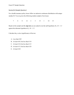


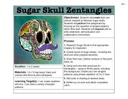


© Copyright 2026