
Intravaginal dehydroepiandrosterone (prasterone), a highly efficient treatment of dyspareunia
CLIMACTERIC 2011;14:282–288
Intravaginal dehydroepiandrosterone
(prasterone), a highly efficient treatment of
dyspareunia
F. Labrie, D. F. Archer*, C. Bouchard{, M. Fortier{, L. Cusan, J.-L. Gomez, G. Girard{, M. Baron**, N. Ayotte{{,
M. Moreau{{, R. Dube´***, I. Coˆte´, C. Labrie, L. Lavoie, L. Berger, L. Gilbert{{{, C. Martel and J. Balser{{{
Climacteric Downloaded from informahealthcare.com by University of Laval on 03/23/11
For personal use only.
EndoCeutics Inc., Quebec City, Canada; *Eastern Virginia Medical School, Obstetrics and Gynecology, Norfolk, USA; {Clinique
de Recherche en Sante´ des Femmes, Quebec City, Canada; {Diex Recherche Inc., Sherbrooke, Canada; **Rapid Medical
Research Inc., Cleveland, USA; {{Clinique Gyne´cologique, Shawinigan, Canada; {{Montreal Clinical Study Center, Montreal,
Canada; ***Centre Hospitalier Affilie´ Universitaire de Que´bec, Quebec City, Canada; {{{McGill University Montreal, Obstetrics
and Gynecology, Montreal, Canada; {{{Veristat Inc., Boston, USA
Key words: PRASTERONE, DHEA, INTRACRINOLOGY, ESTROGENS, ANDROGENS, VAGINAL ATROPHY, DYSPAREUNIA
ABSTRACT
Objective To examine the effect of intravaginal dehydroepiandrosterone (DHEA) on pain at sexual activity
(dyspareunia) identified as the most bothersome symptom of vaginal atrophy in postmenopausal women at
both screening and day 1.
Methods This prospective, randomized, double-blind and placebo-controlled phase III clinical trial studied
the effect of prasterone (DHEA) applied locally in the vagina on the severity of dyspareunia in 114
postmenopausal women who had identified dyspareunia as their most bothersome symptom of vaginal
atrophy, while meeting the criteria for superficial cells 5% and pH 4 5.0 at both screening and day 1.
Results At the standard duration of 12 weeks of treatment, increasing doses of 0.25%, 0.5% and 1.0%
DHEA decreased the percentage of parabasal cells by 48.6 + 6.78%, 42.4 + 7.36% and 54.9 + 6.60%
(p 5 0.0001 vs. placebo for all) with no change with placebo (p ¼ 0.769). The effects on superficial cells and
pH were also highly significant compared to placebo at all DHEA doses. The severity score of pain at sexual
activity decreased by 0.5, 1.4, 1.6 and 1.4 units in the placebo and 0.25%, 0.5% and 1.0% DHEA groups,
respectively, with the p value of differences from placebo ranging from 0.0017 to 5 0.0001.
Conclusions Intravaginal DHEA, through local estrogen and androgen formation, causes a rapid and highly
efficient effect on pain at sexual activity without systemic exposure of the other tissues, thus avoiding the
recently reported systemic effects of estrogens.
INTRODUCTION
Women now spend at least one-third of their lifetime during
postmenopause, with the high probability of suffering from
one or more of the problems related to hormone deficiency,
namely vaginal atrophy, hot flushes, osteoporosis, muscle loss,
fat accumulation, type 2 diabetes, memory loss, cognition loss
and possibly Alzheimer’s disease. An estimated 60 million
women are over 45 years of age in 2010 in the US1.
The rapid and almost complete fall in circulating 17bestradiol at menopause, coupled with the demonstrated
beneficial effects of exogeneous estrogens on menopausal
symptoms2 and bone resorption2–4, has focused most of the
efforts of studies on hormone replacement therapy on
various forms of estrogens as well as to combinations of
estrogens and progestins, in order to avoid the risk
of endometrial cancer induced by estrogens administered
alone.
Correspondence: Dr F. Labrie, EndoCeutics Inc., Quebec City, Canada
ORIGINAL ARTICLE
ª 2011 International Menopause Society
DOI: 10.3109/13697137.2010.535226
Received 30-08-2010
Revised 22-10-2010
Accepted 23-10-2010
Climacteric Downloaded from informahealthcare.com by University of Laval on 03/23/11
For personal use only.
Intravaginal dehydroepiandrosterone for dyspareunia
Recently, a series of reports have indicated that systemic
estrogens þ progestins and estrogens alone increase the risk of
breast5–9, ovarian9–11 as well as endometrial (estrogens
alone)12,13 cancer. The publicity that followed the publication
of the data from the Women’s Health Initiative Study11 has
led to the suggestion by the medical community to use the
lowest estrogen dose possible for the minimal time duration in
order to minimize risks.
In order to avoid the risks of estrogens, we have developed a
novel approach to hormone deficiency after menopause that is
based upon the recent progress achieved in our understanding
of sex steroid physiology in women14–16 and the recognition
that women, at menopause, are not only deprived of estrogen
activity due to an arrest of ovarian estrogen secretion, but
have already been submitted for a few years to a decreasing
exposure to androgens caused by declining dehydroepiandrosterone (DHEA) secretion by the adrenals17.
A very compelling demonstration of the efficacy and safety
of DHEA has recently been obtained in a pivotal phase III,
placebo-controlled, randomized clinical trial in which postmenopausal women suffering from vaginal atrophy received
daily DHEA or placebo intravaginally for 12 weeks. A rapid
and very marked improvement of all the symptoms and signs
of vaginal atrophy was observed, with no change in
circulating estradiol or testosterone. An additional benefit
not seen with estrogens was the finding of a significant
improvement of all domains of sexual dysfunction, namely
desire, arousal, orgasm and pleasure18–20. In order to meet the
recent requirements of the Food and Drugs Administration
(FDA) which indicate the choice of a single self-identified most
bothersome symptom of vaginal atrophy at both screening
and day 1 while meeting the criteria of superficial cells 5%
and pH 4 5.0, we have analyzed the data obtained in women
who met these slightly revised guidelines.
Labrie et al.
(Quebec City, Canada), McGill University (Montreal, Canada), Ethica (Montreal, Canada), Eastern Virginia Medical
School (Norfolk, Virginia, USA) and the Western Institutional
Review Board (Los Angeles, USA).
The inclusion and exclusion criteria were as described by
Labrie and colleagues in reference 18.
A written informed consent was obtained from all subjects
prior to the performance of any study-related procedure. The
subjects had a medical history, a medical examination and a
complete gynecological examination at screening. A partial
gynecological examination was performed to evaluate the
aspect of the mucosa and tolerance to the medication on day 1
and at all visits.
The standard laboratory tests, namely hematology (including complete blood count and coagulation), blood chemistry
and urinalysis, were performed at screening, day 1 and at all
visits.
Vaginal cell smears and endometrial biopsies were examined in a central laboratory. A 100-cell count was performed
to classify cells as superficial, intermediate or parabasal
squamous cell types21,22. Vaginal pH was measured by
applying a pH indicator strip directly to the lateral wall of
the vagina with a forceps.
The women included in the present analysis identified
dyspareunia as the most bothersome symptom at the start of
treatment (both screening and day 1) while having 5% of
superficial cells in the vaginal smear and a pH 4 5.0. The
change in severity of dyspareunia was followed and served to
evaluate the effect of treatment in addition to changes in
vaginal cell maturation and pH. The severity of dyspareunia
was classified by women as being none, mild, moderate or
severe and was analyzed using corresponding values of 0, 1, 2
or 3, respectively, as originally described18. All endpoints had
to demonstrate statistically significant effects relative to
placebo. The primary time-point for analysis was the 12week assessment. Statistics were performed as described18.
METHODS
This study is a phase III, prospective, multicenter, randomized, placebo-controlled and double-blind trial. The original
intend-to-treat (ITT) population included 216 postmenopausal women randomized to receive a daily intravaginal ovule of
the following DHEA concentrations: 0.0% (53 women),
0.25% (3.25 mg DHEA, 53 women), 0.5% (6.5 mg DHEA,
56 women) or 1.0% (13 mg DHEA, 54 women) administered
intravaginally with an applicator at bedtime for 12 weeks18.
From this total population, 114 women self-identified pain at
sexual activity (dyspareunia) as their most bothersome
symptom at both screening and day 1, while meeting the
criteria for superficial cells 5% and pH 4 5.0, thus meeting
the revised FDA guidelines.
The DHEA ovules or suppositories (VaginormTM) containing prasterone in a lipophilic base were manufactured by
Recipharm, Karlskoga, Sweden. The study was divided into
two phases, namely screening followed by a treatment period
of 12 weeks. The protocol was approved by the Institutional
Review Board of the Centre Hospitalier de l’Universite´ Laval
Climacteric
RESULTS
When the statistical analysis was made for patients from the
ITT population who had moderate to severe dyspareunia as
their most bothersome symptom and also met the superficial
cells and pH criteria (superficial cells 5 % and pH 4 5.0) at
both screening and day 1 (baseline), 26, 29, 30 and 29 patients
were eligible in the placebo and 0.25%, 0.5% and 1.0%
DHEA groups, respectively.
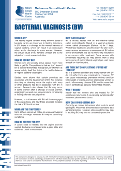
As illustrated in Figure 1, at the standard duration of 12
weeks of treatment, increasing doses of 0.25%, 0.5% and
1.0% DHEA decreased the percentage of parabasal cells by
48.6 + 6.78% (from 65.5 + 6.92% to 16.9 + 3.66%;
p 5 0.0001), 42.4 + 7.36% (from 53.4 + 7.49% to
11.0 + 3.43%; p 5 0.0001) and 54.9 + 6.60% (from
61.8 + 6.88% to 6.9 + 1.77%; p 5 0.0001), respectively,
while no significant effect was observed in the placebo group
(from 46.7 + 8.64% to 47.8 + 7.52%; p ¼ 0.7686). It can be
283
Climacteric Downloaded from informahealthcare.com by University of Laval on 03/23/11
For personal use only.
Intravaginal dehydroepiandrosterone for dyspareunia
Labrie et al.
Figure 1 Effect of daily intravaginal application of 0.0%, 0.25%, 0.5% and 1.0% dehydroepiandrosterone (DHEA) (prasterone) ovules for 12
weeks on the percentage of vaginal parabasal cells in postmenopausal women having identified dyspareunia as their most bothersome symptom
(ERC 210). Data are expressed as mean + standard error of the mean. The p values for the three DHEA doses are comparisons with placebo,
whereas, for the placebo group, comparison is with baseline
seen in the same figure that all doses of DHEA are highly
significantly different from placebo (p 5 0.0001 for all).
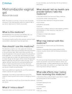
While no significant change was seen at 12 weeks in the
placebo group compared to baseline in the percentage of
superficial cells (Figure 2), increases of 5.3 + 1.39% (from
0.4 + 0.15% to 5.7 + 1.33%; p ¼ 0.0006), 4.8 + 1.20%
(from 0.4 + 11% to 5.2 + 1.19%; p ¼ 0.0004) and
6.1 + 1.54%
(from
0.4 + 0.16%
to
6.5 + 1.53%;
p ¼ 0.0005) were measured in the 0.25%, 0.5% and 1.0%
DHEA groups, respectively. It can be seen in the same figure
that the differences from placebo had respective p values of
0.0052, 0.0111 and 0.0012 for the three increasing doses of
DHEA, respectively.
Vaginal pH, on the other hand, was decreased at 12 weeks
by 1.1 + 0.16 pH units (p 5 0.0001; from 6.6 + 0.10 to
5.5 + 0.19 units), 1.5 + 0.18 pH units (p 5 0.0001; from
6.6 + 0.09 to 5.2 + 0.17 units) and 1.4 + 0.15 pH units
(p 5 0.0001; from 6.5 + 0.11 to 5.1 + 0.12 units) in the
0.25%, 0.5% and 1.0% DHEA-treated groups, respectively
(Figure 3). In the placebo group, pH was decreased by
0.5 + 0.16 units (p ¼ 0.005; from 6.5 + 0.13 to 6.0 + 0.22
units). As indicated in Figure 3, all doses of DHEA are highly
significantly different from placebo with p values of 0.0127,
0.0001 and 0.0001 for the three increasing doses of DHEA,
respectively.
At the 12-week interval, the severity score of the most
bothersome symptom identified at baseline as dyspareunia
was reduced from 2.8 + 0.08 to 2.3 + 0.18 (p ¼ 0.0132),
from 2.8 + 0.08 to 1.4 + 0.22 (p 5 0.0001), from 2.7 + 0.08
to 1.1 + 0.22 (p 5 0.0001) and from 2.6 + 0.09 to
284
1.2 + 0.20 (p 5 0.0001) in the placebo and increasing DHEA
dose groups, respectively (Figure 4). It can be seen in the same
figure that the differences from placebo had respective p
values of 0.0017, 5 0.0001 and 0.0003 for the three
increasing doses of DHEA, respectively.
Concerning safety, no drug-related significant adverse event
was observed in the present study nor in our previous 1-week
pharmacokinetics study with doses of 0.5%, 1.0% and 1.8%
DHEA ovules23,24. No adverse effect was observed on hepatic
tests, hematocrit or any hematological or biochemical parameter.
DISCUSSION
The present data clearly demonstrate the high efficacy of
intravaginal DHEA on pain at sexual activity, the most
frequently vaginal atrophy symptom self-identified by women
in our recent study18,19. In that previous analysis on the ITT
population where 129 women out of 216 had identified
dyspareunia as their most bothersome symptom, the 0.25%,
0.5% and 1.0% doses of DHEA caused 50% (p ¼ 0.0009 vs.
placebo), 60% (p 5 0.0001 vs. placebo) and 58%
(p 5 0.0001 vs. placebo) decreases in the severity of dyspareunia. The effect was already significant at 8, 8, and 2 weeks
for the 0.25%, 0.5% and 1.0% DHEA doses, respectively18.
The present analysis shows that almost super-imposable
effects are observed when the entry criteria (pain at sexual
activity as most bothersome symptom, superficial cells 5%
and pH 4 5.0) are met at both screening and day 1. In fact,
Climacteric
Climacteric Downloaded from informahealthcare.com by University of Laval on 03/23/11
For personal use only.
Intravaginal dehydroepiandrosterone for dyspareunia
Labrie et al.
Figure 2 Effect of daily intravaginal application of 0.0%, 0.25%, 0.5% and 1.0% dehydroepiandrosterone (DHEA) (prasterone) ovules for 12
weeks on the percentage of vaginal superficial cells in postmenopausal women having identified dyspareunia as their most bothersome symptom
(ERC 210). Data are expressed as mean + standard error of the mean. The p values for the three DHEA doses are comparisons with placebo,
whereas, for the placebo group, comparison is with baseline
Figure 3 Effect of daily intravaginal application of 0.0%, 0.25%, 0.5% and 1.0% dehydroepiandrosterone (DHEA) (prasterone) ovules for 12
weeks on vaginal pH in postmenopausal women having identified dyspareunia as their most bothersome symptom (ERC 210). Data are expressed
as mean + standard error of the mean. The p values for the three DHEA doses are comparisons with placebo, whereas, for the placebo group,
comparison is with baseline
using the ITT population meeting the entry criteria at both
screening and day 1, the 0.25%, 0.5% and 1.0% doses of
DHEA caused 50% (p 5 0.0017 vs. placebo), 59%
Climacteric
(p 5 0.0001 vs. placebo) and 54% (p ¼ 0.0003 vs. placebo)
decreases in the severity score of pain at sexual activity at 12
weeks of treatment.
285
Climacteric Downloaded from informahealthcare.com by University of Laval on 03/23/11
For personal use only.
Intravaginal dehydroepiandrosterone for dyspareunia
Labrie et al.
Figure 4 Effect of daily intravaginal application of 0.0%, 0.25%, 0.5% and 1.0% dehydroepiandrosterone (DHEA) (prasterone) ovules for 12
weeks on the severity score of dyspareunia judged by women at baseline as being the most bothersome symptom (ERC 210). Data are expressed as
mean + standard error of the mean. The p values for the three DHEA doses are comparisons with placebo, whereas, for the placebo group,
comparison is with baseline
Current treatments of vaginal atrophy include systemic
hormone therapy, intravaginal estrogens and non-hormonal
lubricants and moisturizers25,26. The risks of estrogens
mentioned above have led to the development of lower doses
of intravaginal estrogens27,28, although serum estrogens are
still increased above normal postmenopausal values with these
low-dose regimens29.
Another approach has been to use ospemifene, a mixed
estrogenic/antiestrogenic SERM (selective estrogen receptor
modulator)30. Ospemifene is a first-generation SERM, a close
analog of tamoxifen and toremifene, with comparable
estrogenic/antiestrogenic relative activities31. Accordingly,
ospemifene has been shown to stimulate uterine weight and
endometrial thickness in the rat31,32. In agreement with its
intrinsic estrogenic activity exposing all tissues following its
systemic administration, ospemifene, in a large scale 12-week
classical study on vaginal atrophy, has shown beneficial effects
on vaginal cell maturation and pH30. Concerning dyspareunia, the daily 30 mg dose of ospemifene did not have a
statistically significant effect compared to placebo on the
severity score (1.02 unit change compared to 0.89 in the
placebo group for a difference of 0.13 unit), while the 0.3 unit
decrease over placebo in the severity score of dyspareunia
observed with the daily 60 mg dose of ospemifene reached
significance with an improvement of 0.3 unit in the severity
score (p ¼ 0.023)30.
By comparison, with the lowest dose (0.25%, 3.25 mg) of
daily intravaginal DHEA, the decrease in the severity score of
dyspareunia was 0.9 units over placebo with a p value of
0.0017. At the 0.5% DHEA dose, the decrease in the severity
score of dyspareunia over the placebo effect was 1.1 units
286
(p 5 0.0001) compared to only 0.3 units for ospemifene
60 mg (p ¼ 0.023) and 0.13 units for ospemifene 30 mg (not
significant). In fact, at the lowest dose of DHEA used (0.25%,
3.25 mg per day), the decrease in the severity score of
dyspareunia was 6.9 times higher (0.9 vs. 0.13 units) than the
non-significant effect observed with the 30 mg dose of
ospemifene, while it was three times more efficient than the
60 mg daily dose of ospemifene. With the 0.5% DHEA dose
(6.5 mg daily), the effect on dyspareunia was 8.5 (1.1 vs. 0.13)
and 3.7 (1.1 vs. 0.3) times more important than the 30 and
60 mg doses of ospemifene, respectively. Since the lubricant was
used only in 31%, 22% and 29% of women in the ospemifene
30 and 60 mg and placebo groups, respectively, during the last
week of treatment and in about one-third at the beginning, the
difference cannot reasonably be explained by the placebo effect
of the lubricant not used in two-thirds of women.
Although 75% of postmenopausal women suffer from
vaginal atrophy33,34, thus affecting their quality of life during
a major part of their lifetime, only 20% or less seek treatment35.
The fear of breast cancer associated with the prescription of
estrogens is the main reason for the lack of acceptance of
estrogen therapy by most women and their physicians. In the
aftermath of the Women’s Health Initiative study, the scientific
challenge is to explore alternative types and formulations of
hormone therapy that would provide all the menopausal
advantages of estrogens while improving women’s quality of
life, minimizing risks, and maximizing benefits36.
The traditional concept of sex steroid physiology in women
was based on the assumption that all estrogens were of
ovarian origin. The relatively recent developments of mass
spectrometry technology have permitted us to gain new and
Climacteric
Climacteric Downloaded from informahealthcare.com by University of Laval on 03/23/11
For personal use only.
Intravaginal dehydroepiandrosterone for dyspareunia
crucial information in this field37–39. In fact, the traditional
concept does not apply to humans and is valid only for animal
species lower than primates, where the ovaries are the only
source of sex steroids.
Since, according to physiology, women are no longer
exposed to systemic estrogens after menopause, it is reasonable to believe that the non-physiologic situation created by
the administration of estrogens could be responsible for at
least some of the side-effects reported by women receiving
traditional estrogen and estrogen/progestin replacement therapy5,11,12,40–44. These side-effects are in addition to the wellrecognized stimulation of the endometrium by unopposed
estrogens and the resulting endometrial hyperplasia and risk
of carcinoma12,13.
It is important to remember that a unique advantage of
DHEA is that this compound is an inactive precursor or
prodrug, which is transformed into active sex hormones
(estrogens and/or androgens) only in the specific cells and
tissues which possess the required enzymes. Due to the
decreasing serum levels of DHEA with age, these tissues,
however, are exposed to lower and lower levels of DHEA45,46,
thus progressively reducing the formation of sex steroids and
causing symptoms in most, but not all, postmenopausal
women. Based upon our data obtained with intravaginal
DHEA18–20,23,24, it is reasonable to believe that supplementation with DHEA in the symptomatic woman simply mimics
the situation of the higher DHEA activity present in healthy
asymptomatic women. Consequently, women with symptoms
of vaginal atrophy and receiving DHEA should not be
different, hormonally speaking, from normal postmenopausal
women having sufficient endogenous DHEA to remain
asymptomatic. This is well supported by the absence of
DHEA-related safety issues in the medical literature, where
Labrie et al.
high doses of DHEA have been administered orally or
percutaneously in a large series of women for up to 2 years
(for review, see reference 47). Moreover, the data from the US
FDA Adverse Event Reporting System and the Center for Food
Safety and Applied Nutrition postmarketing database have
not revealed significant DHEA-related safety concerns.
Moreover, no significant adverse event related to DHEA
was observed in a study performed in 75 women randomized
to receive 3 g of 0.3% DHEA percutaneously twice daily for
12 months39 or 15 women who received daily 3–5 g of 10%
DHEA cream for 1 year48.
Persistent controversial data relate to the search for a
potential correlation between desire and serum testosterone in
healthy women49–51. In fact, most studies have found no
correlation between serum testosterone and arousal49. It is
important to indicate that this lack of correlation can be
explained by the finding that serum testosterone is clearly not
a valid parameter of androgenic activity in women37.
Our recently published study19 has shown for the first time
that local intravaginal treatment with DHEA causes a marked
improvement in all four aspects of women’s sexual dysfunction, namely, desire/interest, arousal, orgasm, and pain at
sexual activity. It thus seems that increased favorable sensitive
outputs from a more healthy vaginal area influence the brain
to feel increased desire/interest without the need for a direct
action of hormones on the brain. The decrease in pain at
sexual activity described above is likely to play a major role in
improving sexual functions in postmenopausal women.
Conflict of interest
F. Labrie is President of EndoCeutics.
Source of funding
Ceutics.
The study was sponsored by Endo-
References
1. Population Division US Census Bureau. Table1: Annual estimates
of the population by sex and five-year age groups for the United
States: April 1, 2000 to July 1, 2007. NC-EST2007–01. May 2008
2. Archer DF, Furst K, Tipping D, Dain MP, Vandepol C. A
randomized comparison of continuous combined transdermal
delivery of estradiol-norethindrone acetate and estradiol alone for
menopause. CombiPatch Study Group. Obstet Gynecol 1999;
94:498–503
3. Christiansen C, Christensen MS, Larsen NE, Transbol IB.
Pathophysiological mechanisms of estrogen effect on bone
metabolism. Dose-response relationships in early postmenopausal
women. J Clin Endocrinol Metab 1982;55:1124–30
4. Women’s Health Initiative. Risks and benefits of estrogen plus
progestin in healthy postmenopausal women. JAMA 2002;288:
321–33
5. Beral V. Breast cancer and hormone-replacement therapy in the
Million Women Study. Lancet 2003;362:419–27
6. Chlebowski RT, Hendrix SL, Langer RD, et al. Influence of
estrogen plus progestin on breast cancer and mammography in
healthy postmenopausal women: the Women’s Health Initiative
randomized trial. JAMA 2003;289:3243–53
Climacteric
7. Collaborative Group on Hormonal Factors in Breast Cancer.
Breast cancer and hormone replacement therapy: collaborative
reanalysis of data from 51 epidemiological studies of 52,705
women with breast cancer and 108,411 women without breast
cancer. Lancet 1997;350:1047–59
8. Li L, Plummer SJ, Thompson CL, et al. A common 8q24 variant
and the risk of colon cancer: a population-based case-control
study. Cancer Epidemiol Biomarkers Prev 2008;17:339–42
9. Lyytinen H, Pukkala E, Ylikorkala O. Breast cancer risk in
postmenopausal women using estrogen-only therapy. Obstet
Gynecol 2006;108:1354–60
10. Lacey JV, Mink PJ, Lubin JH, et al. Menopausal hormone
replacement therapy and risk of ovarian cancer. JAMA 2002;288:
334–41
11. Rossouw JE, Anderson GL, Prentice RL, et al. Risks and benefits
of estrogen plus progestin in healthy postmenopausal women:
principal results from the Women’s Health Initiative randomized
controlled trial. JAMA 2002;288:321–33
12. Beral V, Bull D, Reeves G. Endometrial cancer and hormonereplacement therapy in the Million Women Study. Lancet 2005;
365:1543–51
287
Climacteric Downloaded from informahealthcare.com by University of Laval on 03/23/11
For personal use only.
Intravaginal dehydroepiandrosterone for dyspareunia
13. Gambrell RD Jr, Massey FM, Castaneda TA, et al. Use of the
progestogen challenge test to reduce the risk of endometrial
cancer. Obstet Gynecol 1980;55:732–8
14. Labrie F. Intracrinology. Mol Cell Endocrinol 1991;78:C113–18
15. Labrie F. DHEA after menopause – sole source of sex steroids and
potential sex steroid deficiency treatment. Menopause Management 2010;19:14–24
16. Labrie F, Luu-The V, Be´langer A, et al. Is DHEA a hormone?
Starling Review. J Endocrinol 2005;187:169–96
17. Labrie F. Drug Insight: breast cancer prevention and tissuetargeted hormone replacement therapy. Nature Clinical Practice,
Endocrinology & Metabolism 2007;3:584–93
18. Labrie F, Archer D, Bouchard C, et al. Intravaginal dehydroepiandrosterone (Prasterone), a physiological and highly efficient
treatment of vaginal atrophy. Menopause 2009;16:907–22
19. Labrie F, Archer D, Bouchard C, et al. Effect on intravaginal
dehydroepiandrosterone (Prasterone) on libido and sexual dysfunction in postmenopausal women. Menopause 2009;16:923–31
20. Labrie F, Archer D, Bouchard C, et al. Serum steroid levels during
12-week intravaginal dehydroepiandrosterone administration.
Menopause 2009;16:897–906
21. Meisels A. The maturation value. Acta Cytol 1967;11:249
22. Wied GL. Industrial developments in automated cytology as
submitted by their developers. Anal Quant Cytol Histol
1993;15:358–70
23. Labrie F, Cusan L, Gomez JL, et al. Effect of intravaginal DHEA
on serum DHEA and eleven of its metabolites in postmenopausal
women. J Steroid Biochem Mol Biol 2008;111:178–94
24. Labrie F, Cusan L, Gomez JL, et al. Corrigendum to: Effect of
intravaginal DHEA on serum DHEA and eleven of its metabolites
in postmenopausal women. J Steroid Biochem Mol Biol 2008;
112:169
25. Bachmann GA, Nevadunsky NS. Diagnosis and treatment of
atrophic vaginitis. Am Fam Physician 2000;61:3090–6
26. Nachtigall L, Nachtigall M, Goren J, Loewenstein J. Update on
vaginal atrophy. Menopause Management 2005;14:17–19
27. Bachmann G, Lobo RA, Gut R, Nachtigall L, Notelovitz M.
Efficacy of low-dose estradiol vaginal tablets in the treatment of
atrophic vaginitis: a randomized controlled trial. Obstet Gynecol
2008;111:67–76
28. Simon J, Nachtigall L, Gut R, et al. Effective treatment of vaginal
atrophy with an ultra-low-dose estradiol vaginal tablet. Obstet
Gynecol 2008;112:1053–60
29. Eugster-Hausmann M, Waitzinger J, Lehnick D. Minimized
estradiol absorption with ultra-low-dose 10 mg 17b-estradiol
vaginal tablets. Climacteric 2010;13:219–27
30. Bachmann GA, Komi JO, Ospemifene Study Group. Ospemifene
effectively treats vulvovaginal atrophy in postmenopausal women: results from a pivotal phase 3 study. Menopause
2010;17:480–6
31. Labrie F, Martel C, Gauthier S, Pelletier G, Sance´au JY. Effect of
toremifene and ospemifene, compared to acolbifene, on estrogensensitive parameters in rat and human uterine tissues. Horm Mol
Biol Clin Invest 2010;1:139–46
32. Qu Q, Zheng H, Dahllund J, et al. Selective estrogenic effects of a
novel triphenylethylene compound, FC1271a, on bone, cholesterol level, and reproductive tissues in intact and ovariectomized
rats. Endocrinology 2000;141:809–20
33. NAMS. The role of local vaginal estrogen for treatment of vaginal
atrophy in postmenopausal women: 2007 Position Statement of
288
Labrie et al.
34.
35.
36.
37.
38.
39.
40.
41.
42.
43.
44.
45.
46.
47.
48.
49.
50.
51.
the North American Menopause Society. Menopause 2007;14:
357–69
Wines N, Willsteed E. Menopause and the skin. Australas J
Dermatol 2001;42:149–8; quiz 59
Pandit L, Ouslander JG. Postmenopausal vaginal atrophy and
atrophic vaginitis. Am J Med Sci 1997;314:228–31
Archer DF. Drospirenone-containing hormone therapy for postmenopausal women. Perspective on current data. J Reprod Med
2007;52(2 Suppl):159–64
Labrie F, Be´langer A, Be´langer P, et al. Androgen glucuronides,
instead of testosterone, as the new markers of androgenic activity
in women. J Steroid Biochem Mol Biol 2006;99:182–8
Labrie F, Cusan L, Gomez JL, et al. Comparable amounts of sex
steroids are made outside the gonads in men and women: strong
lesson for hormone therapy of prostate and breast cancer. J
Steroid Biochem Mol Biol 2009;113:52–6
Labrie F, Cusan L, Gomez JL, et al. Changes in serum DHEA and
eleven of its metabolites during 12-month percutaneous administration of DHEA. J Steroid Biochem Mol Biol 2008;110:1–9
Grodstein F, Manson JE, Stampfer MJ. Hormone therapy and
coronary heart disease: the role of time since menopause and age
at hormone initiation. J Womens Health (Larchmt) 2006;15:35–
44
Hsia J, Langer RD, Manson JE, et al. Conjugated equine
estrogens and coronary heart disease: the Women’s Health
Initiative. Arch Intern Med 2006;166:357–65
Pines A, Sturdee DW, MacLennan AH, et al. The heart of the
WHI study: time for hormone therapy policies to be revised.
Climacteric 2007;10:267–9
Riman T, Dickman PW, Nilsson S, et al. Hormone replacement
therapy and the risk of invasive epithelial ovarian cancer in
Swedish women. J Natl Cancer Inst 2002;94:497–504
Ruttimann J. The menopause brain effect: can hormone therapy
help? Endocrine News 2008;15–16
Migeon CJ, Keller AR, Lawrence B, Shepart II TH. Dehydroepiandrosterone and androsterone levels in human plasma. Effect
of age and sex: day-to-day and diurnal variations. J Clin
Endocrinol Metab 1957;17:1051–62
Vermeulen A, Deslypene JP, Schelfhout W, Verdonck L, Rubens
R. Adrenocortical function in old age: response to acute
adrenocorticotropin stimulation. J Clin Endocrinol Metab
1982;54:187–91
Labrie F. DHEA, important source of sex steroids in men and
even more in women. In Martini L, Chrousos GP, Labrie F, Pacak
K, Pfaff D, eds. Neuroendocrinology, The Normal Neuroendocrine System, Progress in Brain Research. New York: Elsevier,
2010:97–148
Labrie F, Diamond P, Cusan L, et al. Effect of 12-month
dehydroepiandrosterone replacement therapy on bone, vagina,
and endometrium in postmenopausal women. J Clin Endocrinol
Metab 1997;82:3498–505
Schover LR. Androgen therapy for loss of desire in women: is the
benefit worth the breast cancer risk? Fertil Steril 2008;90:129–40
van Anders SM, Hamilton LD, Schmidt N, Watson NV.
Associations between testosterone secretion and sexual activity
in women. Horm Behav 2007;51:477–82
van Anders SM, Hampson E. Waist-to-hip ratio is positively
associated with bioavailable testosterone but negatively associated with sexual desire in healthy premenopausal women.
Psychosom Med 2005;67:246–50
Climacteric


© Copyright 2026