
Pityriasis versicolor in Ahvaz, Iran
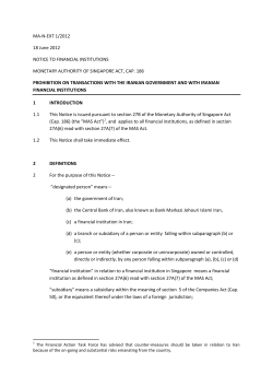
Jundishapur Journal of Microbiology (2009); 2(3): 92-96
92
Original article
Pityriasis versicolor in Ahvaz, Iran
Ali Zarei Mahmoudabadi1, Zahra Mossavi2, Majid Zarrin3
1
Department of Medical Mycoparasitology, School of Medicine, and Infectious and Tropical
Diseases Research Center, Ahvaz Jundishapur University of Medical Sciences, Ahvaz, Iran
2
Department of Dermatology, School of Medicine, Ahvaz Jundishapur University of Medical
Sciences, Ahvaz, Iran
3
Department of Medical Mycoparasitology, School of Medicine, Ahvaz Jundishapur
University of Medical Sciences, Ahvaz, Iran
Received: May 2009
Accepted: August 2009
Abstract
Introduction and objective: Pityriasis versicolor is a chronic superficial mycosis that
caused by several species of Malassezia specially Malassezia globosa. The prevalence of
disease is varying in the world with a rate of 5-50%. Disease is more prevalent in males than
females. The aim of the present study was to review the clinical and epidemiological profile
of pityriasis versicolor in Ahvaz.
Materials and methods: Sellotape method was used for sampling from 500 subjects
suspected to pityriasis versicolor. The presence of clusters of yeasts, budding cells, and
pseudophyae in methylene blue stained samples confirmed disease.
Results: In the present study, 30.6% of subjects were positive for pityriasis versicolor,
62.1% were males, and 37.9% were females. Hypepigmentation lesions were common type
of disease followed by hypopigmentation and erythmatous type.
Conclusion: In conclusion, 30.6% of studied population was positive for tinea versicolor
which is a high prevalence for this disease.
Keywords: Pityriasis versicolor, Tinea versicolor, Malassezia, Hyperpigmentation lesions,
Iran
Introduction
Pityriasis versicolor (Tinea versicolor) is a
mild and chronic superficial mycotic
infection. Disease involved under some
exogenous and endogenous predisposing
factors, which fungus can convert from
yeast to a pathogenic mycelial form.
Disease is usually presented as hypo or
hyperpigmented scaling macules. The
commonest sites of disease are the upper
trunk and neck [1]. Pityriasis versicolor is
common in young adults of both sexes.
Two important exogenous conditions are
high temperature and humidity in hot
season. It has a worldwide distribution with
a high rate (20-50%) in tropical and
subtropical regions [2,3]. Several factors,
such as age, sex, climate, local
environmental factors, malnutrition, and
genetic factors influence course of disease
[4,5].
Jundishapur Journal of Microbiology, School of Medicine, Ahvaz Jundishapur University of Medical Sciences, Ahvaz,
Iran, Phone: +98611 3330074; Fax: +98611 3332036; URL: http://jjm.ajums.ac.ir; E-mail: editorial office: [email protected]
Jundishapur Journal of Microbiology (2009); 2(3): 92-96
Iran is located in subtropical region and
several reports show that tinea versicolor is
more prevalent in Iranian provinces [5-11],
with a higher rate in north and south, which
have warm and humid climate [8-11]. The
frequency of pityriasis versicolor in Iran
varies from 4.4-57.7% in different reports
[5,6,8,12-17]. Afshari [15] reported the
highest frequency of pityriasis versicolor
(57.5%) in Janbazan dormitories in Tehran
whereas lowest frequency (4.4%) reported
by Asadi et al. [14] in Kashan. Disease is
caused by several species of Malassezia
(lipophilic yeasts), which are belong to
normal flora of human body. Malassezia
globosa described as the most common
etiologic agent of pityriasis versicolor
[2,18,19]. Other species, which cause
disease, are M. furfur, M. pachydermatis,
M. sympodialis, M. obtusa, M. restricta and
M. slooffiae [2,18-21]. Recently Lee et al.
[22] presented three new species of
Fig. 1: Tinea versicolor on arm and forearm
Results
In this study, 153 (30.6%) patients were
positive for pityriasis versicolor including
95 (62.1%) males and 58 (37.9%) females
with a male ⁄ female ratio of 1.64:1. The age
of patients were ranged from 6 to 66 years.
The highest prevalence of tinea versicolor
was seen in patients with 17-28 years old
(70.6%) (Fig. 3). In our study, 50% of
93
Malassezia, M. dermatis, M. equi and M.
nana. The intention of the present study was
to review the clinical and epidemiological
profile of pityriasis versicolor in Ahvaz,
Iran.
Materials and methods
In the present study, 500 patients suspected
to pityriasis versicolor attending to a private
dermatology clinic in Ahvaz, Iran (2007)
were sampled (Fig. 1). A questioner
included, sex, age, disease duration, lesion
type and involved area filled for each
patient. Sellotape method was used for
sampling from infected skin of patients [2].
All samples were stained with methylene
blue stain and examined microscopically.
Presence of short and curved pseudohyphae
with clusters of yeasts and budding cells
confirmed the disease (Fig. 2).
Fig. 2: Short and curved pseudohyphae with
clusters of yeasts and budding cells of
Malassezia (methylene blue staining, ×100)
patients had hyperpigmentation followed by
hypopigmentation (36.2%) and erythmatous
(13.8%). Mild lesions were detected in
55.9% of patients, whereas 27.1% and 17%
presented moderate and severe lesions
respectively. Figure 3 shows the duration of
involvement. The disease duration in 41.1%
of patients ranged 1-2 months followed by
3-4, 12-24 and <1 month (Fig. 4). The most
Jundishapur Journal of Microbiology, School of Medicine, Ahvaz Jundishapur University of Medical Sciences, Ahvaz,
Iran, Phone: +98611 3330074; Fax: +98611 3332036; URL: http://jjm.ajums.ac.ir; E-mail: editorial office: [email protected]
Jundishapur Journal of Microbiology (2009); 2(3): 92-96
prominent location of infection on the body
surfaces, was neck (34.6%) followed by
94
trunk (17%) and chest (16.3%) (Table 1).
Fig. 3: Distribution of pityriasis versicolor Fig. 4: Distribution of pityriasis versicolor
according to age
according to disease duration
Table 1: Distribution of location of tinea versicolor on the patient body
Male
Female
Total
Neck &
arm
10
(6.5%)
4
(2.6%)
14
(9.2%)
Head
& face
3
(2.0%)
5
(3.3%)
8
(5.2%)
Forearm
& arm
7
(4.6%)
4
(2.6%)
11
(7.2%)
Trunk
Chest
Neck
15
(9.8%)
11
(7.2%)
26
(17.0%)
12
(7.8%)
13
(8.5%)
25
(16.3%)
39
(25.5%)
14
(9.2%)
53
34.6%
Discussion
Pityriasis versicolor is a worldwide skin
disease, however, its frequency and
occurrence depends on various climatic and
socio-economic state. The frequency and
density of colonization of Malassezia
species in healthy human skin are related to
the subject age and to sebaceous gland
activity in the studied area [23]. In this
study, the highest prevalence of pityriasis
versicolor was observed in 17-28 year age
group (70.6%). The peak of tinea versicolor
is coincided with age. This possibly is due
to hormonal changes and increases in
sebaceous gland activity. In our study, 5.9%
of patients were under 12 years old.
Susceptibility of children was more
common than initially we expected. Tinea
versicolor is a rare disease in children [24].
Tarazooie et al. [2] found only one patient
Neck &
chest
9
(5.9%)
7
(4.6%)
16
(10.5%)
Total
95
(62.1%)
58
(37.9%)
153
(100%)
of pityriasis versicolor in age less than 10
year in Tehran.
Distribution of the patches of pityriasis
versicolor in children is various, and hence
there is a discussion whether this difference
is due to clinical or microscopic appearance
[25]. We found significant differences in
prevalence of pityriasis versicolor between
both sexes. 62.1% of patients were male
and 37.9% were female. Therefore, the
male/female ratio was 1.64:1. Many studies
show dissimilar male to female ratios,
however, they emerge to be almost equal in
both sexes [26,27]. He et al. [4] believe that
the role of sex in susceptibility to disease
and its development is still unclear. In this
study, the most affected areas were neck
with 34.6%, which is followed by trunk
(17%) and chest (16.3%). Distribution of
the patches usually parallels the density of
Jundishapur Journal of Microbiology, School of Medicine, Ahvaz Jundishapur University of Medical Sciences, Ahvaz,
Iran, Phone: +98611 3330074; Fax: +98611 3332036; URL: http://jjm.ajums.ac.ir; E-mail: editorial office: [email protected]
Jundishapur Journal of Microbiology (2009); 2(3): 92-96
sebaceous secretion distribution, [28,29]
with higher incidences on chest, back, and
face. Patches of the face are more prevalent
in children than adults [30]. Aspiroz et al.
[29] found M. restricta associated
particularly with scalp skin, M. sympodialis
with the back, while M. globosa was evenly
distributed on scalp, forehead, and trunk.
Ahvaz is located in subtropical region
with hot and humid conditions from April
to October. Several reports showed that hot
and humid conditions, and hygiene are
susceptible factors for presenting pityriasis
versicolor [4,7]. However Belec et al. [31]
believe that good or poor hygiene of the
clothing had no significant influence on the
prevalence of pityriasis versicolor. The
lesions of tinea versicolor can be
hyperpigmented,
hypopigmented,
leukodermal, erythmatous or dark brown. In
our
study,
50%
of
cases
had
hyperpigmentation
followed
by
hypopigmentation (36.2%) and erythmatous
lesion (13.8%). In conclusion, 30.6% of
studied population was positive for tinea
versicolor which is a high prevalence for
this disease. Therefore, we have to find a
way to control this disease in our area.
Acknowledgment
This study was supported by a grant (No.
84U94) from Jundishapur University of
Medical Sciences, Ahvaz, Iran. The authors
are grateful to the department of medical
mycoparasitology, Ahvaz Jundishapur
University of Medical Sciences for their
help.
References
1) Anaissie EJ, McGinnis MR, Pfaller MA.
Clinical
Mycology.
1ed.,
Elsevier
Sciences, USA, 2003.
2) Tarazooie B, Kordbacheh P, Zaini F, et al.
Study of the distribution of Malassezia
species in patients with pityriasis
versicolor and healthy individuals in
3)
4)
5)
6)
7)
8)
9)
10)
11)
12)
13)
95
Tehran, Iran. BMC Dermatology. 2004; 4:
3-6.
Yazdanpanah MJ, Azizi H, Suizi B.
Comparison between fluconazole and
ketoconazole effectivity in the treatment of
pityriasis versicolor. Mycoses. 2007; 50:
311-3.
He SM, Du WD, Yang S, et al. The genetic
epidemiology of tinea versicolor in China.
Mycoses. 2008; 51: 55-62.
Shamsoddini S, Barik Bin B. Comparison
between the frequency of pityriasis
versicolor in nursing home of Kerman and
the control group. Iran J Infect Dis Trop
Med. 2004; 25: 36-40.
Badiei P, Kord Bacheh P, Zeini F, Shidfar
MR. Survey and diagnosis of superficial
and cetaceous fungal infections in referral
patients in health center in Shiraz. Iran J
Infect Dis Trop Med. 2003; 21: 18-21.
Asadi MA, Droudgar A, Houshyar H.
Prevalence of cutaneous mycoses among
sanitary workers of city municipality of
Kashan, 1998. Feyz, Kashan Uni Med Sci
Health Serv. 1998; 9: 92-9.
Rafiei A, Emmami M, Moghadami M,
Mahmedi M, Shidfar M. Cutaneous
mycosis in Khuzestan province. Sci Med J.
1992; 14: 22-34.
Foladvand MA, Naeimi B. Superficial
Mycoses in fishermen of Bushehr port.
Iran South Med J. 1999; 1: 22-8.
Aziz Jalali MH. Investigation on the
incidence of superficial fungal diseases in
Razi Hospital, Rasht. J Med Council of
Islam Rep Iran. 1991; 4: 245-52.
Shakerian MA, Tirgar Tabari S, Haji
Ahmadi M, Khoshbakht H, Hosseini SD.
Frequency of tinea versicolor in male high
school students, Babol, 2001-4. J Babol
Uni Med Sci. 2006; 30: 77-9.
Salari MH. The epidemiological survey of
bacterial and fungi cutaneous infections of
textile factories workers in Yazd province.
Iran J Infect Dis Trop Med. 2002; 18: 40-5.
Kordbacheh P, Moghaddami M, Asadi
MA. Superficial and cutaneous mycoses at
nurseries and schools of Mahallat city,
central part of Iran. Iran J Pub Health.
1994; 4: 61-71.
Jundishapur Journal of Microbiology, School of Medicine, Ahvaz Jundishapur University of Medical Sciences, Ahvaz,
Iran, Phone: +98611 3330074; Fax: +98611 3332036; URL: http://jjm.ajums.ac.ir; E-mail: editorial office: [email protected]
Jundishapur Journal of Microbiology (2009); 2(3): 92-96
14) Asadi MA, Houshyar H, Dehghani R, et al.
Prevalence of cutaneous and superficial
mycoses in schools of the city of Kashan
and its suburbs Feyz, Kashan Uni Med Sci
Health Serv. 2001; 16: 41-7.
15) Afshari MA. Superficial and cutaneous
mycoses in Janbazan dormitories in
Tehran. Kowsar Med J. 2000; 3: 189-94.
16) Talary S, Asadi M, Yoosefian A.
Prevalence of cutaneous superficial
mycoses among referred patients to
Kashan's university of medical sciences,
medical mycology laboratory in years
(1992-93). Teb Tazkyeh J. 2000; 35: 21-5.
17) Afshari MA. Common fungal diseases
among out-patients of Baghiyatollah (a.s.)
hospital from 1989 to 1995. Kowsar Med
J. 1996; 1: 39-42.
18) Shams M, Rasaee Mj, Moosavi M.
Identification of Malassezia species in
patients
with
pityriasis
versicolor
submitted to the Razi Hospital in Tehran.
Iran Biomed J. 2001; 5: 121-6.
19) Gupta AK, Kohli Y, Faergemann J,
Summerbell RC. Epidemiology of
Malassezia
yeasts
associated
with
pityriasis versicolor in Ontario, Canada.
Med Mycol. 2001; 39: 199-206.
20) Canteros CE, Soria M, Rivas C, et al.
Malassezia species isolated from skin
diseases in a care centre in the city of
Buenos Aires. Rev Argent Microbiol. 2003;
35: 156-61.
21) Mayser P, Tows A, Kramer HJ, Weiss R.
Further characterization of pigmentproducing Malassezia strains. Mycoses.
2004; 47: 34-9.
22) Lee YW, Kim SM, Oh BH, Lim SH, Choe
YB, Ahn KJ. Isolation of 19 strains of
Malassezia dermatis from healthy human
skin in Korea. J Dermatol. 2008; 35: 7727.
23) Marcon MJ, Powell DA. Human infections
due to Malassezia spp. Clin Microbiol
Rev.1992; 5: 101-19.
96
24) Gupta AK, Bluhm R, Summerbell R.
Pityriasis versicolor. J Eur Acad Dermatol
Venereol. 2002; 16: 19-33.
25) Terragni L, Lasgni A, Oriani A, Gelmetti
C. Pityriasis veresicolor in the pediatric
age. Pediatr Dermatol. 1991; 8: 9-12.
26) Nakabayashi A, Sei Y, Guillot J.
Identification of Malassezia species
isolated from patients with seborrhoeic
dermatitis, atopic dermatitis, pityriasis
versicolor and normal subjects. Med
Mycol. 2000; 38: 337-42.
27) Crespo Erchiga V, Ojeda Martos A, Vera
Cassano A. Isolation and identification of
Malassezia spp. In pityriasis versicolor,
seborrheic dermatitis and healthy skin. Rev
Iberoam Micol. 1999; 16: S16-S21.
28) Leeming JP, Notman FH, Holland KT. The
distribution and ecology of Malassezia
furfur and cutaneous bacteria on human
skin. J Appl Bacteriol. 1989; 67: 47-52.
29) Aspiroz C, Moreno LA. Differentiation of
three biotypes of Malassezia species on
normal human skin. Correspondence with
M. globosa, M. sympodialis and M.
restricta. Mycopathologia. 1999; 145: 6974.
30) Terragni L, Lasgni A, Oriani A. Pityriasis
versicolor of the face. Mycoses. 1991; 34:
345-7.
31) Belec L, Testa J, Bouree P. Pityriasis
versicolor in the Central African Republic:
a randomized study of 144 cases. J Med
Vet Mycol. 1991; 29: 323-9.
Address for correspondence:
Ali Zarei Mahmoudabadi, Department of
Medical Mycoparasitology, School of Medicine,
and Infectious and Tropical Diseases Research
Center, Ahvaz Jundishapur University of
Medical Sciences, Ahvaz, Iran
Tel: +98611 3330074; Fax: +98611 3332036
Email: [email protected]
Jundishapur Journal of Microbiology, School of Medicine, Ahvaz Jundishapur University of Medical Sciences, Ahvaz,
Iran, Phone: +98611 3330074; Fax: +98611 3332036; URL: http://jjm.ajums.ac.ir; E-mail: editorial office: [email protected]


© Copyright 2026