
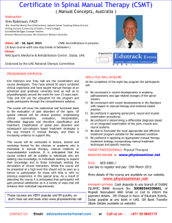
Improving axonal growth and functional recovery after experimental
Brain Research Reviews 36 (2001) 204–212 www.elsevier.com / locate / bres Review Improving axonal growth and functional recovery after experimental spinal cord injury by neutralizing myelin associated inhibitors Fouad Karim a,b ,1 , Dietz Volker b , Martin E. Schwab a , * a Brain Research Institute, University of Zurich and Department of Biology ETH Zurich, Winterthurerstrasse 190, CH-8057 Zurich, Switzerland b ParaCare, University Hospital Balgrist, University of Zurich, CH-8008 Zurich, Switzerland Abstract Injuries of the spinal cord often result in an irretrievable loss of motor and sensory functions of all body parts situated below the lesion site. Functional recovery is restricted mainly due to the limited regeneration and plasticity of injured axons in the adult central nervous system. Over the last few years different experimental approaches have led to axonal growth and functional benefits in animal models. This review focuses on the effects of the neutralization of myelin-associated neurite growth inhibitors, in particular Nogo-A, using the monoclonal antibody IN-1. Acute mAb IN-1 treatment of adult CNS lesioned rats results in extensive plastic changes of neuronal connections and regenerative fiber growth. In two different lesion paradigms (i.e. pyramidal tract lesion and incomplete spinal cord lesion in adult rats), the mAb IN-1-treated animals always showed a higher degree of recovery in various behavioral tests. These observations, together with electrophysiological results, suggest that neuronal CNS circuits of mAb IN-1-treated animals can be rearranged, and that sprouting and regenerating axons form functionally meaningful connections. 2001 Elsevier Science B.V. All rights reserved. Theme: Development and regeneration Topic: Motor systems Keywords: Spinal cord injury; Myelin; Plasticity; Regeneration; Functional recovery; IN-1 Contents 1. Introduction ............................................................................................................................................................................................ 2. Inhibition of neurite growth in the injured mammalian CNS ....................................................................................................................... 3. Treatment approaches in animal models .................................................................................................................................................... 4. Monitoring functional recovery in animal models of spinal cord injury ........................................................................................................ 5. Mechanisms of spontaneous recovery ....................................................................................................................................................... 6. Enhancing functional recovery by neutralizing neurite growth inhibitors...................................................................................................... 7. Neutralization of growth inhibitors — a way to the clinic? ......................................................................................................................... Acknowledgements ...................................................................................................................................................................................... References................................................................................................................................................................................................... 1. Introduction Patients suffering from an acute spinal cord injury (SCI) find themselves in an especially desperate situation, since severed axons of the central nervous system (CNS) are *Corresponding author. Tel.: 141-1-635-3330; fax: 141-1-635-3303. E-mail address: [email protected] (M.E. Schwab). 1 Present address: Faculty of Rehabilitation Medicine, University of Alberta, Edmonton, Alberta, Canada, T6G 2G4. 204 205 206 206 207 208 209 210 210 unable to regenerate spontaneously, and currently treatments to reestablish the lost neuronal connections are not available. It is difficult for a para- or tetra / quadraplegic patient to accept that two fully intact components of her / his body, ‘the control unit’ and ‘the engine’, namely the brain and the limbs, are irretrievably disconnected. For patients with a SCI, the present acute medical treatments focus on limiting the initial damage and on the management of bladder dysfunction, spasticity and neuropathic pain [24]. Functional recovery after a severe SCI is 0165-0173 / 01 / $ – see front matter 2001 Elsevier Science B.V. All rights reserved. PII: S0165-0173( 01 )00096-0 F. Karim et al. / Brain Research Reviews 36 (2001) 204 – 212 restricted, and patients have to learn to make compensatory use of spared motor functions. After incomplete spinal cord injuries intensive physical therapy and to some extent functional electrical stimulation [66] are the only ways to improve the functional outcome. Regular locomotor training, by walking suspended on a treadmill, can even aid a certain group of patients to regain the capability of weight bearing and walking [21,22,93]. Early studies in animals have shown that injured CNS neurons are able to regrow neurites into peripheral nerve grafts [18]. Over the last 15 years, first promising steps were done to understand the factors involved in the lack of regeneration of neurites in the adult CNS tissue and, subsequently, to overcome some of the barriers for regeneration. Thus, encouraging progress is made towards the development of novel therapeutic approaches to treat spinal cord and brain injuries. In this review, we will focus on the approach of neutralizing the neurite growth inhibitory activity associated with CNS myelin, in particular the protein Nogo-A [16], by the application of the monoclonal IN-1 antibody (mAb IN-1) [15]. 2. Inhibition of neurite growth in the injured mammalian CNS Despite clear differences in the regenerative potential of different CNS fiber populations, several experiments have 205 rejected the view of an intrinsic inability of adult CNS fibers to regenerate. This crucial discovery was first made by Tello in 1911 [86], by offering central axons a transplanted piece of a peripheral nerve as a growth permissive substrate. Many years later, these experiments were confirmed [18,72] demonstrating irrevocably that the failure of CNS fibers to regenerate was not mainly due to intrinsic neuronal properties. The concept of a lack of neurotrophic support by the CNS was proposed and has been extensively investigated. Neurotrophic factors were shown to improve neuronal survival, sprouting and short distance growth within the lesioned central nervous system [35,43,48,69,75], but growing axons always avoided white matter. The idea of neurite growth inhibition by CNS tissue was introduced in 1985 [80]. The very potent inhibitory nature of the CNS was attributed to a glial cell subtype: the myelinating oligodendrocytes [14,79,81]. This stands in contrast to the growth promoting properties of the Schwann cells of the peripheral nervous system [12]. Several myelin-associated proteins were shown to possess neurite growth inhibitory properties in vitro (Table 1): Nogo-A [16,78], MAG [51,59] and also certain chondroitin sulfate proteoglycans [25,61,85]. In addition to myelin, a barrier for nerve fibers to regenerate in the CNS which appears after an injury is the glial scar, forming a dense mechanical and probably biochemical barrier for growing axons at the lesion site. The scar consists of reactive astrocytes, microglia, Table 1 Some milestones in the development of the concept of myelin inhibitors and the subsequent treatment approaches Reference Year System Approach Results David and Aguayo [18] 1981 In vivo in rats Axons grow into bridge Schwab and Thoenen [80] Caroni and Schwab [15] 1985 In vitro Transplant peripheral nerve into injured spinal cord Three-compartment chamber providing NGF 1988 In vitro Mukhopadhay et al. [59] McKerracher et al. [51] 1994 1994 In vitro In vitro Bregman et al. [7] Bartsch et al. [4] 1995 1995 In vivo in rats In vivo and in vitro Keirstead et al. [41] 1995 Li et al. [44] Dyer et al. [27] Vanek et al. [88] Huang et al. [39] 1996 1998 1998 1999 In vivo, in chicken with complete transsection In vitro In vivo in rats In vivo in rats In vivo in mice Niederoest et al. [61] 1999 In vitro Chen et al. [16] Cai et al. [13] 2000 2001 In vitro In vitro and in vivo CNS myelin contains defined neurite growth inhibitory proteins (NI-35/250). Antibody (mAB IN-1) was raised against NI-250 MAG application to DRG neurons Fractionating proteins of CNS myelin and apply to neuroblastoma cells Application of the IN-1 antibody after SCI Cells from MAG-deficient mice on myelin and lesion of CST and optic nerve Transient immunological disruption of myelin MAG application to neuronal cell line Local immunological disruption of myelin X-Radiation to destroy myelin Therapeutic vaccination against myelin CSPGs application from myelin on DRG neurons and cerebellar granule cells Cloning of Nogo-A (NI-250) cAMP up- or down-regulation in cell culture or neonatal mice In spite of NGF neurons grow only into peripheral nerve and not optic nerve explants Myelin becomes a more permissive environment MAG inhibits neurite outgrowth Inhibitory activity of MAG on axon extension Increased regeneration and functional recovery No differences in regenerative capabilities on myelin were found either in vivo or in vitro Regeneration of brain stem projections, with electrophysiological evidence for reconnection Growth cone collapse Regeneration of 32% of RST fibers Increased axonal sprouting Regeneration of CST fibers and recovery of placing response Inhibition of neurite outgrowth Nogo-A is a potent neurite growth inhibitor Elevating cAMP blocks MAG inhibition of neurite outgrowth, decrease in cAMP leads to decreased plasticity MAG, myelin associated glycoprotein; NGF, nerve growth factor; DRG, dorsal root ganglion; CSPG, chondroitin sulfate proteoglycans. 206 F. Karim et al. / Brain Research Reviews 36 (2001) 204 – 212 oligodendrocyte precursors, and often, fibroblasts. Furthermore, it contains several potentially repulsive and neurite growth inhibitory factors such as semaphorins, ephrins, tenascin and chondroitin sulfate proteoglycans [26,64,78]. 3. Treatment approaches in animal models For an optimal treatment to partially repair the lesioned CNS, the variety of barriers and inhibitory molecules will probably require a combination of different approaches [28,38,84]. Recently, experimental procedures have led to successful regeneration of injured fibers as well as anatomical reorganization of spared fiber systems. Such results were obtained by grafting peripheral nerve bridges [17], Schwann cells [46,97] or olfactory ensheathing cells [45,70] into the lesion site, as well as by the application of neurotrophic factors [35,43,69,75]. Regeneration of injured axons and increased sprouting of nerve cells by neutralizing or destroying myelin, thereby removing myelin-associated neurite growth inhibitors, has been achieved using various approaches (Table 1): the procedure to focally or transiently destroy myelin by Xradiation [74] or an immunological protocol, consisting of the combination of a myelin specific antibody and serum complement proteins resulted in chickens as well as in rats, in long distance regeneration of spinal axons [27,41,42,83]. A therapeutic approach to immunologically neutralize myelin-associated inhibitors in mice has been achieved by the vaccination with myelin components [39]. Also, this strategy resulted in a strong regenerative growth of a large number of CST axons. In the case of the application of the mAb IN-1, extensive in vitro studies resulted in a significant neurite outgrowth on a CNS substrate [2,15,16,82]. The subsequent in vivo studies confirmed the in vitro results, in that lesioned adult corticospinal tract (CST) axons showed regenerative growth in adult rats, whenever the mAb IN-1 or an IN-1 Fab fragment were applied early following the SCI [7,8,76,77]. After 2 weeks of mAb IN-1 treatment, nerve fibers of the dorsally located CST showed sprouting of fibers around the lesion and scar tissue, and regenerative growth down the spinal cord up to distances of 10–20 mm. In the caudal spinal cord, the regenerated fibers formed dense arborizations suggesting their reconnection to the denervated spinal neuronal circuits [8]. Interestingly, the mAb IN-1 treatment after an CNS injury not only resulted in regenerative nerve fiber growth, it also enhanced sprouting and plastic growth of uninjured fibers at several levels of the motor system [67,87,100]. A major question arising from these studies is that of the functional significance of the observed regeneration and reorganization. It is widely assumed that a small number of preserved, regenerated or newly formed neuronal connections can have large functional impact [60], nevertheless, it is still unclear to which extent anatomical reintegration is needed to result in specific functional benefits. Guidance and target recognition cues have to be present or may have to be re-expressed in the adult and injured CNS, an appropriate amount of fibers has to invade the denervated targets, the fibers have to grow over sufficient distances, and mechanisms of adjustment and fine tuning of the new connections must be operative. To examine these issues, appropriate animal models have to be chosen to inflict a controlled and comparable injury and to quantify the various aspects of the behavioral outcome. Ideally, such models should allow correlating histological and electrophysiological findings with behavior, and they should be valid clinically. In a recent study such a correlation was made and compared with data of human SCI. It was found that the rat represents an adequate model for human SCI [53]. 4. Monitoring functional recovery in animal models of spinal cord injury Various animal models of SCI have been developed to answer injury specific questions (e.g. inflammation, regeneration, spasticity, etc.). In the field of SCI the rat has evolved as the animal of choice [6,55,57]. More recently, however, the availability of knockout and transgenic mice [4] strongly increased the interest for mouse models [40]. For anatomically complete SCI, two models have been developed: the complete transection model, in which the dura mater and the spinal cord are transected with a small blade or scissors, and the complete contusion model, in which a calibrated weight is dropped on to the spinal cord or the spinal cord is compressed by clips, resulting in a total local destruction of gray and white matter, but without lesion of the dura mater [5,96]. Models of complete SCI are particularly useful to examine the effects of implanted bridges and cell grafts [17,70], since they can provide clear evidence about regenerative fiber growth across the lesion. Modeling incomplete spinal cord injuries can be achieved by different strategies. The first one consists of a mild or moderate contusion (weight drop model; [5]) or a compression [71] of the spinal cord without damaging the dura mater. Lesions obtained by such a technique mimic incomplete spinal cord lesions in humans. The use of graded impact / pressure intensity results in different degrees of destroyed gray and white matter. Typically, after a few days, cavities form at the lesion site, which are surrounded by scar tissue [78,84]. Alternative models are specific sections of defined tracts or funiculi with a fine blade, iridectomy scissors or by chemical ablation [7,8,50]. Such specific lesions have the advantage that single tract regeneration and / or reorganization can be monitored. They also allow a more precise behavioral testing, since specific and reproducible deficits occur. For a functional, behavioral analysis the appropriate testing strategy has to correspond to the specific lesion type chosen (Fig. 1). A F. Karim et al. / Brain Research Reviews 36 (2001) 204 – 212 207 Fig. 1. Rat walking at constant speed on a treadmill. Video analysis allows reconstruction of the kinetics of hindlimb movement. Examples of an intact rat and an animal with a moderate spinal cord injury are shown. dorsal lesion of the corticospinal tract will result, for example, in minor deficits of locomotion, but in persistent deficits of grasping or precise limb movements as detected when rats are walking on a grid [54,58]. Lesioning the lateral and ventral funiculi, including the reticulospinal tract and long descending propriospinal interneurons, results in more severe gait impairment [10,34,63]. In incomplete SCI usually spontaneous functional recovery occurs [47,62,89]. In animal models, prediction and quantification of the functional recovery following a specific type of lesion is of crucial importance to draw conclusions about a given experimental intervention. In case of mild injuries, spontaneous recovery is substantial, and the additional functions gained by an experimental intervention require testing paradigms of high resolution. The testing strategies should be adapted to the type and severity of the lesion, and time course studies should offer a transparent picture of the recovery process [6,40,55,57]. 5. Mechanisms of spontaneous recovery Different mechanisms with different time courses are involved in the spontaneous recovery processes, observed after SCI. A first stage represents the recovery of the so-called spinal shock [36,37,47]. Immediately after injury a complex cascade of biochemical reactions occurs in and around the lesion site, leading to ischemic tissue damage, axonal and myelin swelling, intracellular accumulation of Ca 21 , excessive release of the excitatory neurotransmitter glutamate and inflammatory processes [37,78]. These biochemical changes result in a phase of neuronal depression frequently associated with neuropraxia, i.e. a transient failure of axons to transmit impulses. The recovery of this phase is variable and lesion-dependent ranging, in rats, from hours to days [5]. An additional factor contributing to conductance failure of spinal cord tracts following injury is the demyelination of spared axons [78,90]. Spontaneous remyelination of these fibers can occur to some degree, and probably contributes to the functional recovery at later time points [32,73]. Important mechanisms of spontaneous functional recovery are adaptations, and plastic re-organizations of neuronal networks above and below the lesion [20,60,67]. Development of compensatory movement patterns has been observed for grasping after high cervical lesions [1,94] or for walking by involving axial muscles [33]. Another important mechanism for functional recovery represents the activity-dependent adaptation of spinal pattern generating networks; substantial recovery effects were obtained in cats and humans by specific training paradigms. Facilitation of stepping and weight bearing was obtained by regular treadmill training in spinal cats [3,19,49] but not in rats with incomplete SCI [29]. This training was also shown to be beneficial in patients with SCI [21,23,92]. In addition, spinal reflexes implicated in the control of stepping are involved in the recovery of stepping [30,65]. On the supraspinal level, rearrangements of cortical maps have been observed after spinal cord 208 F. Karim et al. / Brain Research Reviews 36 (2001) 204 – 212 injury in animals and humans [9,33,56]. In the spinal cord, sprouting of injured (Brosamle and Fouad, unpublished observation) and of uninjured CST fibers [91] to alternative targets has been shown, a process that may mediate the development of compensatory functional movement patterns. 6. Enhancing functional recovery by neutralizing neurite growth inhibitors Adult CNS tissue, in particular CNS myelin, inhibit neurite growth in a variety of in vitro assays [51,59,79]. Various procedures to neutralize the inhibitory components of myelin have led to regeneration of lesioned axons and to functional changes in different models of spinal cord injury. The delay of myelination in developing chicken resulted in anatomical regeneration and functional recovery compared to that of embryos transected prior to the development of myelin. Also a transient disruption of myelin in the post-hatching chick, using a comparable approach, resulted in regeneration of brainstem projections. Although this did not promote functional recovery, electrophysiological examinations could demonstrate the reintegration of regenerated nerve fibers [41]. Also, a study using autovaccination against myelin in mice to promote axonal regeneration reported recovery in a functional test: Mice with anatomical evidence for regeneration of axons performed superior in the placing test, thereby indicating the reconnection of the CST [39]. Our laboratory has concentrated on the myelin-associated high molecular weight inhibitory protein NI-250 / Nogo-A [15,16,81] and the effect of the monoclonal antibody IN-1 raised against this protein [15]. In two studies, an improved functional recovery after severe spinal cord injury followed by the application of mAb IN-1 over 2 weeks has been reported [7,52]. In both studies a dorsal spinal cord lesion was performed in adult rats at thoracic level (Th8). Control or IN-1 antibody producing hybridoma cells were implanted in the proximity of the ventricular system to allow constant diffusion of the antibodies into the entire CNS. The growth of the hybridoma cells was enabled by the application of cyclosporine over 5–7 days. Six weeks following a dorsal over-hemisection, stride length during walking and contact placing (a reflex known to depend on CST input in adults) significantly improved in the mAb IN-1-treated rats compared to the controls [7]. Pronounced sprouting and regeneration of CST fibers paralleled the improved functional recovery. A subsequent bilateral ablation of the motor cortex abolished the recovered contact placing. In a subsequent study, a more detailed analysis of the locomotor behavior was performed [52]. Using the BBB open field locomotor score [6], significant improvement in the mAb IN-1-treated rats occurred over 2–4 weeks after the injury. Enhanced recovery was also observed in the grid walk test, as well as in the narrow beam test. The behavioral findings were confirmed by electromyographic examinations of muscle activity during treadmill walking, demonstrating the normalization of the stepping pattern in the mAb IN-1-treated rats (Fig. 2) [52]. The histological analysis of these animals showed that rats, especially with higher amounts of spared white matter, benefited from the mAb IN-1 treatment. Spared tissue functions as a bridge for regenerating fibers at the lesion site [8,77], but remaining fibers and tracts also represent the substrate for compensatory mechanisms. Indeed, studies with defined CST lesions at the level of the brain stem have shown that such compensatory processes can be greatly enhanced by mAb IN-1 application. The transection of the pyramidal tract (CST) at the level of the brain stem, where it runs ventrally as a superficially located bundle, allows to deprive half or both sides of the spinal cord of its CST input without lesioning other tracts or the spinal cord itself. Such a lesion results in major and persistent deficits in the fine control of the paw and the digits [95]. A reliable and sensitive method to quantify these deficits is the grasping test where animals are trained to reach for food pellets [87,94]. To test the effect of mAb IN-1, control or IN-1 antibody producing hybridoma cells were implanted into the hippocampus of adult rats that had undergone a unilateral lesion of the pyramidal tract. In the mAb IN-1-treated animals, this resulted in sprouting of cortico-bulbar fibers and establishment of bilateral corticorubral and cortico-pontine projections [87,100], similar to those observed in neonatally lesioned rats [99]. In the spinal cord the remaining CST sprouted across the midline [87]. The anatomical rearrangements were associated with an almost complete functional recovery in the food pellet grasping test [87]. Although limited regeneration of the CST occurred at the lesion site [68] a re-lesioning of these fibers did not disrupt the recovery. This indicates that the improved function was due to compensatory sprouting and plastic rearrangements [100]. Plasticity can also involve fiber systems that are untouched by a lesion, as shown in a study where the pyramidal tract was lesioned bilaterally in adult rats, followed by a control or mAb IN-1 treatment [67]. Whereas control animals (lesion only or lesion with control antibody) showed severe impairments in the grasping test, the mAb IN-1-treated rats recovered to prelesion levels. Anatomically, the rubrospinal tract was observed to increase the number of collaterals in the cervical spinal cord (by more than 100%). These fibers projected to the deep ventral horn, possibly replacing the lost CST fibers [67]. Microstimulation of the forelimb area of the motor cortex in mAb IN-1-treated rats induced EMG responses in the forelimbs, which were never observed in control-Abtreated rats. These responses were abolished by injection of the GABA receptor agonist muscimol into the red nucleus, showing the presence of a new, cortico-rubro-spinal pathway induced by the mAB IN-1 treatment. F. Karim et al. / Brain Research Reviews 36 (2001) 204 – 212 209 Fig. 2. EMG activity of two representative examples of spinal cord lesioned rats treated with a control antibody (hybridoma transplant) (top) or IN-1 antibodies (bottom). Typical abnormalities in the control animal are the uncoupling of left and right tibialis anterior. Animals treated with mAb IN-1 show a much more normal walking pattern with correct left–right alteration and good flexor–extensor alteration (for details see Ref. [52]; courtesy of Dr D. ¨ Merkler, Zurich). Plastic effects of the mAb IN-1 and of new anti Nogo-A antibodies on intact fiber systems were also observed in the intact adult cerebellum, where Purkinje cells upregulated immediate early genes [98], and grew nodal sprouts along their axons [11], in response to these antibodies. Together these results suggest that the Nogo-A protein acts not only as a growth inhibitor for injured axons, but also as a general regulator for neuronal growth and a stabilizing factor for the complex adult neuronal network. 7. Neutralization of growth inhibitors — a way to the clinic? Several important requirements have to be fulfilled before treatments that enhance fiber regeneration and plasticity can be clinically applied. In the case of antibodies against the potent neurite growth inhibitors like Nogo-A, the first requirement is an appropriate antibody administration procedure. The antibody production by implanted hybridoma cells does not allow a controlled application. A major step towards a more defined treatment strategy was the cloning of Nogo, which allows to produce specific antibodies against various regions of the Nogo protein and to design strategies for the cloning of Nogo-A receptors [16,31]. Several antibodies against rat or human Nogo are currently being tested in vivo and in vitro. Application of the purified antibodies via pumps will allow a very controlled application, which can be expanded to weeks or months. It is likely that such a prolonged treatment will result in prolonged and more robust regeneration and sprouting of injured and uninjured nerve fibers. This may result in increased functional benefits especially when long distance regeneration is necessary like in the injured spinal cord. However, long-term application of antibodies might also include certain dangers. Although, no negative side effects of the mAb IN-1 treatment have been detected so far [52], it remains a highly important question whether sprouting and regenerative growth of nerve fibers (including sensory fibers) can result in improper connections leading to malfunctions. Effects of long-term suppression of growth inhibitors on circuits and functions, and the specific role of neuronal activity and training for newly formed connections have to be studied in future experiments. Another important step towards a clinical application is the demonstration of increased plasticity and regenerative growth due to Nogo-A neutralization in a mammalian species closer to man than rodents. This is currently under investigation in our laboratory in monkeys, using specific lesions of the CST in the spinal cord and studying the deficits in hand and leg function and their recovery. To optimize the functional recovery after a spinal cord injury, the anti Nogo-A treatment probably has to be combined with additional neurite growth promoting approaches. To overcome caverns and the scar, a combination with a bridging strategy like Schwann cell, olfactory ensheathing glia or stem cell transplants [38,45,70,97] 210 F. Karim et al. / Brain Research Reviews 36 (2001) 204 – 212 could be of interest. The application of growth promoting factors like NT-3 has already proven to have additive growth enhancing effects [35,38,75]. For the long-term future, a well-balanced combination of such approaches may have to be specifically and individually designed for each patient according to lesion type and extent and the specific functional profile. Acknowledgements We would like to thank our colleagues Drs. D. Merkler and O. Raineteau for discussion and comments. Our laboratories are supported by the Swiss National Foundation (grant no. 4038-043918.95) and the Spinal Cord Consortium of the Christopher Reeve Paralysis Foundation, Springfield, NJ. [13] [14] [15] [16] [17] [18] [19] References [1] B. Alstermark, A. Lundberg, L.G. Petterson, B. Tantisira, M. Walkowska, Motor recovery after serial spinal cord lesions of defined descending pathways in cats, Neurosci. Res. 5 (1987) 68–73. [2] C.E. Bandtlow, M.E. Schwab, NI-35 / 250 / nogo-a: a neurite growth inhibitor restricting structural plasticity and regeneration of nerve fibers in the adult vertebrate CNS, Glia 29 (2000) 175–181. [3] H. Barbeau, S. Rossignol, Recovery of locomotion after chronic spinalization in the adult cat, Brain Res. 412 (1987) 84–95. [4] U. Bartsch, C.E. Bandtlow, L. Schnell, S. Bartsch, A.A. Spillmann, B.P. Rubin, R. Hillenbrand, D. Montag, M.E. Schwab, M. Schachner, Lack of evidence that myelin-associated glycoprotein is a major inhibitor of axonal regeneration in the CNS, Neuron 15 (1995) 1375–1381. [5] D.M. Basso, M.S. Beattie, J.C. Bresnahan, Graded histological and locomotor outcomes after spinal cord contusion using the NYU weight-drop device versus transection, Exp. Neurol. 139 (1996) 244–256. [6] D.M. Basso, M.S. Beattie, J.C. Bresnahan, A sensitive and reliable locomotor rating scale for open field testing in rats, J. Neurotrauma 12 (1995) 1–21. [7] B.S. Bregman, E. Kunkel-Bagden, L. Schnell, H.N. Dai, D. Gao, M.E. Schwab, Recovery from spinal cord injury mediated by antibodies to neurite growth inhibitors, Nature 378 (1995) 498–501. [8] C. Brosamle, A.B. Huber, M. Fiedler, A. Skerra, M.E. Schwab, Regeneration of lesioned corticospinal tract fibers in the adult rat induced by a recombinant, humanized IN-1 antibody fragment, J. Neurosci. 20 (2000) 8061–8068. [9] M. Bruehlmeier, V. Dietz, K.L. Leenders, U. Roelcke, J. Missimer, A. Curt, How does the human brain deal with a spinal cord injury?, Eur. J. Neurosci. 10 (1998) 3918–3922. [10] E. Brustein, S. Rossignol, Recovery of locomotion after ventral and ventrolateral spinal lesions in the cat. I. Deficits and adaptive mechanisms, J. Neurophysiol. 80 (1998) 1245–1267. [11] A. Buffo, M. Zagrebelsky, A.B. Huber, A. Skerra, M.E. Schwab, P. Strata, F. Rossi, Application of neutralizing antibodies against NI35 / 250 myelin-associated neurite growth inhibitory proteins to the adult rat cerebellum induces sprouting of uninjured purkinje cell axons, J. Neurosci. 20 (2000) 2275–2286. [12] R.P. Bunge, Expanding roles for the Schwann cell: ensheathment, [20] [21] [22] [23] [24] [25] [26] [27] [28] [29] [30] [31] [32] [33] [34] myelination, trophism and regeneration, Curr. Opin. Neurobiol. 3 (1993) 805–809. D. Cai, J. Qiu, Z. Cao, M. McAtee, B.S. Bregman, M.T. Filbin, Neuronal cyclic amp controls the developmental loss in ability of axons to regenerate, J. Neurosci. 21 (2001) 4731–4739. P. Caroni, T. Savio, M.E. Schwab, Central nervous system regeneration: oligodendrocytes and myelin as non-permissive substrates for neurite growth, Prog. Brain Res. 78 (1988) 363–370. P. Caroni, M.E. Schwab, Antibody against myelin-associated inhibitor of neurite growth neutralizes nonpermissive substrate properties of CNS white matter, Neuron 1 (1988) 85–96. M.S. Chen, A.B. Huber, M.E. van der Haar, M. Frank, L. Schnell, A.A. Spillmann, F. Christ, M.E. Schwab, Nogo-A is a myelinassociated neurite outgrowth inhibitor and an antigen for monoclonal antibody IN-1, Nature 403 (2000) 434–439. H. Cheng, Y. Cao, L. Olson, Spinal cord repair in adult paraplegic rats: partial restoration of hind limb function, Science 273 (1996) 510–513. S. David, A.J. Aguayo, Axonal elongation into peripheral nervous system ‘bridges’ after central nervous system injury in adult rats, Science 214 (1981) 931–933. R.D. de Leon, J.A. Hodgson, R.R. Roy, V.R. Edgerton, Locomotor capacity attributable to step training versus spontaneous recovery after spinalization in adult cats, J. Neurophysiol. 79 (1998) 1329– 1340. V. Dietz, Human neuronal control of automatic functional movements: interaction between central programs and afferent input, Physiol. Rev. 72 (1992) 33–69. V. Dietz, G. Colombo, L. Jensen, Locomotor activity in spinal man, Lancet 344 (1994) 1260–1263. V. Dietz, G. Colombo, L. Jensen, L. Baumgartner, Locomotor capacity of spinal cord in paraplegic patients, Ann. Neurol. 37 (1995) 574–582. V. Dietz, M. Wirz, A. Curt, G. Colombo, Locomotor pattern in paraplegic patients: training effects and recovery of spinal cord function, Spinal Cord 36 (1998) 380–390. V. Dietz, R.R. Young, The syndromes of spinal cord dysfunction, in: T. Brandt, L.R. Caplan, J. Dichgans, C.H. Diener, C. Kennard (Eds.), Neurological Disorders, Academic Press, New York, 1996, pp. 641–652. C.L. Dou, J.M. Levine, Inhibition of neurite growth by the NG2 chondroitin sulfate proteoglycan, J. Neurosci. 14 (1994) 7616–7628. U. Drescher, The Eph family in the patterning of neural development, Curr. Biol. 7 (1997) 799–807. J.K. Dyer, J.A. Bourque, J.D. Steeves, Regeneration of brainstemspinal axons after lesion and immunological disruption of myelin in adult rat, Exp. Neurol. 154 (1998) 12–22. J.W. Fawcett, Spinal cord repair: from experimental models to human application, Spinal Cord 36 (1998) 811–817. K. Fouad, G.A. Metz, D. Merkler, V. Dietz, M.E. Schwab, Treadmill training in incomplete spinal cord injured rats, Behav. Brain Res. 115 (2000) 107–113. K. Fouad, K.G. Pearson, Modification of group I field potentials in the intermediate nucleus of the cat spinal cord after chronic axotomy of an extensor nerve, Neurosci. Lett. 236 (1997) 9–12. A.E. Fournier, T. GrandPre, S.M. Strittmatter, Identification of a receptor mediating Nogo-66 inhibition of axonal regeneration, Nature 409 (2001) 341–346. J.M. Gensert, J.E. Goldman, Endogenous progenitors remyelinate demyelinated axons in the adult CNS, Neuron 19 (1997) 197–203. S.F. Giszter, W.J. Kargo, M. Davies, M. Shibayama, Fetal transplants rescue axial muscle representation in M1 cortex of neonatally transected rats that develop weight support, J. Neurophysiol. 80 (1998) 3021–3030. T. Gorska, T. Bem, H. Majczynski, W. Zmyslowski, Different forms of impairment of the fore-hindlimb coordination after partial spinal lesions in cats, Acta Neurobiol. Exp. 56 (1996) 177–218. F. Karim et al. / Brain Research Reviews 36 (2001) 204 – 212 [35] R. Grill, K. Murai, A. Blesch, F.H. Gage, M.H. Tuszynski, Cellular delivery of neurotrophin-3 promotes corticospinal axonal growth and partial functional recovery after spinal cord injury, J. Neurosci. 17 (1997) 5560–5572. [36] L.P. Hiersemenzel, A. Curt, V. Dietz, From spinal shock to spasticity: neuronal adaptations to a spinal cord injury, Neurology 54 (2000) 1574–1582. [37] J.W. Holaday, A.I. Faden, Spinal shock and injury: experimental therapeutic approaches, Adv. Shock Res. 10 (1983) 95–98. [38] P.J. Horner, F.H. Gage, Regenerating the damaged central nervous system, Nature 407 (2000) 963–970. [39] D.W. Huang, L. McKerracher, P.E. Braun, S. David, A therapeutic vaccine approach to stimulate axon regeneration in the adult mammalian spinal cord, Neuron 24 (1999) 639–647. [40] L.B. Jakeman, Z. Guan, P. Wei, R. Ponnapan, R. Dzwonczyk, P.G. Popovich, B.T. Stokes, Traumatic spinal cord injury produced by controlled contusion in mouse, J. Neurotrauma 17 (2000) 299–319. [41] H.S. Keirstead, J.K. Dyer, G.N. Sholomenko, J. McGraw, K.R. Delaney, J.D. Steeves, Axonal regeneration and physiological activity following transection and immunological disruption of myelin within the hatchling chick spinal cord, J. Neurosci. 15 (1995) 6963–6974. [42] H.S. Keirstead, S.J. Hasan, G.D. Muir, J.D. Steeves, Suppression of the onset of myelination extends the permissive period for the functional repair of embryonic spinal cord, Proc. Natl. Acad. Sci. USA 89 (1992) 11664–11668. [43] N.R. Kobayashi, D.P. Fan, K.M. Giehl, A.M. Bedard, S.J. Wiegand, W. Tetzlaff, BDNF and NT-4 / 5 prevent atrophy of rat rubrospinal neurons after cervical axotomy, stimulate GAP-43 and Talpha1tubulin mRNA expression, and promote axonal regeneration, J. Neurosci. 17 (1997) 9583–9595. [44] M. Li, A. Shibata, C. Li, P.E. Braun, L. McKerracher, J. Roder, S.B. Kater, S. David, Myelin-associated glycoprotein inhibits neurite / axon growth and causes growth cone collapse, J. Neurosci. Res. 46 (1996) 404–414. [45] Y. Li, P.M. Field, G. Raisman, Repair of adult rat corticospinal tract by transplants of olfactory ensheathing cells, Science 277 (1997) 2000–2002. [46] Y. Li, G. Raisman, Schwann cells induce sprouting in motor and sensory axons in the adult rat spinal cord, J. Neurosci. 14 (1994) 4050–4063. [47] J.W. Little, J.F. Ditunno Jr., S.A. Stiens, R.M. Harris, Incomplete spinal cord injury: neuronal mechanisms of motor recovery and hyperreflexia, Arch. Phys. Med. Rehabil. 80 (1999) 587–599. [48] Y. Liu, D. Kim, B.T. Himes, S.Y. Chow, T. Schallert, M. Murray, A. Tessler, I. Fischer, Transplants of fibroblasts genetically modified to express BDNF promote regeneration of adult rat rubrospinal axons and recovery of forelimb function, J. Neurosci. 19 (1999) 4370– 4387. [49] R.G. Lovely, R.J. Gregor, R.R. Roy, V.R. Edgerton, Effects of training on the recovery of full-weight-bearing stepping in the adult spinal cat, Exp. Neurol. 92 (1986) 421–435. [50] D.S. Magnuson, T.C. Trinder, Y.P. Zhang, D. Burke, D.J. Morassutti, C.B. Shields, Comparing deficits following excitotoxic and contusion injuries in the thoracic and lumbar spinal cord of the adult rat, Exp. Neurol. 156 (1999) 191–204. [51] L. McKerracher, S. David, D.L. Jackson, V. Kottis, R.J. Dunn, P.E. Braun, Identification of myelin-associated glycoprotein as a major myelin-derived inhibitor of neurite growth, Neuron 13 (1994) 805– 811. [52] D. Merkler, G.A.S. Metz, O. Raineteau, V. Dietz, M.E. Schwab, K. Fouad, Locomotor recovery in spinal cord injured rats treated with an antibody neutralizing the myelin associated neurite growth inhibitor Nogo-A, J. Neurosci. 21 (2001) 3665–3673. [53] G.A. Metz, A. Curt, H. van de Meent, I. Klusman, M.E. Schwab, V. Dietz, Validation of the weight-drop contusion model in rats: a comparative study of human spinal cord injury, J. Neurotrauma 17 (2000) 1–17. 211 [54] G.A. Metz, V. Dietz, M.E. Schwab, H. van de Meent, The effects of unilateral pyramidal tract section on hindlimb motor performance in the rat, Behav. Brain Res. 96 (1998) 37–46. [55] G.A. Metz, D. Merkler, V. Dietz, M.E. Schwab, K. Fouad, Efficient testing of motor function in spinal cord injured rats, Brain Res. 883 (2000) 165–177. [56] C.I. Moore, C.E. Stern, C. Dunbar, S.K. Kostyk, A. Gehi, S. Corkin, Referred phantom sensations and cortical reorganization after spinal cord injury in humans, Proc. Natl. Acad. Sci. USA 97 (2000) 14703–14708. [57] G.D. Muir, A.A. Webb, Assessment of behavioural recovery following spinal cord injury in rats, Eur. J. Neurosci. 12 (2000) 3079– 3086. [58] G.D. Muir, I.Q. Whishaw, Complete locomotor recovery following corticospinal tract lesions: measurement of ground reaction forces during overground locomotion in rats, Behav. Brain Res. 103 (1999) 45–53. [59] G. Mukhopadhyay, P. Doherty, F.S. Walsh, P.R. Crocker, M.T. Filbin, A novel role for myelin-associated glycoprotein as an inhibitor of axonal regeneration, Neuron 13 (1994) 757–767. [60] P.W. Nathan, Effects on movement of surgical incisions into the human spinal cord, Brain 117 (1994) 337–346. [61] B.P. Niederost, D.R. Zimmermann, M.E. Schwab, C.E. Bandtlow, Bovine CNS myelin contains neurite growth-inhibitory activity associated with chondroitin sulfate proteoglycans, J. Neurosci. 19 (1999) 8979–8989. [62] L.J. Noble, J.R. Wrathall, Correlative analyses of lesion development and functional status after graded spinal cord contusive injuries in the rat, Exp. Neurol. 103 (1989) 34–40. [63] B.R. Noga, P.A. Fortier, D.J. Kriellaars, X. Dai, G.R. Detillieux, L.M. Jordan, Field potentials mapping of neurons in the lumbar spinal cord activated following stimulation of the mesencephalic locomotor region, J. Neurosci. 15 (1995) 2203–2217. [64] R.J. Pasterkamp, J. Verhaagen, Emerging roles for semaphorins in neural regeneration, Brain Res. Rev. 35 (2001) 36–54. [65] K.G. Pearson, J.E. Misiaszek, Use-dependent gain change in the reflex contribution to extensor activity in walking cats, Brain Res. 883 (2000) 131–134. [66] M.R. Popovic, T. Keller, I.P. Pappas, V. Dietz, M. Morari, Surfacestimulation technology for grasping and walking neuroprosthesis, IEEE Eng. Med. Biol. Mag. 20 (2001) 82–93. [67] O. Raineteau, K. Fouad, P. Noth, M. Thallmair, M.E. Schwab, Functional switch between motor tracts in the presence of the mAb IN-1 in the adult rat, Proc. Natl. Acad. Sci. USA 98 (2001) 6929–6934. [68] O. Raineteau, W.J. Z’Graggen, M. Thallmair, M.E. Schwab, Sprouting and regeneration after pyramidotomy and blockade of the myelin-associated neurite growth inhibitors NI 35 / 250 in adult rats, Eur. J. Neurosci. 11 (1999) 1486–1490. [69] M.S. Ramer, J.V. Priestley, S.B. McMahon, Functional regeneration of sensory axons into the adult spinal cord, Nature 403 (2000) 312–316. [70] A. Ramon-Cueto, M.I. Cordero, F.F. Santos-Benito, J. Avila, Functional recovery of paraplegic rats and motor axon regeneration in their spinal cords by olfactory ensheathing glia, Neuron 25 (2000) 425–435. [71] P.J. Reier, B.T. Stokes, F.J. Thompson, D.K. Anderson, Fetal cell grafts into resection and contusion / compression injuries of the rat and cat spinal cord, Exp. Neurol. 115 (1992) 177–188. [72] P.M. Richardson, V.M. Issa, A.J. Aguayo, Regeneration of long spinal axons in the rat, J. Neurocytol. 13 (1984) 165–182. [73] H. Salgado-Ceballos, G. Guizar-Sahagun, A. Feria-Velasco, I. Grijalva, L. Espitia, A. Ibarra, I. Madrazo, Spontaneous long-term remyelination after traumatic spinal cord injury in rats, Brain Res. 782 (1998) 126–135. [74] T. Savio, M.E. Schwab, Lesioned corticospinal tract axons regenerate in myelin-free rat spinal cord, Proc. Natl. Acad. Sci. USA 87 (1990) 4130–4133. 212 F. Karim et al. / Brain Research Reviews 36 (2001) 204 – 212 [75] L. Schnell, R. Schneider, R. Kolbeck, Y.A. Barde, M.E. Schwab, Neurotrophin-3 enhances sprouting of corticospinal tract during development and after adult spinal cord lesion, Nature 367 (1994) 170–173. [76] L. Schnell, M.E. Schwab, Axonal regeneration in the rat spinal cord produced by an antibody against myelin-associated neurite growth inhibitors, Nature 343 (1990) 269–272. [77] L. Schnell, M.E. Schwab, Sprouting and regeneration of lesioned corticospinal tract fibres in the adult rat spinal cord, Eur. J. Neurosci. 5 (1993) 1156–1171. [78] M.E. Schwab, D. Bartholdi, Degeneration and regeneration of axons in the lesioned spinal cord, Physiol. Rev. 76 (1996) 319–370. [79] M.E. Schwab, P. Caroni, Oligodendrocytes and CNS myelin are nonpermissive substrates for neurite growth and fibroblast spreading in vitro, J. Neurosci. 8 (1988) 2381–2393. [80] M.E. Schwab, H. Thoenen, Dissociated neurons regenerate into sciatic but not optic nerve explants in culture irrespective of neurotrophic factors, J. Neurosci. 5 (1985) 2415–2423. [81] A.A. Spillmann, V.R. Amberger, M.E. Schwab, High molecular weight protein of human central nervous system myelin inhibits neurite outgrowth: an effect which can be neutralized by the monoclonal antibody IN-1, Eur. J. Neurosci. 9 (1997) 549–555. [82] A.A. Spillmann, C.E. Bandtlow, F. Lottspeich, F. Keller, M.E. Schwab, Identification and characterization of a bovine neurite growth inhibitor (bNI-220), J. Biol. Chem. 273 (1998) 19283– 19293. [83] J. Steeves, Cues from developmental models of spinal cord regeneration for the repair of the injured adult CNS, in: M.M. Ingolia N.A and Murray M., (Eds.), Axonal Regeneration in the Central Nervous System, Marcel Dekker, New York, 2001, pp. 129–159. ¨ [84] C.C. Stichel, H.W. Muller, Experimental strategies to promote axonal regeneration after traumatic central nervous system injury, Prog. Neurobiol. 56 (1998) 119–148. [85] C.C. Stichel, G. Wunderlich, M.E. Schwab, H.W. Muller, Clearance of myelin constituents and axonal sprouting in the transected postcommissural fornix of the adult rat, Eur. J. Neurosci. 7 (1995) 401–411. [86] F. Tello, La influencia del neurotropismo en la regeneracion de los centros nerviosos, Trab. Lab. Invest. Biol. 9 (1911) 123–159. [87] M. Thallmair, G.A. Metz, W.J. Z’Graggen, O. Raineteau, G.L. Kartje, M.E. Schwab, Neurite growth inhibitors restrict plasticity and functional recovery following corticospinal tract lesions, Nat. Neurosci. 1 (1998) 124–131. [88] P. Vanek, M. Thallmair, M.E. Schwab, J.P. Kapfhammer, Increased lesion-induced sprouting of corticospinal fibres in the myelin-free rat spinal cord, Eur. J. Neurosci. 10 (1998) 45–56. [89] R.L. Waters, I. Sie, R.H. Adkins, J.S. Yakura, Injury pattern effect on motor recovery after traumatic spinal cord injury, Arch. Phys. Med. Rehabil. 76 (1995) 440–443. [90] S.G. Waxman, Demyelination in spinal cord injury, J. Neurol. Sci. 91 (1989) 1–14. [91] N. Weidner, A. Ner, N. Salimi, M.H. Tuszynski, Spontaneous corticospinal axonal plasticity and functional recovery after adult central nervous system injury, Proc. Natl. Acad. Sci. USA 98 (2001) 3513–3518. [92] A. Wernig, S. Muller, Laufband locomotion with body weight support improved walking in persons with severe spinal cord injuries, Paraplegia 30 (1992) 229–238. [93] A. Wernig, S. Muller, A. Nanassy, E. Cagol, Laufband therapy based on ‘rules of spinal locomotion’ is effective in spinal cord injured persons, Eur. J. Neurosci. 7 (1995) 823–829. [94] I.Q. Whishaw, B. Gorny, J. Sarna, Paw and limb use in skilled and spontaneous reaching after pyramidal tract, red nucleus and combined lesions in the rat: behavioral and anatomical dissociations, Behav. Brain Res. 93 (1998) 167–183. [95] I.Q. Whishaw, S.M. Pellis, B. Gorny, B. Kolb, W. Tetzlaff, Proximal and distal impairments in rat forelimb use in reaching follow unilateral pyramidal tract lesions, Behav. Brain Res. 56 (1993) 59–76. [96] J.R. Wrathall, R.K. Pettegrew, F. Harvey, Spinal cord contusion in the rat: Production of graded, reproducible, injury groups, Exp. Neurol. 88 (1985) 108–122. [97] X.M. Xu, A. Chen, V. Guenard, N. Kleitman, M.B. Bunge, Bridging Schwann cell transplants promote axonal regeneration from both the rostral and caudal stumps of transected adult rat spinal cord, J. Neurocytol. 26 (1997) 1–16. [98] M. Zagrebelsky, A. Buffo, A. Skerra, M.E. Schwab, P. Strata, F. Rossi, Retrograde regulation of growth-associated gene expression in adult rat Purkinje cells by myelin-associated neurite growth inhibitory proteins, J. Neurosci. 18 (1998) 7912–7929. [99] W.J. Z’Graggen, K. Fouad, O. Raineteau, G.A. Metz, M.E. Schwab, G.L. Kartje, Compensatory sprouting and impulse rerouting after unilateral pyramidal tract lesion in neonatal rats, J. Neurosci. 20 (2000) 6561–6569. [100] W.J. Z’Graggen, G.A. Metz, G.L. Kartje, M. Thallmair, M.E. Schwab, Functional recovery and enhanced corticofugal plasticity after unilateral pyramidal tract lesion and blockade of myelinassociated neurite growth inhibitors in adult rats, J. Neurosci. 18 (1998) 4744–4757.


© Copyright 2026