
Mothers know best: redirecting adolescent reward sensitivity toward
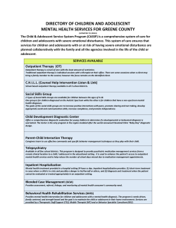
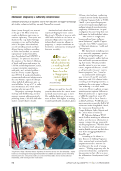
Social Cognitive and Affective Neuroscience Advance Access published April 13, 2015
doi:10.1093/scan/nsv026
SCAN (2015) 1 of 9
Mothers know best: redirecting adolescent reward
sensitivity toward safe behavior during risk taking
Eva H. Telzer,1,2 Nicholas T. Ichien,1 and Yang Qu1
1
Department of Psychology and 2 Beckman Institute for Advanced Science and Technology, University of Illinois, Urbana-Champaign, IL, USA
Despite being one of the healthiest developmental periods, morbidity and mortality rates increase dramatically during adolescence, largely due to
preventable, risky behaviors. Heightened reward sensitivity, coupled with ineffective cognitive control, has been proposed to underlie adolescents risk
taking. In this study, we test whether reward sensitivity can be redirected to promote safe behavior. Adolescents completed a risk-taking task in the
presence of their mother and alone during fMRI. Adolescents demonstrated reduced risk-taking behavior when their mothers were present compared
with alone, which was associated with greater recruitment of the ventrolateral prefrontal cortex (VLPFC) when making safe decisions, decreased
activation in the ventral striatum following risky decisions and greater functional coupling between the ventral striatum and VLPFC when making
safe decisions. Importantly, the very same neural circuitry (i.e. ventral striatum) that has been linked to greater risk-taking can also be redirected toward
thoughtful, more deliberative and safe decisions.
Keywords: adolescence; risk taking; rewards; fMRI; influence; family
Received 6 August 2014; Revised 14 February 2015; Accepted 4 March 2015
We greatly appreciate the assistance of the Biomedical Imaging Center. This research was supported by a grant
from the National Science Foundation (SES 1459719; Telzer) and generous funds from the Department of
Psychology at the University of Illinois.
Correspondence should be addressed to Eva H. Telzer, 603 East Daniel St., Champaign, IL 61820, USA. E-mail:
[email protected].
risky behavior is less rewarding) and redirected to promote cognitive
control?
Because neural regions involved in motivation and cognitive processes undergo significant reorganization during adolescence (Nelson
et al., 2005), the adolescent brain is thought to be highly flexible and
malleable (Crone and Dahl, 2012) and therefore particularly sensitive
to social influences. While prior work has focused on the social contexts which may increase risk taking, for instance peer presence (Chein
et al., 2011), social contexts can also decrease risk taking, such as parental presence. Indeed, parents represent one of the most direct and
proximal sources of influence over teenagers. Although parents tend to
adjust their supervisory practices to allow their adolescent children to
be more independent, adolescents tend to engage in more maladaptive,
risky behaviors during unsupervised time (Richardson et al., 1993;
Beck et al., 2001; Borawski et al., 2003), highlighting the important
role of parents in decreasing adolescent risk taking. While parents may
decrease adolescent risk-taking merely by serving as gatekeepers and
limiting adolescents’ opportunities to make poor decisions, we propose
that parents may actually change the ways in which adolescents think
and reason about risks during both active/deliberative as well as more
automatic decision-making processes.
Parental influence on adolescent risk taking may occur in several
ways. First, due to the relative immaturity of their prefrontal cortex,
adolescents may not yet have the cognitive resources to effectively
avoid risky behaviors and so parents may play an important scaffolding
role, helping their children to regulate their behaviors and engage in
more adaptive decision making. Thus, parents may facilitate improved
cognitive control and thereby reduce their adolescents’ risk taking.
Second, parents may reduce the affective and rewarding nature of
engaging in risky behavior. That is, risk taking may be comparatively
less rewarding and more aversive in the presence of family, and so the
typical heightened reward response following risky behaviors may be
attenuated. Lastly, parents may serve to redirect adolescents’ reward
sensitivity toward safe behavior, such that reward and cognitive control
systems interact to promote safe choices. Most prior work has
focused on the negative contexts (e.g. peer presence) in which
heightened reward sensitivity and immature cognitive control interact
to lead to maladaptive behavior (Chein et al., 2011). However, several
studies have demonstrated that rewards can also lead to improvements
in cognitive control through bottom-up processes that increase
ß The Author (2015). Published by Oxford University Press. For Permissions, please email: [email protected]
Downloaded from http://scan.oxfordjournals.org/ by guest on April 20, 2015
INTRODUCTION
Despite being one of the healthiest developmental periods, morbidity
and mortality rates increase 300% from childhood to adolescence
(National Center for Health Statistics, 2013; Kann et al., 2014) with
over 70% of adolescent deaths each year due to risk-taking behaviors,
such as reckless driving (Centers for Disease Control and Prevention,
2012). Evidence from developmental neuroscience suggests that risktaking behavior increases during adolescence partly due to relatively
early functional development of brain regions involved in reward sensitivity (e.g. ventral striatum) compared with more protracted functional maturation of brain regions supporting cognitive control (e.g.
the prefrontal cortex [PFC]; Somerville et al., 2010). Heightened
reward sensitivity, coupled with ineffective cognitive control, has largely been thought to underlie adolescents’ orientation toward risky
behavior (Steinberg, 2008; Casey et al., 2011). While most prior
work has focused on how this heightened reward sensitivity creates
vulnerabilities for adolescents, leading to increases in risk taking, scholars have begun to question these dual systems models of adolescent
brain development (Crone and Dahl, 2012; Pfeifer and Allen, 2012).
Recent evidence suggests that rewards can play an adaptive role, facilitating cognitive control. For example, rewarding cues (e.g. monetary
rewards; happy faces) enhance inhibitory control (Hardin et al., 2009;
Kohls et al., 2009; Teslovich et al., 2014), perhaps by serving to motivate adolescents to engage in effortful control. Moreover, the ventral
striatum may itself serve a regulatory function (Pfeifer et al., 2011).
Thus, heightened reward sensitivity, which has largely been proposed
to underlie adolescent risk taking (Steinberg, 2008; Casey et al., 2011),
may also shape adolescents’ motivations to engage in greater cognitive
control. In this study, we test whether adolescents’ heightened reward
sensitivity can be redirected to promote cognitive control and safe
behavior during risk-taking. In other words, can the heightened
reward response we typically see during risk taking be reduced (i.e.
2 of 9
SCAN (2015)
activation in brain regions involved in regulation (e.g. ventrolateral
prefrontal cortex [VLPFC]; Geier et al., 2010; Smith et al., 2011). We
therefore examined whether parental presence functions through
bottom-up processing, whereby the parent elicits a reward-related response that boosts adolescents’ motivation to regulate their
behavior. Thus, we tested whether parents increase neural coupling
between the ventral striatum and PFC to facilitate safe behavior
during risk taking.
Risk-taking task
The stoplight task measures risk taking at the behavioral and neural
level (Gardner and Steinberg, 2005; Chein et al., 2011). Adolescents
completed a simulated driving course in which they encountered a
number of stoplights and had to decide whether to ‘stop’ or ‘go’ by
pressing one of two buttons (see Figure 1). A decision to ‘go’ through
the intersection is the fastest option, but participants risk the possibility of crashing, which causes a 6 s delay. If they choose to ‘stop’, participants do not risk crashing, but it results in a short, 3 s delay.
Participants were told that the goal is to get through the driving
course in as short of a time as possible to win more money. The
Fig. 1 The stoplight task.
monetary incentive was used to encourage risk taking (Chein et al.,
2011).
Adolescents completed two rounds of the stoplight task during two
functional brain scans. Similar to the manipulation used by Steinberg
and colleagues with peers (Chein et al., 2011), adolescents played one
round of the game while their mother was watching and one round in
which they were alone. During the mother condition, the participant’s
mother was instructed to speak into the intercom naturally and authentically, informing their child that they would be watching during
the whole scan. Instructions were given to avoid any comments that
might explicitly or intentionally bias their child’s behavior. During the
alone condition, the researcher spoke into the microphone and informed the participant that nobody would be watching during the
round. Run order was counterbalanced across participants.
During each round of the task, participants were presented with 26
intersections. The probability of crashing was kept constant at 30%
(i.e. eight intersections out of the 26 total intersections had cars approaching on the cross street, resulting in a crash if the participant
made a ‘go’ decision), but this was not explicitly revealed to participants. The timing of traffic signals and the presence of a car on the
cross street varied so as to be unpredictable by the participant and to
introduce variable ITIs. At the end of each round, participants were
presented with their overall time and the number of crashes. Due to
learning effects, such that participants show higher behavioral risk
performance during initial rounds of the task followed by decreased
and more stable risk patterns thereafter (Peake et al., 2013), participants played two practice rounds of the task, each with 26 intersections, prior to entering the scanner.
Behaviorally, we examined the percent of decisions that are risky and
safe and examined differences based on maternal presence. In terms of
reaction time differences, the task is not optimized to collect reaction
time. Because of the long inter-trial interval (12 s between stoplights),
participants often make their decision prior to seeing the yellow light.
Although they are instructed to make the decision and subsequent
button response only after seeing the yellow light, many participants
press the button before the yellow light appears. Such button responses
are not recorded. Unfortunately, this precludes our ability to examine
reaction time differences.
Downloaded from http://scan.oxfordjournals.org/ by guest on April 20, 2015
METHODS
Participants
Thirty adolescent-mother dyads participated. Two participants were
excluded due to excessive movement (>3 mm), two participants did
not complete the scan, and one participant’s behavioral responses were
not recorded due to technical issues. Our final sample included 25
fourteen-year-old adolescents and their mother (adolescents:
Mage ¼ 14.43 years; 15 males; mothers: Mage ¼ 43.89 years).
Adolescent participants were predominantly from EuropeanAmerican (n ¼ 18) or African-American (n ¼ 5) backgrounds with
the remaining from Asian-American (n ¼ 1) or Central-American
(n ¼ 1) backgrounds. Mothers reported their highest levels of education as high school (n ¼ 1), some college (n ¼ 8), college (n ¼ 3) and
graduate, medical or law school (n ¼ 13). All participants provided
written assent and consent in accordance with the Institutional
Review Board.
E. H.Telzer et al.
Mothers know best
fMRI data acquisition and analysis
fMRI data acquisition
Imaging data were collected using a 3 Tesla Siemens Trio MRI scanner.
The stoplight task included T2*-weighted echoplanar images (EPI)
(slice thickness ¼ 3 mm; 38 slices; TR ¼ 2 s; TE ¼ 25 ms; matrix ¼ 92 92; FOV ¼ 230 mm; voxel size 2.5 2.5 3 mm3).
Structural scans consisted of a T2*weighted, matched-bandwidth
(MBW), high-resolution, anatomical scan (TR ¼ 4 s; TE ¼ 64 ms;
FOV ¼ 230; matrix ¼ 192 192; slice thickness ¼ 3 mm; 38 slices)
and a T1* magnetization-prepared rapid-acquisition gradient echo
(MPRAGE; TR ¼ 1.9 s; TE ¼ 2.3 ms; FOV ¼ 230; matrix ¼ 256 256;
sagittal plane; slice thickness ¼ 1 mm; 192 slices). The orientation
for the MBW and EPI scans was oblique axial to maximize brain
coverage.
3 of 9
subject contrasts were then submitted to random-effects, group-level
analyses. The following analyses were run at each voxel across the
entire brain volume: StopMother-StopAlone, GoMother-GoAlone
and PassMother-PassAlone. Because of the low frequency of crash
events (i.e. there were only eight possible crash events per run), we
had restricted power to test for CrashMother-CrashAlone. We present
this contrast for the 17 participants who had at least four crash trials
per condition.
We conducted psychophysiological interaction (PPI) analyses
(Friston et al., 1997) to examine functional coupling between the ventral striatum and cognitive control regions. We used the ventral striatum as the seed region, as the striatum has been consistently linked
with the experience of rewards. The striatum was defined structurally
using the WFUpickatlas (Tzourio-Mazoyer, et al., 2002; Maldjian et al.,
2003, 2004). PPI analyses were run using a generalized form of context-dependent PPI. Specifically, the automated gPPI toolbox in SPM
(gPPI; McLaren et al., 2012) was used to i) extract the deconvolved
times series from the ventral striatum region of interest (ROI) for each
participant to create the physiological variables; ii) convolve each trial
type with the canonical HRF, creating the psychological regressor; and
iii) multiply the time series from the psychological regressors with the
physiological variable to create the PPI interaction terms. This interaction term identified regions that covaried in a task-dependent
manner with the striatum. For the first-level model, one regressor
representing the deconvolved BOLD signal was included alongside
each psychological and PPI interaction terms for each condition type
to create a gPPI model. These first levels models created gPPI models
for the following contrasts: Stop Decisions, Go Decisions, and Pass
Outcomes for the Mother Present and Alone conditions separately.
At the group level, we conducted random effect analyses to compare
functional coupling between conditions of interest.
To correct for multiple comparisons, we conducted a Monte Carlo
simulation implemented using 3dClustSim in the software package
AFNI (Ward, 2000). We used our group-level brain mask, which
included only gray matter. Results of the simulation indicated a
voxel-wise threshold of P < 0.005 combined with a minimum cluster
size of 35 voxels for the whole brain, corresponding to P < 0.05, false
wise error (FWE) corrected. We used the MarsBaR toolbox to extract
parameter estimates from significant clusters in the group-level
analyses. To ensure that the neural effects are not driven by differences
in the number of trials in the analyses (e.g. more stop decisions
when mothers are present then when alone), all fMRI analyses
covary for the number of decisions to stop during Alone and
Mother conditions.
RESULTS
Behavioral results
Adolescents made significantly more risky decisions (i.e. ‘go’) when
alone than when their mothers were present [F(1, 24) ¼ 3.9, P < 0.001;
Figure 2]. In prior studies using this same task, the presence of peers
increased risk-taking behavior of adolescent participants (Gardner and
Steinberg, 2005; Chein et al., 2011). In contrast, we find that the presence of mothers reduces risk-taking behavior.
fMRI results
Main effects
Our first fMRI analyses collapsed across conditions to test what regions
were activated when making safe and risky choices irrespective of
maternal presence. In whole brain t-tests, we examined neural activation during Stop decisions, Go decisions and Passes. When adolescents
made decisions to Stop (compared with baseline), they did not show
heightened activation in any region. When making Go decisions
Downloaded from http://scan.oxfordjournals.org/ by guest on April 20, 2015
fMRI data preprocessing and analysis
Neuroimaging data were preprocessed and analyzed using Statistical
Parametric Mapping (SPM8; Wellcome Department of Cognitive
Neurology, Institute of Neurology, London, UK). Preprocessing for
each participant’s images included spatial realignment to correct for
head motion (no participant exceeded 2 mm of maximum image-toimage motion in any direction). The realigned functional data were
coregistered to the high resolution MPRAGE, which was then segmented into cerebrospinal fluid, grey matter and white matter. The
normalization transformation matrix from the segmentation step was
then applied to the functional and T2 structural images, thus transforming them into standard stereotactic space as defined by the
Montreal Neurological Institute and the International Consortium
for Brain Mapping. The normalized functional data were smoothed
using an 8 mm Gaussian kernel, full-width-at-half maximum, to increase the signal-to-noise ratio.
Statistical analyses were performed using the general linear model
(GLM) in SPM8. Each trial was convolved with the canonical hemodynamic response function. High-pass temporal filtering with a cutoff
of 128 s was applied to remove low-frequency drift in the time series.
Serial autocorrelations were estimated with a restricted maximum likelihood algorithm with an autoregressive model order of 1.
In each participant’s fixed-effects analysis, a GLM was created with
four regressors of interest each for the alone condition and mother
condition, modeled as events: two decision regressors (Stop and Go)
and two outcome regressors (Crash and Pass). In addition, the wait
time after stop decisions were modeled as well as the final ‘Game Over’
period at the end of each run, to remove these from the implicit
baseline. This resulted in 12 conditions, six each for alone and
mother: StopMother, GoMother, PassMother, CrashMother,
WaitMother, GameOverMother, StopAlone, GoAlone, PassAlone,
CrashAlone, WaitAlone and GameOverAlone.
Because the task was self-paced, the duration of the decision trials
(stop or go) represented the time from which the traffic light appeared
until the participant made a response, and the duration for the outcome (pass or crash) was 1 s. The onset of the crash event corresponded to another car crashing into the participant’s car. The pass
and wait events had no specific onset time. However, because the crash
events happened at most 2 s after the yellow light, we modeled the pass
and wait events as being 2 s after the yellow light, the point at which the
outcome of the risky decision was clear. Each was modeled with a 1 s
duration. Null events, consisting of the jittered intertrial intervals, were
not explicitly modeled and therefore constituted an implicit baseline.
The parameter estimates resulting from the GLM were used to create
linear contrast images comparing each of four event conditions
(decisions: Go, Stop; outcomes: Crash, Pass) during the Alone round
to the corresponding conditions in the Mother round. The individual
SCAN (2015)
4 of 9
SCAN (2015)
E. H.Telzer et al.
Fig. 2 Behavioral performance. Adolescents made significantly fewer risky choices when their
mothers were present than when alone. Error bars represent SEM.
Interactions between Alone and Mother present condition
Next, we examined differences in neural activation during Stop decisions, Go decisions and Passes when adolescents were alone compared
with mother present (Alone vs Mother).
Decisions
When adolescents made decisions to stop when their mothers were
present compared with alone (StopMother-StopAlone), they showed
increased activation in the left VLPFC and the MPFC (Figure 3a,
Table 2). For descriptive purposes, we extracted parameter estimates
of signal intensity from the VLPFC and MPFC clusters when
adolescents made Stop decisions when alone (StopAlone-baseline)
and when their mother was present (StopMother-baseline). As
shown in Figure 3b, adolescents demonstrated significantly greater activation in the VLPFC and MPFC when making safe decisions in the
presence of their mother compared with alone. When adolescents
made decisions to go through the intersection when their mothers
were present compared with alone (GoMother-GoAlone), they also
showed increased activation in the MPFC (Table 2).
To further probe these effects, we extracted parameter estimates of
signal intensity from the clusters in the MPFC and left VLPFC for the
four conditions relative to baseline (StopAlone, GoAlone, StopMother
and GoMother). We used these parameters in SPSS to conduct interaction analyses. For the VLPFC, we find a significant main effect of
context [Mother vs Alone; F(1,23) ¼ 20.66, P < 0.0001] as well as a
significant interaction [F(1,23) ¼ 6.5, P < 0.01], such that the VLPFC
is recruited significantly more within the Mother condition for Stop
(M ¼ 2.14, SEM ¼ 0.96) than Go (M ¼ 0.41, SEM ¼ 0.54) decisions
[t(24) ¼ 2.2, P < 0.05]. In contrast, when alone, adolescents tend to
recruit the VLPFC more when making Go (M ¼ 1.64, SEM ¼ 0.43)
than Stop (M ¼ 3.06, SEM ¼ 0.62) decisions [t(24) ¼ 2.07, P ¼ 0.05].
Importantly, the VLPFC is activated significantly more than baseline
Fig. 3 (a) Neural activation during decisions to ‘stop’ when adolescents’ mothers were present
compared with alone. For descriptive purposes, parameters estimates of signal intensity were
extracted from the VLPFC and MPFC cluster separately for Stop decisions when Alone and Mother
Present (relative to baseline). Error bars represent SEM.
only in the Mother condition [t(24) ¼ 2.1, P < 0.05] when making safe
decisions. For the MPFC, we find a significant main effect of context
[Mother vs Alone; F(1,23) ¼ 19.44, P < 0.0001], but do not find an
interaction.
Outcomes
When adolescents successfully passed through an intersection without
crashing following a risky decision (PassMother-PassAlone), they
showed significantly less activation in the amygdala and ventral
striatum when their mothers were present compared with alone
(Figure 4a; Table 2). For descriptive purposes, we extracted parameter
estimates of signal intensity from the ventral striatum and amygdala
clusters for successful passes when adolescents were alone (StopAlonebaseline) and when their mother was present (StopMom-baseline). As
shown in Figure 4b, adolescents demonstrated significantly greater activation in the ventral striatum and amygdala following a successful
risky decision when alone than when their mother was present. We do
not find any significant effects for Crash outcomes (CrashMotherCrashAlone). However, because most participants had too few crash
events for statistical tests, we have limited power for this analysis.
Therefore this null result should be interpreted with caution.
PPI analysis
Using the ventral striatum as the seed region, we first extracted parameter estimates of signal intensity during Stop and Go decisions and
Pass outcomes during the Alone and Mother Present conditions and
ran paired-samples t-tests. Adolescents demonstrated greater ventral
striatum activation when making stop decisions when their mother
was present (M ¼ 2.09, SEM ¼ 1.19) than when alone [M ¼ 0.76,
Downloaded from http://scan.oxfordjournals.org/ by guest on April 20, 2015
(compared with baseline), adolescents showed heightened activation in
the ventral striatum, insula, dACC and SMA. In addition, we compared activation between Stop and Go decisions collapsed across
conditions. Adolescents demonstrated greater activation in the
insula, dACC and midbrain when making Go decisions relative to
Stop decisions. The only region that was more active during Stop
compared with Go decisions was the precentral gyrus. Finally, when
adolescents successfully passed through an intersection without
crashing following a risky decision, they demonstrated heightened activation in a large cluster encompassing the superior frontal gyrus,
precuneous and extending into the left temporoparietal junction
(TPJ) (Table 1).
Mothers know best
SCAN (2015)
5 of 9
Table 1 Brain regions activated during the stoplight task irrespective of maternal presence
Contrast
Anatomical region
BA
x
y
z
t
k
12
33
36
3
33
15
5
29
14
14
13
2
2
2
7
43
67
54
4.96
3.48
3.84
4.21
4.24
3.35
191
110
93
621
472
57
Precentral gyrus
42
22
61
4.67
47
Insula
dACC
SMA
Postcentral gyrus
Midbrain
R Precuneus
L Precuneus
39
9
9
45
6
18
9
5
8
20
19
28
76
73
10
43
48
49
14
40
46
5.94
4.73
4.26
5.53
4.18
5.32
5.39
266
1278a
24
9
12
54
6
11
73
73
42
58
55
43
46
34
26
5.97
4.99
4.07
5.49
3.93
2957b
Stop Decisions
–
Go Decisions
Ventral striatum
R Insula
L Insula
dACC
Precentral gyrus
SMA
32/24
Stop-Go Decisions
Go-Stop Decisions
a
a
Pass Outcome
Superior frontal gyrus
R Precuneus
L Precuneus
TPJ
Cerebellum
b
b
b
122
Note. BA refers to putative Broadman’s areas. x, y, and z refer to MNI coordinates; t refers to the t-score at those coordinates (local maxima); k refers to the number of voxels in each
significant cluster. TPJ, temporoparietal junction; SMA, supplementary motor area; dACC, dorsoanterior cingulate cortex. a,b regions are part of the same cluster; – indicates no
significant clusters were identified.
Fig. 4 (a) Neural activation during successful passes when adolescents were alone compared with
mother present. For descriptive purposes, parameters estimates of signal intensity were extracted
from the ventral striatum and amygdala clusters separately for Passes when Alone and Mother
Present (relative to baseline). Error bars represent SEM.
Fig. 5 (a) PPI analysis during stop decisions when mothers were present compared with alone.
Significant activation represents positive coupling with the ventral striatum. (b) For descriptive purposes, parameters estimates of signal intensity were extracted from the VLPFC cluster
separately for Stop decisions when Alone and Mother Present (relative to baseline). Error bars
represent SEM.
Downloaded from http://scan.oxfordjournals.org/ by guest on April 20, 2015
229
165
392
6 of 9
SCAN (2015)
E. H.Telzer et al.
Table 2 Brain regions activated during the stoplight task when adolescents’ were alone compared with mother present
Contrast
Anatomical region
BA
R MPFC
L VLPFC
Precuneus
L Postcentral gyrus
R Postcentral gyrus
32/10
47/11
–
–
VLPFC
47
x
y
z
t
k
9
42
0
42
51
47
35
61
19
7
10
8
19
34
34
4.34
3.01
4.10
4.19
3.95
53
46
60
75
44
39
23
8
3.33
36
9
9
41
44
8
5
4.15
3.97
62
44
6
12
45
22
1
19
5
46
49
4.02
3.52
3.90
117
417
157
3
26
5
5
11
24
4.50
3.26
52
36
27
22
52
3.70
54
27
18
44
48
33
27
3
16
5
17
20
40
20
11
28
3
24
5
8
43
40
55
34
16
3.56
3.78
3.64
4.14
4.97
4.70
4.54
3.55
38
40
53
186
41
114
439
43
30
29
14
4.64
47
30
20
45
0
51
0
12
30
29
62
41
50
2
25
5
20
14
0
22
10
16
52
10
11
4.20
5.61
3.76
3.76
3.45
3.45
5.45
3.28
37
52
54
55
47
103
199
37
3
6
59
22
1
6
5.79
4.46
63
78
3
22
51
41
0
22
13
14
14
3.17
3.88
3.95
41
53
40
Decisions
StopMother-StopAlone
StopAlone-StopMother
StopMother
StopAlone
–
GoMother-GoAlone
R MPFC
L MPFC
32/10
10
GoAlone-GoMother
–
–
–
Midbrain
R ACC
R Precentral gyrus
24/32
Go Alone
Outcomes
PassAlone-PassMother
VS
L Amygdala
PassMother-PassAlone
–
–
PassMother
R Precentral gyrus
PassAlone
L Amygdala
VS
R Insula
R TPJ
L Middle frontal gyrus
R Middle frontal gyrus
Posterior Cingulate Cortex
R Caudate
40
6
PPI with VS
StopMother-StopAlone
R VLPFC
47/11
StopAlone-StopMother
–
StopMother
R VLPFC
L VLPFC
R DLPFC
MPFC
Precentral gyrus
SMA
Caudate
Insula
47
46
32/10
StopAlone
MPFC
Caudate
32/10
GoMother-GoAlone
–
GoAlone-GoMother
–
–
–
–
GoMother
GoAlone
dACC
L Amygdala
STS
(continued)
Downloaded from http://scan.oxfordjournals.org/ by guest on April 20, 2015
GoMother
Mothers know best
SCAN (2015)
7 of 9
Table 2 Continued
Contrast
Anatomical region
BA
–
–
–
–
Temporal pole
Posterior cingulate cortex
21
x
y
z
t
k
51
6
4
40
23
1
4.53
4.14
99
175
54
27
51
51
12
3
14
4
28
26
40
7
8
14
16
23
4
49
4.06
5.29
4.25
3.86
4.38
3.55
54
40
132
104
530
38
PassMother-PassAlone
PassAlone-PassMother
PassMother
PassAlone
R Insula
R Amygdala
R TPJ
L TPJ
Posterior cingulate cortex
SMA
Note. BA refers to putative Broadman’s areas. x, y, and z refer to MNI coordinates; t refers to the t-score at those coordinates (local maxima); k refers to the number of voxels in each
significant cluster. MPFC, medial prefrontal cortex; VLPFC, ventrolateral prefrontal cortex; DMPFC, dorsomedial prefrontal cortex; VS, ventral striatum; SMA, supplementary motor area; TPJ,
temporoparietal junction; STS, superior temporal sulcus; dACC, dorsoanterior cingulate cortex; DLPFC, dorsolateral prefrontal cortex; – indicates no significant clusters were identified.
DISCUSSION
Adolescence is a developmental period marked by exquisite changes in
functional brain development. Heightened reward sensitivity, coupled
with relatively less developed cognitive control, has largely been
thought to underlie adolescents’ engagement in risky behavior
(Steinberg, 2008; Casey et al., 2011). In this study, we test whether
the presence of parents alters adolescent risk taking via changes in
reward sensitivity and cognitive control.
Our findings suggest that maternal presence has a large impact on
adolescents’ risky behavior and brain function. When mothers were
present, adolescents engaged in significantly less risky behavior.
Surprisingly, prior research showing that parental supervision is associated with lower adolescent risk taking has all been correlational
(Richardson et al., 1993; Beck et al., 2001; Borawski et al., 2003).
This is the first study to experimentally demonstrate the protective
role that parental presence plays on reducing adolescent risk taking.
Importantly, we identified the neural mechanisms by which mothers
decrease their adolescent’s risk taking. First, adolescents showed greater
recruitment of the VLPFC when making safe decisions in the presence
of their mothers. The VLPFC is involved in self-control and the ability
to regulate and control one’s prepotent thoughts and behaviors (Baker
et al., 1997; Gray et al., 2002) and plays a causal role in goal-directed
inhibitory control, including the braking of motor responses (Wessel
et al., 2013). These results suggest that mothers boost self-control by
increasing PFC activation, facilitating more deliberative and safe decisions. In addition, adolescents showed heightened activation in the
MPFC when making both safe and risky decisions when their
mother was present. The MPFC is involved in monitoring and computing the reward value of ongoing cognitive tasks (Pochon et al.,
2002), as well as the subjective value of rewards (Kable and
Glimcher, 2007; Levy et al., 2010). Thus, when their mothers are present, adolescents may evaluate the relative reward of running the stoplight vs stopping. The MPFC is also involved in self-related processing
and thinking about close others (Mitchell et al., 2006), and so adolescents may be incorporating their mothers’ perspective to inform their
own behavior and decide whether to engage in risky decisions.
Second, adolescents showed decreased activation in the ventral striatum and amygdala following a risky decision in the presence of their
mothers. The ventral striatum has been consistently linked with hedonic rewards, tends to be more active in adolescents than children or
adults during reward and risk-taking tasks and predicts greater engagement in real-life risk taking behavior (Galvan et al. 2006, 2007). The
amygdala codes for emotionally salient stimuli and tends to be more
active in adolescents than adults during emotion (particularly positive
emotions) and reward processing (Ernst et al., 2005; Hare et al. 2008;
for reviews see Somerville et al., 2011, Nelson et al., 2014). Together,
our findings suggest that mothers reduce the rewarding and salient
nature of engaging in risk taking, perhaps taking the fun out of
being risky. Thus, when adolescents make risky decisions in the presence of their mothers, they respond to risky outcomes with decreased
amygdala and ventral striatum activation than when making the same
risky decisions alone.
Finally, and most novel, maternal presence facilitated functional
coupling between neural regions involved in reward and cognitive
control processing when adolescents made safe but not risky decisions,
suggesting that mothers increased the rewarding nature of engaging in
cognitive control. These functional connectivity results suggest that
within adolescents, those who engage the ventral striatum to a greater
extent also engage the VLPFC more when making safe decisions, but
this only occurs when adolescents’ mothers are present. Theories of
Downloaded from http://scan.oxfordjournals.org/ by guest on April 20, 2015
SEM ¼ 0.46; t(24) ¼ 2.44, P < 0.05], but did not differ when making go
decisions when their mother was present (M ¼ 0.99, SEM ¼ 0.83) compared with alone [M ¼ 0.23, SEM ¼ 0.79; t(24) ¼ 0.6, ns]. For Pass
outcomes, adolescents showed significantly greater activation when
alone (M ¼ 0.58, SEM ¼ 0.38) compared with when their mother was
present [M ¼ 1.10, SEM ¼ 0.40; t(24) ¼ 4.56, P < 0.0001].
Next, we conducted PPI analyses to test for functional connectivity
between the ventral striatum and cognitive control regions. When
adolescents made a decision to stop (StopMother-StopAlone), we
found a significant interaction between the ventral striatum and
VLPFC. For descriptive purposes, we extracted parameter estimates
of signal intensity from the VLPFC cluster that showed a significant
interaction with the VS and plotted the effects separately for
StopMother and StopAlone (vs baseline). As shown in Figure 5, adolescents show VS-VLPFC coupling significantly greater when their
mothers are present than when alone (Table 1). To further probe
this effect, we ran PPI analyses separately for the Mother and Alone
condition for Stop decisions. Adolescents do not show significant
functional coupling between the VS and VLPFC when alone but do
show significant coupling between these regions when their mother
was present (Table 1). No brain regions differentially functionally
interacted with the VS during decisions to Go (GoMother-GoAlone)
or during successful passes (PassMother-PassAlone; see Table 1 for
Mother and Alone conditions separately).
8 of 9
SCAN (2015)
their teenagers’ decision making capacities in ways that allow their
children to engage in more mature cognitive processes. Importantly,
our findings suggest that parents do not just serve as gatekeepers but
actually change the ways in which adolescents think and reason about
risks.
Conflict of Interest
None declared.
REFERENCES
Baker, S.C., Frith, C.D., Dolan, R.J. (1997). The interaction between mood and cognitive
function studied with PET. Psychological Medicine, 27, 565–78.
Beck, K.H., Shattuck, T., Raleigh, R. (2001). Parental predictors of teen driving risk.
American Journal of Health and Behavior, 25(1), 10–20.
Borawski, E.A., Ievers-Landis, C.E., Lovegreen, L.D., Trail, E.S. (2003). Parental monitoring, negotiated unsupervised time, and parental trust: the role of perceived parenting
practices in adolescent health risk behaviors. Journal of Adolescent Health, 33(2), 60–70.
Casey, B.J., Somerville, L.H., Gotlib, I.H., et al. (2011). Behavioral and neural correlates of
delay of gratification 40 years later. Proceedings of the National Academy of Science of the
United States of America, 108(36), 14998–5003.
Chein, J., Albert, D., O’Brien, L., Uckert, K., Steinberg, L. (2011). Peers increase adolescent
risk taking by enhancing activity in the brain’s reward circuitry. Developmental Science,
14, F1–10.
Centers for Disease Control and Prevention Web-based Injury Statistics Query and
Reporting System (WISQARS) (2012), National Center for Injury Prevention and
Control, Centers for Disease Control and Prevention.
Crone, E.A., Dahl, R.E. (2012). Understanding adolescence as a period of social-affective
engagement and goal flexibility. Nature Reviews Neuroscience, 13, 636–50.
Ernst, M., Nelson, E.E., Jazbec, S., et al. (2005). Amygdala and nucleus accumbens in
responses to receipt and omission of gains in adults and adolescents. Neuroimage,
25(4), 1279–91.
Friston, K.J., Buechel, C., Fink, G.R., Morris, J., Rolls, E., Dolan, R.J. (1997).
Psychophysiological and modulatory interactions in neuroimaging. NeuroImage, 6(3),
218–29.
Galvan, A., Hare, T., Voss, H., Glover, G., Casey, B.J. (2007). Risk-taking and the adolescent
brain: Who is at risk? Developmental Science, 10, F8–14.
Galvan, A., Hare, T.A., Parra, C.E., et al. (2006). Earlier development of the accumbens
relative to orbitofrontal cortex might underlie risk-taking behavior in adolescents.
Journal of Neuroscience, 26, 6885–92.
Gardner, M., Steinberg, L. (2005). Peer influence on risk taking, risk preference, and risky
decision making in adolescence and adulthood: an experimental study. Developmental
Psychology, 41(4), 625–35.
Geier, C.F., Terwilliger, R., Teslovich, T., Velanova, K., Luna, B. (2010). Immaturities in
reward processing and its influence on inhibitory control in adolescence. Cerebral
Cortex, 20(7), 1613–29.
Gray, J.R., Braver, T.S., Raichle, M.E. (2002). Integration of emotion and cognition in the
lateral prefrontal cortex. Proceedings of the National Academy of Sciences of the United
States of America, 99, 4115–20.
Hardin, M.G., Mandell, D., Mueller, S.C., Dahl, R.E., Pine, D.S., Ernst, M. (2009).
Inhibitory control in anxious and healthy adolescents is modulated by incentive and
incidental affective stimuli. Journal of Child Psychology and Psychiatry, 50(12), 1550–8.
Hare, T.A., Tottenham, N., Galvan, A., Voss, H.U., Glover, G.H., Casey, B.J. (2008).
Biological substrates of emotional reactivity and regulation in adolescence during an
emotional go-nogo task. Biological Psychology, 63(10), 927–34.
Kable, J.W., Glimcher, P.W. (2007). The neural correlates of subjective value during intertemporal choice. Nature neuroscience, 10(12), 1625–33.
Kann, L., Kinchen, S., Shanklin, S.L., Flint, K.H., Kawkins, J., Harris, W.A. (2014). Youth
risk behavior surveillance – United States, 2013. Morbidity and Mortality Weekly Report.
Surveillance Summaries (Washington, D.C.: 2002), 63(Suppl 4), 1–168.
Kohls, G., Peltzer, J., Herpertz-Dahlmann, B., Konrad, K. (2009). Differential effects of
social and non-social reward on response inhibition in children and adolescents.
Developmental Science, 12(4), 614–25.
Levy, I., Snell, J., Nelson, A.J., Rustichini, A., Glimcher, P.W. (2010). Neural representation
of subjective value under risk and ambiguity. Journal of neurophysiology, 103(2),
1036–47.
Maldijian, J.A., Laurienti, P.J., Burdette, J.H. (2004). Precentral gyrus discrepancy in electronic versions of the Talairach atlas. NeuroImage, 21(1), 450–5.
Maldjian, J.A., Laurienti, P.J., Kraft, R.A., Burdette, J.H. (2003). An automated method for
neuroanatomic and cytoarchitectonic atlas-based interrogation of fMRI data sets.
NeuroImage, 19(3), 1233–39.
McLaren, D.G., Ries, M.L., Xu, G., Johnson, S.C. (2012). A generalized form of context
dependent psychophysiological interactions (gPPI): A comparison to standard
approaches. NeuroImage, 61(4), 1277–1286.
Downloaded from http://scan.oxfordjournals.org/ by guest on April 20, 2015
adolescent risk-taking propose that heightened reward sensitivity largely underlies increased risk-taking during adolescence (Steinberg,
2008), and most prior work has focused on the contexts in which
reward sensitivity leads to maladaptive, risky behavior, for example,
in the presence of peers (Chein et al., 2011). Here, we demonstrate that
safe behavior occurs when the ventral striatum is functionally coupled
with the VLPFC when mothers are present. Thus, heightened coupling
between these regions is associated with safer behavior, suggesting that
maternal presence may promote a reward response that boosts adolescents’ engagement of cognitive control and promotes more safe decision making. Importantly, the very same neural circuitry (i.e. ventral
striatum) that has been linked to greater risk taking may also be related
to thoughtful, more deliberative and safe decisions when mothers are
present. Therefore, maternal presence may serve to redirect adolescents’ reward sensitivity away from risk (i.e. reduced ventral striatum
activation during risky outcomes) and toward more safe behavior (i.e.
increased VS-PFC coupling during safe decisions). Because PPI analyses are not directional, an alternative explanation for functional coupling between the ventral striatum and VLPFC is that engaging in
cognitive control in the presence of their mother is a rewarding process. In other words, recruitment of cognitive control facilitates safe
decisions in the presence of their mother which results in a heighted
sense of reward.
Our findings contribute to conceptualizations of adolescent neurodevelopment. While dual systems models (Steinberg, 2008; Somerville
et al., 2010; Casey et al., 2011) have gained considerable attention,
several alternative perspectives have been proposed (Crone and Dahl,
2012; Pfeifer and Allen, 2012). For example, striatal reactivity is not
always associated with greater adolescent risk taking, as suggested by
dual system models, and, depending on the context, striatal reactivity
can result in lower risk taking behavior (Pfeifer et al., 2011; Telzer
et al., 2013). We build upon these findings and show that ventral
striatum activation is associated with safe decision making when it
occurs in tandem with cognitive control-related neural activation in
the presence of mothers. Together, our findings underscore the importance of understanding the social context in which adolescent decision making occurs and suggests that the striatum plays a diverse
function, sometimes promoting risky behavior but at other times
deterring risky behavior depending upon the motivational context.
While our task did not have a condition to test for the effect of the
person on adolescent risk taking and neural reactivity, our findings are
the opposite of those found by Chein et al. (2010) who used the same
manipulation with peer presence and found that peers increase risk
taking and ventral striatum activation during risky decisions.
Therefore, our effects are likely not due to someone watching vs not
watching, as peers’ presence alters risk taking and related neural patterns in a different way than does parents’ presence. However, without
directly comparing peer and parent influence in the same paradigm, we
cannot be sure that these contexts have opposite effects on adolescents,
and our findings could be due to other factors that differ between the
studies (e.g. characteristics of the samples, age, etc.). Thus, future work
should examine how both parents and peers impact adolescent decision making and neural processing during risk taking as well as the
differential role of parental and authority figures to test whether adults
decrease adolescent risk taking or whether our patterns are specific to
parents.
Mothers in our study did not vocally encourage or discourage any
kind of behavior, actively regulate their child’s decisions or provide
feedback. Instead, mothers simply told their child they would be present and watching. Our findings suggest that parental influence does
not have to be explicit. The mere presence of a parent in the room or
car may help adolescents to slow down, think twice and make more
deliberative and safe decisions. Parents are in a position to regulate
E. H.Telzer et al.
Mothers know best
9 of 9
Smith, A.B., Halari, R., Giampetro, V., Brammer, M., Rubia, K. (2011). Developmental
effects of reward on sustained attention networks. NeuroImage, 56, 1693–704.
Steinberg, L. (2008). A social neuroscience perspective on adolescent risk-taking.
Developmental Review, 28(1), 78–106.
Somerville, L.H., Jones, R.M., Casey, B.J. (2010). A time of change: behavioral and neural
correlates of adolescent sensitivity to appetitive and aversive environmental cues. Brain
and Cognition, 72, 124–33.
Somerville, L.H., Fani, N., McClure-Tone, E.B. (2011). Behavioral and neural representation of emotional facial expressions across the lifespan. Developmental neuropsychology,
36(4), 408–28.
Telzer, E.H., Fuligni, A.J., Lieberman, M.D., Ga´lvan, A. (2013). Ventral striatum activation
to prosocial rewards predicts longitudinal declines in adolescent risk taking.
Developmental Cognitive Neuroscience, 3, 45–52.
Teslovich, T., Mulder, M., Franklin, N.T., et al. (2014). Adolescents let sufficient evidence
accumulate before making a decision when large incentives are at stake. Developmental
Science, 17(1), 59–70.
Tzourio-Mazoyer, N., Landeau, B., Papathanassiou, D., et al. (2002). Automated anatomical labeling of activations in spm using a macroscopic anatomical parcellation of the
mni mri single-subject brain. NeuroImage, 15, 273–89.
Ward, B.D. (2000). Simultaneous inference for fMRI data. Available: http://afni.nimh.nih.
gov/pub/dist/doc/manuals/AlphaSim.pdf (accessed 24 March 2015).
Wessel, J.R., Conner, C.R., Aron, A.R., Tandon, N. (2013). Chronometric electrical stimulation of right inferior frontal cortex increases motor braking. Journal of Neuroscience,
33(50), 19611–9.
Downloaded from http://scan.oxfordjournals.org/ by guest on April 20, 2015
Mitchell, J.P., Macrae., C.N., Banaji, M.R. (2006). Dissociable medial prefrontal contributions to judgments of similar and dissimilar others. Neuron, 50, 655–63.
National Center for Health Statistics (2013), Health, United States, 2013. US Department of
Health and Human Services, Centers for Disease Control and Prevention. Hyattsville,
MD.
Nelson, E.E., Lau, J.Y., Jarcho, J.M. (2014). Growing pains and pleasures: how emotional
learning guides development. Trends in Cognitive Sciences, 18(2), 99–108.
Nelson, E.E., Leibenluft, E., McClure, E.B., Pine, D.S. (2005). The social re-orientation of
adolescence: a neuroscience perspective on the process and its relation to psychopathology. Psychological Medicine, 35(2), 163–74.
Peake, S., Dishion, T.J., Stormshak, E.A., Moore, W.E., Pfeifer, J.H. (2013). Risk-taking and
social exclusion in adolescence: neural mechanisms underlying peer influences on decision-making. NeuroImage, 82, 23–4.
Pfeifer, J.H., Allen, N.B. (2012). Arrested development? Reconsidering dual-systems models of
brain function in adolescence and disorders. Trends in Cognitive Sciences, 16(6), 322–9.
Pfeifer, J.H., Masten, C.L., Moore, W.E., III, et al. (2011). Entering adolescence: resistance
to peer influence, risky behavior, and neural changes in emotion reactivity. Neuron,
69(5), 1029–36.
Pochon, J.B., Levy, R., Fossati, P., et al. (2002). The neural system that bridges reward and
cognition in humans: an fMRI study. Proceedings of the National Academy of Sciences,
99(8), 5669–74.
Richardson, J.L., Radziszewska, B., Dent, C.W., Flay, B.R. (1993). Relationship between
after-school care of adolescents and substance use, risk taking, depressed mood, and
academic achievement. Pediatrics, 92(1), 32–8.
SCAN (2015)


© Copyright 2026