
Treatment of enteritis in chronic granulomatous CASE REPORTS
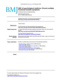
Downloaded from gut.bmj.com on September 9, 2014 - Published by group.bmj.com
Gut 1998;42:127–130
127
CASE REPORTS
Treatment of enteritis in chronic granulomatous
disease with granulocyte colony stimulating factor
B Myrup, N H Valerius, P B Mortensen
Department of
Medicine A, Section of
Gastroenterology,
Rigshospitalet
B Myrup
P B Mortensen
Abstract
Background—In several diseases there is
a relation between deficiency of neutrophil granulocytes and granulomatous
lesions. Recently, in glycogen storage
disease type Ib, this relation has been supported by the beneficial eVect of treatment
of enteritis with granulocyte-macrophage
colony stimulating factor.
Aim—To investigate whether chronic
granulomatous disease could be treated
according to the same principle.
Patients and methods—Inflammatory lesions were monitored in two brothers with
chronic granulomatous disease demonstrated by very low superoxide production
in neutrophil granulocytes. The two patients were treated with recombinant
human granulocyte colony stimulating
factor on three occasions when the disease
was active.
Results—In one patient, remission of an
inflamed stenosis of the colon sigmoideum was shown by granulocyte scintigraphy after one month of treatment with
granulocyte colony stimulating factor. In
the other patient, remission of colon
disease and later of a non-malignant
tumour in the right lung hilum was shown
by colonoscopy and computed tomography scans respectively.
Conclusion—Remission of inflammatory
lesions in two brothers with chronic
granulomatous disease was induced by
granulocyte colony stimulating factor on
three occasions. The mechanism for this
eVect is not known. The result is similar to
the response found in patients with leucocyte deficiency due to glycogen storage
disease type Ib.
suVer from enteritis.7 8 Enteritis is also seen
in other disease states with defective
neutrophils.9–12 In glycogen storage disease type
Ib,13–16 the association between the granulomatous lesion and neutrophil deficiency is supported by the recent demonstration that
patients with enteritis benefit from treatment
with granulocyte-macrophage colony stimulating factor.17 We report three incidents of clinical
remission in two brothers with CGD during
treatment with granulocyte colony stimulating
factor (G-CSF). In parallel with the results
from glycogen storage disease type Ib, this may
serve as a model for the pathogenesis and new
treatment strategies in other granulomatous
diseases.
Case reports
METHODS
Recombinant human G-CSF was obtained
from Roche, Denmark (Neupogen). Each
treatment regimen consisted of subcutaneous
injections of 0.3 mg recombinant human
G-CSF daily for 30 days. Erythrocyte sedimentation rate and orosomucoid and leucocyte
counts were recorded once a week during the
treatment periods. The oxidative metabolism of
neutrophils was assessed by phorbol myristate
acetate stimulated nitroblue tetrazolium slide
test, oxygen consumption, superoxide anion
production, and chemiluminescence as previously described.18 Chemotaxis was measured in
a Boyden chamber using N-formyl-methionylleucyl-phenylalanine
(f-MLP)
as
chemoattractant.19 The eVect of treatment on
the leucocyte population distribution and membrane phenotype was assessed by diVerential
counting and flow cytometric quantification of
cluster of diVerentiation 14, 11b and 16
antigens on granulocytes and monocytes.
(Gut 1998;42:127–130)
PATIENT 1
Department of
Paediatrics, Hvidovre
Hospital, University
Hospital of
Copenhagen, Denmark
N H Valerius
Correspondence to:
B Myrup, Department of
Medicine, Centralsygehuset i
Holbaek, DK-4300 Holbaek,
Denmark.
Accepted for publication
2 September 1997
Keywords: chronic granulomatous disease; enteritis;
granulocyte colony stimulating factor
Chronic granulomatous disease (CGD) is a
group of inherited disorders characterised by
defective superoxide production in circulating
phagocytes leading to impaired bactericidal
activity.1–5 The patients therefore suVer from
frequent bacterial and fungal infections.1 The
diVerent clinical forms of CGD are related to
genetic heterogeneity.6 A number of patients
The patient had suVered from chronic inflammatory bowel disease since the age of 26. He
reported having seven to ten stools per day,
often containing blood. Barium contrast studies showed a stenosis in the rectum and inflammation of the sigmoid and descending colon.
After exclusion of infectious causes, the patient
was treated with prednisolone, salazopyrine,
and azathioprine without clinical improvement. The symptoms did not change over the
following three years. Repeated barium contrast studies gave unchanged results. CGD was
Downloaded from gut.bmj.com on September 9, 2014 - Published by group.bmj.com
128
Myrup, Valerius, Mortensen
80
A
Patients
Chemiluminescence (cpm)
Chemiluminescence (cpm)
40
Controls
30
20
10
0
–1
4
11
18
25
33
70
B
60
50
40
30
20
10
0
39
–1
6
Days from start of treatment
C
Chemotaxis (cells per field)
Chemotaxis (cells per field)
20
300
500
400
300
200
100
0
13
Days from start of treatment
–1
4
11
18
25
33
39
D
200
100
0
–1
6
13
20
Days from start of treatment
Days from start of treatment
Figure 1 N-Formyl-methionyl-leucyl-phenylalanine (f-MLP) induced chemiluminescence in patient 1 (A) and 2 (B) during treatment with granulocyte
colony stimulating factor (days 1–30) in comparison with samples from healthy individuals analysed on the same day. f-MLP induced chemotaxis in
patient 1 (C) and 2 (D) during treatment with granulocyte colony-stimulating factor (day 1–30) in comparison with samples from healthy individuals
analysed on the same day.
30
Molecules per cell (× 1000)
suspected because of a similar diagnosis in the
patient’s brother (patient 2). The diagnosis
was confirmed by a weakly positive nitroblue
tetrazolium test, very low oxidative metabolism of neutrophils and monocytes measured
by chemiluminescence as well as low superoxide production in granulocytes after induction
with f-MLP. Erythrocyte sedimentation rate
and plasma orosomucoid were slightly elevated. The patient received G-CSF treatment
for 30 days after three years of continuous
clinically active disease. He reported a reduction in diarrhoea from ten to four stools per
day and disappearance of bleeding. Colonoscopy and sigmoidoscopy were unsuccessful
because of the severe sigmoidal stenosis at this
stage. Instead, disease activity was monitored
by granulocyte scintigraphy, which was performed before treatment, one month after initiation of treatment, and one, seven and 26
months after termination of treatment. Granulocyte scintigraphy was performed 24 hours
after injection of 111In labelled granulocytes.
Imaging was performed using a General Electric 600xR gamma camera (General Electric
Medical Systems, Buc, France). Before treatment, clear inflammatory activity located in
the colon sigmoideum was observed on the
granulocyte scintigram, which was almost
normalised after one month of treatment. No
scintigraphic relapse was seen one, seven and
26 months after termination of treatment.
Erythrocyte sedimentation rate and plasma
orosomucoid did not change during treat-
20
10
0
–1
4
11
18
25
33
39
Days from start of treatment
Figure 2 Numbers of cluster of diVerentiation 14 antigens
on neutrophils in patient 1 during treatment with
granulocyte colony stimulating factor (days 1–30).
ment. The treatment induced neutrocytosis
(mean 25 (range 15–27) × 109 per litre). Oxidative metabolism measured as f-MLP induced chemiluminescence remained low (fig
1A). f-MLP induced chemotaxis showed a
decrease on initiation of treatment, followed
by a slow increase during the course of
treatment; however, normal values were not
reached (fig 1C). The cluster of differentiation
14 antigen increased from 1250 to 25 000
molecules per neutrophil, indicating a shift in
neutrophil mobilisation and activation of the
bone marrow pool (fig 2).
PATIENT 2
The patient was treated with G-CSF on two
occasions when disease was active. At the age
Downloaded from gut.bmj.com on September 9, 2014 - Published by group.bmj.com
Enteritis in chronic granulomatous disease
of 24 years the patient had recurrent dermal
abscesses, and a granulomatous lesion of a
lymph node at the right lung hilus was found.
He was treated for suspected tuberculosis, and
regression of the lesion occurred during
antituberculosis treatment. Three years later,
CGD was suspected on the basis of clinical
presentation, and the diagnosis was confirmed
by the same methods as used for patient 1. The
patient was admitted to our department at the
age of 28 because of abdominal pain and meteorism. Colonoscopy and barium contrast studies showed an ulcerated tumour of the left
colonic flexure. Biopsy specimens showed
chronic non-specific inflammation. Erythrocyte sedimentation rate and plasma orosomucoid were elevated. The patient was treated
with G-CSF for 30 days. During the treatment
period, he reported relief of abdominal symptoms. A colonoscopy two weeks after initiation
of treatment showed improvement in the
ulcerative inflammatory process of the left
colonic flexure with complete mucosal healing
at four weeks. The patient was asymptomatic
until one year later, when he was readmitted
with cough, expectoration, and haemoptysis.
Radiographs showed prominence of the right
lung hilus, and computed tomography scans
suggested a lung tumour at this site. Bronchoscopy showed no sign of malignant disease, and
biopsy samples from the tumour area at mediastinoscopy were without malignancy. Three
months later a computed tomography scan was
repeated with no detectable change. The
patient was again treated with G-CSF for 30
days. The symptoms of cough and haemoptysis
disappeared two weeks after initiation of
G-CSF treatment. Computer tomography
demonstrated a clear regression of the process
at the right lung hilum. The size of the granulomatous lesion decreased from 4.5 × 2.5 × 2
cm to 1 × 2 × 1 cm. Eight months later it was
undetectable. Erythrocyte sedimentation rate
and plasma orosomucoid were unchanged
during the first treatment period, decreased
during the second treatment period, but did
not normalise. The treatment induced neutrocytosis (mean 24 (range 18–28) × 109 and
mean 31 (range 28–36) × 109 per litre
respectively). Oxidative metabolism measured
as f-MLP induced chemiluminescence and
superoxide production remained low (fig 1B).
f-MLP induced chemotaxis showed a decrease
on initiation of treatment, followed by a slow
increase during the course of treatment (fig
1D). Cluster of diVerentiation antigens were
not measured in this patient.
Discussion
On the basis of the very low but detectable
stimulation of the oxidative metabolism by
f-MLP, it was concluded that both patients suffered from the Xb− form of CGD.2 G-CSF has
been shown both in vivo and in vitro to increase
f-MLP induced chemiluminescence,20 21 but this
seems not to be the case in CGD and therefore
cannot explain the demonstrated clinical effect.
A change in the properties of neutrophils did,
however, occur since the number of cluster of
129
diVerentiation 14 antigens increased substantially. Without activation, neutrophils only
weakly express cluster of diVerentiation 14
antigens.22 Another possibility is that, although
the single neutrophil remained partially deficient in respiratory burst activity, this could be
compensated for by an increased number of
cells as the result of stimulation of granulopoiesis. The finding of reduced chemotaxis during
G-CSF treatment was expected.23 Our result is
in parallel with reports of clinical remission of
enteritis in glycogen storage disease type Ib by
granulocyte-macrophage colony stimulating
factor.17 These inflammatory processes seem to
be modified by a mobilisation of inflammatory
cells from the bone marrow. It thus appears
likely that, in CGD and glycogen storage
disease type Ib, inflammation induced by an
antigen is not resolved.
In conclusion, remission of inflammatory
lesions in two brothers with CGD was induced
by G-CSF on three occasions. The mechanism
for this eVect is not known. The result is similar to the response found in patients with
leucocyte deficiency due to glycogen storage
disease type Ib.
Dr Hans Johnsen, Department of Haematology, Herlev Hospital, Copenhagen Denmark is thanked for analysis of surface
antigens, and valuable comments on the manuscript.
1 Curnutte JT, Dana BA, Whitten DM, Babior BM. Defective
superoxide production by granulocytes from patients with
chronic granulomatous disease. N Engl J Med
1974;290:593–7.
2 Ross D, de Boer M, Borregaard N, et al. Chronic
granulomatous disease with partial deficiency of cytochrome b558 and incomplete respiratory burst: variant of the
X-linked, cytochrome b558-negative form of the disease. J
Leukoc Biol 1992;51:164–71.
3 Rodey GE, Park BH, Windhorst DB, Good RA. Defective
bactericidal activity of monocytes in fatal granulomatous
disease. Blood 1969;33:813–20.
4 Segal AW, Jones OTG, Webster D, Allison AC. Absence of a
newly described cytochrome b from neutrophils of patients
with chronic granulomatous disease. Lancet 1978;2:446–9.
5 Lazarus GM, Neu HC. Agents responsible for infection in
chronic granulomatous disease of childhood. J Pediatr
1975;86:415–17.
6 Mills EL, Quie PG. Inheritance of chronic granulomatous
disease. In: Gallin JI, Fauci AS, eds. Advances in host defense
mechanisms. Vol 3. Chronic granulomatous disease. New York:
Raven Press, 1983:25–54.
7 Werlin SL, Chusid MJ, Caya J, Dechler HW. Colitis in
chronic granulomatous disease. Gastroenterology 1982;82:
328–31.
8 Sloan JM, Cameron CHS, Maxwell RJ, et al. Colitis complicating chronic granulomatous disease. A clinicopathological case report. Gut 1996;38:619–22.
9 Vannier JP, Arnaud-Battandier F, Ricour C, et al. Neutropénie primitive et congénitale et maladie de Crohn.
Archives Francaises de Pediatrie 1982;39:367–70.
10 Ament ME, Ochs HD. Gastrointestinal manifestations of
chronic granulomatous disease. N Eng J Med 1973;288:
382–7.
11 Nusslé, Wyss M, De Peyer R, et al. Primary defect of granulocytes associated with Crohn’s disease. Pediatr Res
1983;17:425.
12 Geelhoed GW, Kane MA, Dale DC, Wells SA. Colon
ulceration and perforation in cyclic neutropenia. J Pediatr
Surg 1973;8:379–82.
13 Roe TF, Thomas DW, Gilsanz V, et al. Inflammatory bowel
disease in glycogen storage disease type Ib. J Pediatr 1986;
109:55–9.
14 Wendel U, Bremer HJ, Willberg B. Inflammatory bowel disease in glycogen storage disease type Ib. J Pediatr 1987;110:
166.
15 Couper R, Kapelushnik J, GriYths AM. Neutrophil
dysfunction in glycogen storage disease Ib: associations
with Crohn’s-like colitis. Gastroenterology 1991;100:549–
54.
16 Sanderson IR, Bisset WM, Milla PJ, Leonard JV. Chronic
inflammatory bowel disease in glycogen storage disease
type Ib. J Inherit Metab Dis 1991;14:771–6.
17 Roe TF, Coates TD, Thomas DW, et al. Treatment of
chronic inflammatory bowel disease in glycogen storage
Downloaded from gut.bmj.com on September 9, 2014 - Published by group.bmj.com
130
Myrup, Valerius, Mortensen
disease type Ib with colony-stimulating factors. N Engl J
Med 1992;326:1666–9.
18 Nielsen H, Valerius NH. Thalidomide enhances superoxide
anion release from human polymorphonuclear and mononuclear leucocytes. Acta Pathol Microbiol Scand 1986;94:
233–7.
19 Borregaard N, Cross AR, Herlin T, et al. A variant form of
X-linked chronic granulomatous disease with normal
nitroblue tetrazolium slide test and cytochrome b. Eur J
Clin Invest 1983;13:243–7.
20 Ohsaka A, Kitagawa S, Sakamoto S, et al. In vivo activation
of human neutrophil functions by administration of recombinant human granulocyte colony-stimulating factor in
patients with malignant lymphoma. Blood 1989;74:2743–8.
21 You A, Kitagawa S, Ohsaka A, et al. Recombinant human
granulocyte colony-stimulating factor as an activator of
human granulocytes: potentiation of responses triggered by
receptor-mediated agonists and stimulation of C3bi receptor expression and adherence. Blood 1989;74:2144–9.
22 Haziot A, Tsuberi BZ, Goyert SM. Neutrophil CD14: biochemical properties and role in the secretion of tumour
necrosis factor-alpha in response to lipopolysaccharide. J
Immunol 1993;150:5556–65.
23 Kownatzki E, Liehl E, Aschauer H, Uhrich S. Inhibition of
chemotactic migration of human granulocytes by recombinant human granulocyte-macrophage colony-stimulating
factor. Immunopharmacology 1990;19:139–43.
Downloaded from gut.bmj.com on September 9, 2014 - Published by group.bmj.com
Treatment of enteritis in chronic
granulomatous disease with granulocyte
colony stimulating factor
B Myrup, N H Valerius and P B Mortensen
Gut 1998 42: 127-130
doi: 10.1136/gut.42.1.127
Updated information and services can be found at:
http://gut.bmj.com/content/42/1/127.full.html
These include:
References
This article cites 20 articles, 6 of which can be accessed free at:
http://gut.bmj.com/content/42/1/127.full.html#ref-list-1
Article cited in:
http://gut.bmj.com/content/42/1/127.full.html#related-urls
Email alerting
service
Topic
Collections
Receive free email alerts when new articles cite this article. Sign up in the
box at the top right corner of the online article.
Articles on similar topics can be found in the following collections
Colon cancer (1442 articles)
Endoscopy (957 articles)
Notes
To request permissions go to:
http://group.bmj.com/group/rights-licensing/permissions
To order reprints go to:
http://journals.bmj.com/cgi/reprintform
To subscribe to BMJ go to:
http://group.bmj.com/subscribe/


© Copyright 2026