
Radiation Safety in the Practice of Cardiology ACC EXPERT CONSENSUS DOCUMENT
892
JACC Vol. 31, No. 4
March 15, 1998:892–913
ACC EXPERT CONSENSUS DOCUMENT
Radiation Safety in the Practice of Cardiology
WRITING GROUP MEMBERS
MARIAN C. LIMACHER, MD, FACC, CHAIR
PAMELA S. DOUGLAS, MD, FACC, GUIDO GERMANO, PHD, WARREN K. LASKEY, MD, FACC,
BRUCE D. LINDSAY, MD, FACC, MARLENE H. MCKETTY, PHD, MARY E. MOORE, MS, MED,
JEANNY K. PARK, MD, FACC, FLORENCE M. PRIGENT, MD, MARY N. WALSH, MD, FACC,
TECHNOLOGY AND PRACTICE EXECUTIVE COMMITTEE
JAMES S. FORRESTER, MD, FACC, CHAIR
PAMELA S. DOUGLAS, MD, FACC, DAVID P. FAXON, MD, FACC, JOHN D. FISHER, MD, FACC,
RAYMOND J. GIBBONS, MD, FACC, JONATHAN L. HALPERIN, MD, FACC, ADOLPH M. HUTTER, JR., MD, FACC,
SANJIV KAUL, MD, FACC, DAVID J. SKORTON, MD, FACC, WILLIAM S. WEINTRAUB, MD, FACC,
WILLIAM L. WINTERS, JR., MD, FACC, MICHAEL J. WOLK, MD, FACC
Contents
Executive Summary . . . . . . . . . . . . . . . . . . . . . . . . . . . . . . . . . . . . . . . . . . . . . . . . . . . . . . . . . . . . . . . . . . . . . .893
I. Preamble . . . . . . . . . . . . . . . . . . . . . .
II. Introduction . . . . . . . . . . . . . . . . . . . .
III. Background . . . . . . . . . . . . . . . . . . . . .
A. What Is Radiation? . . . . . . . . . . . . . .
B. Radiation Quantities and Units . . . . . . .
C. Regulatory Agencies and Advisory Groups .
.
.
.
.
.
.
.
.
.
.
.
.
.
.
.
.
.
.
.
.
.
.
.
.
.
.
.
.
.
.
.
.
.
.
.
.
.
.
.
.
.
.
.
.
.
.
.
.
.
.
.
.
.
.
.
.
.
.
.
.
.
.
.
.
.
.
.
.
.
.
.
.
.
.
.
.
.
.
.
.
.
.
.
.
.
.
.
.
.
.
.
.
.
.
.
.
.
.
.
.
.
.
.
.
.
.
.
.
.
.
.
.
.
.
.
.
.
.
.
.
.
.
.
.
.
.
.
.
.
.
.
.
.
.
.
.
.
.
.
.
.
.
.
.
.
.
.
.
.
.
.
.
.
.
.
.
.
.
.
.
.
.
.
.
.
.
.
.
.
.
.
.
.
.
.
.
.
.
.
.
.
.
.
.
.
.
.
.
.
.
.
.
.
.
.
.
.
.
.
.
.
.
.
.
.
.
.
.
.
.
.
.
.
.
.
.
.
.
.
.
.
.
.
.
.
.
.
.
.
.
.
.
.
.
.
.
.
.
.
.
.
.
.
.
.
.
.
.
.
.
.
.
.
.
.
.
.
.
.
.
.
.
.
.
.
.
.
.
.
.
.
.
.
.
.
.
.894
.894
.895
.895
.895
.896
. . . .
. . . .
. . . .
Risks.
. . . .
.
.
.
.
.
.
.
.
.
.
.
.
.
.
.
.
.
.
.
.
.
.
.
.
.
.
.
.
.
.
.
.
.
.
.
.
.
.
.
.
.
.
.
.
.
.
.
.
.
.
.
.
.
.
.
.
.
.
.
.
.
.
.
.
.
.
.
.
.
.
.
.
.
.
.
.
.
.
.
.
.
.
.
.
.
.
.
.
.
.
.
.
.
.
.
.
.
.
.
.
.
.
.
.
.
.
.
.
.
.
.
.
.
.
.
.
.
.
.
.
.
.
.
.
.
.
.
.
.
.
.
.
.
.
.
.
.
.
.
.
.
.
.
.
.
.
.
.
.
.
.
.
.
.
.
.
.
.
.
.
.
.
.
.
.
.
.
.
.
.
.
.
.
.
.
.
.
.
.
.
.
.
.
.
.
.
.
.
.
.
.
.
.
.
.
.896
.896
.897
.897
.898
V. Concepts of Protection . . . . . . . . . . . . . . . . . . . . . . . . . . .
A. Principle of “as Low as Reasonably Achievable” (ALARA) . . . .
1. X-ray intensity and energy. . . . . . . . . . . . . . . . . . . . . .
2. Distance and intensity . . . . . . . . . . . . . . . . . . . . . . . .
3. Scatter X-rays . . . . . . . . . . . . . . . . . . . . . . . . . . . . .
B. Personnel Shielding Options . . . . . . . . . . . . . . . . . . . . . .
C. Monitoring Personnel Exposures. . . . . . . . . . . . . . . . . . . .
1. Dose Equivalent, Effective Dose Equivalent and Effective Dose
2. Film badges . . . . . . . . . . . . . . . . . . . . . . . . . . . . . .
3. Monitoring in the nuclear cardiology laboratory . . . . . . . . .
4. Response to overexposure . . . . . . . . . . . . . . . . . . . . . .
.
.
.
.
.
.
.
.
.
.
.
.
.
.
.
.
.
.
.
.
.
.
.
.
.
.
.
.
.
.
.
.
.
.
.
.
.
.
.
.
.
.
.
.
.
.
.
.
.
.
.
.
.
.
.
.
.
.
.
.
.
.
.
.
.
.
.
.
.
.
.
.
.
.
.
.
.
.
.
.
.
.
.
.
.
.
.
.
.
.
.
.
.
.
.
.
.
.
.
.
.
.
.
.
.
.
.
.
.
.
.
.
.
.
.
.
.
.
.
.
.
.
.
.
.
.
.
.
.
.
.
.
.
.
.
.
.
.
.
.
.
.
.
.
.
.
.
.
.
.
.
.
.
.
.
.
.
.
.
.
.
.
.
.
.
.
.
.
.
.
.
.
.
.
.
.
.
.
.
.
.
.
.
.
.
.
.
.
.
.
.
.
.
.
.
.
.
.
.
.
.
.
.
.
.
.
.
.
.
.
.
.
.
.
.
.
.
.
.
.
.
.
.
.
.
.
.
.
.
.
.
.
.
.
.
.
.
.
.
.
.
.
.
.
.
.
.
.
.
.
.
.
.
.
.
.
.
.
.
.
.
.
.
.
.
.
.
.
.
.
.
.
.
.
.
.
.
.
.
.
.
.
.
.
.
.
.
.
.
.
.
.
.
.
.
.
.
.
.
.
.
.
.
.
.
.
.
.
.
.
.
.
.
.
.
.
.
.
.
.
.
.
.
.
.
.
.
.
.
.
.
.
.
.
.
.
.
.
.
.
.
.
.
.
.
.
.
.
.
.
.
.
.
.
.
.
.
.
.
.
.
.
.
.
.
.
.
.
.
.
.
.
.
.
.
.
.
.
.
.
.
.
.
.
.
.
.
.
.
.
.
.
.
.
.
.
.898
.898
.898
.899
.899
.900
.900
.900
.900
.901
.901
VI. Radiation Exposure in Cardiovascular Practice. . . . . . . . . . . . . . . . . . . . . . . . .
A. Radiation Exposure During Diagnostic and Interventional Cardiac Catheterization . .
B. Radiation Exposure During Pediatric Cardiovascular Procedures . . . . . . . . . . . .
C. Radiation Exposure During Electrophysiology Studies and Pacemaker Implantations.
D. Radiation Exposure in Nuclear Cardiology . . . . . . . . . . . . . . . . . . . . . . . . .
E. Anticipated Exposure From Techniques in Development . . . . . . . . . . . . . . . . .
.
.
.
.
.
.
.
.
.
.
.
.
.
.
.
.
.
.
.
.
.
.
.
.
.
.
.
.
.
.
.
.
.
.
.
.
.
.
.
.
.
.
.
.
.
.
.
.
.
.
.
.
.
.
.
.
.
.
.
.
.
.
.
.
.
.
.
.
.
.
.
.
.
.
.
.
.
.
.
.
.
.
.
.
.
.
.
.
.
.
.
.
.
.
.
.
.
.
.
.
.
.
.
.
.
.
.
.
.
.
.
.
.
.
.
.
.
.
.
.
.
.
.
.
.
.
.
.
.
.
.
.
.
.
.
.
.
.
.
.
.
.
.
.
.
.
.
.
.
.
.902
.902
.903
.903
.904
.906
IV. Personal Health Risks . . . . . . . . . . . . . . . . . . . . .
A. Cancer . . . . . . . . . . . . . . . . . . . . . . . . . . .
B. Cataracts . . . . . . . . . . . . . . . . . . . . . . . . . .
C. Radiation-Related Risks Before Conception—Genetic
D. Radiation Risks During Pregnancy . . . . . . . . . . .
.
.
.
.
.
.
.
.
.
.
.
.
.
.
.
.
.
.
“Radiation Safety in the Practice of Cardiology” was approved by the American
College of Cardiology Board of Trustees in October 1997. This Expert Consensus
Document was endorsed by the North American Society of Pacing and Electro©1998 by the American College of Cardiology
Published by Elsevier Science Inc.
physiology (NASPE) the Society for Cardiac Angiography and Interventions
(SCAI) and the American Society of Nuclear Cardiology in January 1998.
Address for reprints: Educational Services, American College of Cardiology,
9111 Old Georgetown Road, Bethesda, Maryland 20814-1699.
0735-1097/98/$19.00
PII S0735-1097(98)00047-3
JACC Vol. 31, No. 4
March 15, 1998:892–913
893
LIMACHER ET AL.
RADIATION SAFETY IN CARDIOLOGY
VII. Recommendations for Limiting Radiation Exposure. . . . . . . . . . . . . . . . . . . . .
A. Fluoroscopy and Angiography in Adult and Pediatric Cardiac Laboratories . . . . .
1. Equipment factors . . . . . . . . . . . . . . . . . . . . . . . . . . . . . . . . . . . .
2. Operator-dependent practices. . . . . . . . . . . . . . . . . . . . . . . . . . . . . .
3. Shielding. . . . . . . . . . . . . . . . . . . . . . . . . . . . . . . . . . . . . . . . . .
B. Recommendations for Radiation Protection in the Nuclear Laboratory. . . . . . . .
C. Radiation Safety Training. . . . . . . . . . . . . . . . . . . . . . . . . . . . . . . . . .
D. Recommendations for Radiation Protection of Women Staff Members Who Are or
1. Minimizing radiation exposure to the pregnant worker . . . . . . . . . . . . . . .
2. Recommended maximum dose . . . . . . . . . . . . . . . . . . . . . . . . . . . . .
3. Legal issues . . . . . . . . . . . . . . . . . . . . . . . . . . . . . . . . . . . . . . . .
. . . .
. . . .
. . . .
. . . .
. . . .
. . . .
. . . .
Desire
. . . .
. . . .
. . . .
. . . . . . . . . . . . .
. . . . . . . . . . . . .
. . . . . . . . . . . . .
. . . . . . . . . . . . .
. . . . . . . . . . . . .
. . . . . . . . . . . . .
. . . . . . . . . . . . .
to Become Pregnant .
. . . . . . . . . . . . .
. . . . . . . . . . . . .
. . . . . . . . . . . . .
.
.
.
.
.
.
.
.
.
.
.
.
.
.
.
.
.
.
.
.
.
.
.
.
.
.
.
.
.
.
.
.
.
.
.
.
.
.
.
.
.
.
.
.
.
.
.
.
.
.
.
.
.
.
.
.
.
.
.
.
.
.
.
.
.
.
.
.
.
.
.
.
.
.
.
.
.
.
.
.
.
.
.
.
.
.
.
.
.
.
.
.
.
.
.
.
.
.
.
.906
.906
.906
.906
.906
.906
.907
.908
.909
.909
.909
VIII. Summary . . . . . . . . . . . . . . . . . . . . . . . . . . . . . . . . . . . . . . . . . . . . . . . . . . . . . . . . . . . . . . . . . . . . . . . .910
Appendix I. X-Ray Production Overview . . . . . . . . . . . . . . . . . . . . . . . . . . . . . . . . . . . . . . . . . . . . . . . . . . . . . . . . .910
Appendix II. Regulatory Agencies Governing Medical Radiation . . . . . . . . . . . . . . . . . . . . . . . . . . . . . . . . . . . . . . . . . . .911
References . . . . . . . . . . . . . . . . . . . . . . . . . . . . . . . . . . . . . . . . . . . . . . . . . . . . . . . . . . . . . . . . . . . . . . . . . . .912
Executive Summary
The medical use of radiation accounts for approximately
90% of the human-made radiation dose to which the U.S.
population is exposed. Radiation exposure is a concern for
those who participate in many aspects of cardiology practice.
Invasive and interventional cardiologists have frequent exposure through fluoroscopy and cineangiography. Electrophysiologists rely heavily on fluoroscopy for procedures. Cardiologists and critical care specialists utilize fluoroscopy for
placement of central catheters and temporary pacemakers.
Nuclear cardiologists are exposed to radioactive materials. In
addition to practitioners, cardiologists-in-training, nurses, laboratory technologists and support personnel are also exposed
to radiation in the cardiac laboratory setting and through
patients who have been injected with radioactive materials. As
the responsible physicians, cardiologists are expected to be
knowledgeable about radiation risks and optimal protective
methods. They also bear the primary responsibility for the
safety and education of cardiologists-in-training.
This document was developed to 1) summarize currently
available information regarding risks of radiation exposure
during the practice of a variety of cardiology procedures; 2)
recommend techniques for reducing radiation exposure in
cardiology laboratories in general; and 3) make recommendations for safely conducting procedures, in particular, during
times conception is being planned or during pregnancy. The
focus of this document is on the health care worker at risk for
exposure to radiation during procedures in the catheterization,
electrophysiology and nuclear cardiology laboratories. Radiation exposure to patients is not addressed.
Significant levels of radiation exposure pose serious health
hazards to medical personnel if standard safety precautions are
not taken. These hazards include cancer, cataracts, genetic
risks and radiation risks to the fetus during pregnancy. The
most important somatic risk of low dose ionizing radiation is
cancer induction. While the risk to medical personnel associ-
ated with the acute radiation exposure per case in cardiology
laboratories is not of sufficient magnitude to be a major
concern, the cumulative risk associated with a lifetime of
exposure could become significant, especially if appropriate
precautions are not taken.
The advent of complex and prolonged coronary interventional procedures has further increased levels of radiation
exposure, although proper procedures and experience can
improve exposure rates per case. The use of digital imaging,
although potentially capable of reducing radiation dose, requires additional constraints on input dosing, fluoroscopy and
personnel exposure. The main differences between cardiac
diagnostic and interventional procedures are the increased
duration of the procedure and the altered ratio of fluoroscopic
to cine time during cardiac intervention, generally resulting in
increased duration of fluoroscopy. The estimated annual exposure for those who primarily perform electrophysiologic
procedures is lower because of the reduced need for cineangiography and high intensity fluoroscopic imaging. Major
concern about radiation exposure in the electrophysiology
laboratory generally focuses on ablation procedures because
they can require prolonged fluoroscopy time. Data on the
occupational exposure of pediatric cardiologists are sparse but
suggest that technical limitations in working with the smaller
patient may adversely affect radiation exposure to physicians
and the medical personnel who assist them. Reductions in
exposure can be achieved through training.
Radiation safety concerns also exist for personnel in the
nuclear cardiology laboratory, although risks can be minimized
by complying with established safety practices. Brachytherapy,
the local application of radioactive sources at the coronary
endoluminal surface, is a novel and potentially efficacious
modality of transcatheter therapy in patients with coronary
disease. Should such therapies prove to be effective in largescale clinical trials, considerable effort must be devoted to the
training and credentialing in the use of these isotopes.
894
LIMACHER ET AL.
RADIATION SAFETY IN CARDIOLOGY
Medical personnel may be concerned about their exposure
to radiation if they are not appropriately protected during
pregnancy because of the risks of fetal death, malformations,
growth retardation, congenital defects, mental retardation and
cancer induction. Exposures within the recommended limits
(0.5 rem per pregnancy, 0.05 rem/month of pregnancy) are not
thought to impart a significant risk for the pregnant worker or
her unborn child. A woman who chooses to continue working
in the cardiology laboratories may do so when pregnant or
planning conception as long as appropriate protection and
monitoring practices are followed. However, if a pregnant
worker chooses to limit her exposure during pregnancy, her
decision should be supported without repercussions. Appropriate planning will permit the pregnant cardiologist-intraining to meet all procedural requirements, even if she limits
exposure during pregnancy.
The ALARA principle, which emphasizes utilizing techniques and procedures to keep exposure to a level as low as
reasonably achievable, should be followed to minimize the risk
of radiation exposure to medical professionals. Personnel
shielding options (e.g., two-piece wraparound aprons, thyroid
shields and eye protection) should be used to effectively
attenuate scatter X-ray levels. Two monitoring badges are
recommended to be worn during X-ray exposure (one inside
the lead apron at the waist level [which also serves as a “fetal”
monitor for the pregnant worker] and one outside the lead
apron at the collar). Angiographers, especially those who
perform peripheral angiography, should consider the use of
sterilizable ring badges to monitor hand exposure. Workers in
the nuclear cardiology laboratory should wear a badge at the
waist or chest level and also a ring badge when handling
radioactive doses. When radiation badges or reviews of personal exposure history indicate that exposure exceeds recommended (or personally acceptable) limits, it is vital that critical
reviews of equipment performance and laboratory and individual practices be conducted, in addition to a possible temporary
reduction in the number of cases.
Given the large number of cardiac procedures involving
radiation being performed in the United States by an increasing number of workers, the principles for reducing and monitoring radiation exposure should be known and followed by
every practitioner, trainee and assistant in every laboratory.
Many steps, as detailed in the tables in this article, can be taken
to minimize radiation exposure to the worker in the cardiology
laboratories. When properly applied, the combination of limiting radiation exposure and following optimal practices, which
include appropriate equipment selection and use, maintaining
distance from X-ray sources and using shielding, will prevent
excessive exposure in every operator, regardless of caseload
and complexity.
I. Preamble
The present document is an expert consensus. This type of
document is intended to inform practitioners, payers and other
interested parties of the opinion of the American College of
JACC Vol. 31, No. 4
March 15, 1998:892–913
Cardiology (ACC) concerning evolving areas of clinical practice and/or technologies that are widely available or are new to
the practice community. Topics chosen for coverage by Expert
Consensus Documents are so designated because the evidence
base and experience with the technology or clinical practice are
not sufficiently well developed to be evaluated by the formal
ACC/American Heart Association (AHA) Practice Guidelines
process. Thus, the reader should view Expert Consensus
Documents as the best attempt of the ACC to inform and
guide clinical practice in areas where rigorous evidence is not
yet available. Where feasible, Expert Consensus Documents
will include indications and contraindications. Some topics
covered by Expert Consensus Documents will be addressed
subsequently by the ACC/AHA Practice Guidelines process.
II. Introduction
To date, there has been a lack of widely distributed
recommendations to limit radiation exposure and its potential
biological risks in cardiovascular practice. Hospitals may provide instruction and establish limitations for personal exposure, but less is provided for individuals considering conceiving
a child or for women who are pregnant. Few training programs
and hospitals have well developed policies regarding radiation
risk and exposure recommendations, particularly for pregnant
women. A survey of diagnostic radiology residency programs
revealed that 87% of the 53% responding to the survey had a
written policy regarding parental leave and radiation safety. Of
those, 44% addressed the possibility of adjusting rotation schedules to avoid angiography and 42% to adjust fluoroscopy rotations
during pregnancy (1).
In 1996, the ACC Ad Hoc Committee on Women in
Cardiology conducted a Career Development and Professional
Life Survey of all women and a sample of men in the ACC
(ACC Professional Life Survey, 1996). Included in the questionnaire were the following questions:
●
●
●
Have you altered your training or practice focus to reduce
the risk of occupational radiation exposure? In what way?
Women only—Have you performed procedures requiring
fluoroscopy or angiography while pregnant?
Women only—Where have you obtained information
regarding risk of radiation exposure due to cardiac
procedures during pregnancy?
Of 505 women respondents, 44% indicated they had altered
their training or practice to reduce risk of radiation exposure,
compared to 17% of 539 male respondents. Women indicated
that they most frequently selected a career/training track with
minimal radiation exposure (53%) while 50% planned conception/pregnancy during a time of nonradiation exposure and 4%
chose not to have children (more than one answer could be
selected). The most frequent references for information regarding radiation risks were medical texts or journals (33%),
consulting a physicist knowledgeable about medical radiation
(27%), consulting an obstetrician (18%) or consulting radiologists or cardiologists (15% each). Only 19% referred to
JACC Vol. 31, No. 4
March 15, 1998:892–913
LIMACHER ET AL.
RADIATION SAFETY IN CARDIOLOGY
895
Table 1. Radiologic Quantities*
Radiation Quantity
(conventional term)
Description of Quantity
Conventional Units
(abbreviation)
SI Units
(abbreviation)
Symbol
Equivalent Expressions—
Relationship Between Quantities
Exposure
Amount of ionization per mass of air
due to X and gamma rays
Roentgen (R)
coulombs/kg (C/kg)
X
1R 5 2.58 3 1024 C/kg
Absorbed dose
Amount of energy imparted by
radiation per mass
Radiation absorbed
dose (rad)
Gray (Gy)
D
1 rad 5 100 erg/g
1 rad 5 10 mGy
100 rad 5 1 Gy
1 J/kg 5 1 Gy
Kerma
Kinetic energy released per unit mass
Radiation absorbed
dose (rad)
Gray (Gy)
K
K(mGy) 5 3.39 3 1024 3 X(C/kg)
Dose equivalent
A measure of radiation-specific
biological damage in humans
rem
Sievert (Sv)
H
H(Sv) 5 WR 3 D (Gy)
H(rem) 5 QF 3 D (rad)
1 rem 5 10 mSv
100 rem 5 1 Sv
Effective dose equivalent
A measure of radiation- and organ
system-specific damage in humans
rem
Sievert (Sv)
HE
HE(Sv) 5 ¥WTHT(Sv)
HE(rem) ; ¥WTHT(rem)
Activity
Amount of radioactivity expressed as
the nuclear transformation rate
Curie (Ci)
Bequerel (Bq)
A
1 Ci 5 3.7 3 1010 Bq
1 Bq 5 1 s21 (dps)
37 kBq 5 1 mCi
37 MBq 5 1 mCi
37 GBq 5 1 Ci
*Data from Bushberg et al. (58). dps 5 disintegration per second; HT 5 equivalent dose to organ or tissue; QF 5 quality factor; WR 5 radiation weighting factor;
WT 5 tissue weighting factor; 1 erg 5 1027 joule.
hospital or training program policies and 29% never obtained
any information (ACC Professional Life Survey, 1996).
Of the 87 men indicating they had altered their training or
practice focus to reduce risk of radiation exposure, 47%
indicated that they selected a career/training track with minimal
radiation exposure and 15% planned conception during a time of
nonradiation exposure (ACC Professional Life Survey, 1996).
Many young practitioners, particularly women, select a
specific area of cardiology that limits radiation exposure in
training and practice, based on what is likely an inadequate
knowledge of actual risk (ACC Professional Life Survey, 1996).
Whether talented individuals are choosing not to pursue any
form of career in cardiology because of perceived risks due to
radiation is not known. A summary of existing knowledge
regarding personal and fetal risks would be valuable for
trainees and program directors to enable individuals to make
informed career decisions as well as to reduce risk during
laboratory procedures. Therefore, the purpose of this document is 1) to summarize currently available information regarding risks of radiation exposure during the practice of a
variety of cardiology procedures; 2) to recommend techniques
for reducing radiation exposure in cardiology laboratories in
general; and 3) to make recommendations for safely conducting procedures, in particular, during times conception is being
planned or during pregnancy. Although there is often a direct
relationship between reduction in radiation exposure to patients and reduction in exposure to workers, this document will
focus on the health care worker at risk for exposure to
radiation during procedures in the catheterization, electrophysiology and nuclear cardiology laboratories. This document
will not address radiation exposure to patients.
III. Background
A. What Is Radiation?
Both ionizing and nonionizing radiation are used in medical
practice; however, ionizing radiation is of primary concern to
cardiologists because of its risk of producing biological injury.
Ionizing radiation is any electromagnetic or particulate energy
capable of producing ions by interaction with matter and
includes X-rays from X-ray producing equipment and gamma
rays from radioactive material. Whereas X-ray units only
produce radiation when they are energized, radioactive material used in nuclear cardiology emits radiation continuously.
Nonionizing radiation (which will not be discussed in this
document) includes ultrasound, magnetic resonance imaging
(MRI), and radiofrequency electromagnetic radiation, which
in turn includes laser beams and microwaves.
B. Radiation Quantities and Units
The basic radiation quantities and units are summarized in
Table 1. Throughout this document, units will be expressed in
traditional units, followed by the International System of Units
(SI) in parentheses.
Exposure is a measure of the amount of ionization produced
in a unit mass of air and is proportional to the quantity of X or
gamma photons incident upon the air mass. In traditional
units, the roentgen (R) is the unit of exposure and is a defined
amount of electric charge collected in a unit mass of air. The SI
term for exposure is coulombs/kilogram:
1 R 5 2.58 3 1024 coulombs/kilogram of air.
896
LIMACHER ET AL.
RADIATION SAFETY IN CARDIOLOGY
The absorbed energy per unit mass of material is expressed as
the rad (radiation absorbed dose) in traditional units or gray
(Gy) in SI units.
1 rad 5 100 ergs/gram;
1 Gy 5 1 joule/kilogram 5 100 rad;
1 rad 5 0.01 Gy.
The amount of energy absorbed by different materials for
the same exposure of X or gamma radiation varies depending
on the energy of the radiation and atomic number of the
absorber. Dose Equivalent is the term used for purposes of
radiation protection and expresses on a common scale the
radiation effects from different types of radiation. Some types
of radiation produce more biological damage per unit dose
than other types of radiation. The Dose Equivalent is expressed as rem in traditional units and as sievert (Sv) in SI
units. A rem (or sievert) is equal to a rad (or gray) multiplied
by a quality factor (QF) and other modifying factors. Since the
quality factor of X-rays and beta/gamma rays is 1, quantitatively, 1 rem 5 1 rad, for the procedures involved in cardiology
practice.
The activity of a radioactive substance is expressed as the
number of nuclear disintegrations per unit time. The Curie
(Ci) is the traditional unit of radioactivity, defined as 3.7 3
1010 nuclear disintegrations per second (dps). The Becquerel
(Bq) is the SI unit of radioactivity and is defined as one
disintegration per second (Table 1).
There are three main characteristics of radionuclide materials: 1) physical, biological and effective half-life; 2) level of
energy, expressed in kiloelectron volt (keV); and 3) type of
radiation emission, i.e., alpha, beta, gamma or positron. The
physical half-life of a given radioisotope is defined as the time
required for one-half of the original number of atoms in a
sample to decay. Physical half-life is not affected by temperature, atmospheric pressure or chemical composition. The
biological half-life is defined as the time required for the body
to eliminate 50% of any administered radioactive dose through
biological processes. Effective half-life is defined as the time
required for the body to eliminate 50% of any administered
radioactive dose through a combination of biological processes
and physical radioactive decay. This parameter takes into
consideration both the biologic elimination and the physical
radioactive decay. It is the effective half-life that is used in
dosimetry in nuclear cardiology. The level of energy emitted
and type of emission are unique to each radionuclide. An
overview of X-ray production is presented separately in Appendix I.
C. Regulatory Agencies and Advisory Groups
National and international groups of radiation scientists
have reported the effects of radiation dose and made recommendations for limiting radiation exposure. The National
Council on Radiation Protection and Measurement (NCRP) is
the national advisory body whose recommendations are fol-
JACC Vol. 31, No. 4
March 15, 1998:892–913
lowed in the United States. The International Commission for
Radiological Protection (ICRP) is the international authority
that has made recommendations on exposure limits (2). The
U.S. Nuclear Regulatory Commission (NRC) is a federal
agency that regulates the use and production of reactor
by-product radioactive materials. NRC regulations often incorporate recommendations by the ICRP and/or the NCRP. The
NRC publishes its radiation protection regulations in Title 10,
Code of Federal Regulations as “Standards for Protection
Against Radiation” (3). The Food and Drug Administration
(FDA) regulates manufacturing of all medical X-ray equipment. Each state regulates the users of medical X-ray equipment. In some states (agreement states), the NRC has transferred its regulatory and licensing function to a state regulatory
organization (see Appendix II).
IV. Personal Health Risks
Recognition of biological effects and risks from radiation
exposure has ultimately resulted in the development of recommendations for limits of exposure and dose. The biological
effects of radiation depend on the amount of energy absorbed
by the cells and where in the cell the energy is absorbed.
Biological effects are divided into deterministic and stochastic
effects. Deterministic effects include the following: erythema,
desquamation, cataracts, decreased white blood count, organ
atrophy, fibrosis and sterility. The onset of any of these somatic
effects depends on the absorbed dose, dose rate and the extent
of the body area exposed. These effects have a dose threshold,
and the intensity of the effect increases with increasing dose.
Stochastic effects include cancer and genetic risk. With stochastic effects, the probability of biological effect increases with
increasing dose, but the intensity of the effect is not a function
of the absorbed dose. For example, a cancer produced by 100
rads is no worse than the same cancer induced by 10 rads (4).
Much of the information available on the effects of radiation exposure in humans is based on observations of the
survivors of the Hiroshima and Nagasaki bombings at the end
of World War II. For medical personnel who work in cardiac
laboratories, the major concerns about radiation exposure are
the potential risk of cancer, cataracts, genetic birth defects in
offspring and damage to the fetus of a pregnant physician or
staff member if the appropriate protective measures are not
practiced.
A. Cancer
The most important somatic risk of low dose ionizing
radiation is cancer induction (4). Recent extrapolations based
on observations from Hiroshima and Nagasaki indicate that
the risk of fatal cancer due to whole-body X-ray exposure is
approximately 0.04% per rem (4% per Sv) for levels encountered in medical settings (5). Whether there is a lower threshold dose for the induction of cancer remains uncertain, but the
BEIR V report recommended that safety guidelines should
assume that a linear dose response occurs down to minute
JACC Vol. 31, No. 4
March 15, 1998:892–913
LIMACHER ET AL.
RADIATION SAFETY IN CARDIOLOGY
Table 2. Ranges of Staff Radiation Effective Dose Equivalent in
Cardiac Catheterization Laboratories*
Group
mSv/yr
(range)
mrem/yr
(range)
Physicians
Nurses
Technologists
Assistant technicians
2– 60
8 –16
2
0 –2
200 – 6,000
800 –1,600
200
0 –200
*Data from reference 6. Group averages (in mSv/year) based on direct
measurements with thermoluminescent dosimeters (TLDs) worn on the collar
outside and above protective aprons. Traditional units (mrem/year) provided for
consistency.
exposures (5). The Dose Equivalent to physicians and medical
personnel averages less than 5 rem (50 mSv) per year as
measured from a collar badge worn outside a lead apron
(Table 2) (6). Since much of the body is shielded by the lead
apron, a monitoring dosimeter worn outside lead shielding
may overestimate the risk to the whole body by a factor of
about 6. A Dose Equivalent of 5 rem (50 mSv) per year should
be associated with a low incremental increase in risk (0.2% per
year) compared to the lifetime risk of spontaneously occurring
fatal cancer (estimated at 1 in 5 or 20% for the United States
population) (7).
While the risk to medical personnel associated with the
acute radiation exposure per case in catheterization laboratories is not of sufficient magnitude to be a major concern, the
cumulative risk associated with a lifetime of exposure could
become significant, especially if appropriate precautions are
not taken. Table 3 summarizes the risk of developing fatal
cancer with increasing lifetime radiation exposure (8). Assuming that the annual Dose Equivalent, measured by film badge,
of a busy interventional cardiologist using a thyroid shield is
3 rem (30 mSv) per year, the cumulative occupational Dose
Equivalent, measured outside a lead apron, would be in the
range of 90 rem (900 mSv) over 30 years, which would be
associated with a projected additional lifetime risk of developing cancer of about 3.6% [90 3 0.04%] in addition to the 20%
estimated current risk of developing cancer in a lifetime. The
estimated annual exposure for those who focus primarily on
electrophysiologic procedures is lower, estimated to be less
Table 3. Estimated Probabilities for Developing Fatal Cancer From
Lifetime Dose Equivalent*
Lifetime Dose
Equivalent (rem)†
Fatal Cancer
0.1
1.0
10.0
100.0
0.004%
0.04%
0.4%
4.0%
*Data from Moore (8). †As measured outside lead shielding, without use of
thyroid shielding; Note that the true dose equivalent may be overestimated by a
factor of nearly 6. Values represent the risk expressed as percent; 1 rem 5
10 mSv.
897
than 1 rem (10 mSv) per year, because of the reduced need for
cineangiography and high intensity fluoroscopic imaging.
Cardiologists’ hands receive the highest X-ray exposure
during catheterization and electrophysiologic procedures because the hands are closest to the X-ray beam. Yet, hand
exposures are frequently not monitored. Long-term “low” level
radiation (i.e., of the order received in cardiology procedures)
can pose a serious health risk, as has been demonstrated in the
history of early radiation workers before radiation safety
practices such as shielding and collimation were in use (9). The
first cancers due to X-irradiation were skin cancer of the hand
reported in physicians, dentists, physicists and X-ray technologists in the late 1890s and early 1900s. Most cases occurred
after years of radiation dermatitis and a long latent period.
Dentists who routinely held X-ray films in place in their
patients’ mouths were found to develop skin changes and
cancers, many leading to amputation of the involved digits (4).
Physicians performing fluoroscopy and cine angiography
should take precautions to protect their hands. Leaded latex
gloves provide only limited shielding capability, attenuating
only 20% to 30% of the X-ray beam. Use of leaded gloves may
be counterproductive if the gloved hands are directly imaged,
since the automatic dose rate control on most equipment will
increase the intensity to compensate for the radiopaque image
in the field, thereby increasing the dose. Therefore, training is
the key to reducing and maintaining low exposures to the
hands. If an operator’s hands are visible on the TV monitor or
the cine film, then practices should be altered.
B. Cataracts
Cataract formation is considered a deterministic effect of
radiation exposure, i.e., its onset depends on the absorbed dose
and rate of dose accumulation. If given in a single dose, the
minimum amount associated with the development of a progressive cataract is about 200 rads (2 Gy) (4). Higher total
doses can be tolerated when administered over longer time.
Radiotherapy patients receiving 250 to 650 rads (2.5 to 6.5 Gy)
in divided fractions have been reported to develop cataracts
after an average latent period of eight years (4). On the other
hand, cumulative exposures up to 750 rads (7.5 Gy) have
resulted in no evidence of cataracts. A cardiologist adhering to
the recommended Dose Equivalent to the lens of less than 15
rem/year (150 mSv/year) (10) may accumulate up to 450 rem
(4.5 Sv) to the lens after working 30 years. Although there are
limited data defining the actual risk of cataracts for cardiologists and workers in cardiology laboratories, the risk for
radiation-induced cataract formation is likely to be small.
Nonetheless, appropriate eye protection is warranted, including coverage to protect from splash exposure. Leaded eyeglasses may reduce the risk of future cataract development.
C. Radiation-Related Risks Before Conception—
Genetic Risks
The natural incidence of spontaneous genetic mutation is
estimated to be 6% in humans (11), but there is still uncer-
898
LIMACHER ET AL.
RADIATION SAFETY IN CARDIOLOGY
tainty about the quantitative additional effects of radiation.
The only detected genetic impact on the offspring of survivors
of the bombings of Hiroshima and Nagasaki was a slight
change in the ratio of males to females (5). Because of the lack
of definitive human data, estimates of potential genetic effects
from radiation exposure on humans are extrapolated largely
from experiments in mice. The analysis of experimental data in
animals has led to the conclusion that while spontaneous
mutations exist in nature, exposure to 100 rad (1 Gy) doubles
the frequency of genetic mutations in humans (doubling dose)
(5). This contrasts with data from survivors of the atomic
bombs, which suggest that a value of 100 rad (1 Gy), equivalent
to 100 rem of X-ray exposure, represents the lower 95%
confidence limit for the human doubling dose (5). Assuming
that appropriate shielding from lead aprons is employed,
available data suggest that the annual gonadal Dose Equivalent
of an invasive cardiologist is in the range of 70 to 160 mrem
(0.7 to 1.6 mSv) per year (12). The risk of serious birth defects
in the future offspring of irradiated parents is estimated to be
2 3 1025 to 3 3 1025 per rem (or per 10 mSv) (5,13). If one
assumes a Dose Equivalent of 200 mrem (2 mSv) per year, the
cumulative gonadal exposure over a period of 20 years would
be 4 rem (40 mSv). It can be projected that the risk of serious
birth defects would be 8 3 1025 to 1.2 3 1024 for 4 rem and
4 3 1026 to 6 3 1026 for 200 mrem (0.2 rem).
D. Radiation Risks During Pregnancy
Medical personnel may be concerned about their exposure
to radiation if they are not appropriately protected during
pregnancy because of the risks of fetal death, malformations,
growth retardation, congenital defects, mental retardation and
cancer induction. The estimated radiation dose to an adult that
could potentially cause temporary or permanent sterility in
that adult is approximately 500 rads (5 Gy). Embryonic death
may occur at a dose of 10 to 50 rad (100 to 500 mGy) (14).
These doses are far in excess of the gonadal radiation exposure
normally received by properly shielded radiation workers (6)
(Table 2). The maximum permitted dose for the fetus of a
pregnant worker is 50 mrem (0.5 mSv) per month, or a total
gestational Dose Equivalent of 500 mrem (5 mSv) (15). In
practice, if one assumes a fetal exposure equivalent to waist
level radiation Dose Equivalent of 3 mrem (0.030 mSv) per
week measured under a 0.5-mm lead apron, the total gestational exposure would be about 120 mrem (1.2 mSv) for 40
weeks of gestation. According to the United Nations Scientific
Committee on the Effects of Atomic Radiation (UNSCEAR),
the risk of a congenital malformation, or of developing a
malignancy after irradiation in utero, with doses of the order of
1 rem over the course of gestation is estimated at 1 in 500
(0.2%) (16). The corresponding risk for in utero exposure to
120 mrem (1.2 mSv) of X-radiation would be 1 in 4,166
(0.024%). Although case-control studies suggested an increase
in childhood malignancies in children exposed to prenatal
radiation (17), none of the cohort studies confirm these
findings (6).
JACC Vol. 31, No. 4
March 15, 1998:892–913
Fetal exposure to high doses of radiation can also potentially cause mental retardation. In Japanese survivors of the
atomic bomb, the prevalence of marked mental retardation
was highest in those irradiated between weeks 8 and 15
gestational age (5), corresponding to the time the fetal central
nervous system is developing. The magnitude of this risk is
approximately 1 chance in 25 (4%) per 10 rem (100 mSv) (5),
but it remains unclear whether there is a threshold dose. The
risk for mental retardation at the 0.05-rem/month limit for
pregnant workers would be assumed to be much lower and
could be reduced further if radiation exposure were severely
limited between weeks 8 and 15 gestational age. A reduction in
IQ seems a more robust measure of detriment. The Japanese
survivor information would suggest a loss of approximately 20
to 30 IQ points per rem. The important time for inducing such
effects would again be weeks 8 to 15 gestational age (18,19).
V. Concepts of Protection
A. Principle of “as Low as Reasonably Achievable”
(ALARA)
The need for and potential benefit from obtaining a cardiac
test or intervention involving exposure to radiation must
always outweigh the risks involved in performing the procedure. Nonetheless, the risks should be minimized by utilizing
techniques and procedures that keep exposure to a level as low
as reasonably achievable (ALARA) (20). The principle of
ALARA is the overriding axiom for all radiation workers. Its
successful implementation requires applying the three cardinal
principles of increasing distance, decreasing time and use of
shielding in diverse settings and procedures. Practicing the
ALARA principle involves understanding the factors responsible for levels of radiation exposure, as well as the best
judgment of the responsible practitioner who must continually
balance the specific techniques utilized with the quality of the
images obtained.
1. X-ray intensity and energy. Intensity and energy of any
X-ray exposure are two major factors to consider in radiation
safety. Intensity refers to the number of X-ray photons in the
X-ray beam, or the number of X-ray photons entering or
exiting the patient. It should not be confused with penetration
ability. Factors controlling the X-ray beam intensity are the
mA (milliamp), kV (kilovoltage) and pulse width (time). The
greater the number of electrons accelerated through the X-ray
tube, or the greater the mA, the higher the X-ray intensity.
There is a linear relationship between mA and intensity: When
mA is doubled, the number of X-ray photons produced are
doubled, assuming that kV and time are kept constant. To
increase film density while maintaining contrast, mA is increased but kV remains unchanged.
The penetrating ability of the beam is determined by the
energy of the beam, which is controlled by voltage applied
across the X-ray tube. The higher the voltage the more energy
the electrons acquire and the more they can lose as they travel
toward the anode. When an electron is accelerated though a
JACC Vol. 31, No. 4
March 15, 1998:892–913
LIMACHER ET AL.
RADIATION SAFETY IN CARDIOLOGY
potential difference of 1 volt it acquires the energy of 1
electron volt (1 eV). When it is accelerated through a potential
difference of 100,000 volts (100 kV), it acquires 100 keV of
energy, which it can lose as it slows down and release as a
100-keV X-ray.
Since the relationship between kV and intensity is exponential, a 15% increase in kV is equivalent to doubling the mAs.
This “15% rule” is used to adjust techniques to maintain
desired film density (21). While the number of photons increase, the primary effects of increasing kV are the increased
penetration of the beam with concurrent reduction in energy
absorbed and increased scatter. This reduction in the energy
absorbed by bone and tissue reduces the contrast between
them and results in lower image contrast.
2. Distance and intensity. Primary beam X-rays travel in
straight but divergent directions as they exit the X-ray tube
port and collimator. Because this divergence increases with
distance, the number of X-ray photons per unit area decreases
exponentially as the distance increases from the primary X-ray
source, which may be considered a point source. Conversely,
there is an exponential increase in the number of photons/area
at closer distances to the source. The inverse square law can be
used to calculate changes in primary beam exposure at different distances from the X-ray tube target. The law states that
the intensity of X-rays is inversely proportional to the distance
squared:
899
Table 4. Effect of Distance From Patient on Exposure From
Common Radioisotopes*
Exposure for Typical
Procedures (mR)†
99m
Distance From
Patient (cm)
Exposure Rate
(mR/h per mCi)
Thallium
(3.5 mCi)
Tc
(30 mCi)
1
5
15
30
100
698
28
3
0.8
0.07
1,629
65
7
1.8
0.2
13,960
558
60
16
1.4
*Data from reference 59. †The exposures are calculated for standard dose of
3.5 mCi of thallium or 30 mCi of 99mTc, assuming the imaging time is 40 minutes.
1 mCi 5 3.7 3 107 Bq. The energy of the photonic emission (70 keV for 201Tl,
140 keV for 99mTc and 511 keV for 18FDG) plays a minor role in the actual
absorption of radiation emitted from the patient, but it may make a major
difference if body shielding (lead aprons) is employed. For example, approximate
exposure rates from a patient injected with 20 mCi of 99mTc are:
Distance From Patient (ft)
Exposure Rate (mR/h)
1
3
6
3 (with 0.5-mm lead apron)
5– 8
1–2
0.1– 0.3
0.5
X } 1/d2,
where X 5 exposure, and d 5 distance. If the distance from the
source is doubled, then the exposure is reduced to 1⁄4. Conversely, if this distance were decreased by a factor of 3, then the
exposure would be increased by a factor of 9.
The intensity of the X-ray beam is measured with an
ionization chamber, because it is measuring the number of
ionizations produced by X-rays in air. Since the number of
photons in air also refers to exposure in air, the roentgen or
milliroentgen is the unit of measurement for intensity. Often,
exposure is used to indicate intensity.
The inverse square law also applies to radioisotope sources
in nuclear cardiology, as illustrated in Table 4. Clearly, direct
contact with radiation sources should be minimized: For
instance, vials containing radioisotopes should be shielded and
handled with tongs. Radioactive materials should be transported in a shielded container, preferably on a cart to avoid
hand carrying. Imaging rooms should be large enough to
permit the technologist to operate the console/computer at a
reasonable distance from the patient. A separate waiting area
should be considered for patients injected with radioactive
material. Depending on its location, the waiting area may need
to be shielded in order to comply with the 100-mrem/year limit
for nonradiation workers who may have workstations near the
nuclear cardiology waiting area.
3. Scatter X-rays. When X-rays enter the patient, some are
absorbed totally by certain tissues, some are partially absorbed
and change direction, and others penetrate the patient and
enter the image intensifier. The X-rays that change direction
and exit all sides of the patient, including back toward the
X-ray tube, are scattered X-rays. For this reason, forward, side
and backscatter are terms frequently used. Side scatter is of
concern during clinical procedures. The larger the beam size
entering the patient, the higher the amount of scatter. The
percent scatter at a 90-degree angle and at one meter from the
patient is 0.1% of the intensity of the beam entering the patient. With a 5-R/min exposure rate entering the patient, the
scatter at 1 meter from the patient would be 5 mR/min.
However, exposure from scatter is highly angular dependent.
Thus, the classic inverse square law may not necessarily apply
to scatter radiation.
Some factors affecting scatter levels are high kV and mA,
wide open collimators and large distances between X-ray tube
and image intensifier. Since patient size and density are not
controllable, collimation and keeping the image intensifier as
close to the patient as possible are two operational methods
that can be used to reduce scatter levels.
Even though scatter radiation is not emanating from a point
source, the inverse square law can be used to estimate the level
of scatter at distances other than 1 meter. For example, if the
physician is standing at 12 inches instead of 39 inches from the
patient, then the exposure rate is increased 11-fold.
The information in Table 2 reveals that physicians have the
greatest radiation exposure in the cardiac catheterization
laboratory and that nurses also receive considerable yearly
exposure. Technologists and assistants receive considerably
lower radiation exposure (6).
900
LIMACHER ET AL.
RADIATION SAFETY IN CARDIOLOGY
B. Personnel Shielding Options
It should be noted that personnel shielding is designed to
effectively attenuate scatter X-ray levels, not primary beam
exposures. As the X-rays are scattered they undergo loss of
energy and penetration ability. A 0.5-mm lead apron is approximately equivalent to two half-value layers for the scatter
radiation associated with a 100-kV beam. The half-value layer
is the thickness of a given material that reduces the intensity of
the radiation to 50%. The effectiveness of attenuation decreases with increasing kV.
Wraparound two-piece aprons, thyroid shields and eye
protection are important personnel shields. Even though the
primary operator may not often turn away from the radiation
source, changes in beam angle to obtain angulated views and
functioning as an assistant will expose more of the body,
making the use of wraparound lead shielding desirable. Many
are custom made for the individual because of the long hours
of wear. The lead content of the aprons should be verified.
Some manufacturers use 0.25-mm of lead to achieve 0.5-mm
lead when the apron overlaps in the front. However, there is
only 0.25 to 0.3 mm of lead in the back of the apron. Also, the
neck and armholes should be small enough to prevent irradiation of mammary tissues. Pregnancy aprons and skirts are
designed with additional lead and growing room. As a result,
these shields are heavier than standard ones. Care needs to be
taken to avoid back injuries when handling and wearing these
aprons.
Lead aprons should be tested for defects before being used.
The Joint Commission on Accreditation of Health Organizations (JCAHO) requires annual testing for defects and records
of test results.
Additional shielding available includes ceiling-mounted
lead acrylic face shields, table side drapes and mobile “doortype” shields, which are all effective options to reduce exposures to staff during long procedures.
C. Monitoring Personnel Exposures
According to NRC regulations, occupational exposure
needs to be monitored for individuals working in laboratories
producing exposures in excess of 10% of the applicable limits
(3).
1. Dose Equivalent, Effective Dose Equivalent and Effective
Dose. The absorbed dose is the energy per unit mass deposited in the tissue and organs of the body. The Dose Equivalent
is the result of modification of the absorbed doses to reflect the
fact that some types of radiation are more effective in producing biological effects than are others. The Effective Dose
Equivalent was introduced to allow for a consistent approach
to estimating risks when different organs receive different
levels of Dose Equivalent. This could occur either with partial
body exposures or by exposure to internally deposited radioactive materials or simply because the X-ray energy is not
sufficient to penetrate evenly throughout the body.
The Effective Dose Equivalent is used to assess the total
risk of two specific radiation effects: risk of death from cancer
JACC Vol. 31, No. 4
March 15, 1998:892–913
Table 5. Maximum Allowable Radiation Limits for Medical
Radiation Workers From All Sources*
Whole body
Skin
Hands, feet
Lens of the eye
Fetus (pregnant worker)
Other, including thyroid
Cumulative exposure§
5 rem/yr (50 mSv/yr)
30†–50‡ rem/yr (300 –500 mSv/yr)
50‡–75† rem/yr (500 –750 mSv/yr)
5†–15‡ rem/yr (50 –150 mSv/yr)
0.5 rem/yr (5 mSv/yr); 0.05 rem/mo (0.5 mSv/mo)
15 rem/yr (150 mSv/yr)
1 rem 3 age (10 mSv 3 age)
*Data from references 10, 49 and 50. †Limits established by some states that
apply to X-ray and radioisotope workers that may differ from the NRC limits.
Some states use 75 rem as the annual limit for extremities. ‡Federal limits for
radioisotopes. §NCRP recommendation.
and risk of severe hereditary effects for two generations. In
ICRP Publication 60 (2) and NCRP Report 116 (22), Effective
Dose has been modified to include a nonfatal cancer component and severe genetic effects over all generations. Committed Effective Dose Equivalent is a simple summation of all the
Effective Dose that will be delivered over the next 50 years
from the intake of a given quantity of radioactive material.
Film badge readings used to be reported as one Dose
Equivalent value per badge. Film badge processors modified
their reports when the NRC mandated that whole-body occupational dose limits be specified as either Effective Dose
Equivalent or the sum of Deep Dose Equivalent and committed Dose Equivalent, as skin/extremity Shallow Dose Equivalent and as Eye Dose Equivalent.
The NRC defines Effective Dose Equivalent (HE) as the
sum of the product of the Dose Equivalent to the organ or
tissue (HT) and the weighting factors (WT) applicable to each
of the body organs or tissues that are irradiated. The NRC uses
a WT of 1 for external exposures. The recorded Deep Dose
Equivalent, or the Dose Equivalent at a tissue depth of 1 cm,
is acceptable as the Effective Dose Equivalent by the NRC.
The maximal allowable exposures for medical radiation
workers from all sources are listed in Table 5. Until, or unless,
the federal and state regulatory agencies adopt the NCRP
recommendation of expressing dose limits in terms of Effective
Dose, records of radiation doses must be maintained as
specified in the respective regulations.
2. Film badges. Personnel dosimetry monitors include
those using X-ray film (film badges) or thermoluminescent
dosimeters (TLDs), which use lithium fluoride crystals. Both
detectors are placed in holders containing different filters. This
allows the dosimetry laboratory to identify the type and energy
of the radiation. Monitors are typically worn for one month
before being submitted for processing. The laboratory processing the film badges compares the density of the film in the
badge worn by an individual exposed to an unknown amount of
radiation to film densities from known exposures. The solid
lithium fluoride crystal atoms in a TLD absorb X-rays and their
electrons are raised to a higher energy state after exposure to
ionizing radiation. When the crystals are later heated, the
excited electrons return to their normal energy levels and emit
light in the process. The amount of light emitted is propor-
JACC Vol. 31, No. 4
March 15, 1998:892–913
tional to the amount of radiation the crystal received. TLDs
can be calibrated to provide tissue equivalent doses. A particular advantage of the TLD is that the response is largely
independent of the X-ray energy. However, they are more
costly than film badges. Film badges can be rechecked at a
future date if a reading is ever questioned, whereas TLDs can
be read only once. Pocket ionization chambers are other
devices that can be read directly, thereby permitting case-bycase exposure readings. This may be of particular use to the
pregnant worker who may wish to evaluate exposure on a
frequent basis. Some pocket dosimeters have an audible
warning device to warn the operator of intense radiation field
exposure. The pocket dosimeters should be considered as
additional rather than replacement monitoring tools to film
badges or TLDs.
Film badge wearers should be certain that the front of the
film badge is placed in line with the scattered X-rays for
maximum accuracy. If the film badge is clipped with the back
of the holder toward the beam, or if it is attached with only the
edge toward the beam, the recorded results may not accurately
reflect the actual exposures. Film badge results will be affected
by extreme heat (left in a car on a hot summer day) and
moisture. The damage from being placed in a washer or dryer
is not repairable.
In practice, if a single badge is worn it is usually placed
outside the apron at collar level. This monitors exposure to
head, lens of the eye and neck and is important to ensure that
lens and thyroid dose equivalents are within recommended
limits. In the nuclear cardiology laboratory, the badge is to be
worn at the level of highest exposure—at the chest or waist.
When two badges are worn (as recommended), one is worn
outside the apron at the neck and one is worn under the apron
at the waist. The second badge monitors the effectiveness of
the lead apron. During pregnancy the under-apron waist badge
will monitor fetal exposures. A third useful badge is the ring
badge, which is particularly important in the nuclear laboratory
when working with radiopharmaceutical injections. Cardiac
angiographers may not be able to wear ring badges and
maintain a sterile environment; however, some badges are
available that can be sterilized or can be worn during scrubbing. Since the hands are often the closest part of the body to
the beam and subject to the highest exposure, particularly
during angiography involving the pelvic and femoral vessels,
individual angiographers should consider the feasibility of
wearing a ring badge. When wearing a ring badge, the label
(which indicates the TLD position) should be placed palm side
down to accurately assess hand exposure.
For wearers of a single film badge, the radiation risk can be
estimated from the film badge’s recorded Deep Dose Equivalent for individuals wearing lead aprons and one film badge
worn at the neck outside the apron, by dividing the film badge
Deep Dose Equivalent value by 5.6 (6).
For wearers of two film badges, the radiation risk can be
estimated from the Deep Dose Equivalent recorded by the film
badge worn at the waist under the lead apron and the film
LIMACHER ET AL.
RADIATION SAFETY IN CARDIOLOGY
901
badge at the neck outside the apron by the following equations
(6).
For Effective Dose Equivalent: risk of death from cancer/
heredity defects:
HE ~ estimate! 5 1.5HW 1 0.04HN,
where W 5 waist, and N 5 neck.
For Effective Dose: risk of death from cancer/heredity defects,
a component of the risk of nonfatal cancer, hereditary effects over
all generations:
E~ estimate! 5 0.5HW 1 0.025HN.
The NCRP recommends that an occupational worker’s
cumulative Effective Dose Equivalent should not exceed that
person’s age multiplied by 10 (Table 5) (6). The NCRP
recommends that regulatory agencies should express dose
limits in terms of Effective Dose.
3. Monitoring in the nuclear cardiology laboratory. The
annual Effective Dose Equivalent in the nuclear laboratory
must be measured and recorded. The individual’s exposure
history should be obtained whenever possible. The film badge
worn is the most widely used personal dosimeter used in the
nuclear cardiology laboratory. It should be worn at chest or
waist level. Ring badges (frequently using TLDs) should be
worn by nuclear cardiology personnel handling radioisotopes.
Routine monitoring of hands, clothes, work and decay areas
must be performed. Radiation survey meters must be used to
evaluate external radiation from various sources, such as
radiopharmaceutical packaging or disposal materials, and in
the event of a spill to evaluate contamination. Liquid scintillation counters or well counters should be used to measure
wipe tests for routine weekly contamination assessment. Wipe
tests are performed to assess removable contamination not
detected by portable survey meters. Internal exposures are
monitored with bioassays. In most instances, however, internal
exposure usually does not need to be measured in personnel
performing only nuclear cardiology testing, unless volatile
radioisotopes (i.e., iodine) are used in the area or if staff
become internally contaminated. Bioassays should be done if
any pregnant staff members are internally or externally contaminated.
4. Response to overexposure. When properly applied, the
combination of limiting radiation exposure and following
optimal practices, which include appropriate equipment selection and use, maintaining distance from X-ray sources and
using shielding, should prevent excessive exposure in every
operator, regardless of caseload and complexity. Thus, when
radiation badges or review of personal exposure history indicates that exposure exceeds recommended (or personally
acceptable) limits, it is vital that critical reviews of equipment
performance and laboratory and individual practices be conducted, in addition to a possible temporary reduction in
number of cases. The measures outlined in Section VII to
reduce risks of radiation exposure are strongly recommended.
To merely remove a worker from the laboratory without
determining causes of the increased exposure is punitive,
902
LIMACHER ET AL.
RADIATION SAFETY IN CARDIOLOGY
encourages poor compliance with monitoring requirements
and ultimately endangers all laboratory personnel and possibly
patients. The responsibility for such a review is shared by the
hospital, laboratory director, radiation safety officer/medical
physicist and the individual operator. All parties should participate in the problem solving required to prevent future
excessive exposures. Techniques and technologies should be
carefully examined and updated or revised on a regular basis so
that no operator is endangered or prohibited from practice.
Documenting fluoroscopy time and cine time per case and per
type of procedure can be valuable in assessing patterns that
may be contributing to increases in radiation exposure.
VI. Radiation Exposure in
Cardiovascular Practice
A. Radiation Exposure During Diagnostic and
Interventional Cardiac Catheterization
Any attempt to compare published studies of radiation
exposure levels during diagnostic or interventional cardiac
catheterization procedures must be performed with circumspection due to the lack of standardization of data acquisition
and the uncontrolled variables of patient size, equipment
differences, radiographic technique and advances in technology. In one recent study, the hospital radiation badges that
most commonly exceeded established limits were worn by
personnel in the cardiology division (23). A prospective study
of radiation practices suggested that cardiologists were probably inconsistent in their use of badges and appropriate
shielding (23). The implications are that cardiologists are
exposed to significant levels of radiation that could pose a
health hazard if they do not abide by standard safety precautions.
Most catheterization laboratories monitor collar-level radiation exposure (outside the lead apron), and many also
monitor waist-level exposure under the apron. The mean
collar-level exposure per case for physicians who perform
coronary angiography and percutaneous transluminal coronary
angioplasty (PTCA) has been reported to be 4 to 16 mrem
(0.04 to 0.16 mSv) (24 –27). The use of suspended leaded
acrylic shields was variable in these studies. The significant
impact of operator technique on the level of exposure can be
seen in the reduction of waist-level exposures (under the lead
apron) from 3.3 mrem (0.033 mSv) to 1.4 mrem (0.014
mSv)/operator per week when operators restricted use of the
left anterior oblique view (which results in much higher scatter
at the cardiologist’s position than the right anterior oblique
view) (12). In this study, left anterior oblique views resulted in
2.6 to 6.1 times the operator dose of equivalently angled right
anterior oblique views. Moreover, although cine generates far
more radiation per second than fluoroscopy, the authors found
that fluoroscopy was a greater source of total radiation by a
ratio of 6.3 to 1 because of its protracted use (12). Other
studies have found that waist-level exposure beneath a 0.5-mm
lead apron was 1 to 2 mrem (0.01 to 0.02 mSv) per case for
JACC Vol. 31, No. 4
March 15, 1998:892–913
Table 6. Air Scatter Levels (mrem/h)* Measured During
Fluoroscopy for Radiofrequency Ablation†‡
X-Ray Projection
Subject
Cardiologist
Femoral position
Beam open: no shield
Subclavian position
Beam open: no shield
Beam collimated: no shield
Beam open: shield used
Monitoring personnel
Beam open: no shield
Nurse
Beam open: no shield
PA
RAO
LAO
47
21
80
70
36
2
30
11
7
210
100
5
15
9
30
8
5
4
*Assumes QF 5 1 was used for measurements reported. †Measurements do
not assume use of pulse mode fluoroscopy. ‡Data from Lindsay et al. (29).
LAO 5 left anterior oblique; PA 5 posteroanterior; RAO 5 right anterior
oblique; shield 5 leaded-acrylic shield positioned between beam and operator.
diagnostic coronary angiography and PTCA, representing approximately a 95% reduction in exposure from measurements
outside the apron (28). In addition, use of lead eyeglasses
decreases radiation exposure to the lens to about 2.6 mrem
(0.026 mSv) per case, representing a 35% reduction compared
with measurements outside the glasses (28). These data underscore the dramatic attenuating effects of protective lead aprons
and the effects of radiographic projection, proper collimation
and entry exposure rates from the primary beam. Other
procedural modifications, including use of last image hold
capability and pulsed fluoroscopy, should further reduce exposure.
Studies of radiation exposure in cardiac catheterization
laboratories have usually focused on the primary operator
because the exposure of other medical personnel is lower.
There are limited data pertaining to the exposure of physicians
who assist during cardiac catheterizations and for technologists
and nurses. The mean radiation doses of waist-level (under
apron) and collar-level (outside apron) exposures during
PTCA were 0.5 mrem (0.005 mSv) and 3 mrem (0.03 mSv) per
case, respectively, for an assisting physician in one study (28).
These values represent 10% to 30% of the primary operator’s
exposure. This is consistent with the observation that attending
physicians generally have lower exposure levels than
physicians-in-training (25) who often spend more time in the
position of the primary operator and work more slowly. It
should be recalled that the inverse square law is a potent factor
influencing nonprimary operator and support staff exposure.
As shown in Table 6, the exposure of a nurse stationed a few
feet from the primary beam was 2% to 11% of the exposure for
the primary operator, depending on the angulation of the
beam relative to the nurse’s position (29). Appropriate use of
portable shielding and positioning of the nurses and technologists can reduce their exposure to very low levels.
The advent of complex and prolonged coronary interventional procedures has further increased levels of radiation
JACC Vol. 31, No. 4
March 15, 1998:892–913
exposure. This also pertains to the area of digital imaging,
which places unique constraints on input dosing, fluoroscopy
and personnel exposure. From a radiologic standpoint, the
main differences between cardiac diagnostic and interventional
procedures are the increased duration of the procedure and
the altered ratio of fluoroscopic to cine time during cardiac
intervention (30). The requirement for high contrast video
systems has, on the one hand, resulted in the development of
high detection quantum efficiency (DQE) image intensifiers
and reduced input exposure to the image intensifier. On the
other hand, the requirement for enhanced resolution in the
coronary tree has led manufacturers to provide stations for
fluoroscopic exposures approaching those of cine technique
(“high contrast” fluoroscopy). These inherently conflicting
elements must be balanced in order to optimize procedural
outcome and minimize personnel exposures. Additional approaches include pulse-mode fluoroscopy, progressive television scanning (31) and fiber optic coupling of the output
phosphor of the image intensifier to the television pickup tube,
allowing for lower image intensifier input radiation requirements. The current enthusiasm for “filmless” cardiac catheterization facilities represents another potential means of reducing overall radiation exposure. However, at the present time,
the caveats noted above with respect to extended fluoroscopic
procedures become even more important to reduce personnel
exposure in these environments.
B. Radiation Exposure During Pediatric
Cardiovascular Procedures
Although pediatric cardiologists have been performing cardiac catheterizations in infants and children for over 30 years,
data regarding their occupational exposure are sparse (32–34).
Examination of radiation safety issues has focused primarily on
the exposure of the pediatric patient (35–38). Radiation exposure during arrhythmia ablation procedures in children, as
measured by fluoroscopy duration, has been shown to be
comparable to adults (39,40). However, results of studies
pertaining to diagnostic and therapeutic catheterization in
adults may not be uniformly applicable to pediatrics because of
differences in the types of procedures that are performed and
the size of the patient, which affect both the intensity of the
beam required to obtain a satisfactory image and the degree of
scatter radiation. Radiation scatter is greater for larger patients than for smaller ones for a given angiographic projection, and it can be difficult to make optimal use of shielding
when variably angulated or biplane views are required for
analysis or treatment of congenital abnormalities. These technical limitations may adversely affect radiation exposure to
physicians and the medical personnel who assist them.
The actual measurements and estimated radiation exposure
to medical personnel reported in the literature vary widely,
reflecting differences in the duration of fluoroscopy, exposure
rate and the area of the body at which exposure was measured.
Li et al. (32) measured radiation doses to all staff involved with
pediatric cardiac catheterization procedures (0 to 14 years
LIMACHER ET AL.
RADIATION SAFETY IN CARDIOLOGY
903
old). Measurements were made at the hand, neck and the chest
under the lead apron. As the one closest to the patient
throughout the procedures, physician exposure was higher
than that for the assistant, technologist or nurse. The exposures reported per case for the physician were 1.4 to 34.8 mrem
(0.014 to 0.348 mSv) (mean 8.8 mrem [0.088 mSv]) at the lens
and 1.5 to 66.3 mrem (0.015 to 0.663 mSv) (mean 18.2 mrem
[0.182 mSv]) at the thyroid. Wu et al. (33) also reported levels
of physician exposure to be greatest at the left knee (3.4 6
1.9 mrem [0.034 mSv]), with the thyroid, umbilicus and left third
finger each receiving an average dose ,1 mrem (,0.01 mSv).
Attenuation of scattered X-rays by lead aprons was 60 to 90%.
Henderson et al. (34) studied the radiation exposure of
anesthesiologists participating in pediatric cardiac catheterizations. Average use of fluoroscopy was 225 minutes per month.
Dosimeter readings placed at forehead level averaged 103 mrem
(1.03 mSv) (range 30 to 180 mrem [0.30 to 1.8 mSv]) for the
first month and dropped to 38 mrem (0.38 mSv) (range 20 to
70 mrem [0.2 to 0.7 mSv]) during the second month. Heightened awareness on the part of the anesthesiologists led to
self-initiated improvements in radiation safety practice and is
the most likely explanation for the significant decrease in
exposure from the first to the second month (34).
There are several considerations affecting radiation exposure to medical personnel that are unique to pediatric procedures. These include the need for greater magnification for
optimal visualization of structures in small patients, the use of
higher frame rates during cineangiography, interference from
patient motion and changes in the indicators for performance
of pediatric cardiac catheterizations. The use of twodimensional echocardiography has replaced diagnostic cardiac
catheterization in diagnosing many congenital heart lesions.
Cardiac catheterization in pediatric patients is presently restricted to answering complex diagnostic questions or performing interventional techniques, including balloon angioplasty,
placement of stents or occlusion devices, coil embolization and
ablation of arrhythmias. Thus, the increased complexity of
interventional procedures has increased imaging requirements
and could adversely affect radiation exposure unless there are
compensatory reductions in the radiation dose by technical
advances or improved safety practices.
C. Radiation Exposure During Electrophysiology
Studies and Pacemaker Implantations
The radiation exposure to medical personnel from scattered
radiation during electrophysiology studies has been estimated
using TLDs or by recordings from an electrometer/ion chamber (29,41,42). Radiation levels due to scattered radiation for
left anterior oblique right anterior oblique, and anterior views
have been recorded at the position occupied by a physician
performing the procedure and for monitoring personnel positioned approximately 8 feet from the chest. Table 6 lists the
secondary beam measurements obtained from these positions
(29). Appropriate collimation of the X-ray field reduced the
exposure to the patient and to medical personnel by 40%. As
904
LIMACHER ET AL.
RADIATION SAFETY IN CARDIOLOGY
expected, the exposure is greatest for the physician who
manipulates the catheter. These recordings were obtained at
waist level with the assumption that the physician would be
positioned at the patient’s right side to maneuver the catheters
inserted through the femoral or subclavian veins. Exposure
rates for the physician are considerably higher during manipulation of a catheter inserted through the subclavian vein
because of closer proximity to the primary beam.
The calculated Effective Dose Equivalent to the physician
who manipulates catheters from the femoral area during an
ablation procedure is 1.8 mrem (0.018 mSv) per case with an
exposure of 55 minutes of fluoroscopy, which was the mean
fluoroscopy time reported in one study (29). This calculation
assumes 1) the field of image is appropriately collimated; 2)
the physician maintains a distance of 30 inches from the
patient’s chest; and 3) standard leaded aprons and thyroid
collars are worn. The calculated Effective Dose Equivalent is
2.8 mrem (0.028 mSv) per case if a thyroid collar is not used.
A physician who performs 250 ablation procedures per year
will incur a predicted Dose Equivalent of 450 mrem (4.5 mSv)
per year, which is 9% of the recommended annual limit for
radiation workers. Effective Dose Equivalent to the eye is
about 8 mrem (0.080 mSv) unless an effort is made to reduce
exposure by means of a leaded acrylic shield or leaded glasses.
The calculated Dose Equivalent for personnel at the monitoring station was 0.2 to 0.6 mrem (0.002 to 0.006 mSv) per case
or 54 –162 mrem (0.054 to 0.16 mSv) per 250 cases depending
on the position of the individual relative to the image intensifier. Estimates for radiation exposure rates for medical personnel assisting in a busy electrophysiology laboratory are
provided in Table 6 (29).
The concern about radiation exposure in the electrophysiology laboratory has generally focused on ablation procedures
because they can be protracted. It can be anticipated that as
experience is gained by a physician the duration of fluoroscopy
required to perform a procedure, and consequently the radiation exposure, will decrease. A recent study compared the
radiation exposure during ablation procedures performed in
500 patients between 1990 and 1995 (43). Despite the ongoing
training of new fellows in clinical electrophysiology, there was
a significant decrease in the amount of fluoroscopy used during
ablation procedures. The fluoroscopy time per case for ablation of patients who underwent ablation of atrioventricular
node reentry or an accessory pathway decreased from 49
minutes in the period from 1990 to 1993 to 37 minutes in 1994
to 1995 as the experience and technique of the trainers
improved. While diagnostic electrophysiology studies and
pacemaker implantations also require fluoroscopic guidance,
these procedures generally employ only 5 to 10 minutes of
fluoroscopy or less.
D. Radiation Exposure in Nuclear Cardiology
The radiation exposure for patients and staff in the nuclear
cardiology laboratory is relatively low. Each laboratory is
under the supervision of a Radiation Safety Officer who is
JACC Vol. 31, No. 4
March 15, 1998:892–913
Table 7. Commonly Used Isotopes in Nuclear Cardiology
Isotope
201
Thallium-201 ( Tl)
Technetium-99m (99mTc)
Indium-111 (111In)
Fluorine-18 (18F)
Rubidium-82 (82Rb)
Nitrogen-13 (13N)
Half-Life
Energy (keV)
73 h
6h
67.2 h
110 min
76 s
10 min
68 – 83, 162
140
171, 246
511
511
511
responsible for enforcing federal and state regulations. Nuclear physicians and technologists are trained in radiation
safety and are required to promote radiation safety in the
laboratory.
The most commonly used isotopes in nuclear cardiology are
listed in Table 7 and protocols describing their use are detailed
in Table 8. Thallium-201 (201Tl) is used to assess myocardial
perfusion and viability, but its long half-life significantly limits
the dose administered. The shorter half-life of technetium-99m
(99mTc) allows a higher injectable dose. The higher dose
combined with higher energy of 99mTc offers a significant
advantage over 201Tl with regard to image quality. Two 99mTclabeled myocardial perfusion agents are commonly in use
today: 99mTc-sestamibi (Cardiolite) and 99mTc-tetrofosmim
(Myoview). These two compounds can be employed for firstpass ventriculography as well. 99mTc-pertechnitate is used in
gated equilibrium blood pool “MUGA” studies and first-pass
ventriculography. Technetium-99m pyrophosphate has been
used for infarct avid imaging with pyrophosphate accumulation
in necrotic myocardium, enabling the identification of patients
with acute myocardial infarction. Indium-111 (111In)-labeled
antibodies (Myoscint) are currently available for detecting the
presence and location of myocardial injury in patients with
myocardial infarction, myocarditis, allograft rejection or
trauma.
Several positron-emitting (PET) radionuclides are in fairly
wide clinical use. Rubidium-82 (82Rb) and N-13 ammonia
(13N) are both perfusion tracers, and deoxyglucose when
labeled with fluorine-18 (18F) has been used in the assessment
of myocardial viability. Fluorodeoxyglucose (18FDG) is
cyclotron-produced, but its relatively long half-life allows delivery to off-site facilities. 82Rb is generator-produced; 13N is
cyclotron-produced. Other tracers have been demonstrated to
have utility in the assessment of myocardial perfusion and
metabolism but are not widely used clinically and are beyond
the scope of this report.
Imaging with a positron-emitting radionuclide is based on
the fact that a positron interacts with an electron to yield
photons emitted in opposite directions with 511 keV energy.
Hence these photons have significantly higher energy than
more conventional nuclear cardiology isotopes. Traditionally,
tomographic systems utilized with PET tracers have consisted
of a ring of detectors around the body that allows coincidence
detection (i.e., the detection of two photons by opposing
detectors within a given time window). More recently, new
generations of dual-head single-photon emission computed
JACC Vol. 31, No. 4
March 15, 1998:892–913
905
LIMACHER ET AL.
RADIATION SAFETY IN CARDIOLOGY
Table 8. Common Nuclear Cardiology Protocols*
Isotope
First-pass radionuclide angiography
Planar/SPECT equilibrium gated blood pool
Planar/SPECT perfusion
Planar/SPECT perfusion (with reinjection)
99m
Planar/SPECT perfusion
Planar/SPECT perfusion (2-day stress–rest)
99m
Planar/SPECT perfusion (1-day stress–rest with reinjection)
99m
Tc-MPA
Planar/SPECT perfusion (1-day rest–stress with reinjection)
99m
Tc-MPA
Dual-isotope SPECT perfusion (rest–stress)
201
Tc
Tc
201
Tl
201
Tl
99m
99m
Tl
Tc-MPA
99m
Tc
18
FDG
111
In
99m
Technetium-99m pyrophosphate
Metabolic FDG imaging (SPECT or PET)
Indium-111 antimyosin antibodies
Tc-MPA
Tc-MPA
Injected Dose
(mCi)
Acquisition
Duration (min)
25
20 –30
2.5–3.5
31
1.5@2– 4 h
20 –25
20 –301
20 –30 next day
10 –151
25–30@2– 4 h
8 –121
22–25@2– 4 h
2.5–31
22–25@, 1 h
15
10
2.0
1–2
2–30
10 – 40
10 – 401
10 – 40@2– 4 h
10 –30
10 –301
10 –30 next day
10 –301
10 –30@2– 4 h
10 –301
10 –30@2– 4 h
15–301
20 – 40@, 1 h
10 – 40
15– 45
10 – 40
*Study duration depends on the use of single or multidetector cameras, as well as on the number of views acquired
(planar studies). FDG 5 fluorodeoxyglucose; MPA 5 myocardial perfusion agent (99mTc-sestamibi, 99mTc-tetrofosmin);
PET 5 positron emission tomography; SPECT 5 single-photon emission computed tomography (tomographic).
tomographic (SPECT) cameras allow for imaging of PET
tracers such as 18FDG. This is done using either an ultra high
energy collimator or coincidence detection. Hence, PET myocardial agents may become routinely used in the nuclear
cardiology laboratory.
The reported average occupational exposure in a nuclear
medicine or nuclear cardiology department is significantly less
than maximal permissible doses. The mean annual Dose
Equivalent for nuclear medicine personnel is 100 to 140 mrem
(1 to 1.4 mSv) (10). The occupational exposure varies with the
type of activity performed, with maximal exposure occurring
for radiopharmacists and individuals injecting radioisotopes
(44) (Table 9). Exposure is lower for technologists working in
a nuclear medicine department as compared to a positron
emission tomography imaging (PET) facility (45).
Circumstances of radiation exposure in the nuclear cardiology laboratory can be divided into four categories: 1) tracer
preparation (for information on specific radioisotope emissions, see Table 7); 2) administration of tracer and patient
monitoring; 3) imaging; and 4) quality control. The most
important source of exposure in nuclear cardiology is the
handling of radioactive material, which includes drawing a
dose from a radioactive vial, eluting the generator, preparing a
radioactive kit, labeling red blood cells for radionuclide angiography (RNA) or injecting a radioactive material. The use
of unit-doses, as opposed to the in-house use of 99mTc generators, decreases radiation exposure to the radiopharmacist or
person eluting the generator. Patients injected with radioisotopes should be considered unshielded sources during the time
there is radioactivity in the body. Thus, exposure to staff by a
patient is dependent upon the dose injected and the half-life of
the isotope used. Physicians, nurses and technologists are
exposed at the time of physical examination or patient prepa-
ration for stress testing if the radioisotope has already been
injected. After tracer injection, staff exposure occurs when
taking blood pressure, examining the patient or removing the
electrodes or intravenous tubing which is radioactive. Positioning a patient and acquiring or processing studies on a computer
console located close to the imaging table represent increased
opportunities for exposure for the nuclear personnel. Positioning may be more difficult with sicker patients and with planar
as compared to SPECT imaging. As computer technology and
camera-gantry design improve, patient positioning is made
easier, thereby reducing exposure time for the technologist.
Table 9. Average Yearly Effective Dose Equivalent for Nuclear
Medicine Personnel*
Maximum annual Effective Dose
Equivalent (regulatory
limit)
Reported clinical exposures
Bloe and Williams (45)
Nuclear medicine (n 5 846)
PET laboratory (n 5 6)
Radiopharmacist (n 5 103)
Owens et al. (44)
Nuclear cardiology*
Nuclear medicine*
Nuclear pharmacist*
Injection operator*
Total Effective Dose
Equivalent (whole body)
(mrem)
Hand
(mrem)
5,000
50,000
178
412
181
988
1,745
14,490
144
72
288
300
72
60
21,200
996
*Extrapolated from average monthly doses. In the nuclear cardiology
laboratory (44), the monthly average was 60 radionuclide ventriculographic and
380 myocardial perfusion studies with either 201Tl or 99mTc agents. n 5 sample
size.
906
LIMACHER ET AL.
RADIATION SAFETY IN CARDIOLOGY
Other sources of exposure include performance of camera
quality control assessment, manipulation of flood sources or
phantoms and decontamination of a spill. Personnel exposure
will also occur when handling waste or contaminated supplies.
Additionally, if the nuclear cardiology laboratory is located
inside a nuclear medicine department, workers may be exposed to contamination from airborne radioactivity from isotopes used for aerosol and ventilation studies if proper protective mechanisms are not in place. These isotopes include
99m
Tc DTPA, 133Xenon and 81mKrypton. Sodium iodide, used
for thyroid scans, is very volatile.
Some nuclear cardiology laboratories store waste materials
for decay, since 99mTc and 201Tl have relatively short half-lives.
Regulations require that the material be stored for 10 halflives, after which less than 0.1% of the activity remains.
Additionally, the waste material should be surveyed before
disposal or shipping. To avoid exposure, the radioactive waste
holding area should be appropriately shielded, secured and
surveyed weekly.
E. Anticipated Exposure From Techniques
in Development
Brachytherapy, the local application of radioactive sources
at the coronary endoluminal surface, is a novel and potentially
efficacious modality of transcatheter therapy in patients with
coronary disease. Preliminary results in humans using both
beta and gamma sources indicate a striking effect on angiographic restenosis rates compared to conventional PTCA
(46 – 48). Should such therapies prove to be effective in large
scale clinical trials, considerable effort must be devoted to the
training and credentialling in the use of these isotopes. Furthermore, since patient and operator exposure will vary widely
depending on the type of energy used, considerable data still
need to be reported on the absorbed dose for the patient,
operator and support personnel for each isotope used.
VII. Recommendations for Limiting
Radiation Exposure
A. Fluoroscopy and Angiography in Adult and
Pediatric Cardiac Laboratories
Recommendations for the maximum allowable radiation
exposure for physicians and other medical personnel who
perform and assist with invasive cardiovascular procedures
have been published by the Society for Cardiac Angiography
and Interventions (49,50) (summarized in Table 5). These
recommendations correspond with NRC and state regulatory
exposure limits. It is incumbent on physicians who perform
these procedures to ensure that all personnel adhere to
standard safety precautions to avoid excessive radiation exposure. Personnel who assist in the procedure should be stationed as far as practical from the patient and should be
appropriately shielded. Precautions that substantially limit
radiation exposure are summarized in Table 10.
JACC Vol. 31, No. 4
March 15, 1998:892–913
1. Equipment factors. Technical design changes available
to reduce patient and operator dose during fluoroscopy include pulsed digital imaging, fluoroscopy, high efficiency image
intensifiers, solid state coupling, thin copper filters, frame
averaging and last image hold.
2. Operator-dependent practices. Fluoroscopy should be
used as sparingly as possible to position catheters, and pulsed
digital fluoroscopy should be used when available. Pulsed
digital fluoroscopy maintains image quality while reducing
exposures approximately 50% when compared to continuous
fluoroscopy (51,52). Since cine is a high dose imaging mode, it
should be used efficiently for image recording. Redundant
views should be avoided. Magnification should be used only
when necessary as the increase in entrance dose to the patient
is approximately equal to the ratio of the area of the input
phosphor in the magnified mode to the area of the input
phosphor used for the nonmagnified mode (i.e., 7 inch2 divided
by 9 inch2) (21). In other words, the dose is 1.7 times higher in
the 7-inch mode compared to the 9-inch mode. This increased
dose to the patient results in increased scatter levels. Proper
use of shutters to collimate the beam will reduce scatter
exposure levels because the amount of scatter originating from
the patient is directly related to the area of the beam. By
reducing scatter, collimation also improves image quality.
It is especially important that operators take advantage of
the inverse square law. The benefit of increasing the distance
between the operator and the primary beam should not be
underestimated. If the operator increases his or her distance
from the beam from 2 feet to 4 feet, there is a reduction in
waist level exposure to one-fourth the original level. This effect
is particularly dramatic for other medical personnel whose
radiation exposure is low if they are properly positioned at
distances greater than 8 feet from the patient.
3. Shielding. Physicians should also make full use of personal shielding in using lead aprons, thyroid collars and leaded
eye protection. Lead aprons and thyroid shields should be
fluoroscoped at least annually to check for cracks and holes.
Table side drapes and ceiling suspended leaded acrylic shields
are important components of radiation protection. A leaded
acrylic shield that is properly positioned can reduce exposure
to the operator’s thorax and head by about 90% (29). C-arm
position changes necessitate repositioning the ceiling mounted
shield to maintain its effectiveness. Designs vary for the table
side drapes. Some are available with a hinge to allow moving a
section of the drape to maintain shielding effectiveness when
the X-ray tube position is changed. The cumulative effects of
these precautions markedly reduce exposure to radiosensitive
tissues and should limit annual radiation doses to levels well
below published safety guidelines.
B. Recommendations for Radiation Protection in
the Nuclear Laboratory
The three cardinal principles of radiation protection (increase distance, decrease time, use shielding) should be routinely applied to the nuclear cardiology laboratory. The specific
JACC Vol. 31, No. 4
March 15, 1998:892–913
Table 10. Recommendations for Reducing Radiation Exposure in
the Catheterization and Electrophysiologic Laboratories
Equipment factors—incorporate as many as possible
1. Pulsed progressive fluoroscopy
2. Additional copper filters
3. Digital-only cine acquisition
4. Last image hold feature
5. Image looping
6. High frequency generator
Operator-dependent functions
1. Minimize patient exposure
2. Limit number and length of cine runs
3. Use least amount of fluoroscopy time possible
4. Minimize use of magnification
5. Use proper collimation of primary beam
6. Utilize appropriate shielding above and below table:
—side table drapes
— ceiling mounted shield
—mobile “door” shields
7. Maintain distance from primary beam
8. Keep image intensifier as close to patient as possible
9. Maintain source to entrance distance as long as practical (recommended
distance . 50 cm)
10. Select highest kilovoltage that provides the needed contrast
11. Avoid use of “boost” or high dose modes that can increase radiation
exposure tenfold
Laboratory maintenance
1. Conduct periodic inspections and testing of X-ray unit(s)
2. Inspect X-ray lead shields and lead aprons/thyroid shields at least annually
or more frequently to detect cracks
Operator shielding
1. Wear two-piece lead apron that wraps around body, covering back to
protect from scattered X-rays
2. Select proper-fitting lead aprons:
—minimize armhole openings
— cover entire torso
— extend to midthigh
—wear properly designed and weighted maternity aprons when
pregnant or planning pregnancy
3. Wear thyroid shield
4. Wear eye protection: leaded eyeglasses with temple shields protect against
splash exposure and reduce lens exposure to radiation
5. Adhere to universal precautions for blood-borne pathogens
Monitoring
1. Wear two badges:
— one under lead apron at waist level
— one outside lead shields at collar level
2. Consider use of a sterilizable ring on arm closest to X-ray tube to
approximate hand exposure, especially if performing peripheral
angiography
3. Regularly review personal exposure
4. Adjust techniques and practices if exposure exceeds recommended levels
5. Record fluoroscopic and cine times for each case to allow correlation with
film badge readings
Training
1. Participate in annual didactic safety training sessions
2. Ensure that all new personnel and trainees acquire radiation safety
training before working in laboratories
3. Establish laboratory equipment and practice standards to reduce
interindividual variations in adherence to recommendations
LIMACHER ET AL.
RADIATION SAFETY IN CARDIOLOGY
907
steps to utilize these principles in reducing radiation exposure
in the nuclear cardiology laboratory are listed in Table 11.
In general, all personnel should participate in annual
didactic training sessions. New personnel must be adequately
trained prior to working in the laboratory. Finally, laboratory
practice standards should be established to increase adherence
to recommended guidelines and regulatory requirements.
To increase distance, direct contact with radioactive sources
should be avoided by wearing gloves, handling vials with tongs,
transporting materials on a cart and keeping vials and syringes
in a leaded storage container. During the stress test, workers
should maintain adequate distance (at least 1 meter) from
patients. If increasing distance is not possible, consideration
should be given to observing from behind a transparent leaded
shield of 0.5 mm lead-equivalent. A separate waiting room may
have to be provided for patients who have been injected. The
room may have to be shielded in order to comply with the
100 rem per year limits for nonradiation workers who may be
located in the vicinity.
To reduce time of exposure, whenever possible patients
should be questioned and examined before injecting radioactive materials into them. Work in the vicinity of radioactive
sources should be accomplished as rapidly as possible, especially in the hot lab.
Workers do not need to wear lead aprons during routine
clinical studies. Some tasks, such as preparing a 99mTc generator, warrant the use of a table lead “L” shield and may
warrant the use of a lead apron.
The basic rules for avoiding contamination resemble the
“universal precautions” used to protect personnel from bloodborne pathogens and are listed in Table 11.
C. Radiation Safety Training
All personnel who enter areas of radiation exposure must
be instructed on the health risks associated with ionizing
radiation and the means of minimizing exposure. Any individual who regularly enters a cardiology laboratory should receive
annual radiation safety training. Personnel who occasionally
enter the laboratories or who are not directly working in a
restricted area, but who may be in contact with patients
injected or treated with radiopharmaceuticals, including
nurses, aides, house officers and transport personnel, should
also receive training. In addition, information on risk of
exposure and minimizing exposure should be easily accessible
by all personnel.
It is strongly recommended that formal didactic sessions be
incorporated into the training of physicians and other medical
personnel who work in catheterization, electrophysiology and
nuclear laboratories. The content should include basic principles of radiation physics, radiation biology, radiation safety
practices, monitoring procedures and potential health risks.
Training sessions should be completed before beginning any
participation in cardiology laboratories and annually thereafter. Training sessions also should routinely address concerns
that may exist about exposure to radiation prior to conception
908
LIMACHER ET AL.
RADIATION SAFETY IN CARDIOLOGY
Table 11. Recommendations for Reducing Radiation Exposure in
the Nuclear Cardiology Laboratory
Laboratory design features
1. Observe patients from behind a transparent, leaded shield of 5-mm leadequivalent material
2. Provide a separate waiting room for patients who have received an
injection; determine the need for room shielding; have a large enough
room to permit adequate distance between patients and personnel
Operator-dependent functions
1. Avoid direct contact with radiation sources:
—wear gloves, remove gloves before leaving the area
— handle shielded vials of radioactive substances with tongs
— keep vials and syringes in leaded storage holders
—transport shielded radioactive materials on a cart
2. Maintain adequate distance from the patient (who is a radiation source
once injected)—recommended minimum 5 1 meter
3. Limit duration and frequency of visits to the hot laboratory
4. Work as rapidly and carefully as possible with radioisotopes
5. Assess and examine patients before injection of radioactive materials
6. Do not eat, drink, smoke, apply cosmetics or linger in an area where
radioactive materials are used or stored
7. Do not place personal belongings (books, purses, clothing) on laboratory
work surfaces
8. Never pipette by mouth; use a syringe, propipettor or other remote
control device
Laboratory procedures
1. Always line trays, benches and hoods with absorbent paper before using
radioactive materials to confine spills and facilitate decontamination
2. Contain and clean all spills immediately; report all spills and other
incidents to the Radiation Safety Officer immediately
3. Handle all used materials with similar precautions to those for
radioisotopes and shield storage areas for radioactive waste
4. Use and store radioactive materials such that unauthorized persons are
prevented from using or removing such material
5. Alternate personnel handling of flood sources and phantoms
6. No food, drink or personal effects should be stored in laboratory refrigerators
7. Limit storage of radioactive solutions to clearly labeled shielded
containers; these containers shall be labeled, “CAUTION—
RADIOACTIVE MATERIAL” and must show the isotope, dose and date
8. Use unit doses of radiopharmaceuticals, if possible
Operator shielding
1. Wearing of lead aprons is not warranted during clinical studies but can be
considered for use during elution of generators, during cleaning up spills
and when manipulating a phantom
2. A table leaded “L” shield should be used for elution of a 99mTc generator
3. Wear goggles when necessary
4. Wear laboratory coat when handling radioactive materials
5. Wash hands and monitor hands with a survey meter after working with
radioactive materials
6. Adhere to universal precautions for blood-borne pathogens
Monitoring
1. Wear a film badge or TLD on the chest at all times in the laboratory
2. Wear a ring badge if handling radioisotopes
3. Monitor all injuries occurring while handling radioactive materials for
contamination
4. Conduct weekly wipe tests to assess contamination of laboratory surfaces
Training
1. Ensure that all new personnel and trainees acquire radiation safety
training before working in laboratories
2. Provide sufficient training opportunities with “dummy” materials before
permitting a new worker to handle radioisotopes
3. All workers should participate in annual didactic safety training sessions
4. Establish laboratory practice standards to reduce interindividual variations
in adherence to recommended guidelines
JACC Vol. 31, No. 4
March 15, 1998:892–913
and during pregnancy. Women who are familiar with the risks,
precautions and monitoring capabilities can feel comfortable
and supported when deciding how to integrate family planning
and work duties.
D. Recommendations for Radiation Protection of
Women Staff Members Who Are or Desire to
Become Pregnant
As discussed in Section IVB, the risk of genetic alteration of
reproductive cells by radiation exposure is low. Women and
men who adhere to recommended radiation safety precautions
(Tables 10, 11 and 12) should feel reassured their exposure will
not endanger their future children. The risks of radiation
exposure to the fetus can be minimized by additional shielding
and monitoring measures outlined in Table 12. It should be
made clear that physicians may safely perform or assist studies
during pregnancy, but ultimately, each woman has the prerogative to determine whether or not she will do so. With
planning, the cardiologist-in-training can meet procedural
requirements even if she limits her radiation exposure during
pregnancy.
The NCRP recommendations state that the dose to the
fetus from occupational exposure of a declared pregnant
worker should not exceed 0.5 rem (5 mSv) over the entire
pregnancy and 0.05 rem (0.5 mSv) during any single month of
the pregnancy (15). In addition, the NRC regulations state that
any substantial variation above a uniform monthly exposure
rate to a pregnant woman should be avoided (3). This estimated exposure should include all occupational exposures if
the individual holds several jobs. The pregnant worker should
declare her pregnancy although this declaration is voluntary,
and provide an estimated date of conception so the accumulated dose to the fetus can be subtracted from 0.5 rem (5 mSv)
to determine the dose the fetus will be allowed to receive
during the remainder of the pregnancy. If the dose is determined to be 0.45 rem (4.5 mSv) or greater, it is the Radiation
Safety Officer’s responsibility to ensure that the fetus receives
only 0.05 rem (0.5 mSv) during the remainder of the pregnancy. Personal monitoring with film badge or dosimeter after
declaration of pregnancy is indicated if the individual may
receive 10% of the 0.05 rem (0.5 mSv) fetal dose. The dose to
the fetus is considered equivalent to the sum of the Deep Dose
Equivalent recorded on the mother’s film badge and the dose
to the fetus from any radionuclides found in the mother and
fetus. The Deep Dose Equivalent will be monitored by a film
badge located at waist level under any protective garment. In
addition, the internal dose to the fetus should be determined
by bioassay if the declared pregnant woman’s internal exposure
is likely to exceed in one year the committed Effective Dose
Equivalent of 0.05 rem (0.5 mSv) (53). This may occur if the
mother is exposed to an internal uptake of radioactive material, as may happen in the nuclear medicine department when
performing aerosol studies or manipulating iodides or if she
has undergone treatment or testing with radioactive pharmaceuticals before or during pregnancy. A description of the
JACC Vol. 31, No. 4
March 15, 1998:892–913
Table 12. Recommendations for Radiation Protection of Women
Who Are or Desire to Become Pregnant
Exposure
1. Pregnant women should be able to safely perform all duties provided that
strict attention is paid to exposure limits
2. Exposure should not exceed 0.5 rem (5 mSv) over entire pregnancy or
0.05 rem (0.5 mSv) in any single month
3. Consider reducing exposure during gestational weeks 8 –15 to reduce risk
of fetal mental retardation
Shielding
1. Use same precautions as all operators (see Table 10)
2. Use maternity aprons
3. Other shielding options are not recommended, such as:
—lead underwear
— double aprons, which may cause imbalance and injury
4. Inspect lead aprons fluoroscopically on a monthly basis
Monitoring
1. Two film badges should be worn in the catheterization or
electrophysiology laboratory:
— one at neck outside of lead
— one at waist inside of lead
2. In the nuclear laboratory, two film badges should be worn:
— one at chest level
—one at waist level (if lead is worn, only the waist badge is worn under
lead)
3. Badges should be monitored monthly, although weekly is ideal
4. Women in the catheterization and electrophysiology laboratories,
particularly with a personal exposure history for the current year
. 0.1 rem, should monitor exposure on a case-by-case basis using a
pocket ionization chamber and maintain a record of readings; this does
not substitute for a badge that monitors cumulative exposure
Counseling
1. Every woman has the prerogative to choose between continuing her
professional activities within exposure recommendations listed above or
restricting them during all or part of her pregnancy
2. Such decisions should be based on knowledge of risks, monitoring options
and shielding techniques and, most important, personal history of
radiation exposures
3. Every woman should have the opportunity to discuss these issues privately
and confidentially with an unbiased knowledgeable individual (e.g., a
radiation safety officer). At the time the pregnancy is declared, the woman
and radiation safety officer will review previous exposure records, evaluate
monthly exposure history and plan activities and monitoring during the
pregnancy
4. Cardiology laboratory directors should recognize that pregnancy by itself
should not limit activities in the laboratory but should support female
personnel who choose to reduce their exposure by limiting their radiationrelated activities
calculation of internal doses is beyond the scope of this
document.
1. Minimizing radiation exposure to the pregnant worker.
Since the maximal permissible dose for the fetus is 0.5 rem
(5 mSv), the maximal occupational dose per pregnancy of a
pregnant worker can be considered to be 0.5 rem (5 mSv). At
the time of declaration of pregnancy, several measures should
be taken. The pregnant worker should meet with the radiation
safety officer. The risk to the fetus from radiation should be
explained and the necessity of abiding by the radiation protection rules reinforced. Previous records should be examined in
order to evaluate if the fetus has received any exposure. In all
LIMACHER ET AL.
RADIATION SAFETY IN CARDIOLOGY
909
laboratories, a second badge should be worn at the waist level
under the lead apron to monitor fetal exposure. This “fetal”
badge is worn in addition to the badge worn at the collar or
chest level. Film badges should be monitored monthly. At
present, some monitoring companies provide weekly reading
for waist (fetal) badges so maternal exposure can be rapidly
adjusted, if necessary. The consideration of modifying duties,
especially during gestational weeks 8 to 15, should be discussed
(4,14). If the average exposure measured by the underapron
waist badge is below the monthly limit for the fetus, then the
daily routine need not be changed.
There are no written policies regarding modification of
duties in pregnant workers in nuclear cardiology laboratories
(54,55). Any change in duty is certainly easier to accomplish in
a large clinic than a small clinic. Benedetto (55) recommends
removing a pregnant technologist from any duty involving the
handling of therapeutic doses, the elution of generators and
preparation of radiopharmaceutical kits. However, handling
doses below 15 mCi of 99mTc or 201Tl does not impart a
significant exposure to pregnant workers. One can switch
temporarily to unit dose delivery from a radiopharmacy instead of in-house preparation of radiopharmaceuticals. Manipulation of phantoms and flood sources should be minimized. Performing intrinsic uniformity testing that requires
the use of a point source of radioactivity instead of extrinsic
uniformity testing requiring a flood source, should be considered. The advantages and drawbacks of wearing a lead
apron in the nuclear laboratory should be discussed between
the radiation safety officer and the pregnant worker. Lead
aprons are not fully effective at shielding from the radiation
from 99mTc and, if worn, these aprons should cover the back
and side as well as the front. These “maternity” aprons are
so heavy that they may cause back pain and injury. They
may, however, be used for specific tasks involving significant
radiation.
2. Recommended maximum dose. Debate has arisen regarding the NRC regulations limiting radiation exposure to the
pregnant worker. The maximal permissible dose to the fetus of
a pregnant worker is 0.5 rem (5 mSv), whereas the maximal
dose to the general public, hence to the fetus of the general
public, is 0.1 rem (1 mSv)! The ICRP has recommended that
the maximal permissible dose to the fetus of a pregnant worker
be equal to that of the public, that is, 0.1 rem (1 mSv).
According to the ICRP, such a limit can be achieved by
applying a limit of 0.2 rem (2 mSv) to the woman’s abdomen.
Others recommend a maximal Dose Equivalent of 0.13 rem
(1.3 mSv) to the abdomen (56). Overall, provided that the
cardinal rules of radiation protection are respected, the risk
from occupational exposure to the fetus should not exceed the
risk to the fetus in the general population.
3. Legal issues. The legal status of women who choose to
work in an environment where they are exposed to radiation is
unambiguous. Title VII of the Civil Rights Act of 1964, as
amended by the Pregnancy Discrimination Act, forbids fetal
protection policies and specifies that unless pregnant employees differ from others in their ability to work, they must be
910
LIMACHER ET AL.
RADIATION SAFETY IN CARDIOLOGY
treated the same as other employees for all employmentrelated purposes. Decisions about the welfare of future
children are left to the prospective parents. The provisions
of the Pregnancy Discrimination Act prohibit an employer
from discriminating against a woman because of her capacity to become pregnant unless her reproductive potential
prevents her from performing the duties of her job. These
principles were reviewed and upheld by the United States
Supreme Court in a decision delivered by Justice Blackmun
in 1991 (57).
VIII. Summary
Given the large number of cardiac procedures involving
radiation being performed in the United States by an increasing number of workers, the principles for reducing radiation
and monitoring exposure should be known and followed by
every practitioner, trainee and assistant in every laboratory.
The rapid development of new technologies in the cardiology
laboratories and increasing volumes and case complexity all
suggest that radiation protection is of vital and increasing
importance. This document has reviewed the health risks of
radiation exposure and provided practice-specific recommendations for minimizing those risks. In particular, the concerns
of women who are planning to become or who already are
pregnant have been addressed. It is hoped that future research
will further modify procedures to reduce risks to the lowest
possible level.
Appendix I. X-Ray Production Overview
In order to understand why certain procedures or equipment designs reduce exposures, it is necessary to understand
how X-rays are produced and the impact of production on
image quality.
X-rays are produced electronically by accelerating electrons
from a cathode to an anode mounted within an evacuated glass
“bulb.” Heating the cathode releases electrons. Applying kilovoltage (“kV”) across the two electrodes accelerates the electrons toward the anode. These traveling electrons are called
“tube current,” or the mA (milliamp). Pulse width refers to the
duration of each pulse of the beam.
X-ray production results in the formation of an isotropic
source of X-rays. The glass X-ray tube is encased, or “housed,”
in lead and steel shielding that has a small opening called a
window or port. X-rays penetrating the lead housing are
termed leakage radiation. X-rays passing through the port
constitute the primary beam. A box-like device attached at the
port contains an adjustable shutter or collimating mechanism
that shapes and changes the size of the primary beam. Aluminum and/or copper filters are placed in the port to absorb
the low energy component of the X-ray beam. This use of
filters reduces skin dose, since the low energy X-rays are
primarily absorbed by the skin. Low energy X-rays are not
used for imaging since their energy is too low to penetrate
the patient.
JACC Vol. 31, No. 4
March 15, 1998:892–913
Fluoroscopy uses either continuous or pulsed X-ray beam.
Patient and staff exposures are reduced when pulsed progressive fluoroscopy is used in place of continuous fluoroscopy with
interlaced TV cameras. In order to pulse the beam, some
pulsed fluoroscopy systems use a grid in the X-ray tube to
control the number of electron pulses traveling to the anode.
Other systems pulse the generator. Rapidly pulsing the beam
and maintaining image quality requires fast, accurate, and
consistent X-ray output. Conventional 12-pulse three-phase
generators utilize mechanical devices responding to computer
controls. Additional beam on time is frequently needed
while the mechanical arm of the conventional autotransformer moves into the position required to produce the
kilovoltage designated by the computer. High frequency
generators with fast closed loop circuits are electronic
devices that respond very quickly, accurately, precisely and
consistently to the computer’s commands. As a result, high
frequency generators are frequently used with pulsed progressive digital fluoroscopy.
Pulsed progressive fluoroscopy refers to the use of pulsed
beams as described above and of progressively scanned TV
cameras in place of interlaced TV cameras. Progressive scanning permits reducing the standard 60 pulses per second to 30
pulses per second. This reduction in pulses reduces the exposure to the patient, to the staff and to the image intensifier. The
manufacturers have addressed this reduction in entrance exposure to the image intensifier (EEII) by improving the image
intensifier designs, increasing mAs/frame, increasing frame
averaging and other techniques.
Once the X-ray beam is produced, it passes through the
patient and the fraction of the beam that is not absorbed exits
the image intensifier, which is the first component of the
imaging chain. The imaging chain consists of the image
intensifier (II), the TV camera and TV monitor. With digital
imaging, the computer becomes an integral component of the
imaging chain. For film cine, a 35-mm cine camera is added to
the chain. Absorption and conversion efficiency of the II, the
characteristics of the TV camera, TV monitor, computer
design and programs are major factors affecting analog and
digital fluoroscopy image quality.
In general, the higher the EEII, the better the image
quality. The EEII is the exit exposure from patient. Because
the size and density of the patients vary, the relationship
between the two is such that skin entrance exposures vary until
the preset EEII is obtained. A common misconception is that
the patient skin entrance exposure is the same as EEII.
Caution is needed in assuming that any percent reduction in
the EEII means that the patient exposure is reduced by that
percentage. If the EEII is higher than the preset value, the
automatic brightness control (ABC) system reduces the exposure until the preset value at the II entrance is reached. The
patient’s exposure will also be reduced, but the percentage
reduction may not equal that of the II. Patient size and density
as well as beam filtration will affect the percentage reduction at
the patient’s skin surface.
JACC Vol. 31, No. 4
March 15, 1998:892–913
When X-rays enter the II, they are converted to light and
then to electrons, which are accelerated through a potential
difference of about 30,000 volts. They strike the II’s anode and
are converted back to light. The number of electrons accelerated and the brightness of the light output is directly related to
the number of X-ray photons entering the input phosphor of
the II. Optical lenses, light sensor, cine and TV cameras are
located at the output end of the II.
Manufacturers set the EEII for each imaging mode to
obtain the required light brightness at the output phosphor. To
maintain the EEII, some manufacturers use a light sensing
device, and others monitor the voltage variations across the TV
camera. Both of these methods are part of a feedback system,
the ABC, that signals the generator to adjust pulse width, mA,
and/or kV to maintain the designated EEII. Many new fluoroscopy units have the option of selecting normal and magnified image modes, low or high dose modes in conventional
and/or digital fluoroscopic systems.
The ABC system and the automatic dose control system
(ADC) work together. The ABC’s purpose is to maintain the
required EEII. The ADC’s purpose is to limit X-ray tube
exposure output not to exceed a preset maximum to ensure
compliance with federal and state regulations. To verify compliance with these regulations, measurement of maximum
exposure rates depends on the type of fluoroscopy unit. For
under the table X-ray tube fluoroscopic units, the maximum
output is measured at 1 cm above the tabletop. For C-arm
fluoroscopy units, the maximum output is measured at 30 cm
from the entrance of the II, not the X-ray tube source. The
first measurement technique corresponds with measuring
skin entrance exposure rates, but the measurement point for
C-arms does not. It is a measurement reference point only.
The regulatory limits for maximum exposure rates are
10 R/min for routine fluoroscopy and 20 R/min for high
dose rate fluoroscopy. There is no limit when in the record
mode.
The ABC system will signal the generator to produce the
designated pulse width, kV and mA needed to achieve the
preset EEII. If the patient is very large and dense, or if
the distance between the X-ray tube and the II is increased, the
ABC may drive the generator to its maximum factors. The
ADC will limit the achievable pulse width, mA, and/or kV so as
not to exceed regulatory exposure limits. As a result, the
maximum pulse width, mA, and kV may not be adequate for
good image quality when the II is at its maximum distance from
the X-ray tube. The distance between the tube and the II is
increased whenever the angle needs to be changed (i.e., going
from anteroposterior to left anterior oblique). Once angulation change has been completed, returning the II as close
as possible to the patient will improve image quality and
reduce both patient entrance exposures and scatter levels to
the staff.
There are two methods of magnification: geometric and
electronic. If the II is moved away from the patient (and
therefore the X-ray tube source), the image is geometrically
LIMACHER ET AL.
RADIATION SAFETY IN CARDIOLOGY
911
magnified and the dose output goes up. This is called the air
gap technique of magnification.
Selecting a magnified mode on the II results in a smaller
section of the II being exposed, and the image is magnified
by the II. The increase in the EEII is proportional to the
inverse square ratio of the areas of the fields of view (FOVs)
when the normal 9-inch FOV is changed to the magnified
6-inch FOV:
Exposure rate at 6-inch FOV 92 inch
5
5 2.25.
Exposure rate at 9-inch FOV 62 inch
Since a smaller portion of the input phosphor is being exposed,
less light is created at the output phosphor. In order to
maintain enough light output for acceptable image quality, the
ABC system will cause the X-ray tube mA to increase significantly. Increased mA produces increased patient exposures
and increased scatter. The increased magnification produces
better spatial resolution. This is another example of the
conflict between image quality and dose reduction. It is
common to have two or three magnified FOVs with new
fluoroscopy units.
Appendix II
Table A1. Regulatory Agencies Governing Medical Radiation
Name (abbreviation)
Nuclear Regulatory
Commission (NRC)
Individual states
(agreement states)
Individual states
(nonagreement states)
Food and Drug
Administration (FDA)
Department of
Transportation (DOT)
Environmental Protection
Agency (EPA)
Role
Regulates special nuclear material
(plutonium enriched in 233U and 235U,
and by-product material of nuclear
fission). Their regulations are found in
Title 10 of the Code of Federal
Regulations (10 CFR) (1). The most
important parts for medicine are Parts
19, 20, 30 and 35
Regulate the same radioisotopes as the NRC
as well as naturally occurring and
accelerator-produced radioisotopes and
X-ray equipment used
Regulate naturally occurring and acceleratorproduced radioisotopes and X-ray
equipment used. (Do not regulate same
isotopes as the NRC.)
Regulates radiopharmaceutical development
and manufacturing and the performance
and radiation safety requirements
involved in the production of X-ray
equipment. Their regulations are
contained in Title 21 of the Code of
Federal Regulations
Regulates the transportation of radioactive
materials
Regulates the release of radioactive material
to the environment
Information about risks from ionizing radiation used in setting limits come
from the following sources: United Nations Scientific Committee on the Effects
of Atomic Radiation (UNSCEAR) (16) and Committee on Biological Effects of
Ionizing Radiations (BEIR V) (5).
912
LIMACHER ET AL.
RADIATION SAFETY IN CARDIOLOGY
Staff, American College of Cardiology
DAVID J. FEILD, Executive Vice President
DAVID P. BODYCOMBE, SCD, Director, Research and Information
Management
DAWN P. PHOUBANDITH, MSW, Manager/Practice Guidelines
GWEN C. PIGMAN, MLS, Assistant Director, Griffith Resource Library
RENEE L. HEMSLEY, Research and Information Management
CLAUDIA N. FLATAU, MPH, Survey Research Analyst
References
1. Manaster BJ, Hulen R. Pregnancy and maternity policies in radiology
residencies: the 1993 survey of the American Association for Women
Radiologists. Acad Radiol 1995;2:604 – 6.
2. International Commission on Radiological Protection. 1990 Recommendations of the ICRP. New York: Pergamon Press, 1991; ICRP Pub. No. 60.
3. US Nuclear Regulatory Commission: Standards for protection against
radiation. Washington (DC): NRC, 1996; 10 CFR Part 20.
4. Hall EJ. Radiobiology for the Radiologist. 4th ed. Philadelphia: JB Lippincott,
1994.
5. Committee on the Biological Effects of Ionizing Radiation (BEIR V).
Health effects of exposure to low levels of ionizing radiation. National
Academy of Science. Washington (DC): National Research Council, 1990.
6. National Council on Radiation Protection and Measurements. Use of
personal monitors to estimate effective dose equivalent and effective dose to
workers for external exposure to LOW-LET Radiation. Bethesda (MD):
NCRP, 1995; NCRP Report No. 122.
7. Boring CC, Squires TS, Tony T. Cancer statistics. CA J Clini 1991;41:19 –35.
8. Moore RJ. Imaging Principles of Cardiac Angiography. Rockville (MD):
Aspen, 1993.
9. Hendee WR, Ritenour ER. Medical Imaging Physics. 3rd ed. St. Louis:
Mosby-Year Book, 1992.
10. National Council on Radiation Protection and Measurements. Radiation
protection for medical and allied health personnel. Bethesda (MD): NCRP,
1989; NCRP Report No. 105.
11. International Commission on Radiological Protection. Radiologic protection of the worker in medicine and dentistry. New York: Pergamon Press,
1989; ICRP Pub No. 57.
12. Pitney MR, Allan RM, Giles RW, et al. Modifying fluoroscopic views
reduces operator radiation exposure during coronary angioplasty. J Am Coll
Cardiol 1994;24:1660 –3.
13. International Commission on Radiological Protection. Recommendations of
the ICRP. New York: Pergamon Press, 1977; ICRP Pub No. 26.
14. Brent RL. The effects of embryonic and fetal exposure to ionizing radiation:
counseling the patient and worker about these risks. In: Mossman KL, Mill
WA, editors. The Biological Basis of Radiation Protection Practice. Baltimore
(MD): Williams & Wilkins, 1992:3.
15. National Council on Radiation Protection and Measurements. Limitation of
exposure to ionizing radiation. Bethesda (MD): NCRP, 1993; NCRP Report
No. 116.
16. UNSCEAR. United Nations Scientific Committee on the Effects of Atomic
Radiation. Genetic and somatic effects of ionizing radiation: report to the
General Assembly. New York. United Nations. 1986.
17. Stewart A, Webb D, Hewitt D. A survey of childhood malignancies. BMJ
1958;1:1495–508.
18. Schull WJ, Otake M, Yoshimaru H. Effect on Intelligence Test Score of
Prenatal Exposure to Ionizing Radiation in Hiroshima and Nagasaki: A
Comparison of the T65DRk and DS86 Dosimetry Systems. Hiroshima:
Radiation Effects Research Foundation, 1988:3– 88.
19. Schull WJ. Effects of Atomic Radiation—A Half Century of Studies From
Hiroshima and Nagasaki. New York: Wiley-Liss. 1995.
20. National Council on Radiation Protection and Measurements. Implementation of the principle of as low as reasonably achievable (ALARA) for
medical and dental personnel. Bethesda (MD): NCRP, 1990; NCRP Report
No. 107.
21. Bushong SC. Radiologic Science for the Technologist: Physics, Biology and
Protection. 6th ed. St. Louis (MO): Mosby-Year Book, 1997.
22. National Council on Radiation Protection and Measurements. Limitation of
exposure to ionizing radiation. Bethesda (MD): NCRP, 1993; NRCP Report
No. 116.
JACC Vol. 31, No. 4
March 15, 1998:892–913
23. McKetty MH. Study of radiation doses to personnel in a cardiac catheterization laboratory. Health Phys 1996;70:563–7.
24. Finci L, Meier B, Steffenino G, Roy P, Rutishauser W. Radiation exposure
during diagnostic catheterization and single- and double-vessel percutaneous
transluminal coronary angioplasty. Am J Cardiol 1987;60:1401–3.
25. Renaud L. A 5-year follow-up of the radiation exposure to in-room
personnel during cardiac catheterization. Health Phys 1992;62:10 –5.
26. Zorzetto M, Bernardi G, Morocutti G, Fontanelli A. Radiation exposure to
patients and operators during diagnostic catheterization and coronary
angioplasty. Cathet Cardiovasc Diagn 1997;40:348 –51.
27. Hujskens CJ, Hummel WA. Data analysis on radiation exposures in cardiac
angiography. Radiat Protect Dosim 1995;57:475– 80.
28. Dash H, Leaman DM. Operator radiation exposure during percutaneous
transluminal coronary angioplasty. J Am Coll Cardiol 1984;4:725– 8.
29. Lindsay BD, Eichling JO, Ambos HD, Cain ME. Radiation exposure to
patients and medical personnel during radiofrequency catheter ablation for
supraventricular tachycardia. Am J Cardiol 1992;70:218 –23.
30. Cascade PN, Peterson LE, Wajszczuk WJ, Mantel J. Radiation exposure to
patients undergoing percutaneous transluminal coronary angioplasty. Am J
Cardiol 1987;59:996 –7.
31. Holmes DR, Bove AA, Wondrow MA, Gray JE. Video x-ray progressive
scanning: new technique for decreasing x-ray exposure without decreasing
image quality during cardiac catheterization. Mayo Clin Proc 1986;61:
321– 6.
32. Li LB, Kai M, Takano K, Ikeda S, Matsuura M, Kusama T. Occupational
exposure in pediatric cardiac catheterization. Health Phys 1995;69:261– 4.
33. Wu JR, Huang TY, Wu DK, Hsu PC, Weng PS. Radiation exposure of
pediatric patients and physicians during cardiac catheterization and balloon
pulmonary valvuloplasty. Am J Cardiol 1991;68:221–5.
34. Henderson KH, Lu JK, Strauss KJ, Treves ST, Rockoff MA. Radiation
exposure of anesthesiologists. J Clin Anesth 1994;6:37– 41.
35. Schueler BA, Julsrud PR, Gray JE, Stears JG, Wu KY. Radiation exposure
and efficacy of exposure-reduction techniques during cardiac catheterization
in children. AJR Am J Roentgenol 1994;162:173–7.
36. Leibovic SJ, Fellows KE. Patient radiation exposure during pediatric cardiac
catheterization. Cardiovasc Intervent Radiol 1983;6:150 –3.
37. Martin EC, Olson AP, Steeg CN, Casarella WJ. Radiation exposure to the
pediatric patient during cardiac catheterization and angiocardiography:
emphasis on the thyroid gland. Circulation 1981;64:153– 8.
38. Waldman JD, Rummerfield PS, Gilpin EA, Kirkpatrick SE. Radiation
exposure to the child during cardiac catheterization. Circulation 1981;64:
158 – 63.
39. Van Hare GF, Witherell CL, Lesh MD. Follow-up of radiofrequency
catheter ablation in children: results in 100 consecutive patients. J Am Coll
Cardiol 1994;23:1651–9.
40. Park JK, Halperin BD, McAnulty JH, Kron J, Silka MJ. Comparison of
radiofrequency catheter ablation procedures in children, adolescents, and
adults and the impact of accessory pathway location. Am J Cardiol 1994;74:
786 –9.
41. Calkins H, Niklason L, Sousa J, El-Atassi R, Langberg J, Morady F.
Radiation exposure during radiofrequency catheter ablation of accessory
atrioventricular connections. Circulation 1991;84:2376 – 82.
42. Kovoor P, Ricciardello M, Collins L, Uther JB, Ross DL. Radiation
exposure to patient and operator during radiofrequency ablation for supraventricular tachycardia. Aust N Z J Med 1995;25:490 –5.
43. Park TH, Eichling JO, Schechtman KB, Bromberg BI, Smith JM, Lindsay
BD. Risk of radiation induced skin injuries from arrhythmia ablation
procedures. Pacing Clin Electrophysiol 1996;19:1363–9.
44. Owens TP, Hung JC. The effect of job duties in contributing to radiation
exposure of nuclear medicine technologist. J Nucl Med Technol 1995;231:
87–90.
45. Bloe F, Williams A. Personnel monitoring observations. J Nucl Med Technol
1995;23:82– 6.
46. Teirstein PS, Massullo V, Jani S, et al. Catheter-based radiotherapy to
inhibit restenosis after coronary stenting. N Engl J Med 1997;336:1697–
703.
47. Condado JA, Waksman R, Gurdiel O, et al. Long-term angiographic and
clinical outcome after percutaneous transluminal coronary angioplasty
and intracoronary radiation therapy in humans. Circulation 1997;96:727–
32.
48. King SB, Williams DO, Chougule P, et al. Intracoronary beta radiation
JACC Vol. 31, No. 4
March 15, 1998:892–913
49.
50.
51.
52.
inhibits late lumen loss following balloon angioplasty: results of the BERT-1
trial [abstract]. Circulation 1997;96:1-219.
Johnson LW, Moore RJ, Balter S. Review of radiation safety in the cardiac
catheterization laboratory. Cathet Cardiovasc Diagn 1992;25:186 –94.
Society for Cardiac Angiography Laboratory Performance Standards Committee. Guidelines for radiation protection in the cardiac catheterization
laboratory. Cathet Cardiovasc Diagn 1984;10:87–92.
den Boer A, de Feyter PJ, Hummel WA, Keane D, Roelandt JR. Reduction
of radiation exposure while maintaining high-quality fluoroscopic images
during interventional cardiology using novel x-ray tube technology with extra
beam filtering. Circulation 1994;89:2710 – 4.
Herrmann K, Helmberger T, Waggershauser T, Schatzl M, Allmendinger H,
Reiser M. Initial experiences with pulsed fluoroscopy on a multifunctional
fluoroscopic unit [in German]. Rofo Fortschr Geb Rontgenstr Neuen Bildgeb
Verfahr 1996;165:475–9.
LIMACHER ET AL.
RADIATION SAFETY IN CARDIOLOGY
913
53. Draft Regulatory Guide DG-8011 Radiation dose to the embryo fetus.
Washington (DC): US Nuclear Regulatory Commission. Office of the
Nuclear Regulatory Research, 1992.
54. Kane D, Sims E, Stecker L, et al. The declared pregnant worker in nuclear
medicine. J Nucl Med Technol 1996;24:83–91.
55. Benedetto A. Employment in nuclear medicine during pregnancy. J Nucl
Med Technol 1986;14:218 –24.
56. Mountford PJ, Steele HR. Fetal dose estimates and the ICRP abdominal
dose limit for occupational exposure of pregnant staff to technetium-99m
and iodine-131 patients. Eur J Nucl Med 1995;22:1173–9.
57. Automobile Workers v. Johnson Controls, 499 U.S. 177 (1991) (U.S. Supreme
Court).
58. Bushberg JT, Seibert JA, Ledidholdt EM, Boone JM. The Essential Physics
of Medical Imaging. Baltimore (MD): Williams & Wilkins, 1994.
59. Bureau of Radiological Health, editor. Radiological Health Handbook.
Rockville (MD): Bureau of Radiological Health, 1970.


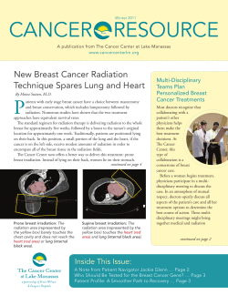


© Copyright 2026