
Evaluation of non–lead-based protective radiological material in spinal surgery MD ,
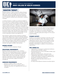
The Spine Journal 6 (2006) 577–582
Technical Review
Evaluation of non–lead-based protective radiological
material in spinal surgery
Gaetano J. Scuderi, MDa, Georgiy V. Brusovanik, MDb,*, David R. Campbell, MDc,
Robert P. Henry, MDd, Brain Kwone, Alexander R. Vaccaro, MDf
a
Private Practice, 2055 Military Trail #204, Jupiter FL 33458, USA Jupiter, FL, USA
Orthopaedic Surgery Resident, Department of Orthopedic Surgery, University of Hawaii, 1356 Lusitana Street,
6th Floor, Honolulu, HI 96813, USA University of Hawaii, Honolulu, HI, USA
c
Private Practice, 2055 Military Trail #204, Jupiter FL 33458, USA, Jupiter, FL, USA
d
Department of Radiology, Jackson Memorial Hospital, 1611 NW 12th Ave, Miami, FL 33136, USA
e
Department of Orthopaedic University of British Columbia, D6 Heather Pavilion, Vancouver General Hospital, Vancouver BC,
V5Z 3J5, Canada, British Columbia, Canada
f
Department of Orthopaedic Surgery, Thomas Jefferson University and the Rothman Institute, 925 Chestnut Street,
5th Floor, Philadelphia, PA 19107, USA
Received 6 June 2005; accepted 11 September 2005
b
Abstract
BACKGROUND CONTEXT: Traditionally, lead-based garments are the standard method of intraoperative radiation protection during fluoroscopy. Unfortunately, the lead used is heavy, lacks durability, is difficult to launder, and its disposal is associated with environmental hazards.
PURPOSE: An evaluation of the protective radiation efficiency of three commercially available
radiation protective garments compared with a standardized lead protective shield.
STUDY DESIGN/SETTING: Measured radiation transmission through lead and three commercially available lightweight radiological protective garments (Xenolite, EarthSafe, and Demron)
was performed using a standard, calibrated dosimeter.
METHODS: Radiation transmission, attenuation, lead equivalencies as well as garment weight
comparisons were measured. The tests were repeated through a range of voltage and tube current
settings that are common to clinical radiological applications (60–120 keV).
RESULTS: All materials tested demonstrated effectiveness at common clinically relevant energy
exposures (100 keV). EarthSafe and Xenolite demonstrated 0.5 mm lead equivalency protection at
80 and 100 keV X-ray energies but not at higher energy levels (O100 keV), which is where most
radiological procedures are performed utilizing more advanced technological imaging equipment.
Demron was best able to effectively shield ionizing radiation at higher energy levels (O100
keV). The lightweight nature of these lead-free materials may result in less fatigue and musculoskeletal complaints by the wearer.
CONCLUSIONS: Of the tested lead-free garments, Demron appears to offer equivalent levels of
protection to standard lead-based shields within traditional energy zones but with less weight than
standard lead-based shields. Ó 2006 Elsevier Inc. All rights reserved.
Keywords:
Protective radiation garments; Lead; Radiation transmission; Lead equivalencies; Lead-free garments; Ionizing
radiation shield
Introduction
FDA device/drug status: not applicable.
Nothing of value received from a commercial entity related to this
manuscript.
* Corresponding author. Department of Orthopedic Surgery, University
of Hawaii, 1356 Lusitana Street, 6th Floor, Honolulu, HI 96813. Tel.: (808)
586-2920; fax: (808) 956-9481.
1529-9430/06/$ – see front matter Ó 2006 Elsevier Inc. All rights reserved.
doi:10.1016/j.spinee.2005.09.010
As more spinal pathology is being treated with minimally invasive procedures, the spinal surgeon must rely
on other means of identifying surgical anatomy besides direct visualization. The most common method today for indirect visualization of the spinal elements is via bi-planar
578
G.J. Scuderi et al. / The Spine Journal 6 (2006) 577–582
fluoroscopy. Technological advances have allowed better
indirect visualization of spinal anatomy and have enabled
the spinal surgeon to accomplish complicated spinal procedures minimizing the magnitude of tissue trauma [1]. This
has not come without increased risk. Radiation exposure
can be up to 10 times higher in spinal surgery compared
with other procedures [2].
The use of fluoroscopy is also vital in other areas of
medicine including pain management, interventional cardiology, urology, general orthopedics, and of course radiology. Despite this, little attention by surgical clinicians has
been paid to the potential seriousness and means of protecting operating personnel from the harmful ionizing radiation
that occurs intraoperatively. Traditionally, clinicians have
relied on cumbersome protective lead-reinforced garments
despite numerous issues surrounding their use [5].
These lead-based garments serve as the standard radiation attenuating shields for operating personnel. Unfortunately, lead has difficulties associated with its use. A lead
apron is relatively heavy and is frequently associated with
musculoskeletal complaints with extended use. Additionally, there are environmental issues with its disposal secondary to its known toxicity. Lead within protective
aprons may exude into soil and permeate water supplies.
In this study, we evaluated three commercially available
non–lead-based radiological protective garments purported
to be similar in function to standard lead aprons in attenuating radiation. These garments are 30–40% lighter than
lead and therefore may be helpful in terms of compliance
and in minimizing musculoskeletal complaints associated
with extended apron use.
Material and methods
All testing was performed at Massachusetts General
Hospital, Boston, Massachusetts. A polycarbonate test
stand (Radcal Corporation, Monrovia, CA; Part. No.
10T5) was placed beneath an X-ray tube and aligned such
that the X-rays emitted from the tube were centered (a laser
is used as a guide) on sample material/filters placed on top
of a stand. The stand was secured to the X-ray table. An
ionization chamber (Model No. 10X5-60; Radcal Corporation) was also set up on an adjustable metal stand so that it
was centered beneath the sample material/filters. The ionization chamber was then connected to a Dosimeter (Model
No. 9010; Radcal Corporation) by a cable with a converter
(Model No. 9060; Radcal Corporation).
The Dosimeter, which displays the Exposure in milliRoentgens (mR or millirem), was calibrated to read 0.00
mR. The X-ray machine was then set to the desired voltage
(kVp) and tube current (mAs). The protocol called for the
following voltages and currents: (1) 60 kVp and 10.0
mAs; (2) 80 kVp and 8.0 mAs; (3) 100 kVp and 6.4
mAs; (4) 120 kVp and 5.0 mAs; (5) 130 kVp and 3.2 mAs.
The procedure for testing each of the samples was as
follows: A dosimeter reading for 0 mm or air was initially
recorded (in this experiment, for example @60 kVp and 10
mAs, the dosimeter read 34.24 mR). Testing was then performed to calculate the aluminum (Al) half value layer. An
aluminum filter (0.51 mm) was placed on top of the BRH
test stand and aligned properly so that the majority of the
incident X-rays hit the center of the plate. The X-ray machine was switched on, and the dosimeter reading was
taken. The Al thickness was increased gradually by adding
more filters. The filters were added one by one until the radiation was halved (until the reading on the dosimeter
showed approximately half the initial value of 34.24 mR).
The reading in mR for the corresponding Al thickness
was recorded. The dosimeter was zeroed each time an extra
Al plate was added. Four Al plates were required to cut the
radiation by half.
The aluminum plates were removed and the dosimeter
zeroed. The sample to be tested was then placed on the
BRH test stand and the dosimeter was zeroed again. The
X-ray machine was switched on, and the dosimeter reading
was noted. The sample was removed, and a similar procedure was followed for other samples.
Standard lead was then tested after the designated numbers of samples were tested. A lead foil of 1 mil (1 mil51/
1,000th inch) thickness was placed on the stand, and the
X-ray machine was switched on. The dosimeter reading
was noted. The thickness of lead foils was increased until
the final dosimeter reading was the same as that of the sample. If many layers of the same sample were used, the
dosimeter reading of all the combined layers was taken.
Results
Table 1 lists and compares the transmission, attenuation,
and lead equivalencies of all material tested. The results for
lead, summarized in Table 1, were obtained experimentally
by testing lead foils; transmission data were collected for
19.68 mil (0.5 mm) thick lead foils. As one may glean from
the table, % attenuation diminishes with higher energy
(keV). At lower energies, attenuation approaches 100%.
At 60 keV, attenuation for all materials is O99%. The discrepancy in radiation attenuation is magnified at higher energies (Fig. 1).
The three non–lead-based radioactive shields tested included EarthSafe, Xenolite, and Demron. EarthSafe was
noted to have over 0.5 mm lead equivalency protection at
80 and 100 keV X-ray energies but not at energies at the
higher end of the spectrum (O100 keV), which is where
most radiological procedures are performed.
Xenolite followed similar trends as EarthSafe and did
not have the required 0.5 mm lead equivalency protection
at the energy extremes. Demron was effective both in the
lower energy range (below 100 keV) as well as the higher
energy range (100, 120, and 130 keV). It should be noted
from Figure 1A and 1B that although Demron did not give
0.5 mm lead equivalency protection at energies lower than
100 keV, the difference in the transmission percentages
G.J. Scuderi et al. / The Spine Journal 6 (2006) 577–582
Table 1
Comparison of transmission, attenuation, and lead equivalencies
Energy
(KeV)
Demron
60
80
100
120
130
EarthSafe
60
80
100
120
130
Xenolite
60
80
100
120
130
Lead
60
80
100
120
130
Transmission
(%)
Attenuation
(%)
Lead
equivalency
(mm)
0.55
2.21
3.14
4.00
4.50
99.45
97.79
96.86
96.00
95.50
0.39
0.44
0.54
0.55
0.54
0.46
1.47
3.67
6.78
8.43
99.54
98.53
96.33
93.22
91.57
0.42
0.53
0.51
0.42
0.39
0.4
1.75
3.46
5.89
7.18
99.6
98.25
96.54
94.11
92.82
0.44
0.5
0.52
0.45
0.44
0.23
1.80
3.50
4.80
5.20
99.77
98.2
96.5
95.2
94.8
0.5
0.5
0.5
0.5
0.5
between lead and Demron was negligible at lower energies.
The differences in percent transmission between Demron,
Xenolite, and EarthSafe became significant at higher
energies.
Lead equivalencies and transmission percentages of lead
at 100, 120, and 130 keV X-ray energies are summarized in
Table 2. The number of layers required to achieve 0.5 mm
lead equivalency above 100 keV, and their weights for each
product are tabulated in Table 3. A sample piece of 10.2
cm2 area was taken from each of the garments and weighed
separately using a digital balance. Although a single layer
of EarthSafe or Xenolite weighs less than a single layer
of Demron, the number of layers required to achieve 0.5
mm lead equivalency above 100 keV is more than that of
Demron. It took approximately four layers of Demron to
achieve the required 0.5 mm lead equivalency above 100
keV; whereas five layers were needed for EarthSafe and
Xenolite to achieve the same. To offer the same attenuation
protection, Demron is approximately 15.8% lighter than
EarthSafe and approximately 3.5% lighter than Xenolite.
Discussion
Exposure to ionizing radiation (alpha, beta, X-rays and
gamma rays) is clearly dangerous to humans. Skin lesions
are a known consequence of high-level exposure, and numerous malignancies have been associated with chronic
low-level exposure [12]. We are becoming increasingly
579
aware of the risks of occupational exposure in the medical
arena. A study by Wall constructed radiation risk projection
models for computer tomography examinations and found
that a lifetime risk of fatal cancer can reach 1 in 1,000
for children, depending on the frequency of examinations
[3]. Although not well studied, it is felt that the health effects of radiation exposure are linear with no threshold
[9]. With the widespread use of computer tomography,
plain roentgenography, and the increasing use of ionizing
radiological equipment by nonmedical personnel, the potential problems of radiation exposure may be more widespread than reported [4].
To accomplish enhanced visualization, the newer generation fluoroscopes produce higher amounts of energy. X-ray
energy is measured in kVp. A typical chest X-ray utilizes
an average energy of 80 kVp for a period of 0.1 second.
Fluoroscopes, on the other hand, emit energies up to 130
kVp, and because they are continuous, emit for substantially longer periods of time [10].
The physician must take appropriate measures to reduce
the risk of radiation exposure to the patient, the operating
staff, and himself. Concepts such as time, distance, and
shielding are the most common ways of reducing radiation
exposure. By reducing the time of exposure, maximizing
the distance from the anode, and utilizing shielding apparatus, medical personnel may limit the amount of radiation
exposure to a minimum [11,12].
Exposure rates to the patient of 1,200 to 4,000 mrem/min
have been recorded [6]. The close proximity of operating
room personnel to fluoroscopy equipment may result in higher dosages of backscatter, a concept that has received little attention in the past [7]. Exposure rates from scatter at 2 feet
from the anode have been recorded at approximately 5
mrem/min. A heightened risk of radiation exposure to the assistant surgeon has been demonstrated [8]. Health-care
workers may lower radiation exposure in a limited number
of ways. Decreasing the exposure time clearly reduces the
dose received from the radiation source. However, this is
not always practical, especially in long fluoroscopic procedures common in orthopedics and cardiovascular surgery. Increasing the distance from the source, another well-known
factor in limiting radiation exposure, similarly may not be
practical for the surgeon and assistants as surgical technique
may require proximity.
The simplest way to minimize exposure is via shielding,
which creates a barrier between the source and the individual. Traditionally this has resulted in the cumbersome donning of heavy and awkward undergarments that include
lead. Although they provide good attenuation to radiation
exposure, they are heavy, uncomfortable, and often crack,
leaving the wearer unknowingly exposed. Lead-based polymer composites are made by incorporating lead or other
heavy metal compounds into a resin or polymer matrix. Attempts have been made to obtain similar protection with
composite materials that reduce weight. These materials
have had variable results in laboratory testing, are more
580
G.J. Scuderi et al. / The Spine Journal 6 (2006) 577–582
A
B
9.00
8.00
Transmission (%)
7.00
6.00
5.00
4.00
3.00
2.00
1.00
0.00
50
60
70
80
90
100
110
120
130
140
X-Ray Energy (kVP)
Fig. 1. Comparison of percent transmission of X-rays.
expensive, and have been associated in some cases with an
unpleasant odor. Additionally, because they contain lead,
they cannot be laundered, and are prone to cracking, which
renders the garment unusable. An additional problem for
the health-care facility is the problem of apron disposal.
Lead is considered to be the gold standard in X-ray
protection.
The American Society for Testing Materials (ASTM)
guidelines have recommended 0.5-mm lead equivalent garments for standard protection. However, the ASTM has not
devised a standard protocol for testing radiological equipment in the health-care field. Traditionally, private companies have tested their product in a single radiological dose
(usually 100 kVp), or at the specific dose that the lightest
material composite can achieve 0.5 mm of lead equivalency. We chose to evaluate five different levels of kVp
(60–130). This is the field where approximately 99% of radiologic energy is utilized in the medical environment.
Data from studied manufacturers are published only at
100 kVp. These data correlated with the findings of our
study at 100 kVp. However, by evaluating other levels of
radiation exposure we were able to elucidate a trend for
different composites at both lower and higher energies.
At various levels of energy (or keV), lead has been shown
to predictably reduce X-ray transmission as a function of
thickness. For example, a 0.25-mm lead foil has known
values of X-ray transmission reduction at various levels
of energy. When an attenuating material reduces a certain
G.J. Scuderi et al. / The Spine Journal 6 (2006) 577–582
Table 2
Lead equivalencies and % transmission of lead at 100, 120,
and 130 keV X-ray energies
100 kVp
Lead equivalency (mm)
0.25 Apron
0.35 Apron
0.50 Apron
Transmission (%)
0.25 Apron
0.35 Apron
0.50 Apron
120 kVp
130 kVp
0.24
0.39
0.54
0.25
0.39
0.55
0.25
0.39
0.54
11.50
5.73
3.14
13.69
7.22
4.00
14.85
8.01
4.50
keV by the same percentage as a 0.25-mm piece of lead
foil, that material is described as having a lead equivalence
of 0.25 mm of lead at that keV. Low-lead or lead-free composites will market their product’s attenuation ability at either a percent reduction in transmission or as a function of
lead equivalence. These values are usually referenced at
a certain energy level or kVp. The usual kVp quoted is
80 or 100 kVp. In order to communicate the effectiveness
of X-ray attenuation, various terms such as lead equivalency and percent transmission are often used. The percent
transmission refers to the amount or percent of radiation
transmission that is allowed to pass through a material at
a certain amount of energy or keV. With all attenuators,
the higher the keV, the more difficult it is to attenuate,
and hence the higher the transmission. We focused on
a range of energy levels currently encountered in the performance of minimally invasive spinal procedures.
In the United States, most state regulations require that
the X-ray aprons have a lead equivalence of 0.5 mm of
lead. As such, the wearer could predict his or her level of
protection at various levels of energy or keV. The difficulty
arises with the introduction of lead substitutes. Convention
has dictated that companies advertise their lead substitute
products at 0.5 mm of lead equivalency at 100 keV. When
a manufacturer claims that their reduced or lead-free apron
has a lead equivalence of 0.5 mm at 80 kVp, the wearer
should not assume that the apron will perform similar to
the same thickness of lead at other kVp values because it
is not lead. This is especially pertinent for medical personnel who are exposed to higher X-ray energies during
lengthy or complicated fluoroscopic procedures. The clinician/purchaser of radiological protective gear needs to be
aware of this fact in order to properly evaluate the efficiency of differing materials used for lead substitution.
Table 3
Comparison of weights
Product name
No. of layers
(area of 1 layer 5 4 in2)
Weight (g)
Demron
EarthSafe
Xenolite
4
5
5
16.4
19.5
17
581
In this study, we compared three popular reduced lead or
lead-free composites with lead and their ability to attenuate
X-ray radiation at various energies. The most common lead
substitutes for medical radiation exposure protection are
EarthSafe, Xenolite, and Demron. The manufacturers of
each product claim that their composite material possesses
similar attenuation coefficients as traditional lead-based
garments. Although the exact composition of these materials is proprietary and is protected by patents, both EarthSafe and Xenolite are composed of varying amounts of tin,
antimony, arsenic, and cadmium in addition to other unknown materials. The manufacturer of Demron discloses
the presence of bismuth, barium, tungsten, iodine, as well
as proprietary nanocomposites which contribute to its structure and ability to attenuate radiation.
In summary, all materials tested in this study demonstrated effectiveness in radiation attenuation at 100 keV. This
level of protection may not be relevant in today’s newer fluoroscopic procedures, which use radiation sources of higher
energy for longer periods of time. It appears from this study
that Demron is best able to shield ionizing radiation of higher
energy. Demron was the only material to maintain very low
percent transmission over 120 and 130 keV, and was the only
composite that provided satisfactory protection at energies
superseding 100 keV. In fact, the composite material in Demron attenuated ionizing radiation better than lead at higher energies. Additionally, Demron’s physical characteristics as
a flexible fabric may enable the life of the material to be significantly prolonged. Both EarthSafe and Xenolite contain
tin, which may hinder flexibility. Because it is lead-free,
Demron is nontoxic, washable, and easily disposable without
environmental concerns. The reduced weight of Demron
compared with lead may reduce the incidence of musculoskeletal complaints for medical personnel involved in
lengthy fluoroscopic procedures.
References
[1] Kruger R, Faciszewski T. Radiation dose reduction to medical staff
during vertebroplasty: a review of techniques and methods to mitigate
occupational dose. Spine 2003;28:1608–13.
[2] Rampersaud YR, Foley KT, Shen AC, Williams S, Solomito M. Radiation exposure to the spine surgeon during fluoroscopically assisted
pedicle screw instrumentation. Spine 2000;25:2637–45.
[3] Wall BF. Radiation protection dosimetry for diagnostic radiology patients. Radiat Prot Dosimetry 2004;109:409–19.
[4] Mettler FA Jr. Medical perspective on ways to improve radiation protection standards. Health Phys 2004;87:289–92.
[5] Yaffe MJ, Mawdsley GE, Lilley M, Servant R, Reh G. Composite
materials for x-ray protection. Health Phys 1991;60:661–4.
[6] Norris TG. Radiation safety in fluoroscopy. Radiol Techniques
2002;73:511–33.
[7] Brateman L. Radiation safety considerations for diagnostic radiology
personnel. Radiographics 1999;19:1037–55.
[8] Tasbas BA, Yagmurlu MF, Bayrakci K, Ucaner A, Heybeli M. Which
one is at risk in intraoperative fluoroscopy? Assistant surgeon or orthopaedic surgeon? Arch Orthop Trauma Surg 2003;123:242–4. Epub
2003 May 10.
[9] Birch R, Marshall M, Ardran GM. Catalogue of spectral data for diagnostic X-rays. London: Hospital Physicists’ Association, 1979.
582
G.J. Scuderi / The Spine Journal 6 (2006) 577–582
[10] Johlin FC, Pelsang RE, Greenleaf M. Phantom study to determine radiation exposure to medical personnel involved in ERCP fluoroscopy
and its reduction through equipment and behavior modifications. Am
J Gastroenterol 2002;97:893–7.
[11] Kuon E, Schmitt M, Dahm JB. Significant reduction of radiation exposure to operator and staff during cardiac interventions by analysis
125 Years
Ago in Spine
of radiation leakage and improved lead shielding. Am J Cardiol
2002;89:44–9.
[12] Simons GR, Orrison WW Jr. Use of a sterile, disposable, radiationabsorbing shield reduces occupational exposure to scatter radiation
during pectoral device implantation. Pacing Clin Electrophysiol
2004;27(6 Pt 1):726–9.
stretch test was Forst’s doctoral thesis [1]. That Laza
K. Lazarevic had published the same observation the
year before in the Serbian literature [2] did not keep
the sign from being named for Laséque.
References
J.J. Forst, a pupil of E.C. Laséque, first
reported the sign of nerve root irritation that had been
observed by his mentor. The description of the sciatic
[1] Forst JJ. Contribution à l’étude clinique de la sciatique. Paris,
Thèse No. 33, 1881.
[2] Lazarevic LK. Srpski Arh. 7:23. Cited in: Rang M. Anthology of
orthopaedics 1966. Edinburgh: Churchill Livingstone, 1966:150.


© Copyright 2026