
Document 141532
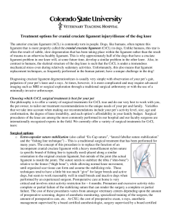
Failure of Cast Immobilization for
Thumb Ulnar Collateral Ligament
Avulsion Fractures
Mark Dinowitz, MD, Thomas Trumble, MD, Douglas Hanel, MD,
Nicholas B. Vedder, MD, Mary Gilbert, MA, Seattle, WA
To determine if small avulsion fractures of the thumb ulnar collateral ligament (UCL) with
minimal (_< 2,0 ram) displacement can successfully be treated by cast immobilization, the
authors reviewed 9 patients with minimally displaced fractures initially treated by casting.
Despite immobilization within an average of 2 days of the initial injury (range, 0-6 days), a
minimum of 6 weeks of immobilization in a cast, and adequate rehabilitation, all 9 patients
had persistent thumb pain, especially with activities requiring strong pinch. After undergoing
open reduction and internal fixation, the patients had relief of thumb pain and pinch strength
improved from 36% of the contralateral side to 89% (p < .01). Grip strength increased from
77% to 93% (p < .05), but the ranges of motion of the thumb metacarpophalangeal and
interphalangeal joints were not significantly altered. Minimally displaced UCL avulsion fractures frequently have significant rotation that prevents successful fracture healing even with
prompt cast immobilization. (J Hand Surg 1997;22A:1057-1063.)
Acute injuries to the thumb metacarpophalangeal
(MP) joint ulnar collateral ligament (UCL) can occur
with an avulsion fracture in approximately 20%-30%
of cases3, 2 Open reduction and internal fixation is the
standard treatment when the fracture involves
15%-20% of the joint surface or when there is sufficient displacement to suggest that the ligament
attachment site cannot heal properly. 3-8 These criteria
emphasize 2 points: the need to restore the articular
surface and the need to allow for the healing of the
ligament in the correct position. Unlike ligamentous
disruptions without a fracture, the majority of the
UCL avulsion fractures do not have a Stener lesion in
which the distal end of the ligament folds back on
From the Division of Hand and Microsurgery, Department of
Orthopaedics, Universityof Washington,HarborviewMedical Center,
Seattle,WA.
Received for publication Dec. 12, 1995; accepted in revised form
March6, 1997.
No benefitsin any formhavebeen receivedor will be receivedfroma
commercialpartyrelateddirectlyor indirectlyto the subjectof thisarticle.
Reprintrequests:ThomasTrumble,MD, Departmentof Orthopaedics,
Mailstop356500, Universityof Washington,Seattle,WA98195.
itself and is trapped by the adductor aponeurosis.l, 9,10
The question that remains is how much displacement
can be tolerated in small avulsion fractures that do
not compromise a significant portion of the articular
surface? Even small amounts of rotation can result in
interposition of soft tissue or cartilage between the
fracture fragments. Similarly, for pure ligamentous
injuries of the UCL, it is not known whether complete disruptions without displacement can heal satisfactorily. In our initial experience, healing through
cast immobilization alone for patients with small,
minimally displaced articular fractures was hampered
by persistent thumb pain and decreased pinch
strength despite adequate rehabilitation exercises. On
closer examination, it appeared that many of these
small avulsion fractures had rotated such that avulsion fragment's articular surface's faced the fracture
surface of the larger fragment, making fracture healing unlikely. This retrospective study reviews the
cases of 9 patients initially treated with immobilization alone for small, minimally displaced avulsion
fractures of the thumb UCL in order to demonstrate
the problems associated with this injury.
The Journal of Hand Surgery
1057
1058
Dinowitz et al. / Casting Fails in Thumb UCL Fractures
Materials and Methods
Demographics
Between 1987 and 1993, 9 patients with minimally
displaced (< 2 mm) small (< 2 mm in diameter) UCL
avulsion fractures were treated (Figs. 1, 2). Their
average age was 27 years (range, 18-43 years). There
were 7 men and 2 women, and the dominant hand
was involved in 5 patients. Average time from injury
to initial treatment with cast immobilization averaged
2 days (range, 0-6 days). None of the injuries were
work related. The injuries occurred during the following activities: skiing (3), biking (3), falls (2), skateboarding (1). Three patients were students, 3 were
manual laborers, and 3 were manager-professionals.
All patients' affected thumbs were initially placed in
a spica cast for 6-7 weeks and all patients subsequently started an exercise program stressing active
and passive flexion, especially combined flexion of
the MP and interphalangeal (IP) joints. All patients
continued to complain of pain and decreased pinch
strength. In none of the patients was radiographic evi-
Figure 2. This close-up of a posteroanterior radiograph of
same injury shown in Figure 1 demonstrates rotation and
slight (< 2.0 mm) displacement so that the articular surface of the fragment (arrow) is facing the fracture surface
of the remainder of the proximal phalanx.
dence of fracture union demonstrated at the time that
patients were evaluated for further treatment by surgical repair at an average of 6.5 months (range, 4-13
months) after the injury. Our records indicated that
none of these injuries were successfully treated by
cast immobilization alone.
During this same period, 15 patients sustained dramatically displaced UCL avulsion fractures as Stener
lesions and they were successfully treated by acute
UCL repair using the same techniques. 10 Three patients had true nondisplaced fractures as defined by the
lack of separation or rotation of the UCL fragment
from the proximal phalange shown on any of the radiographs and they were successfully treated by casting.
Eighty-five patients were diagnosed as having soft tissue injuries of the UCL; 48 of these patients had complete ligament disruptions requiring surgical repair.
Clinical Evaluations
Figure 1. The fracture (arrow) appears well aligned and
minimally displaced in this lateral oblique radiographic
view.
The patients were evaluated prior to surgery and at
the time of final follow-up evaluation. They were
surveyed as to whether they had no pain (level 0),
pain with heavy activities (level 1), or pain with
activities of daily living (ADLs) (level 2). MP and IP
joint arcs of motion were measured preoperatively and
at the time of final follow-up examination according
to the guidelines for evaluation of permanent impairment by the American Medical AssociationJ 1 Key
pinch and tip pinch strengths were measured using a
pinchmeter (North Coast Medical Inc., San Jose,
CA) for both the injured and uninjured side in order
The Journal of Hand Surgery / Vol. 22A No. 6 November 1997
1059
to report the results as a percentage of the contralateral side. In a similar fashion, maximal grip strength
was measured using a Jamar Dynamometer (JAS P.
Marsh Corp., Skokie, IL).
PostoperativeManagement
After surgery, the patients' thumbs were placed in
a spica cast for 4 weeks, with the volar aspect of the
cast distal to the thumb IP joint removed to allow
early IP joint range of motion (ROM). A removable
plastic splint was used to protect the UCL for 2-3
weeks after removal of the cast. The patients were
started on active and passive ROM exercises emphasizing combined IP and MP joint flexion as soon as
the cast was removed. After surgery, the patients
were followed for an average of 36 months (range,
24-56 months). Radiographs documented healing
within 6-7 weeks after surgery.
:,;:'
Technique
An S-shaped incision is used, with the distal
aspect of the incision placed along the border of the
glabrous skin of the thumb. There is a predictable
branch of the radial sensory nerve in the dorsal skin
flap that is protected. The adductor aponeurosis is
incised longitudinally and the avulsion fracture is
identified near the volar margin of the proximal phalanx. Frequently, a small arthrotomy is necessary to
accurately reduce the avulsion fracture, which will
often be rotated 90 ~ or more (Fig. 3). Prior to the
final reduction, we place a nonabsorbable suture
(3-0 Ticron, Davis and Geck American Cyanamid
Co., Manoti, PR) resembling one half of a modified
Kessler tendon suture in the UCL. The suture is
passed as close to the fragment as possible and then
brought through the base of the proximal phalanx by
2 Keith needles (Figs. 4, 5). The needles are passed
perpendicular to the fracture line and the 2 suture
ends are brought out through a small incision on the
radial side of the thumb, which avoids the need for a
pull-out button and the attendant difficulties with skin
irritation, tza3 This technique, unlike tension band
wires, which can result in rotation of these small fracture fragments,6 allows the force vector of the suture to
stabilize the fracture. The fragment is pinned in place
anatomically with .028-inch Kirschner wires (Kwires) (Fig. 6). Although one could excise the avulsion fragment and repair just the ligament, doing so
would greatly hamper the surgeon's efforts to accurately reconstruct the centers of rotation of the MP
Figure 3. The rotated position of the ulnar collateral ligament avulsion fracture fragment is demonstrated.
joint. A recent Biomechanical study has demonstrated
that even an error of 1-2 mm results in significant
changes in joint rotation and stability. 14 Fluoroscopy
is used to confirm that the rotational deformity has
been corrected. After anatomic reduction has been
verified, the suture is tied down on the radial side of
the proximal phalanx. The K-wires are buried beneath
the skin so that they are palpable on the radial side of
the thumb. The wires can be easily removed under
local anesthetic 6-8 weeks after surgery by an incision of the radial aspect of the thumb. Because the
wires are buried, they do not interfere with mobilization of the joints during rehabilitation.
Statistics
The change between preoperative and postoperative parametric data was evaluated using the student's t-test. The nonparametric evaluation of pain
was performed using Wilcoxon's signed-rank test.
Results
The average time from injury to initial consultation was 2 days (range, 0-6 days). After 6-7 weeks
1060 Dinowitz et al. / Casting Fails in Thumb UCL Fractures
it
Figure 4. A suture is placed in the ligament just proximal to the avulsion fragment. Two suture ends are passed through
the proximal phalanx by 2 Keith needles drilled through the proximal phalanx.
of cast immobilization and appropriate therapy, all 9
patients continued to have pain (level 2) limiting
their performance of ADLs, especially opening jars
and turning keys, despite having an average of 7
months in which to recover (range, 4-13 months).
We chose 4 months as the minimum period to consider the cast treatment a failure because this is the
period that it takes even the surgically repaired complete disruptions of the ligament to regain most of
the strength and obtain maximal relief of pain. Final
Figure 5. The sutures are tied on the radial side of the proximal phalanx after the avulsion fragment has been derotated,
reduced, and secured with 2.028-inch Kirschner wires.
The Journal of Hand Surgery / Vol. 22A No. 6 November 1997
1061
Strength
The preoperative key pinch strength averaged 8 lb.
(range, 6-12 lb.), or 36% of that of the contralateral
side, and tip pinch averaged 4 lb. (range, 2-6 lb.), or
33% of that of the contralateral side. Postoperative
key pinch strength significantly increased to an average of 22 lb. (range, 13-24 lb., p < .01), which was
89% of that of the contralateral side. Similarly, the
tip pinch strength improved to 9 lb. (range, 4-15 lb.),
or 74% of that of the contralateral side (p < .05). The
grip strength of the injured extremity averaged 68 lb.
(range, 25-112 lb.), which was 76% of that of the
uninjured side. Grip strength improved to an average
of 83 lb. (93% of that of the contralateral side; range,
45-120 lb.). This increase in grip strength was significant when compared to the preoperative strength
(p < .04).
Figure 6. On this postoperative posteroanterior radiograph, the reduction of the articular fracture can be seen
secured by 2 .028-inch Kirschner wires. In addition, a
suture was placed into the collateral ligament and tied on
the radial side of the proximal phalanx.
Radiographs
follow-up evaluation was at an average of 36 months
after surgery (range, 24-56 months).
By 6-7 weeks after surgery, all patients' ligaments
had achieved solid union. One of the 9, who had an
old radial collateral ligament injury, had early radiographic signs of joint narrowing that was present
prior to the surgery. In none of the patients, however,
did sclerosis or osteophytes develop.
Pain
At final average follow-up evaluation, only 1 of 9
patients reported pain at work and with some ADLs.
This is a significant improvement from the preoperative status (p < .05).
Intraoperative Findings
Motion
The preoperative MP joint arc of motion was 40 ~
(range 15~176 which was 61% of the contralateral thumb (Table 1). The IP joint ROM was 44 ~
(range, 35~176 or 77% of that of the contralateral
side. At final follow-up examination, the MP joint
motion increased slightly to an average of 42 ~
(range, 35~176
which was 65% of that of the
contralateral side, and the IP joint motion increased
to 47 ~ or 82% of that of the contralateral side
(range, 30~176
In all cases, the majority of the UCL ligament was
attached to the avulsion fragment. Five of the patients
had concomitant injuries to the palmar plate of the
thumb MP joint, 2 of whom had small avulsion fractures associated with the palmar plate injury. These
palmar plate injuries were repaired with sutures at the
time of the UCL repair. Because all the significant
portions of the UCL were attached to the fragment,
the joint was not stable until the fragment was reapproximated. Although the suture in the ligament
helped to protect the fixation of the small fragment, it
did not act as a tensioning suture to stabilize the joint.
Table 1. Thumb Range of Motion and Strength
MP joint range of motion (~
IP joint range of motion (~
Key pinch (lb.)
Tip pinch (lb.)
Grip (lb.)
Preoperative
(% contralateral)
Postoperative
(% contralateral)
40 + 16 (61%)
44 + (77%)
8 + 2 (36%)
4 + 2 (33%)
68 + 26 (76%)
42 + l0 (65%)
47 + 10 (82%)
22 + 6 (89%)
9 + 4 (74%)
83 + 30 (93%)
IP, interphalangealjoints; ME metacarpophalangeal.
1062 Dinowitz et al. / Casting Fails in Thumb UCL Fractures
Discussion
This report focuses on minimally displaced UCL
avulsion fractures. Despite the delay in treatment, the
results are similar to those in reports of acute UCL
repairs. Derkash et al., 15 in a review of 123 thumb
injuries, noted that mild stiffness and pain was common, although pain or stiffness was severe enough to
be a problem in only 4% and 5%, respectively, of
patients; however, 41% of the patients were lost to
follow-up monitoring. Kessler found that 5 of 13
patients had persistent pain with activities even
though their physician rated their treatment as having
good results, j6 Using a rating of excellent to poor,
Gerber et al. 17 noted that in 90% of the 47 patients,
treatment had good to excellent results. If these criteria are used, 8 of the 9 patients in our study had good
or excellent results. Using the criteria of satisfactory
versus unsatisfactory, Lamb et al. ~8 found that in 17
of 21 patients, treatment had satisfactory results,
while Frank and Dobyns ~9reported that results for 22
of 24 patients were satisfactory. For all 9 of the
patients in our study, treatment results would be rated
as satisfactory using these criteria. Bowers and Hurst 7
reviewed the world literature involving the treatment
of 197 UCL injuries and noted that 138 of 197
injuries were surgically repaired and that for 131,
there were good results following surgery, although
their rating system was not defined. Interestingly, they
noted that patients with UCL avulsion fractures without surgery had poor results (8 of 9), while patients
with surgical repair had good results (25 of 26).
Delay to Treatment
Smith 2 and Strandel120 both noted a decrease in the
functional results when the ligament or fracture
repair was delayed by 3 or more weeks. Smith 2 noted
that 6 of 31 patients with delayed treatment had
recurrent ligament instability, which did not occur in
the patients in our study. The results in our study,
including a return of nearly 90% of preinjury pinch
strength, suggest that delayed repair is possible
although probably not optimal in small avulsion
fractures of the UCL.
Method of Repair
Obviously, there are many successful ways to
repair avulsion fractures of the UCL. We believe that
tension-band wiring did not result in a force vector
perpendicular to the fracture plane and therefore
caused problems with rotation of the small fracture
fragment that did not have significant interdigitation
of the fracture surfaces. 6 The technique of passing
both suture ends through the proximal phalanx
avoided the need for a pull-out button and provided
support for the K-wire fixation without causing rotation of the fracture fragment. ~2 Suture anchors can be
effective in UCL repairs, but with avulsion fractures,
the suture anchor position is usually distal to the
fracture, creating a rotational moment. 21 By tying the
suture on the radial side of the thumb proximal phalanx, we were able to avoid placing the ligament
under tension when tying the suture, whereas we had
to keep the thumb somewhat radially abducted when
using the suture anchors to maintain enough exposure to securely tie the suture.
References
1. Hintermann B, Holzach PJ, Schutz M, Matter P. Skier's
thumb: the significance of bony injuries. [See comments.]
Am J Sports Med 1993;21:800-804.
2. Smith RJ. Post-traumatic instability of the metacarpophalangeal joint of the thumb. J Bone Joint Surg 1977;59A:
14-21.
3. Kozin SH, Bishop AT. Gamekeeper's thumb: early diagnosis and treatment. Orthop Rev 1994;23:797-804.
4. Louis DS, Huebner JJ Jr, Hankin FM. Rupture and displacement of the ulnar collateral ligament of the metacarpophalangeal joint of the thumb: preoperative diagnosis. J
Bone Joint Surg 1986;68A: 1320o1326.
5. Bronstein A J, Koniuch MP, von Holsbeeck M. Ultrasonographic detection of thumb ulnar collateral ligament
injuries: a cadaveric study. J Hand Surg 1994; 19A:304-312.
6. Jupiter JB, Sheppard JE. Tension wire fixation of avulsion
fractures in the hand. Clin Orthop 1987;214:113-120.
7. Bowers WH, Hurst LC. Gamekeeper's thumb: evaluation
by arthrography and stress roentgenography. J Bone Joint
Surg 1977;59A:519-524.
8. Osterman LA, Hayken, GD, Bora FW. A quantitative evaluation of thumb function after ulnar collateral repair and
reconstruction. J Trauma 1981 ;21:854-861.
9. Heyman P, Gelberman RH, Duncan K, Hipp JA. Injuries of
the ulnar collateral ligament of the thumb metacarpophalangeal joint: biomechanical and prospective clinical studies on the usefulness of valgus stress testing. Clin Orthop
1993;292:165-171.
10. Stener B. Displacement of the ruptured ulnar collateral ligament of the metacarpophalangeal joint of the thumb: a
clinical anatomic study. J Bone Joint Surg 1962;44B:
869-879.
11. Engleberg AL. Guidelines to evaluation of permanent
impairment. 4th ed. Chicago: American Medical Association, 1992:13-94.
12. Saetta JP, Phair IC, Quinton DN. Ulnar collateral ligament
repair of the metacarpophalangeal joint of the thumb: a
study comparing two methods of repair. J Hand Surg
1992;17B:160-163.
The Journal of Hand Surgery / Vol. 22A No. 6 November 1997
13. Reck T, Landsleitner B, Richter H, Geldmacher J. A new
method of transosseous pull-out fixation in ligamentous
injuries of the metacarpophalangeal joint of the thumb
[transl. Handchir Mikrochir Plast Chir 1991;23:90-92.
14. Bean C, Trumble TE, Tencer A. Reconstruction of the
thumb metacarpophalangeal joint. J Hand Surg 1998 (in
press).
15. Derkash RS, Matyas JR, Weaver JK et al. Acute surgical
repair of the skier's thumb. Clin Orthop 1987;216:29-33.
16. Kessler I. Complete avulsion of the ulnar collateral ligament of the metacarpophalangeal joint of the thumb. Clin
Orthop 1961 ;29:196-200.
17. Gerber C, Senn E, Matter P. Skier's thumb: surgical treatment of recent injuries to the ulnar collateral ligament of
18.
19.
20.
21.
1063
the thumb's metacarpophalangeal joint. Am J Sports Med
1981;9:171-177.
Lamb DW, Abemathy PJ, Fragiadakis E. Injuries of the
metacarpophalangeal joint of the thumb. Hand 1971;3:
164-168.
Frank WE, Dobyns J. Surgical pathology of the ulnar collateral ligament of the thumb. Clin Orthop 1972;83:
102-114.
Strandell G. Total rupture of the ulnar collateral ligament
of the metacarpophalangeal joint of the thumb. Acta Chir
Scand 1959;118:72-80.
Bovard RS, Derkash RS, Freeman JR. Grade III avulsion
fracture repair on the UCL of the proximal joint of the
thumb. Orthop Rev 1994;23:167-169.


© Copyright 2026