
Persistent HPV infection in postmenopausal age women *, S.R. Johnson E.M. Smith
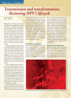
International Journal of Gynecology and Obstetrics (2004) 87, 131–137
www.elsevier.com/locate/ijgo
ARTICLE
Persistent HPV infection in postmenopausal
age women
E.M. Smitha,b,*, S.R. Johnsona,b, J.M. Ritchiec, D. Feddersena, D. Wangd,
L.P. Turekd, T.H. Haugend
a
Department of Epidemiology, College of Public Health, University of Iowa,
Iowa City, IA 52242, USA
b
Department of Obstetrics/Gynecology, College of Medicine, University of Iowa,
Iowa City, IA 52242, USA
c
Department of Biostatistics, College of Public Health, University of Iowa,
Iowa City, IA 52242, USA
d
Veterans Affairs Medical Center and Department of Pathology, College of Medicine,
University of Iowa, Iowa City, IA 52242, USA
Received 29 January 2004; received in revised form 7 July 2004; accepted 14 July 2004
KEYWORDS
Human papillomavirus;
Postmenopausal;
Persistence;
Cervical cancer
ABSTRACT
Objective: Persistence of human papillomavirus (HPV) is associated with an
increased risk of developing cervical SIL and cancer in young women. Because this
association in older, postmenopausal age women has received little attention, we
evaluated persistence of HPV among women in this age group. Methods: Women
(n=105) ages 45–64 were examined annually for 7 years to evaluate HPV in cervical
cytologic specimens. PCR, dot blot hybridization and DNA sequencing were used to
detect HPV types. Results: The cumulative prevalence of HPV was 34%, and 24% had
HPV high-risk oncogenic types which are associated with genital cancers. The most
common oncogenic types were HPV-16 (72%) and HPV-31 (16%). The persistence rate
of HPV infection was 16%. No specific risk factors were associated with repeat viral
positivity. Conclusion: Postmenopausal women are infected with persistent oncogenic HPV at a substantial rate, supporting the need for continued screening in
postmenopausal women to detect preneoplastic genital lesions.
D 2004 International Federation of Gynecology and Obstetrics. Published by Elsevier
Ireland Ltd. All rights reserved.
* Corresponding author. Department of Epidemiology, 2800 SB, College of Public Health, Iowa City, IA 52242, United States. Tel.: +1
319 384 5014; fax: +1 319 384 5031.
E-mail address: [email protected] (E.M. Smith).
0020-7292/$ - see front matter D 2004 International Federation of Gynecology and Obstetrics. Published by Elsevier Ireland Ltd.
All rights reserved.
doi:10.1016/j.ijgo.2004.07.013
132
1. Introduction
Human papillomavirus (HPV) is a necessary cause
for the development of cervical neoplasms and a
large portion of other genital tumors [1]. In the
United States, the incidence of invasive cervical
cancer increases up to age 45 (16/105) where after
the rate plateaus and does not drop in older ages
[2]. Premenopausal women who test positive for
HPV persistently over time have been shown to be
at highest risk of developing preneoplastic genital
disease [3–7]. Only a few studies have evaluated
healthy women over age 40 to detect the prevalence or persistence of HPV infection in the lower
genitals, and the frequency of HPV infection has
been reported to vary widely [8–11]. Whereas in a
cross-section of women and based on a single HPV
assessment, Ferenczy et al. [9] detected only 1%
with viral infection; in a prospective study [11], we
reported a cumulative prevalence of 14% over a 2year period. A comparison of women of all ages in
Spain, Columbia and Brazil [10] showed that HPV
frequency not only varied widely among countries
but also among women ages 40 to 60+ living in
different parts of the world (1–22%). In two of the
countries, the prevalence rate of HPV was similar in
the 60+ age group to that of the youngest age b25
years old.
Oral contraceptive (OCs) steroid hormones have
been shown to increase the risk of cervical
dysplasia and cancer in the presence of an HPV
infection in younger age women [1,12]. In contrast, the risk associated with use of hormone
replacement therapy (HRT) is relatively unknown.
Despite recent reports of increased risk of breast
cancer and other diseases, many postmenopausal
women are continuing to use HRTs to deter side
effects of menopause and osteoporosis. The
adverse effects of using HRTs for long duration
and subsequent development of cervical squamous
epithelial neoplasia (SIL) are unknown and it is
unclear whether their effects will mimic those
found for OCs. The in vitro evidence indicates that
there is greater HPV gene expression and cell
transformation by the viral DNA in the presence of
progesterones [13–15].
The previous results of this study population [8]
were based on a shorter time interval, 2 years, and
used a less-sensitive HPV-typing technique than
employed in the current study. The aims of this
investigation were to determine the prevalence of
HPV and oncogenic types in postmenopausal women
annually over a 7-year interval between 1990 and
1996, the prevalence and types of HPV associated
with persistent viral infection and the association
E.M. Smith et al.
between HRT regimens and detection of HPV over
time.
2. Methods
2.1. Patient population
Patient characteristics and details of the study
design have been described previously [16]. Briefly,
women were enrolled in this ancillary study at the
University of Iowa between 1989 and 1990 as part
of the Postmenopausal Estrogen and Progestin
Intervention (PEPI) randomized clinical trial
(RCT). Participants were between ages 45 and 64
prior to randomization to treatment, not currently
using hormone replacement therapy at least 6
months prior to enrollment and had experienced
menopause at least 1 year before or had a
hysterectomy at least 2 months prior to study
entry. Those who had a hysterectomy were
included because they were also at risk of condylomata and genital cancers other than the cervix
and were also associated with HPV infection. None
of the participants had a genital dysplasia or cancer
at the time of enrollment. Women were administered a University-approved human subjects review
form for this ancillary study to the PEPI randomized
clinical trial (RCT) before enrollment. Participants
were followed annually for HPV specimens and Pap
smears at baseline and three follow-ups with a 1year hiatus after the RCT was completed, and prior
to the funding of the follow-up period with an
additional three annual follow-ups for a total of one
baseline and six follow-up HPV and Pap smear
specimens. These specimens were collected annually between 1990 and 1993 and again between
1994 and 1996. Investigators and laboratory staff
were blinded to HRT treatment group, Pap smear
and risk factor results until the HPV study was
completed.
2.2. Hormone replacement groups
Women in this study were randomly assigned to one
of five treatment groups: (1) placebo, (2) 0.625
mg/day conjugated equine estrogen (CEE) daily, (3)
0.625 mg/day CEE daily plus cyclic medroxyprogesterone (MPA), 10 mg/daily for days 1–12 of each
month, (4) 0.625 mg/day CEE daily plus MPA 2.5 mg
daily or (5) 0.625 mg/day CEE daily plus cyclic
micronized progesterone (MP) 200 mg/daily for
days 1–12. For the purpose of this study, HRT
comparisons were made between the placebo
group (#1) and the estrogen only HRT group (#2)
Persistent HPV infection in postmenopausal age women
or with the estrogen/progestin HRT groups combined (#3–5).
2.3. Pap smear and HPV specimen collection
Prior to and after randomization to placebo or an
HRT, participants received Pap smear evaluations
and a cervical swab to collect HPV DNA at 12month intervals. Randomization procedures, timing
of treatment and annual visit information have
been described previously [16]. Unblinding of HRT
treatment group did not occur until the end of the
third year of the study. Only participants who
remained on protocol were included in the PEPI
RCT and in this 7-year ancillary study. This included
105 of the 125 women originally enrolled in the
clinical trial. A single nurse practitioner for the
study (D.F.) collected all specimens through the
study. Women first had a Pap smear retrieved and
then, using a cotton tip swab, had cells for HPV
evaluation collected from the transitional zone,
endocervix, ectocervix and pooled secretions in
the posterior vaginal fornix. A speculum was
placed, and the swab was inserted carefully
through to the cervix/vagina, avoiding the perianal
skin surface. This sample was prepared and frozen
until tested. Pap smears were reviewed by a single
SmithKline cytopathologist who was involved in the
clinical trial and blinded to the HRT treatment
group assignment of individuals. The Bethesda
system was used to report Pap smear results.
Included in the abnormal Pap smear category was
atypical squamous cells of undetermined significance (ASCUS), low-grade SIL (L-SIL), high-grade
SIL (H-SIL), adenocarcinoma or squamous cell
carcinoma.
2.4. HPV DNA detection and typing
Description of processing the cervical cells has been
described in detail elsewhere [8,11]. Briefly, after
processing the cells, the HPV DNA was PCR-amplified with MY09/MY11 HPV L1 primers [17], using
DNA from approximately 30,000 cells as template.
An additional 3V primer that facilitates HPV-51
amplification was included [3]. Primers to amplify a
294-bp portion of the h-globin gene were used as an
internal control [18]. Each set of PCR amplifications
included positive and negative controls. Amplification of HPV DNA was tentatively judged positive if
electrophoresis on a 1% agarose gel showed an
approximately 450 bp band by staining with ethidium bromide or if a signal was detected by nylon
membrane bdot blotQ hybridization with [a-32P]
dATP-labeled generic HPV probes. All specimens
133
were B-globin-positive by gel electrophoresis for all
assessments.
Automated DNA sequence analysis was used to
confirm and type the HPV DNA. Amplified DNAs
were purified using the Wizard PCR Preps DNA
Purification Kit (Promega, Madison, WI) and
sequenced on a PE Applied Biosystems automated
sequencer (Perkin Elmer Cetus, Foster City, CA).
When insufficient DNA was present for sequencing,
HPV DNA sequences were reamplified for 25 cycles
in a second PCR reaction using 1 Al of the original
PCR product and the MY09/MY11 primers or using
MY09/GP5+ [19] as heminested primers. The DNA
sequences were compared with those of HPV known
isolates on the GenBank database using the BLAST
sequence analysis program [20]. A sample was
identified as a specific HPV type if it matched a
type on the database by more than 90%. Several of
the mucosal oncogenic, high-risk HPV (HPV-HR)
types, those most commonly associated with an
increased risk of cervical SIL and carcinoma and
those most closely related to them, were found in
this study. Sequencing also identified mucosal nononcogenic and cutaneous HPVs and sequences
matching GenBank accession numbers of as yet
unnumbered types. The mucosal non-oncogenic
and cutaneous HPV types are considered low-risk
(HPV-LR) because of their usual association with
benign lesions, or because their DNA sequence is
closer genetically to these benign types than to the
high-risk types. All tentatively positive samples
gave interpretable sequence results. Single-strand
conformational polymorphism (SSCP) analysis was
performed on samples to detect multiple HPV
types. This was performed in women who were
detected with different HPV types during the study
period. Procedures for SSCP have been described
previously [11]. Samples which showed a less than
95% sequence homology with any HPV sequence on
the GenBank database were considered to be a
different type. The single-stranded DNA was
resolved by a 6% polyacrylamide electrophoresis
in buffer containing 10% glycol. Isolated bands were
characterized further by DNA sequencing.
2.5. Statistical analyses
The Kruskal–Wallis test was used to test for group
differences at baseline in continuous variables and
the Pearson Chi Square test or Fisher’s Exact Test
was used to test for group differences in categorized variables. Logistic regression was used to
assess the association between baseline risk factors
and HPV detection at baseline. To deal with the
fact that each patient had repeat HPV results, the
generalized estimating equation (GEE) methodol-
134
ogy [21–23] for longitudinal data using a logit link
was used to assess the association between HPV
detection over time at follow-up visits only and
possible risk factors including HRT regimen, baseline HPV status and time. Within the GEE methodology, the independence bworkingQ correlation
matrix was used. Although the correlation was not
specified explicitly, the intrasubject correlation
was taken into account in the analyses. A total of
95% confidence intervals were calculated using the
standard errors from the corresponding logistic
regression or GEE models and the normal approximation. All calculations were conducted using the
SAS statistical package [24].
3. Results
Prior to randomization into the PEPI clinical trial in
1990, the median age of the 105 participants was
56 years, average education was 13.4 years and
almost three-quarters had a family income less
than $50,000. Most women were currently married
(80%). The majority of women were 19 years of age
or older at first sexual intercourse (63%) and had
one male partner during their life (59%). The
median age among those who had a hysterectomy
prior to study enrollment (26%) was 46 years and
among those with a natural menopause (74%), it
was 51 years. Among the 63% who had previously
taken oral contraceptives (OCs), duration averaged
58 months, whereas among the 41% who had taken
HRTs prior to the study enrollment, the average was
37 months. Among prior users of hormone replacement (n=43), 42% used estrogen only, 56% use
combination HRTs and one woman could not recall
what type of hormone(s) she had used.
Table 1 shows demographic characteristics and
risk factors associated with HPV detection at baseline. Results are not described by HRT treatment
group because there were no significant differences across groups by risk factor including baseline
HPV-positive and HPV-HR rates. Detection of HPV at
baseline was significantly associated with an
increase in number of sex partners (z2 vs. V1
lifetime male partners; OR=2.8; 95% CI, 1.0–8.1).
Risk associated with a history of an HPV-related
disease, past user of OCs or current smoker was not
related to an increased frequency of HPV detection
in this older age group.
The frequency of HPV positivity, persistence,
HPV-HR, HPV-LR and types are shown in Table 2.
Over a 7-year period, more than one-third of the
postmenopausal women was infected with HPV at
least once. The cumulative prevalence of HPV-HR
infection was 24% and included HPV-16, 18, 31, 33,
E.M. Smith et al.
Table 1
baseline
Risk factors associated with HPV infection:
Risk
n=105
%HPV+
Age a
46–55
56–65
46
59
13.0
20.3
Education a
V12
N12
48
57
16.7
17.5
Age first intercourse
V18
N18
39
66
18.0
16.7
No. of partners b
V1
z2
63
42
11.1
26.2
No. of pregnancies
0–3
4–5
z6
50
35
20
18.0
8.6
30.0
OC duration a
0
1–12
z13
39
25
41
17.9
16.0
21.1
HRT b
Never
Ever
62
43
19.4
14.0
HRT duration
0
1–12
z13
62
14
29
19.4
21.4
10.3
Age at menopause
40–49
50–55
27
53
14.8
22.6
Age at hysterectomy
30–45
46–55
13
14
7.7
14.3
Smoking
Never/Ex
Current
92
13
17.4
15.4
History of HPV-related disease c
No
98
Yes
7
16.3
28.6
Abnormal pap during study
No
83
Yes
22
15.7
22.7
a
Years.
pb0.05.
c
Includes condylomata acuminata, L-SIL, H-SIL, VIN/
cancer.
b
39, 45, 51 and 58. Sequencing also identified 19%
with HPV-LR types: HPV-6, 11, 38, 44, 53, 61, 62
and sequences matching GenBank accession num-
Persistent HPV infection in postmenopausal age women
Table 2
Cumulative HPV prevalence and types
HPV statusa
n=105
Percentage (%)
HPV+b
HPV persistencec
HPV+ by time period
BL
FU 1
FU 2
FU 3
FU 4
FU 5
FU 6
HPV-HR
16
Otherd
HPV-LR
62
Othere
36
17
34.3
16.2
18
12
11
12
10
10
5
25
18
10
20
6
17
17.1
11.9
10.9
12.2
14.3
16.4
8.3
23.8
17.1
9.5
19.0
5.7
16.2
a
4 with double/multiple infections; 12 with different
HPV types during interval.
b
Ever positive, any time interval.
c
HPV+ N1 interval.
d
Includes HPV-18, 31, 33, 39, 45, 51, 58.
e
Includes HPV-6, 11, 38, 44, 53, 61, AF042004, U01532,
U12480, U12490.
bers AF042004, U01532, U12480 and U12490. The
annual prevalence of HPV detection ranged
between 8% and 17% (Table 2) and between 3%
and 11% for those with HPV-HR types. There was no
trend toward an increase or decrease prevalence
rate over time. Among women ever detected with
HPV, 44% were detected with oncogenic types, 31%
with HPV-LR types and 25% with both high- and lowrisk HPV types. The most common HPV type was
HPV-16 which accounted for 50% of the infections,
followed by HPV-62 (17%) a low-risk type. Four
women were detected with multiple types. There
was no difference in the frequency of HPV-HR
detection in women with (23%) and without (26%)
a hysterectomy.
Persistent infection, defined as HPV-positive at
two or more assessments at any visit during the 7year interval, occurred in 16% of the participants
(Table 2) or 47% of the infected women. A total of
41% had persistence infection with HR types. Only 2%
(n=2) tested positive at every visit and both women
were infected with LR and HR types at different time
intervals. Another 18% of the study group had a
transient infection of HPV detection at a single
period. Those with persistent infection had a higher
risk of reporting an abnormal cytology: OR=2.3 (0.6–
8.6). There were no statistically significant differences in the frequency of other risk factors shown in
Table 1 in the persistently HPV-HR women, compared to those who were persistently HPV-negative
at all time intervals. Persistent HR cases also had a
history of using oral contraceptives longer than
135
those without infection (80 months versus 38
months) but the difference was not statistically
significant.
The probability of being detected with the virus
over time at follow-up visits after adjusting for
other significant risk factors, prior hysterectomy
and number of pregnancies, showed that baseline
HPV status was a significant factor ( p=0.02) for
predicting subsequent HPV infection. The odds of
having a subsequent positive HPV result among
women initially detected with HPV were three
times greater compared to those who were initially
HPV negative (OR=3.0; CI, 1.2–7.7). The odds did
not change even if a prior history of an HPV-related
disease was included in the model.
During the 7-year interval, 23% (n=22) of women
had an abnormal Pap smear at least once: 16
ASCUS, 5 L-SIL and 1 H-SIL (Table 1). Among these
women, all those who harbored the virus (36.4%)
had only HR types: 4 were diagnosed with ASCUS, 3
with L-SIL, and 1 with H-SIL. All other women with
ASCUS or L- SIL were HPV-negative at all time
periods. Among those who had exclusively normal
Pap smears throughout the study (79%), 7% of those
had persistently HPV-HR types and another 5% had
persistently HPV-LR types.
Fig. 1 shows the cumulative prevalence of HPV
by HRT treatment groups. The HPV-positivity rate in
nonusers of HRTs was 23% compared to 30% among
users of estrogen-only HRTs and 40% in users of
estrogen/progestin HRTs. Despite the increase in
frequency of HPV positivity across HRT groups, the
difference between HRT nonuser and user groups
Figure 1
regimen.
Cumulative prevalence of HPV by HRT
136
was not statistically significant, even after adjusting for number of pregnancies, prior hysterectomy
or a prior history of an HPV-related disease, which
were shown to be associated with an increased risk
of HPV positivity (data not shown).
4. Discussion
This study found that the prevalence rate of HPV
DNA in the cervix or vagina of postmenopausal
women was high. Over a third of the women were
detected with the virus at least once during a 7year period and two-thirds of those were detected
with oncogenic types. This finding is contrary to
the long-standing belief that only young women,
particularly those bage 25, have high rates of HPV
infection and that rates are much lower in middle
and older age [9,25]. This high rate of HPV
detection also is surprising because our study
group would appear to be at low risk of HPV
infection based on their lack of risk factors for
HPV including an average of only one lifetime
male sex partner. Previous studies have included
few if any older women, generally defined as over
age 40, have focused on higher-risk older populations, and have rarely followed-up women in this
age group at multiple time periods to examine
viral persistence [10]. In contrast, our investigation included women ranging between the ages of
45 and 64 at baseline to ages 52 and 71 at the
final follow-up period.
Two issues considered of major importance:
persistence of HPV-HR types and the relationship
between short- and long-term persistence [4,26],
which were also found to be relatively stable in our
study, between 7% and 10%. In younger women, it
has been shown that persistent viral detection
represents a more accurate measure of risk for
development of cervical neoplasia than do tests
taken at a single point in time [3,4,27]. Like
younger aged women, the cumulative prevalence
of HPV among postmenopausal women was much
higher than at individual time points. The known
association between detection of HPV oncogenic
types in women with an abnormal Pap smear was
supported by our data with only HPV-HR types
detected among those with an abnormal cytology,
whereas those who had normal Pap smears throughout the study had a much lower rate of HR type
persistence, 36% versus 7%. A small percentage of
women in our investigation were persistently
positive for only LR types (4%). None of these
women was identified with an abnormal Pap smear.
In contrast, in a primarily premenopausal age
group, women detected with low-risk types were
E.M. Smith et al.
at greater risk of being diagnosed with an abnormal
Pap smear [3,4]. However, these investigations
retested women over shorter time intervals and
may have missed some of the intermittent oncogenic types that we detected over a longer
interval.
We recognize that the lack of HRT associated
findings of increased HPV detection in this investigation may be due to our small sample size. Yet
we believe that our findings, performed in a
tightly controlled randomized clinical trial, are
sufficiently intriguing and compelling that larger
studies should investigate these HPV-related issues
among older women. HPV replication in vitro
increases in the presence of steroid hormones
and glucocorticoids [15,28]. Steroid hormone
exposure potentiates HPV-induced neoplastic
transformation of cells in culture [15,28]. These
molecular experiments help to explain the known
association between use of OCs and increased risk
of HPV-related cervical cancer. Our in vivo findings
of HRT steroids showed a slight increased frequency of HPV detection between nonusers of
HRTs compared to women who took estrogen only
or in combination with progestin, and possibly is
suggesting that these hormones up-regulate the
HPV gene. In a previous, larger study that
examined HPV over two periods, we showed a
significant association between HPV detection and
duration in estrogen/progestin HRT use (OR=1.8,
1.1–3.1) compared to never users. Further clarification is needed to determine whether women
using HRTs will have a higher risk of developing
cervical and other gynecologic cancers.
The misconception that HPV prevalence is low in
the cervix of older women may be detrimental to
their health because many women after their
childbearing years no longer have Pap smears or
routine gynecologic examinations. Coupled with
the relatively high frequency of HPV-HR detected
over this 7-year study in a group of women at low
risk of HPV-related genital cancers, this study
suggests that being detected with HPV is a significant predictor of subsequent HPV positivity and
may be predictive of the development of preneoplastic lesions as it is in younger women. The fact
that the incidence of cervical neoplasia does not
decrease with increasing older age also supports
the argument that, because HPV is a necessary
cause of this cancer and because HPV-positivity
predicted subsequent infection, testing older
women for the virus may be a cost-effective
method of disease prevention. Thus, the continual
need for Pap smears and pelvic exams in menopausal age women need to be emphasized to
patients and providers.
Persistent HPV infection in postmenopausal age women
Acknowledgment
This study was supported by NIH NHLBI grant no. 5
U01 HL40195 (SRJ, DF), Cancer Research Foundation Award (EMS, JMR), Veterans Affairs Merit
Review Funds (LPT, THH).
References
[1] Munoz N, Bosch FX. HPV and cervical neoplasia:
review of case–control and cohort studies. IARC Publ
1992;119:251—61.
[2] L.A. Ries, C.L. Kosary, B.F. Hankey, B.A. Miller, A. Harras,
B.K. Edwards, eds. SEER Cancer Statistics Review, 1973–
1994. USPH, HHS, NIH, Bethesda, Md. Publ No. 97-2789.
[3] Hildesheim A, Schiffman MH, Gravitt PE, Glass AG, Greer
CE, Zhang T. Persistence of type-specific human papillomavirus infection among cytologically normal women. J Infect
Dis 1994;169:235—40.
[4] Schlecht NF, Kulaga S, Robitaille J, Ferreira S, Santos M,
Miyamura RA, Duarte-Franco E, Rohan TE, Ferenczy A, Villa
LL, Franco EL. Persistent human papillomavirus infection as
a predictor of cervical intraepithelial neoplasia. J Am Med
Assoc 2001;286:3106—14.
[5] Liaw K-L, Glass AG, Manos MM, Greer CE, Scott DR, Sherman
M, et al. Detection of human papillomavirus DNA in
cytologically normal women and subsequent cervical squamous intraepithelial lesions. J Natl Cancer Inst 1999;91:
954—60.
[6] Chua KL, Hjerpe A. Persistence of human papillomavirus
(HPV) infections preceding cervical carcinoma. Cancer
1996;77:121—7.
[7] Ho GY, Bierman R, Beardsley L, Chang CJ, Burk RD. Natural
history of cervicovaginal papillomavirus infection in young
women. N Engl J Med 1998;338:423—8.
[8] Smith EM, Johnson SR, Figuerres EJ, Mendoza M, Fedderson
D, Haugen TH, et al. The frequency of human papillomavirus detection in postmenopausal women on hormone
replacement therapy. Gynecol Oncol 1997;65:441—6.
[9] Ferenczy A, Gelfand MM, Franco E, Mansour N. Human
papillomavirus infection in postmenopausal women with
and without hormone therapy. Obstet Gynecol 1997;90:
7—11.
[10] Munoz N, Kati I, Bosch FX, Eluf-Neto J, De SanJose S,
Ascunce N, Gili M, et al. Risk factors for HPV DNA detection
in middle-aged women. J STD 1999;23:504—10.
[11] Smith EM, Ritchie JM, Levy BT, Zhang W, Wang D-H, Haugen
TH, Turek LP. Prevalence and persistence of human
papillomavirus in postmenopausal age women. Cancer
Detec Prev 2003;27:472—80.
[12] Hildesheim A, Reeves WC, Brinton LA, Lavery C, Brenes M,
DeLa Guardia ME, et al. Association of oral contraceptive
use and human papillomavirus in invasive cervical cancers.
Int J Cancer 1990;45:860—4.
137
[13] Chan WK, Klock G, Bernard H-U. Progesterone and glucocorticoid response elements occur in the long control
region of human papillomaviruses. J Virol 1989;63:3261—9.
[14] Crook T, Storey A, Almond N, Osborn K, Crawford L. Human
papillomavirus type 16 cooperates with activated ras and
fos oncogenes in hormone dependent transformation of
primary mouse cells. Proc Natl Acad Sci U S A 1988;85:
8820—24.
[15] Cripe TP, Haugen TH, Turk JC, Tabatabai F, Schmid III PG,
Durst M, et al. Transcriptional regulation of the human
papillomavirus-16 E6-E7 promoter by a keratinocytedependent enhancer, and by viral E2 trans-activator and
repressor gene products: implications for cervical carcinogenesis. EMBO J 1987;6:3745—53.
[16] Espeland MA, Bush TL, Mebane-Sims I, Stefanick ML,
Johnson R, Waclawiw M. The postmenopausal estrogen/
progestins interventions trial: rationale, design, and conduct. Control Clin Trials 1995;16:54S—65S.
[17] Ting Y, Manos MM. Detection and typing of genital human
papillomavirus. In: Innis M, Gelfand D, Sninsky J, White T,
editors. PCR Protocols: A Guide to Methods and Applications. San Diego, CA7 Academic Press, 1990. p. 356—67.
[18] Chehab FF, Doherty M, Cai SP, Kan YW, Cooper S, Rubin EM.
Detection of sickle cell anaemia and thalassaemias. Nature
1987;329:293—4 [letter, published erratum appears in
Nature 1987 Oct 22–28;329(6141):678].
[19] De Roda Husman AM, Walboomers JM, van den Brule AJ,
Meijer CJ, Snijers PJ. The use of general primers GP5 and
GP6 elongated at their 3Vends with adjacent highly
conserved sequences improves human papillomavirus
detection by PCR. J Gen Virol 1995;76:1057—62.
[20] Altschul SF, Gish W, Miller W, Myers EW, Lipman DJ. Basic
local alignment search tool. J Mol Biol 1990;215:403—10.
[21] Zeger SL, Liang KY. Longitudinal data analysis for discrete
and continuous outcomes. Biometrics 1986;42:121—30.
[22] Liang KY, Zeger SL. Longitudinal data analysis using
generalized linear models. Biometrika 1986;73:13—22.
[23] Zeger SL, Liang KY, Albert PS. Models for longitudinal data:
a generalized estimating equation approach. Biometrics
1988;44:1049—60.
[24] SAS System for Windows. Version 8.0. Cary, NC7 SAS
Institute; 1999.
[25] Burk RD, Kelly P, Feldman J, Bromberg J, Vermund SH,
Dehovitz JA, et al. Declining prevalence of cervicovaginal
human papillomavirus infection with age is independent of
other risk factors. Sex Transm Dis 1996;23:333—41.
[26] Wheeler CM, Greer CE, Becher TM, Hunt WC, Anderson SM,
Manos MM. Short-term fluctuations in the detection of
cervical human papillomavirus DNA. Obstet Gynecol
1996;88:261—8.
[27] Mittal R, Pater A, Pater MM. Multiple human papillomavirus
type 16 response elements functional for transformation,
transient expression, and DNA–protein interactions. J Virol
1993;67:5656—9.
[28] Pater MM, Hughes GA, Hyslop DE, Nakshatri H, Pare A.
Glucocorticoid-dependent oncogenic transformation by
type 16 but not by type 11 human papillomavirus DNA.
Nature 1988;335:832—5.


© Copyright 2026