
Identification and Treatment of Benign Skin Lesions Objectives
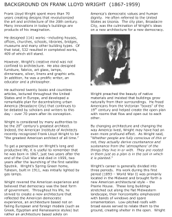
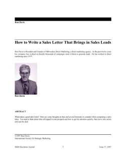
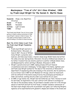
Identification and
Treatment of Benign
Skin Lesions
Wend L.
Wendy
L Wright,
W ight MS,
MS RN,
RN ARNP
ARNP, FNP
FNP, FAANP
Family Nurse Practitioner
Wright & Associates Family Healthcare
Amherst, New Hampshire
Partner – Partners in Healthcare Education, LLC
Objectives
Upon completion of this lecture, the health
care provider will be able to:
1. Identify common benign dermatologic lesions in
the primary care setting
2 Discuss treatment of various lesions
2.
3. Discuss implementation of cryosurgery into your
practice
©Wright, 2010
Wendy Wright, ARNP, FNP, FAANP
Financial Disclosures
• Consultant: Orasure Technologies
Common
Dermatologic
Lesions
©Wright, 2010
Skin Tags (Achrochordons)
• Very commonly encountered benign
lesions
• Seen in approximately 25% of men and
women
• Most common locations: axilla, neck,
inguinal region
• Usually begin in 2nd decade and peak by
the 5th decade of life
©Wright, 2010
Wright, 2010
©Wright, 2010
Skin Tags (Achrochordons)
• Appearance
– Begins as a tiny flesh-toned or brown lesion
– May increase to 1 cm in size
– Hallmark: polypoid mass on a long narrow
stalk
– Bleeds very easily; particularly because they
often get caught on a necklace or clothing
©Wright, 2010
1
Diagnosis?
Linked with____________?
Skin Tags
©Wright, 2010
Skin Tags (Achrochordons)
©Wright, 2010
Dermatofibroma
• Common, benign asymptomatic lesions
• May be slightly itchy; Retract beneath the
skin when you try to elevate them
• 1-10
1 10 lesions occurring on the extremities;
most common location is the anterior
surface of the lower leg
• Etiology: fibrous reaction to trauma, virus
or an insect bite
• Treatment
– Shave excision
– Cryosurgery
– Electrocautery
©Wright, 2010
Dermatofibroma
– Multiple lesions: Systemic lupus
©Wright, 2010
Dermatofibroma
• Treatment
– Elliptical excision
– Shave excision
– Cryosurgery
©Wright, 2010
Wright, 2010
©Wright, 2010
2
Verruca Vulgaris
Verruca Vulgaris
• Common warts
• Benign lesions of the epidermis caused by a
virus
• Transmitted by touch and commonly appear
at sites of trauma, on the hands, around the
periungual regions from nail biting and on the
plantar surfaces of the feet
• Appearance
– Smooth, flesh colored papules which
evolve into a dome-shaped growth with
black dots on the surface
– Black dots are thrombosed capillaries and
can be visualized with a 15 blade
• Treatment
– OTC products: Compound W; Duoplant
©Wright, 2010
Verruca Vulgaris
©Wright, 2010
Verruca Vulgaris
• Treatment
–
–
–
–
–
–
–
Liquid nitrogen
Cryosurgery
Electrocautery
Duct Tape
Blunt dissection (plantar lesions)
Tagamet 600 mg bid x 2 - 4 weeks
Imiquimod
©Wright, 2010
Condyloma Acuminata
• AKA: genital warts
• Etiology: human papillomavirus
– More than 30 strains of HPV infect the genital
tract
– Usually caused by HPV subtypes 6, 11, 40 –
45, and 51
©Wright, 2010
Condyloma Acuminata
• Characteristics
– Cauliflower appearing lesion
– White or flesh toned
– May be associated with abnormal pap
smear
• Transmitted through sexual contact
• Incubation period of 1 – 6 months
©Wright, 2010
Wright, 2010
©Wright, 2010
3
Condylomata Acuminata
Condyloma Acuminata
• Treatment
– Cryosurgery
– Imiquimod 5% cream
– TCA
– Electrodessication
– Laser
– Podofilox
©Wright, 2010
Seborrheic Keratoses
•
•
•
•
©Wright, 2010
Seborrheic Keratoses
Most common benign skin lesion
Unknown origin
No p
potential for malignancy
g
y
Characteristics
– Smooth surface with tiny round, embedded
pearls
– May be rough, dry and cracked
– Appear stuck on the surface
©Wright, 2010
Seborrheic Keratosis
©Wright, 2010
Wright, 2010
©Wright, 2010
Can Mimic a Malignant Melanoma
©Wright, 2010
4
Seborrheic Keratoses
• Treatment
– Lesions are only removed for cosmetic
purposes or for a biopsy if pathology is
unknown
– If removed, shave excision
– Cryosurgery
Molluscum Contagiosum
• Infection caused by the pox-virus
• Most commonly seen on the face, trunk and
axillae
• Self-limiting
• Spread by auto-inoculation
• Incubation period: 2-7 weeks after
exposure
• Contagious until gone
©Wright, 2010
Molluscum Contagiosum
©Wright, 2010
Molluscum Contagiosum
• Asymptomatic lumps
• May have 1 - hundreds
• Physical Examination
–
–
–
–
2-5mm p
papule
p
with an umbilicated center
Flesh toned - white in color
Most often around the eye in children
Scaling and erythema around the periphery of the lesion
is not unusual
– If in the genital area of a child-should consider sexual
abuse
©Wright, 2010
Molluscum Contagiosum
• Plan
Molluscum Contagiosum
• Plan
– Diagnostic:
• None or KOH prep looking for inclusion bodies
– Therapeutic:
• Conservative treatment is the best for children
–Curettage
–Cryosurgery
–Tretinoin
©Wright, 2010
Wright, 2010
©Wright, 2010
– Therapeutic:
• Salicylic Acid (Occlusal)
• Laser
• TCA
• Imiquimod
©Wright, 2010
5
Molluscum Contagiosum
• Plan
– Educational
• May resolve on own in 6 - 9 months
• Contagious until lesions are gone
• Benign
• Recurrence very common
©Wright, 2010
Actinic Keratoses
Actinic Keratoses
• Common sun-induced premalignant lesions
• Incidence: Increases with age, light
complexion
• Clinical presentation: Slightly roughened
area that often bleeds when excoriated
– Progresses to an adherent yellow crust
– Size 3-6 mm
– Common location: scalp, temples, forehead,
hands
©Wright, 2010
Actinic Keratosis
• Prognosis
– Can spontaneously regress if sun exposure is
eliminated
– Good
G d prognosis
i if treated
t t d adequately
d
t l
– Small percentage transform into a squamous
cell carcinoma which can metastasize
• 60% of all squamous cell carcinomas began
as an actinic keratosis
©Wright, 2010
Actinic Keratosis
• Keratin may
accumulate and
transform lesion
into a cutaneous
horn
• Frequently seen on
the pinna of the ear
©Wright, 2010
Wright, 2010
©Wright, 2010
Actinic Keratoses
• Treatment
– Cryosurgery
• preferred method
–
–
–
–
–
–
Surgical
g
Removal
Tretinoin
5-fluorouracil
Acid peels
Sunscreen
Imiquimod
©Wright, 2010
6
Cryosurgery
Cryosurgery
• Definition: The process of applying extreme
cold to a lesion for the purpose of
destruction
• Indications
– Seborrheic keratoses
– Actinic keratoses
– Skin tags
– Verruca vulgaris
• Indications
– Plantar warts
– Condyloma acuminatum
– Molluscum contagiosum
• Advantages
– Minimal discomfort
– Minimal scarring
– No sutures needed
©Wright, 2010
Cryosurgery
©Wright, 2010
Cryosurgery
• Disadvantages
– May not be effective for all lesions, particularly
warts
– Some individuals report moderate pain during
the procedure
• Equipment
– Cotton applicators
– Vaseline petroleum jelly
– Dressing
• Equipment
– Betadine
– 4X4 gauze
– Freeze kit or nitrous oxide cryosurgery unit
©Wright, 2010
Cryosurgery
©Wright, 2010
Cryosurgery
• Procedure
– Position the patient for provider comfort
– Cleanse the lesion with betadine
– Cover the lesion with a water soaked dressing for 5-10
minutes
– Using the cotton applicators, surround the lesion with
vaseline petroleum jelly
– Choose the appropriate wand for the lesion
– Freeze the lesion for the appropriate amount of time
©Wright, 2010
Wright, 2010
©Wright, 2010
7
Cryosurgery
Cryosurgery
©Wright, 2010
Cryosurgery
©Wright, 2010
Cryosurgery
• Time Frame
– Utilize charts provided with your particular cryokit/device
– I.e. 20 – 40 seconds
– Liquid nitrogen – much, much less than this
• Time Frame
– Another method is to apply the freeze until
a frost ring appears approximately 1-2 mm
around the lesion
©Wright, 2010
Cryosurgery
Cryosurgery
• Procedure
• Red Flags
– Apply additional pressure for deeper tissue
penetration
– Cover
C
with
ith a dressing
d
i
• Follow-up
– Monitor for redness, discharge, fever, pain,
streaking
– Recheck lesion in 4 - 6 weeks
©Wright, 2010
Wright, 2010
©Wright, 2010
– Malignant lesions
– Facial lesions
– Infected lesion
• CPT Code
– 17000: Destruction, any method, all benign
– 17110: Destruction, flat warts or molluscum
contagiosum
©Wright, 2010
8
Thank You
Case Study
• 47 year-old female presents with the
following:
– 2 verruca vulgaris lesions on the soles of her feet
– 1 actinic keratosis on her right hand
– Assessments:
• 1. Warts
– CPT: 17110: 1-14 lesions (cryo)
• 2. Actinic keratosis
– CPT: 17000 (1 lesion) – destroy benign/premalig lesion
– Mod 57 (see and perform destruction today)
–
©Wright, 2010
I Would Be Happy To
E t t i A
Entertain
Any
Questions That You
May Have!!
©Wright, 2010
Wendy L. Wright, MS, RN, ARNP, FNP, FAANP
(W) 603-249-8883
(H) 603-472-6776
(F) 603-472-2597
Email: [email protected]
©Wright, 2010
Wright, 2010
9


© Copyright 2026