
Pruritic urticarial papules and plaques of pregnancy Obstetric case repOrts Discussion
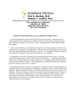
Journal of Obstetrics and Gynaecology, April 2012; 32: 301–302 © 2012 Informa UK, Ltd. ISSN 0144-3615 print/ISSN 1364-6893 online Obstetric Case Reports Pruritic urticarial papules and plaques of pregnancy E. Giugliano, E. Cagnazzo, T. Servello, E. Mossuto, R. Marci & A. Patella Department of Biomedical Sciences and Advanced Therapy, Section of Obstetrics and Gynaecology, University of Ferrara, Ferrara, Italy J Obstet Gynaecol Downloaded from informahealthcare.com by 94.163.23.201 on 02/28/12 For personal use only. DOI: 10.3109/01443615.2011.652704 Correspondence: E. Giugliano, Department of Biomedical Sciences and Advanced Therapy, Section of Obstetrics and Gynaecology, Giovecca Street 203, University of Ferrara. E-mail: [email protected] Introduction The term pruritic urticarial papules and plaques of pregnancy (PUPPP) was first proposed by Lawley et al. in 1979 to describe benign dermatoses of pregnancy characterised by erythema, urticarial plaques and papules. It is a skin disorder whose incidence is estimated to occur in one in 200 pregnancies (0.5%) (Aronson et al. 1998). In the present study, an unusual case of PUPPP is reported. The interesting presentation of this case may render new insights regarding the aetiological factors of this disease. Case report A 22-year-old Moroccan woman in her second pregnancy, at 32 weeks’ gestation, was admitted to our Department of Obstetrics for elevated glycaemic values (310 mg/dl). She had suffered from type 1 diabetes since she was 8 years old. During her first pregnancy, she was hospitalised for hyperglycaemic coma (440 mg/dl), causing spontaneous abortion at the 20th week of gestation. The course of the first part of the present pregnancy was normal. Blood glucose levels were normal with insulin therapy. In the 3rd trimester of pregnancy, blood glucose tended to increase in spite of the fact that insulin therapy had been repeatedly adapted. Therefore, the patient was hospitalised to perform intensive monitoring of maternal and fetal wellbeing. During the admission, the patient developed intense pruritus and abdominal erythema. The rash was described as erythematous papules spreading below her umbilicus within abdominal stria (Figure 1). For 5 days, the rash remained localised, but then it began to spread centrifugally outwards on the abdomen. The papules coalesced to form plaques with halos of erythema. The intensity of the pruritus escalated. The maternal liver function was normal, therefore a tentative diagnosis of pruritic urticarial papules and plaques of pregnancy (PUPPP) was made. A dermatologist was consulted on therapeutic options. The treatment included Betamethasone 0.5 mg cpr twice/day. Considering the improvement of symptoms, biopsy was not performed on a papula, but the rash did not completely regress. Given the persistently high blood glucose values in spite of insulin therapy, caesarean section at 34 weeks’ gestation was performed, and a boy of 2,780 g was born without complication. The newborn was admitted to neonatal intensive care for hypoglycaemia, without consequences. After delivery, the patient continued oral Betamethasone 0.5 mg daily for 4 days. The pruritus and rash completely resolved on this regimen without reoccurrence. At 6 weeks postpartum, the symptoms completely resolved. Discussion PUPPP is a disorder that predominantly occurs in a first pregnancy (Powell 2000). Skin changes typical for PUPPP are erythematous, urticarial plaques and 1–2 mm papules are usually surrounded by a narrow and pale halo (Lawley et al. 1979). The distribution of lesions in PUPPP is a notable clinical feature. The primary location of the eruption is on the abdomen, frequently occurring in the striae gravidarum, which first become itchy, then erythematous and finally urticarial. The eruption then extends on the buttocks and medial thighs. In many patients, it remains limited to these areas, but in might become generalised (Aronson et al. 1998). Although the abdomen is the most commonly affected site, the periumbilical skin is often relatively spared. Sparing of the palms, soles and face are typical of the rash’s presentation. However, there might be a considerable variation in the morphology of the eruption (Ahmadi and Powell 2005). The rash usually regresses within 1 week postpartum. The maternal and fetal prognosis is unaffected. Recurrence of the eruption in subsequent pregnancies is rare. Treatment of PUPPP is focused on the relief of pruritus. The most common agents used are antipruritic agents, skin emollients and topical corticosteroids. Refractory cases may require oral corticosteroid therapy (Catanzarite and Quirk 1990). Although this syndrome has been known for several years, its pathogenesis is still unclear. Some authors suggest an immunological mechanism; others highlight the hormonal abnormalities; others hypothesise a mechanical factor (Aractinigi et al. 1998; Beckett and Goldberg 1991; Vaughan Jones et al. 1999). In our case, the hypothesis of the autoimmune mechanism was plausible, as the patient suffered from type 1 diabetes. Her pathology worsened during pregnancy (in the first pregnancy she had an abortion for hyperglycaemic coma; in the second pregnancy, the blood glucose levels remained steadily increased, despite medical treatment). Therefore, we can speculate that the pregnancy triggered an uncontrolled immune response, also responsible for the skin disorders. The type of treatment confirms this hypothesis, in fact immunomodulatory drugs as corticosteroids determined the resolution of symptoms. Resolution of the skin disorders after delivery is another factor in favour of this hypothesis. Furthermore, the specificity of these skin lesions with pregnancy may also make us reconsider this dermatological syndrome. Figure 1. Erythematous papules spreading below umbilicus within an abdominal stria. 302 Obstetric case reports PUPPP may not simply be regarded as a dermatological disease, but its skin manifestations may represent a sign of a pregnancy at risk. Accordingly, the mechanism of occurrence of these skin disorders must have considerable clinical interest; however, there are still too many doubts about the aetiology of PUPPP. Therefore, further studies are necessary to clarify its pathogenesis. Declaration of interest: The authors report no conflicts of interest. The authors alone are responsible for the content and writing of the paper. J Obstet Gynaecol Downloaded from informahealthcare.com by 94.163.23.201 on 02/28/12 For personal use only. References Ahmadi S, Powell FC. 2005. Pruritic urticarial papules and plaques of pregnancy: Current status. Australasian Journal of Dermatology 46:53–60. Aronson IK, Bond S, Fiedler VC et al. 1998. Pruritic urticarial papules and plaques of pregnancy: Clinical and immunopathologic observations in 57 patients. Journal of the American Academy of Dermatology 39:933–939. Aractinigi S, Berkane N, Bertheau P et al. 1998. Fetal DNA in skin of polymorphic eruption of pregnancy. Lancet 352:1898–1901. Beckett MA, Goldberg NS. 1991. Pruritic urticarial papules and plaques of pregnancy and skin distension. Archives of Dermatology 127:125–130. Catanzarite V, Quirk JG Jr. 1990. Papular dermatoses of pregnancy. Clinical Obstetrics and Gynecology 33:754–758. Lawley TJ, Hertz KC, Wade TR et al. 1979. Pruritic urticarial papules and plaques of pregnancy. Journal of the American Medical Association 241:1696–1699. Powell FC. 2000. Pruritic urticarial papules and plaques of pregnancy and multiple pregnancies. Journal of the American Academy of Dermatology 43:730–731. Vaughan Jones SA, Hern S, Nelson-Piercy C et al. 1999. A prospective study of 200 women with dermatoses of pregnancy correlating clinical findings with hormonal and immunopathological profiles. British Journal of Dermatology 141:71–81. A pregnancy complicated with fetal growth restriction in a patient with dystrophic epidermolysis bullosa E. Ozkaya, E. Baser, G. Akgul & T. Kucukozkan Department of Obstetrics and Gynaecology, Dr Sami Ulus Maternity and Children’s Health Research and Training Hospital, Ankara, Turkey DOI: 10.3109/01443615.2011.653595 Correspondence: E. Baser, Dr. Sami Ulus Maternity and Children’s Health Research and Training Hospital, Babur sokak No: 44 Altindag, Ankara, Turkey. E-mail: [email protected] Epidermolysis bullosa (EB) is a group of inherited skin diseases. We present a case of pregnancy in a patient with dystrophic EB complicated by fetal growth restriction (FGR). A 26-year-old primigravid patient previously diagnosed as dystrophic EB with genital mucosal involvement was referred to our hospital. The patient delivered a growth restricted neonate at 40 weeks’ gestation, by caesarean section, under general anaesthesia. There may be an association between genital involvement of EB and fetal growth restriction, therefore genital examination should be performed before and during pregnancy in these patients. Case report A 26-year-old primigravid woman diagnosed as dystrophic EB, was referred to our hospital in her 17th gestational week. On family history, she had one sister affected with EB who was deceased because of infection. Results of first visit laboratory tests (biochemistry, complete blood count, TORCH panel) were within normal ranges. Triple test results demonstrated low risk for chromosomal anomalies and open neural tube defects. At the 22nd gestational week, she underwent a 2nd trimester ultrasound scan, which revealed normal fetal anatomy with appropriate growth for gestational age. She had regular visits to a dermatologist in an associated hospital. There was no exacerbation in skin lesions at 34 weeks. Weekly non-stress tests (NST) were reactive, also amniotic fluid index (AFI) and fetal growth were within normal ranges. The patient was lost to follow-up for 6 weeks. She returned to our hospital at the 40th gestational week, complaining of painful regular uterine contractions. On pelvic examination, she had minimal cervical effacement and 1 cm dilatation. On systemic examination, she had widespread skin erosions, including the vulval region and vaginal mucosa, compatible with genital involvement of EB. Ultrasound examination revealed anhydramnios with fetal measurements compatible with 35 gestational weeks, with an estimated fetal weight of 2,338 g. On Doppler examination, there was loss of end-diastolic flow in umbilical artery and cerebro-placental pulsatility index ratio was 1. These findings strongly suggested placental insufficiency with brain sparing effect. Non-stress test (NST) revealed regular uterine contractions with persistent variable decelerations. Caesarean section was performed due to non-reassuring fetal status. A female neonate was delivered weighing 2,410 g. First minute Apgar score was 6 and at 5 min, the Apgar score was 7. The neonate was transferred to the neonatal intensive care unit for close observation. The patient’s postpartum period was uneventful and without any complications. Skin lesions were treated with topical agents and antibiotics, with the supervision of a dermatologist. (The caesarean incision on the 2nd postoperative day is shown in Figure 1). The woman was discharged from the hospital on the 7th postoperative day. Skin lesions were stable during the puerperium. The neonate also did not have any important complications and was discharged from the hospital at the same day, together with the mother. Comment We present a pregnancy complicated with fetal growth restriction (FGR) in a patient with dystrophic EB. It is unclear if EB has an adverse effect on an otherwise healthy pregnancy. Such pregnancies usually end in term delivery. There have, however, been instances where the pregnancy results in pre-term delivery following pre-term premature rupture of the membranes (PPROM) (Anum et al. 2009). A case of pregnancy complicated with FGR, similar to our case, was reported previously in a patient with genital involvement of dystrophic EB (Bianca et al. 2003). The reported case delivered a growth restricted neonate at 36 weeks’ gestation. A question was raised in our minds whether EB is specifically Introduction Epidermolysis bullosa (EB) is a group of autosomally inherited skin diseases, where a small trauma to skin and mucous membranes causes severe lesions such as blisters and erosions. Three major types of EB are defined according level of skin separation, which is intraepidermal in simplex, intra-lamina lucida in junctional and sub-basal lamina in dystrophic form. Dystrophic EB is associated with conjunctival, genital and anal involvement and pregnancies have rarely been reported in this type of disease (Bianca et al. 2003; Buscher et al. 1997; Price and Katz 1988). However, the effects of this disease on an otherwise healthy pregnancy are not well understood. Herein, we report a case of pregnancy complicated by fetal growth restriction (FGR), in a patient with dystrophic EB. Figure 1. Caesarean incision on the second postoperative day.


© Copyright 2026