ABC
docz
Explore
Log in
Create new account
Download
Report
health and fitness
disease
cancer
126 SIGN • Diagnosis and management of colorectal cancer A national clinical guideline
} Investment in r esear ch saves l ives and money Today:
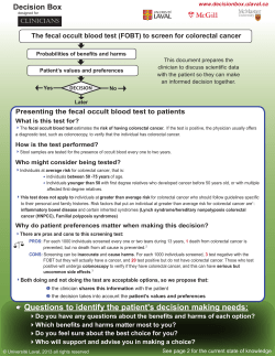
Decision Box
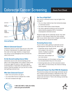
Colorectal Cancer Screening Basic Fact Sheet Are You at High Risk?
References
Collaborative Group of the Americas on Inherited Colorectal Cancer
13. Appropriateness of Colonoscopy: Surveillance After Curative Resection of Colorectal Cancer 1
CONSENSUS STATEMENT OVERVIEW The Consensus Statement: “Cytoreductive Surgery and Hyperthermic Intraperitoneal
Colorectal Cancer Overview
Living With Colorectal Cancer An Information and Resources Guidebook
Volume 3, Year 2012, Edition 10, March 10
© Copyright 2026
About abcdocz
DMCA / GDPR
Report