
Uterine sarcomas: A review ⁎ Emanuela D'Angelo, Jaime Prat
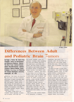
ARTICLE IN PRESS
YGYNO-973334; No. of pages: 9; 4C: 3, 6
Gynecologic Oncology xxx (2009) xxx–xxx
Contents lists available at ScienceDirect
Gynecologic Oncology
j o u r n a l h o m e p a g e : w w w. e l s ev i e r. c o m / l o c a t e / y g y n o
Review
Uterine sarcomas: A review
Emanuela D'Angelo, Jaime Prat ⁎
Department of Pathology, Hospital de la Santa Creu i Sant Pau, Autonomous University of Barcelona, Sant Antoni M. Claret, 167, 08025 Barcelona, Spain
a r t i c l e
i n f o
Article history:
Received 29 June 2009
Available online xxxx
Keywords:
Uterine sarcomas
Leiomyosarcoma
Endometrial stromal sarcoma
Undifferentiated endometrial sarcoma
Adenosarcoma
Carcinosarcoma
a b s t r a c t
Objective. Uterine sarcomas are rare tumors that account for 3% of uterine cancers. Their histopathologic
classification was revised by the World Health Organization (WHO) in 2003. A new staging system has been
recently designed by the International Federation of Gynecology and Obstetrics (FIGO). Currently, there is no
consensus on risk factors for adverse outcome. This review summarizes the available clinicopathological data
on uterine sarcomas classified by the WHO diagnostic criteria.
Methods. Medline was searched between 1976 and 2009 for all publications in English where the studied
population included women diagnosed of uterine sarcomas.
Results. Since carcinosarcomas (malignant mixed mesodermal tumors or MMMT) are currently classified
as metaplastic carcinomas, leiomyosarcomas remain the most common uterine sarcomas. Exclusion of
several histologic variants of leiomyoma, as well as “smooth muscle tumors of uncertain malignant
potential,” frequently misdiagnosed as sarcomas, has made apparent that leiomyosarcomas are associated
with poor prognosis even when seemingly confined to the uterus. Endometrial stromal sarcomas are
indolent tumors associated with long-term survival. Undifferentiated endometrial sarcomas exhibiting
nuclear pleomorphism behave more aggressively than tumors showing nuclear uniformity. Adenosarcomas
have a favorable prognosis except for tumors showing myometrial invasion or sarcomatous overgrowth.
Adenofibromas may represent well-differentiated adenosarcomas. The prognosis of carcinosarcomas (which
are considered here in a post-script fashion) is usually worse than that of grade 3 endometrial carcinomas.
Immunohistochemical expression of Ki67, p53, and p16 is significantly higher in leiomyosarcomas and
undifferentiated endometrial sarcomas than in endometrial stromal sarcomas.
Conclusions. Evaluation of H&E stained sections has been equivocal in the prediction of behavior of
uterine sarcomas. Immunohistochemical studies of oncoproteins as well as molecular analysis of nonrandom translocations will undoubtedly lead to an accurate and prognostically relevant classification of
these rare tumors.
© 2009 Elsevier Inc. All rights reserved.
Contents
Introduction . . . . . . . . . . . . . . . . . . . . .
Leiomyosarcoma . . . . . . . . . . . . . . . . . . .
Clinical features . . . . . . . . . . . . . . . . .
Pathological features . . . . . . . . . . . . . . .
Immunohistochemistry and molecular biology . . .
Prognosis and treatment . . . . . . . . . . . . .
Smooth muscle tumors of uncertain malignant potential
Endometrial stromal tumor . . . . . . . . . . . . . .
Endometrial stromal nodule . . . . . . . . . . .
Low-grade endometrial stromal sarcoma . . . . .
Immunohistochemistry and molecular biology . . .
Prognosis and treatment . . . . . . . . . . . . .
Undifferentiated endometrial sarcoma . . . . . . . . .
Clinicopathological features . . . . . . . . . . . .
Immunohistochemistry . . . . . . . . . . . . . .
Prognosis and treatment . . . . . . . . . . . . .
. . . . .
. . . . .
. . . . .
. . . . .
. . . . .
. . . . .
(STUMP).
. . . . .
. . . . .
. . . . .
. . . . .
. . . . .
. . . . .
. . . . .
. . . . .
. . . . .
.
.
.
.
.
.
.
.
.
.
.
.
.
.
.
.
.
.
.
.
.
.
.
.
.
.
.
.
.
.
.
.
.
.
.
.
.
.
.
.
.
.
.
.
.
.
.
.
.
.
.
.
.
.
.
.
.
.
.
.
.
.
.
.
.
.
.
.
.
.
.
.
.
.
.
.
.
.
.
.
.
.
.
.
.
.
.
.
.
.
.
.
.
.
.
.
.
.
.
.
.
.
.
.
.
.
.
.
.
.
.
.
.
.
.
.
.
.
.
.
.
.
.
.
.
.
.
.
.
.
.
.
.
.
.
.
.
.
.
.
.
.
.
.
.
.
.
.
.
.
.
.
.
.
.
.
.
.
.
.
.
.
.
.
.
.
.
.
.
.
.
.
.
.
.
.
.
.
.
.
.
.
.
.
.
.
.
.
.
.
.
.
.
.
.
.
.
.
.
.
.
.
.
.
.
.
.
.
.
.
.
.
.
.
.
.
.
.
.
.
.
.
.
.
.
.
.
.
.
.
.
.
.
.
.
.
.
.
.
.
.
.
.
.
.
.
.
.
.
.
.
.
.
.
.
.
.
.
.
.
.
.
.
.
.
.
.
.
.
.
.
.
.
.
.
.
.
.
.
.
.
.
.
.
.
.
.
.
.
.
.
.
.
.
.
.
.
.
.
.
.
.
.
.
.
.
.
.
.
.
.
.
.
.
.
.
.
.
.
.
.
.
.
.
.
.
.
.
.
.
.
.
.
.
.
.
.
.
.
.
.
.
.
.
.
.
.
.
.
.
.
.
.
.
.
.
.
.
.
.
.
.
.
.
.
.
.
.
.
.
.
.
.
.
.
.
.
.
.
.
.
.
.
.
.
.
.
.
.
.
.
.
.
.
.
.
.
.
.
.
.
.
.
.
.
.
.
.
.
.
.
.
.
.
.
.
.
.
.
.
.
.
.
.
.
.
.
.
.
.
.
.
.
.
.
.
.
.
.
.
.
.
.
.
.
.
.
.
.
.
.
.
.
.
.
.
.
.
.
.
.
.
.
.
.
.
.
.
.
.
.
.
.
.
.
.
.
.
.
.
.
.
.
.
.
.
.
.
.
.
.
.
.
.
.
.
.
.
.
.
.
.
.
.
.
.
.
.
.
.
.
.
.
.
.
.
.
.
.
.
.
.
.
.
.
.
.
.
.
.
.
.
.
.
.
.
.
.
.
.
.
.
.
.
.
.
.
.
.
.
.
.
.
.
.
.
.
.
.
.
.
.
.
.
.
.
.
.
.
.
.
.
.
.
.
.
⁎ Corresponding author. Fax: +34 93 291 93 44.
E-mail address: [email protected] (J. Prat).
0090-8258/$ – see front matter © 2009 Elsevier Inc. All rights reserved.
doi:10.1016/j.ygyno.2009.09.023
Please cite this article as: D'Angelo E, Prat J, Uterine sarcomas: A review, Gynecol Oncol (2009), doi:10.1016/j.ygyno.2009.09.023
.
.
.
.
.
.
.
.
.
.
.
.
.
.
.
.
.
.
.
.
.
.
.
.
.
.
.
.
.
.
.
.
.
.
.
.
.
.
.
.
.
.
.
.
.
.
.
.
0
0
0
0
0
0
0
0
0
0
0
0
0
0
0
0
ARTICLE IN PRESS
2
E. D'Angelo, J. Prat / Gynecologic Oncology xxx (2009) xxx–xxx
Adenosarcoma . . . . . . . . . . . . . .
Clinical features . . . . . . . . . . .
Pathological features . . . . . . . . .
Adenosarcoma versus adenofibroma . .
Immunohistochemistry . . . . . . . .
Prognosis and treatment . . . . . . .
Carcinosarcoma (malignant mixed mullerian
Clinical features . . . . . . . . . . .
Pathological features . . . . . . . . .
Histogenesis . . . . . . . . . . . . .
Immunohistochemistry . . . . . . . .
Prognosis and treatment . . . . . . .
Conflict of interest statement . . . . . . .
Acknowledgments . . . . . . . . . . . .
References . . . . . . . . . . . . . . . .
. . . .
. . . .
. . . .
. . . .
. . . .
. . . .
tumor)
. . . .
. . . .
. . . .
. . . .
. . . .
. . . .
. . . .
. . . .
.
.
.
.
.
.
.
.
.
.
.
.
.
.
.
.
.
.
.
.
.
.
.
.
.
.
.
.
.
.
.
.
.
.
.
.
.
.
.
.
.
.
.
.
.
.
.
.
.
.
.
.
.
.
.
.
.
.
.
.
.
.
.
.
.
.
.
.
.
.
.
.
.
.
.
.
.
.
.
.
.
.
.
.
.
.
.
.
.
.
.
.
.
.
.
.
.
.
.
.
.
.
.
.
.
.
.
.
.
.
.
.
.
.
.
.
.
.
.
.
.
.
.
.
.
.
.
.
.
.
.
.
.
.
.
Introduction
Uterine sarcomas are rare tumors that account for approximately
1% of female genital tract malignancies and 3% to 7% of uterine cancers
[1]. Although the aggressive behavior of most cases is well recognized,
their rarity and histopathological diversity has contributed to the
lack of consensus on risk factors for poor outcome and optimal treatment [2].
Histologically, uterine sarcomas were first classified into carcinosarcomas, accounting for 40% of cases, leiomyosarcomas (40%),
endometrial stromal sarcomas (10% to 15%), and undifferentiated
sarcomas (5% to 10%). Recently, carcinosarcoma has been reclassified
as a dedifferentiated or metaplastic form of endometrial carcinoma.
Despite this, and probably because it behaves more aggressively than
the ordinary endometrial carcinoma, carcinosarcoma is still included
Table 1
FIGO staging for uterine sarcomas (2009).
Stage
.
.
.
.
.
.
.
.
.
.
.
.
.
.
.
.
.
.
.
.
.
.
.
.
.
.
.
.
.
.
.
.
.
.
.
.
.
.
.
.
.
.
.
.
.
.
.
.
.
.
.
.
.
.
.
.
.
.
.
.
.
.
.
.
.
.
.
.
.
.
.
.
.
.
.
.
.
.
.
.
.
.
.
.
.
.
.
.
.
.
.
.
.
.
.
.
.
.
.
.
.
.
.
.
.
.
.
.
.
.
.
.
.
.
.
.
.
.
.
.
.
.
.
.
.
.
.
.
.
.
.
.
.
.
.
.
.
.
.
.
.
.
.
.
.
.
.
.
.
.
.
.
.
.
.
.
.
.
.
.
.
.
.
.
.
.
.
.
.
.
.
.
.
.
.
.
.
.
.
.
.
.
.
.
.
.
.
.
.
.
.
.
.
.
.
.
.
.
.
.
.
.
.
.
.
.
.
.
.
.
.
.
.
.
.
.
.
.
.
.
.
.
.
.
.
.
.
.
.
.
.
.
.
.
.
.
.
.
.
.
.
.
.
.
.
.
.
.
.
.
.
.
.
.
.
.
.
.
.
.
.
.
.
.
.
.
.
.
.
.
.
.
.
.
.
.
.
.
.
.
.
.
.
.
.
.
.
.
.
.
.
.
.
.
.
.
.
.
.
.
.
.
.
.
.
.
.
.
.
.
.
.
.
.
.
.
.
.
.
.
.
.
.
.
.
.
.
.
.
.
.
.
.
.
.
.
.
.
.
.
.
.
.
.
.
.
.
.
.
.
.
.
.
.
.
.
.
.
.
.
.
.
.
.
.
.
.
.
.
.
.
.
.
.
.
.
.
.
.
.
.
.
.
.
.
.
.
.
.
.
.
.
.
.
.
.
.
.
.
.
.
.
.
.
.
.
.
.
.
.
.
.
.
.
.
.
.
.
.
.
.
.
.
.
.
.
.
.
.
.
.
.
.
.
.
.
.
.
.
.
.
.
.
.
.
.
.
.
.
.
.
.
.
.
.
.
.
.
.
.
.
.
.
.
.
.
.
.
.
.
.
.
.
.
.
.
.
.
.
.
.
.
.
.
.
.
.
.
.
.
.
.
.
.
.
.
.
.
.
.
.
.
.
.
.
.
.
.
.
.
.
.
.
.
.
.
.
.
.
.
.
.
.
.
.
.
.
.
.
.
.
.
.
.
.
.
.
.
.
.
.
.
.
.
.
.
.
.
.
.
.
.
.
.
.
0
0
0
0
0
0
0
0
0
0
0
0
0
0
0
in most retrospective studies of uterine sarcomas, as well as in the
2003 World Health Organization (WHO) classification [3].
The 1988 International Federation of Gynecology and Obstetrics
(FIGO) criteria for endometrial carcinoma have been used until now to
assign stages for uterine sarcomas in spite of the different biologic
behavior of both tumor categories. Recently, however, a new FIGO
classification and staging system has been specifically designed for
uterine sarcomas in an attempt to reflect their different biologic
behavior (Table 1) [4]. Briefly, three new classifications have been
developed: (1) staging for leiomyosarcomas and endometrial stromal
sarcomas; (2) staging for adenosarcomas; and (3) staging for carcinosarcomas (MMMT). Whereas in the first classification stage I sarcomas
are subdivided according to size, subdivision of stage I adenosarcomas
takes into account myometrial invasion. On the other hand, carcinosarcomas will continue to be staged as endometrial carcinomas.
Leiomyosarcoma
Clinical features
Definition
(1) Leiomyosarcomas and endometrial stromal sarcomasa
I
Tumor limited to uterus
IA
Less than or equal to 5 cm
IB
More than 5 cm
II
Tumor extends beyond the uterus, within the pelvis
IIA Adnexal involvement
IIB Involvement of other pelvic tissues
III
Tumor invades abdominal tissues (not just protruding into the abdomen)
IIIA One site
IIIB More than one site
IIIC Metastasis to pelvic and/or para-aortic lymph nodes
IV
IVA Tumor invades bladder and/or rectum
IVB Distant metastasis
(2) Adenosarcomas
I
Tumor limited to uterus
IA
Tumor limited to endometrium/endocervix with no myometrial invasion
IB
Less than or equal to half myometrial invasion
IC
More than half myometrial invasion
II
Tumor extends beyond the uterus, within the pelvis
IIA Adnexal involvement
IIB Tumor extends to extrauterine pelvic tissue
III
Tumor invades abdominal tissues (not just protruding into the abdomen).
IIIA One site
IIIB More than one site
IIIC Metastasis to pelvic and/or para-aortic lymph nodes
IV
IVA Tumor invades bladder and/or rectum
IVB Distant metastasis
(3) Carcinosarcomas
Carcinosarcomas should be staged as carcinomas of the endometrium.
a
Note: Simultaneous endometrial stromal sarcomas of the uterine corpus and ovary/
pelvis in association with ovarian/pelvic endometriosis should be classified as
independent primary tumors.
After excluding carcinosarcoma (MMMT), leiomyosarcoma has
become the most common subtype of uterine sarcoma. However, it
accounts for only 1–2% of uterine malignancies. Most occur in women
over 40 years of age who usually present with abnormal vaginal
bleeding (56%), palpable pelvic mass (54%), and pelvic pain (22%).
Signs and symptoms resemble those of the far more common
leiomyoma and preoperative distinction between the two tumors
may be difficult. Nevertheless, malignancy should be suspected by the
presence of certain clinical behaviors, such as tumor growth in
menopausal women who are not on hormonal replacement therapy
[5]. Occasionally, the presenting manifestations are related to tumor
rupture (hemoperitoneum), extrauterine extension (one-third to
one-half of cases), or metastases. Only very rarely does a leiomyosarcoma originate from a leiomyoma.
Pathological features
The histopathologic diagnosis of uterine leiomyosarcoma is usually
straightforward since most clinically malignant smooth muscle
tumors of the uterus show the microscopic constellation of hypercellularity, severe nuclear atypia, and high mitotic rate generally
exceeding 15 mitotic figures per 10 high-power-fields (MF/10 HPF)
[6–7] (Fig. 1a). Moreover, one or more supportive clinicopathologic
features such as peri- or postmenopausal age, extrauterine extension,
large size (over 10 cm), infiltrating border, necrosis, and atypical
mitotic figures are frequently present [8].
Epithelioid and myxoid leiomyosarcomas, however, are two
rare variants which may be difficult to recognize microscopically as
their pathologic features differ from those of ordinary spindle cell
leiomyosarcomas. In fact, nuclear atypia is usually mild in both tumor
Please cite this article as: D'Angelo E, Prat J, Uterine sarcomas: A review, Gynecol Oncol (2009), doi:10.1016/j.ygyno.2009.09.023
ARTICLE IN PRESS
E. D'Angelo, J. Prat / Gynecologic Oncology xxx (2009) xxx–xxx
3
Fig. 1. (a) Leiomyosarcoma, spindle-cell variant; (b) myxoid leiomyosarcoma; (c) epithelioid leiomyosarcoma; (d) endometrial stromal sarcoma.
types and the mitotic rate is often b3 MF/10 HPF [9] (Figs. 1b, c). In
epithelioid leiomyosarcomas, necrosis may be absent and myxoid
leiomyosarcomas are often hypocellular. In the absence of severe
cytologic atypia and high mitotic activity, both tumors are diagnosed
as sarcomas based on their infiltrative borders [10].
The minimal pathological criteria for the diagnosis of leiomyosarcoma are more problematic and, in such cases, the differential
diagnosis has to be made, not only with a variety of benign smooth
muscle tumors that exhibit atypical histologic features and unusual
growth patterns (Table 2), but also with smooth muscle tumors of
uncertain malignant potential (STUMP) (Table 3). Application of the
2003 WHO diagnostic criteria [4] has allowed distinguishing these
unusual histologic variants of leiomyoma frequently misdiagnosed as
Table 2
Benign smooth muscle tumors of the uterus.
Leiomyoma variants that
may mimic malignancy
Smooth muscle proliferations
with unusual growth patterns
• Mitotically active leiomyoma
• Cellular leiomyoma
• Hemorrhagic leiomyoma and
hormone-induced changes
• Leiomyoma with bizarre nuclei
(atypical leiomyoma)
• Myxoid leiomyoma
• Epithelioid leiomyoma
• Leiomyoma with massive
lymphoid infiltration
• Disseminated peritoneal leiomyomatosis
• Benign metastasizing leiomyoma
• Intravenous leiomyomatosis
• Lymphangioleiomyomatosis
well-differentiated or low-grade leiomyosarcomas in the past. Indeed,
in a recent population-based study of uterine sarcomas from Norway
[11], of 356 tumors classified initially as leiomyosarcomas, diagnosis
was confirmed in only 259 cases (73%), whereas 97 (27%) were
excluded on review and reclassified as leiomyomas or leiomyoma
variants. Follow-up information, however, revealed that 4 of 48
excluded tumors (1 cellular leiomyoma and 3 STUMPs) developed
metastases.
Immunohistochemistry and molecular biology
Recently, several immunohistochemical and molecular genetic
studies on uterine leiomyosarcomas have been reported [12,13–19].
Leiomyosarcomas usually express smooth muscle markers such as
desmin, h-caldesmon, smooth muscle actin, and histone deacetylase
8 (HDCA8). However, it is important to keep in mind that epithelioid
and myxoid leiomyosarcomas may show lesser degrees of immunoreaction for these markers. Also, leiomyosarcomas are often immunoreactive for CD10 and epithelial markers including keratin and EMA
Table 3
Smooth muscle tumors of uncertain malignant potential (STUMP).
Pathologic criteria
•
•
•
•
Tumor cell necrosis in a typical leiomyoma
Necrosis of uncertain type with ≥ 10 MF / 10 HPFs, or marked diffuse atypia
Marked diffuse or focal atypia with borderline mitotic counts
Necrosis difficult to classify
Please cite this article as: D'Angelo E, Prat J, Uterine sarcomas: A review, Gynecol Oncol (2009), doi:10.1016/j.ygyno.2009.09.023
ARTICLE IN PRESS
4
E. D'Angelo, J. Prat / Gynecologic Oncology xxx (2009) xxx–xxx
(the latter being more frequently positive in the epithelioid variant).
Conventional leiomyosarcomas express estrogen receptors (ER),
progesterone receptors (PR), and androgen receptors (AR) in 3040% of cases. Whereas a variable proportion of uterine leiomyosarcomas has been reported as being immunoreactive for c-KIT, no c-KIT
mutations have been identified [20].
Recent studies have shown statistically significant higher levels of
Ki67 in uterine leiomyosarcomas compared with benign smooth
muscle tumors [15–19]. Mutation and overexpression of p53 have
been described in a significant minority of uterine leiomyosarcomas
(25-47%) but not in leiomyomas [15,18,19]. Intermediate rates have
been found in atypical leiomyomas and STUMPs. Overexpression of
p16 has been described in uterine leiomyosarcomas and may prove to
be a useful adjunct immunomarker for distinguishing between benign
and malignant uterine smooth muscle tumors [13–15].
The vast majority of uterine leiomyosarcomas are sporadic.
Patients with germline mutations in fumarate hydratase are believed
to be at increased risk for developing uterine leiomyosarcomas as well
as uterine leiomyomas [21,22]. The oncogenic mechanisms underlying the development of uterine leiomyosarcomas remain elusive.
Overall, uterine leiomyosarcoma is a genetically unstable tumor that
demonstrates complex structural chromosomal abnormalities and
highly disturbed gene regulation which likely reflects the end-state of
accumulation of multiple genetic defects. Extrapolating from experiences in soft tissue leiomyosarcomas, it is unlikely that recurrent
disease-driven genetic aberrations (i.e. gene mutation or translocation events) will be uncovered. In comparison with other more
common uterine malignancies, uterine leiomyosarcomas bear some
resemblance to type 2 endometrial carcinomas and high-grade serous
carcinomas of ovary/fallopian tube origin, based on their genetic
instability, frequent p53 abnormalities, aggressive behavior, and
resistance to chemotherapy. Therefore, therapies that exploit the
underlying genetic instability of uterine leiomyosarcomas may prove
to be an effective therapeutic strategy.
they act independently of stage which still is the most significant
prognostic factor for uterine sarcomas.
Treatment of leiomyosarcomas includes total abdominal hysterectomy and debulking of tumor if present outside the uterus. Removal
of the ovaries and lymph node dissection remain controversial as
metastases to these organs occur in a small percentage of cases and
are frequently associated with intra-abdominal disease [2]. Ovarian
preservation may be considered in premenopausal patients with
early-stage leiomyosarcomas [2]. Lymph node metastases have been
identified in 6.6% and 11% of two series of patients with leiomyosarcoma who underwent lymphadenectomy [2,30]. In the first series,
the 5-year disease-specific survival rate was 26% in patients who had
positive lymph nodes compared with 64.2% in patients who had
negative lymph nodes (p b 0.001) [30]. The influence of adjuvant
therapy on survival is uncertain. Radiotherapy may be useful in
controlling local recurrences and chemotherapy with doxorubicin or
docetaxel/gemcitabine is now used for advanced or recurrent disease,
with response rates ranging from 27% to 36% [31,32]. Some patients
may respond to hormonal treatment [33].
Smooth muscle tumors of uncertain malignant potential (STUMP)
Uterine smooth muscle tumors that show some worrisome
histological features (i.e., necrosis, nuclear atypia, or mitoses), but
do not meet all diagnostic criteria for leiomyosarcoma, fall into the
category of STUMP (Table 3) [3,34]. The diagnosis of STUMP, however,
should be used most sparingly and every effort should be made to
classify a smooth muscle tumor into a specific category [3,34]. Most
tumors classified as STUMP have been associated with favorable
prognosis and, in these cases, only follow-up of the patients is
recommended [35]. In fact, in a recent study of 41 cases of STUMP, the
recurrence rate was 7%. One of the two recurrences was in the form of
STUMP and the other as leiomyosarcoma [36].
Endometrial stromal tumor
Prognosis and treatment
Leiomyosarcomas are very aggressive tumors. It has become
apparent that tumors diagnosed according to the 2003 WHO criteria
are associated with poor prognosis even when confined to the uterus
[11,23] and even when diagnosed at an early stage; recurrence rate
has ranged from 53% to 71% [1]. First recurrences were in the lungs in
40% of patients and in the pelvis in only 13%. Overall survival rate
ranged from 15% to 25% with a median survival of only 10 months in
one study. In the Norwegian series [11], patients with leiomyosarcomas limited to the uterus had poor prognosis with a 5-year overall
survival of 51% at stage I and 25% at stage II (by the 1988 FIGO staging
classification). All patients with spread outside the pelvis died within
5 years.
There has been no consistency among various studies regarding
correlation between survival and patient age, clinical stage, tumor
size, type of border (pushing versus infiltrative), presence or absence
of necrosis, mitotic rate, degree of nuclear pleomorphism, and
vascular invasion [2,12,23–29]. One study, however, found tumor
size to be a major prognostic parameter [2]: five of 8 patients with
tumors b5 cm in diameter survived, whereas all patients with tumors
N5 cm in diameter died of tumor. In this study of 208 uterine
leiomyosarcomas, the only other parameters predictive of prognosis
were tumor grade and stage [2]. Histologic grade, however, has not
been consistently identified as a significant prognostic parameter. In
the report from Norway [11], including 245 leiomyosarcomas
confined to the uterus, tumor size and mitotic index were significant
prognostic factors and allowed for separation of patients into 3 risk
groups with marked differences in prognosis. Ancillary parameters
including p53, p16, Ki 67, and Bcl-2 have been used in leiomyosarcomas trying to predict outcome [23]. However, it is not clear whether
Endometrial stromal tumors are the second most common pure
mesenchymal tumors of the uterus even though they account for less
than 10% of all such tumors. According to the latest WHO classification
[3], the term endometrial stromal tumor is applied to neoplasms
typically composed of cells that resemble endometrial stromal cells of
the proliferative endometrium [3]. They are divided into: endometrial
stromal nodules, low-grade endometrial stromal sarcomas, and
undifferentiated endometrial sarcomas.
Endometrial stromal nodule
These rare tumors are composed of cells reminiscent of proliferative-phase endometrial stromal cells. They occur at any age during
reproductive or later years. Most are incidental findings in a
hysterectomy specimen while others present with abnormal uterine
bleeding.
The tumors are typically round and well-circumscribed but not
encapsulated. They are usually solitary, ranging from under 1 to 22
(mean 7) cm. If located in the endometrium, they are frequently
polypoid; however, they may be intramyometrial or subserosal. They
have a uniform soft, yellow cut surface which does not show the
whorled pattern characteristic of a leiomyoma. Cysts may be present.
The main distinguishing feature of endometrial stromal nodules is
their expansile, non-infiltrating, smooth margin that contrasts with
the infiltrating irregular margin of stromal sarcomas [37]. Focal
irregularities in the form of lobulated or finger-like projections into
the adjacent myometrium not exceeding 3 mm and not exceeding 3 in
number may be seen [38]. Vascular invasion should not be present.
Endometrial stromal nodules have an excellent prognosis and
patients are cured by hysterectomy [39]. Conservative treatment with
Please cite this article as: D'Angelo E, Prat J, Uterine sarcomas: A review, Gynecol Oncol (2009), doi:10.1016/j.ygyno.2009.09.023
ARTICLE IN PRESS
E. D'Angelo, J. Prat / Gynecologic Oncology xxx (2009) xxx–xxx
excision of the mass is performed only when complete examination of
the margins can be done which only occurs in rare instances [40].
Low-grade endometrial stromal sarcoma
Endometrial stromal sarcomas account for approximately 0.2% of
all malignant uterine tumors and 10-15% of uterine malignancies with
a mesenchymal component. They occur in women between 40 and 55
years of age. Some cases have been reported in patients with ovarian
polycystic disease, after estrogen use, or tamoxifen therapy. Patients
commonly present with abnormal uterine bleeding, pelvic pain, and
dysmenorrhea but as many as 25% of them are asymptomatic [41]. At
presentation, extrauterine pelvic extension, most commonly involving the ovary, is found in up to 1/3 of patients. Thus, when evaluating
an ovarian tumor microscopically consistent with an endometrial
stromal tumor, it is important to exclude a prior history of uterine
endometrial stromal tumor and to suggest inspection of the uterus, as
the latter are far more common.
Grossly, there is irregular nodular growth involving the endometrium, myometrium, or both. The main mass is frequently associated
with varying degrees of permeation of the myometrium, including
worm-like plugs of tumor that fill and distend myometrial veins,
frequently extending to parametrial veins and lymphatics. Microscopically, endometrial stromal sarcomas exhibit only mild nuclear
atypia. Mitotic activity is typically b5 MF/10 HPF. Necrosis is rarely
seen (Fig. 1d).
Immunohistochemistry and molecular biology
Endometrial stromal nodules and low-grade endometrial stromal
sarcomas are typically immunoreactive for vimentin, muscle-specific
actin, alpha-smooth muscle actin, and frequently keratin [42–44].
Most endometrial stromal tumors as well as normal endometrial
stromal cells stain for CD10. However, smooth muscle tumors, mixed
mullerian tumors or even rhabdomyosarcomas may also be immunoreactive for CD10 [42–44]. Thus, this antibody should not be used
in isolation when evaluating the cell of origin in a uterine
mesenchymal tumor. Not uncommonly, endometrial stromal tumors
can exhibit diffuse alpha-smooth muscle actin reactivity, while
desmin and h-caldesmon are generally negative or at most focally
positive [44]. Other muscle markers including myosin and HDCA8 are
also helpful in this differential diagnosis [45]. Areas of smooth
muscle differentiation are reactive for all smooth muscle markers as
well as for CD10. Areas of sex cord-like differentiation may be
reactive for inhibin, calretinin, CD99, WT-1, and Melan A. [46]
Endometrial stromal tumors frequently contain ER and PR and they
also frequently express beta-catenin [47]. Endometrial stromal
sarcomas often carry the translocation t(7;17) with involvement of
two zinc finger genes, JAZF1 and JJAZ1, suggesting a genetic basis for
tumor development [48].
Prognosis and treatment
Endometrial stromal sarcomas are indolent tumors with a
favorable prognosis [38]. Tumor behavior is characterized by late
recurrences even in patients with stage I disease; thus, long term
follow-up is required. About one third of patients develop recurrences, most commonly in the pelvis and abdomen, and less
frequently in the lung and vagina [41].
The outcome in patients with endometrial stromal sarcomas
depends largely on the extent of the tumor at the time of diagnosis.
Surgical stage higher than I is a univariate predictor of unfavorable
outcome. Generally endometrial stromal sarcomas have good prognosis, with 5- and 10-year actuarial survival for patients with stage I
tumors of 98% and 89%, respectively [41]. Several other features may
help predict outcome. Clinicopathologic factors reported in the older
5
literature to be of potential prognostic importance included age, race,
size, FIGO stage, depth of myometrial invasion, tumor grade, mitotic
activity, and DNA ploidy [49–52]. However, in the largest study of
low-grade endometrial stromal sarcomas, mitotic activity and
cytologic atypia were not found to be predictive of tumor recurrence
in stage I tumors (most common scenario), while size correlated
poorly with outcome as tumors b4 cm in diameter also recurred [41].
In another recent study [11], prognosis of endometrial stromal sarcomas confined to the uterus (83 cases) was related to mitotic index
and tumor cell necrosis.
Treatment of endometrial stromal sarcomas is largely surgical in
the form of hysterectomy and bilateral salpingo-oophorectomy. These
tumors are often sensitive to hormones and it has been stated that
patients retaining their ovaries have a higher risk of recurrence [53];
however, there is no complete agreement on this issue [50,53–56].
Although lymph node metastases have been found in 7% of 384
women with low-grade endometrial stromal sarcoma, this finding
does not affect the excellent overall survival of these patients [54].
Patients may receive also adjuvant radiation or hormonal treatment
with progestational agents or aromatase inhibitors [57,58].
Undifferentiated endometrial sarcoma
Clinicopathological features
The diagnosis of undifferentiated endometrial sarcoma is applied
to tumors that exhibit myometrial invasion, severe nuclear pleomorphism, high mitotic activity, and/or tumor cell necrosis, and lack
smooth muscle or endometrial stromal differentiation [3]. Grossly,
they are often polypoid and show a fleshy, gray to white cut surface
and prominent areas of hemorrhage and necrosis. On microscopic
examination, there is destructive myometrial invasion while the
intravascular worm-like plugs characteristic of low-grade endometrial stromal sarcomas are typically absent. They have marked
cellular pleomorphism and brisk mitotic activity, almost always
exceeding 10 MF/10HPF and sometimes approaching 50 MF/10HPF.
Extensive necrosis is frequently present. These tumors should be
diagnosed only after extensive sampling has excluded smooth or
skeletal muscle differentiation or even small foci of carcinoma, as
this finding would result in a diagnosis of carcinosarcoma. The
histological appearance of this tumor is more like the mesenchymal
elements of a carcinosarcoma than a typical endometrial stromal
tumor [3]. Occasional tumors have a component of low-grade
endometrial stromal sarcoma indicating that the high-grade component is presumably of endometrial stromal derivation. A recent
study [59] has divided high-grade tumors into two categories based
on nuclear uniformity and has proposed that undifferentiated
endometrial sarcomas showing nuclear regularity may represent
an intermediate subcategory of endometrial stromal tumors (formerly classified as high-grade endometrial stromal sarcomas) that
shares some immunohistochemical and molecular features with
low-grade endometrial stromal sarcoma and is associated with
better outcome than undifferentiated sarcomas exhibiting nuclear
pleomorphism [59].
Immunohistochemistry
Undifferentiated endometrial sarcomas lack immunoreaction for
ER and PR, but a high proportion is EGFR immunoreactive [3]. CD10
expression is not helpful in the differential diagnosis with other
uterine sarcomas because undifferentiated endometrial sarcoma as
well as leiomyosarcoma, rhabdomyosarcoma, and carcinosarcoma
may express this marker. Smooth muscle markers and myogenin or
myoD1 may be used to rule out leiomyosarcoma or rhabdomyosarcoma respectively, or to identify a rhabdomyosarcomatous component of a carcinosarcoma.
Please cite this article as: D'Angelo E, Prat J, Uterine sarcomas: A review, Gynecol Oncol (2009), doi:10.1016/j.ygyno.2009.09.023
ARTICLE IN PRESS
6
E. D'Angelo, J. Prat / Gynecologic Oncology xxx (2009) xxx–xxx
Prognosis and treatment
Undifferentiated endometrial sarcomas have very poor prognosis and most patients die of disease within two years of the
diagnosis. In a recent study [11], vascular invasion was the only
statistically significant prognostic factor, with a 5-year crude survival of 83% and 17% when vascular invasion was absent or present,
respectively (P = 0.02). Local recurrences and distant metastases are
associated with a high mortality. Treatment is primarily surgical
with or without addition of adjuvant radiotherapy or chemotherapy
[60,61].
Adenosarcoma
Clinical features
The rare mullerian adenosarcoma is a mixed tumor of low
malignant potential with distinctive clinicopathologic features [62].
It occurs mainly in the uterus of postmenopausal women but also in
adolescents and young adults and in extrauterine locations [62]. The
most common presenting symptom is abnormal vaginal bleeding but
some patients present with pelvic pain, an abdominal mass or
vaginal discharge. Some patients have taken tamoxifen therapy or
have had prior radiation therapy. Most commonly, adenosarcomas
arise from the endometrium, including the lower uterine segment,
but rare tumors arise in the endocervix and within the myometrium,
probably from adenomyosis. Rarely, adenosarcomas have an extra-
uterine location and involve the ovary, pelvic tissues, or intestinal
serosa.
Pathological features
The uterine cavity is typically filled and distended by a soft
polypoid and sometimes large mass which may project through the
cervical os. The cut surface may show variably sized cysts or clefts.
There is often focal hemorrhage and necrosis. The margin of the tumor
is usually well defined.
Microscopically, it shows an intimate admixture of benign but
sometimes atypical glandular epithelium and low-grade sarcoma,
usually of endometrial stromal type. Typically, the glands are cystic
and the stroma concentrates around them forming periglandular cuffs
(Figs. 2a, b). The histologic picture is reminiscent of a phyllodes tumor
of the breast. Although the mean mitotic rate is 9 MF/10 HPF [62], in
the presence of hypercellular periglandular cuffs, only 2 MF/10 HPF
are enough for the diagnosis [62]. Most adenosarcomas show only
mild to moderate nuclear atypia in the stromal component.
Heterologous mesenchymal elements (usually rhabdomyosarcoma,
but also cartilage, fat, and other elements) are found in 10–15%
of cases. Vaginal or pelvic recurrence, estimated to occur in about
25–30% of cases at 5 years, is associated almost exclusively with
myometrial invasion and sarcomatous overgrowth [62]. Myometrial
invasion is found in 15% of cases, but deep invasion in only 5%.
Sarcomatous overgrowth defined as the presence of pure sarcoma,
usually of high-grade and without a glandular component, occupying
Fig. 2. (a) Adenosarcoma; (b) periglandular cuffing in adenosarcoma; (c) carcinosarcoma; (d) Rhabdomyosarcoma component in carcinosarcoma.
Please cite this article as: D'Angelo E, Prat J, Uterine sarcomas: A review, Gynecol Oncol (2009), doi:10.1016/j.ygyno.2009.09.023
ARTICLE IN PRESS
E. D'Angelo, J. Prat / Gynecologic Oncology xxx (2009) xxx–xxx
at least 25% of the tumor, has been reported in 8–54% of uterine and
30% of ovarian adenosarcomas [62].
Adenosarcoma versus adenofibroma
Adenosarcomas are low-grade neoplasms classified halfway along
the spectrum of mixed mullerian tumors, with adenofibromas at one
end and carcinosarcomas at the other. Whereas the histologic
diagnosis of the latter is usually straightforward, distinction between
adenosarcoma and its rarer benign counterpart, the adenofibroma,
may be difficult. A recent study [63] has shown that some clinically
malignant tumors without sarcomatous overgrowth may exhibit only
moderate stromal cellularity with focal periglandular cuffs, low
mitotic count (b2 MFs/10 HPF) and mild nuclear atypia. The finding
of such cases raise the question whether or not adenofibroma exists as
a tumor entity. In this study, immunoreaction for several tumor
markers was similar both in typical adenosarcomas and adenofibromas associated with favorable outcome. Thus, it was suggested that
some of so-called “adenofibromas” may in fact represent exceedingly
well differentiated adenosarcomas [63].
Immunohistochemistry
In most adenosarcomas without sarcomatous overgrowth, the
immunophenotype of the stromal component resembles that of an
endometrial stromal sarcoma. In cases with sarcomatous overgrowth,
the mesenchymal component exhibits a higher Ki-67 proliferation,
p53 immunoreaction and there is usually loss of expression of ER, PR
and CD10. The immunophenotype is similar to that of a high-grade
uterine sarcoma [63,64] and DNA is aneuploid [65].
Prognosis and treatment
Except when associated with myometrial invasion or sarcomatous
overgrowth, the prognosis of adenosarcoma is far more favorable than
that of carcinosarcoma; however, about 25% of patients with
adenosarcoma ultimately die of their disease [62]. Recurrences
usually occur in the vagina, pelvis, or abdomen. They may be late,
for which reason long-term follow-up is needed. Local recurrences
and distant metastases, which occur in 5% of cases, are almost always
composed of pure sarcoma (70%). Treatment of choice is total abdominal hysterectomy with bilateral salpingo-oophorectomy. In the
series from Norway [11], which included 23 adenosarcomas, tumor
cell necrosis was the strongest prognostic factor (P = 0.006).
Carcinosarcoma (malignant mixed mullerian tumor)
Clinical features
Carcinosarcoma, also referred to as “malignant mixed mullerian
tumor,” is a biphasic neoplasm composed of distinctive and separate,
but admixed, malignant-appearing epithelial and mesenchymal elements (Fig. 2c). It accounts for almost half of all uterine sarcomas
[65,66]. Although they occur typically in post-menopausal women, a
small number has been reported in patients less than 40 years of age.
Most women present with abnormal vaginal bleeding and uterine
enlargement. The serum level of CA125 is elevated in most cases. At
presentation, extrauterine spread (stages III–IV) is found in up to 1/3 of
cases. Up to 37% of patients with carcinosarcomas have a history of
pelvic irradiation. These tumors tend to occur in younger women, often
contain heterologous elements, and are found at advanced stage [67].
Pathological features
Carcinosarcomas are typically large, bulky polypoid masses, filling
the uterine cavity and prolapsing through the cervical os. The cut
7
surface is usually fleshy and often shows areas of hemorrhage,
necrosis, and cystic change. Myometrial invasion is frequently seen.
Rare tumors may arise in the uterine cervix. On microscopic
examination, the carcinomatous component is usually serous (twothirds of cases) or endometrioid (one-third) but, rarely, it may be
clear cell, mucinous, or squamous cell carcinoma. In a recent study,
10% of the carcinomatous components were FIGO grade 1, 10% grade
2, and 80% grade 3 [66]. The sarcomatous components are heterogeneous. The homologous components of carcinosarcoma are usually
spindle cell sarcoma without obvious differentiation; many resemble
fibrosarcomas or pleomorphic sarcomas. Almost all are high grade
sarcomas. The most common heterologous elements are malignant
skeletal muscle or cartilage resembling either pleomorphic rhabdomyosarcoma or embryonal rhabdomyosarcoma [66] (Fig. 2d).
Histogenesis
Recently, it has been proposed that carcinosarcomas may
represent metaplastic carcinomas [66–68]. Findings that support
this hypothesis include: (a) frequent association of carcinosarcomas
with otherwise typical endometrial adenocarcinomas within the same
hysterectomy specimen; (b) frequent recurrence of carcinosarcomas
as pure adenocarcinomas; (c) occasional recurrence of apparently
pure endometrial adenocarcinomas as carcinosarcomas; and (d)
similar metastatic pattern of carcinosarcomas and endometrial
adenocarcinomas. Nevertheless, from a managerial viewpoint, it
should be emphasize that carcinosarcomas have distinctive clinical
and pathological features which warrant their separation from
endometrial carcinomas; i.e., they are highly aggressive tumors and
fatal in the vast majority of cases. Unlike metaplastic carcinomas in
other sites, there is usually no merging of the two components of
carcinosarcomas at either histological or ultrastructural [69] levels
and heterologous mesenchymal elements are common.
Immunohistochemistry
The immunophenotype parallels that of the individual elements;
i.e., the serous component should express cytokeratins, epithelial
membrane antigen (EMA), and p53, while the rhabdomyoblastic
elements should express desmin, myogenin, or MyoD1. However, it is
well known that the sarcomatous component can express cytokeratins (as in leiomyosarcomas) and the epithelial component is often
immunoreactive for vimentin (as in endometrial carcinomas). Such
findings reflect the common mesodermal origin of these tumors. The
homologous component can also express CD10, a marker used
initially for the diagnosis of endometrial stromal tumors. In most
cases, immunohistochemistry is not needed for diagnosis and should
only be used to confirm the presence of rhabdomyoblasts.
Prognosis and treatment
Carcinosarcomas are highly aggressive tumors, far more aggressive
than usual endometrial carcinomas. The overall 5-year survival for
patients with carcinosarcoma is around 30% and for those with stage I
(confined to the corpus) approximately 50% [1,66–69]. This is in
contrast with that of other high grade endometrial cancers for which
5-year survival in stage I disease is approximately 80% or better
[70,71]. Surgical stage and, particularly, depth of myometrial invasion
are the most important prognostic indicators. Myometrial invasion
beyond the inner third is seen in 80% of tumors and 40% show deep
myometrial invasion. However, confinement to an endometrial polyp
in absence of myometrial invasion does not preclude extrauterine
spread. Lymphatic and blood vessel invasion are found in most cases.
Metastatic and recurrent tumors may exclusively be carcinomatous,
sarcomatous, or mixed, but they are often predominantly carcinomatous [66,69]. Tumors containing serous and clear cell carcinoma are
Please cite this article as: D'Angelo E, Prat J, Uterine sarcomas: A review, Gynecol Oncol (2009), doi:10.1016/j.ygyno.2009.09.023
ARTICLE IN PRESS
8
E. D'Angelo, J. Prat / Gynecologic Oncology xxx (2009) xxx–xxx
thought to be associated with higher frequency of metastases, deep
myometrial invasion, lymphatic or vascular space invasion, and
cervical involvement [68]. In common with the older literature, a
recent study has found that the presence of heterologous elements is a
statistically significant poor prognostic factor in stage I patients [66].
Appropriate treatment includes total abdominal hysterectomy
with bilateral salpingo-oophorectomy, removal of pelvic and aortic
lymph nodes, omentectomy, and peritoneal cytology. The role of
adjuvant radiotherapy and chemotherapy is uncertain but some
studies have demonstrated the advantage of radiotherapy for diseasespecific survival in early-stage tumors as well as local control in
advanced-stage tumors. Taxanes and cisplatin-based chemotherapy
as well as ifosphamide, along with whole pelvic irradiation, may lead
to increased survival in patients with metastatic carcinosarcomas
[72–74].
Conflict of interest statement
The authors declare that there are no conflicts of interest.
Acknowledgments
This study is supported by Grants RTICC RD06/0020/0015 and FIS
080410 Department of Health, Spain. It is also supported by Marato
TV3 050432.
References
[1] Major FJ, Blessing JA, Silverberg SG, et al. Prognostic factors in early stage uterine
sarcoma: a Gynecologic Oncology Group study. Cancer 1993;71:1702–9.
[2] Giuntoli II RL, Metzinger DS, DiMarco CS, Cha SS, Sloan JA, Keeney GL, et al.
Retrospective review of 208 patients with leiomyosarcoma of the uterus:
prognostic indicators, surgical management, and adjuvant therapy. Gynecol
Oncol 2003;89:460–9.
[3] World Health Organization classification of tumours. In: Tavassoli FA, Devilee P,
editors. Pathology and genetics of tumours of the breast and female genital organs.
Lyon: IARC Press; 2003.
[4] FIGO staging for uterine sarcomas. Int J Gynaecol Obstet 2009;104:179.
[5] Perri T, Korach J, Sadetzki S, Oberman B, Fridman E, Ben-Baruch G. Uterine
leiomyosarcoma: does the primary surgical procedure matter. Int J Gynecol Cancer
2009;19:257–60.
[6] Zaloudek CJ, Norris HJ. Mesenchymal tumors of the uterus. In: Fenoglio CM, Wolff
M, editors. Progress in Surgical Pathology, Vol. III. New York: Masson-Publishing
Inc.; 1981. p. 1–35.
[7] Evans HL, Chawla SP, Simpson C, Finn KP. Smooth muscle neoplasms of the uterus
other than ordinary leiomyoma. A study of 46 cases with emphasis on diagnostic
criteria and prognostic factors. Cancer 1988;62:2239–47.
[8] Perrone T, Dehner LP. Prognostically favorable “mitotically active” smooth-muscle
tumors of the uterus. A clinicopathologic study of 10 cases. Am J Surg Pathol
1988;12:1–8.
[9] Kurman RJ, Norris HJ. Mesenchymal tumors of the uterus. VI. Epithelioid smooth
muscle tumors including leiomyoblastoma and clear-cell leiomyoma. A clinical
and pathological analysis of 26 cases. Cancer 1976;37:1853–65.
[10] Atkins K, Bell S, Kempson M, Hendrickson M. Myxoid smooth muscle tumors of
the uterus. Modern Pathol 2001;132A:14.
[11] Abeler VM, Royne O, Thoresen S, Danielsen HE, Nesland JM, Kristensen GB. Uterine
sarcomas in Norway. A histopathological and prognostic survey of a total
population from 1970 to 2000 including 419 patients. Histopathology 2009;54:
355–64.
[12] Mayerhofer K, Obermair A, Windbichler G, et al. Leiomyosarcoma of the uterus: a
clinicopathologic multicenter study of 71 cases. Gynecol Oncol 1999;74:196–201.
[13] Atkins KA, Arronte N, Darus CJ, Rice LW. The use of p16 in enhancing the histologic
classification of uterine smooth muscle tumors. Am J Surg Pathol 2008;32:98–102.
[14] Bodner-Adler B, Bodner K, Czerwenka K, Kimberger O, Leodolter S, et al.
Expression of p16 protein in patients with uterine smooth muscle tumors: an
immunohistochemical analysis. Gynecol Oncol 2005;96:62–6.
[15] Chen L, Yang B. Immunohistochemical analysis of p16, p53, and Ki-67 expression
in uterine smooth muscle tumors. Int J Gynecol Pathol 2008;27:326–32.
[16] Mittal K, Demopoulos RI. MIB-1 (Ki-67), p53, estrogen receptor, and progesterone
receptor expression in uterine smooth muscle tumors. Hum Pathol 2001;32:
984–7.
[17] O'Neill CJ, McBride HA, Connolly LE, McCluggage WG. Uterine leiomyosarcomas
are characterized by high p16, p53 and MIB1 expression in comparison with usual
leiomyomas, leiomyoma variants and smooth muscle tumours of uncertain
malignant potential. Histopathology 2007;50:851–8.
[18] Jeffers MD, Farquharson MA, Richmond JA, McNicol AM. P53 immunoreactivity
and mutation of the p53 gene in smooth muscle tumours of the uterine corpus.
J Pathol 1995;177:65–70.
[19] Akhan SE, Yavuz E, Tecer A, et al. The expression of Ki-67, p53, estrogen and
progesterone receptors affecting survival in uterine leiomyosarcomas. A clinicopathologic study. Gynecol Oncol 2005;99:36–42.
[20] Raspollini MR, Pinzani P, Simi L, Amunni G, Villanucci A, et al. Uterine
leiomyosarcomas express KIT protein but lack mutation(s) in exon 9 of c-KIT.
Gynecol Oncol 2005;98:334–5.
[21] Lehtonen HJ, Kiuru M, Ylisaukko-Oja SK, Salovaara R, Herva R, et al. Increased risk
of cancer in patients with fumarate hydratase germline mutation. J Med Genet
2006;43:523–6.
[22] Ylisaukko-oja SK, Kiuru M, Lehtonen HJ, Lehtonen R, Pukkala E, et al. Analysis of
fumarate hydratase mutations in a population-based series of early onset uterine
leiomyosarcoma patients. Int J Cancer 2006;119:283–7.
[23] D'Angelo E, Spagnoli LG, Prat J. Comparative clinicopathologic and immunohistochemical analysis of uterine sarcomas diagnosed using the World Health
Organization classification system. Hum Pathol 2009;40:1571–85.
[24] Koivisto-Korander R, Butzow R, Koivisto AM, Leminen A. Clinical outcome and
prognostic factors in 100 cases of uterine sarcoma: experience in Helsinki
University Central Hospital 1990-2001. Gynecol Oncol 2008;111:74–81.
[25] Wang WL, Soslow RA, Hensley M, et al. Histopathologic prognostic factors in stage
I uterine leiomyosarcomas (Ut-LMS): a clinicopathologic study of 28 cases. Mod
Path 2007:217A.
[26] Denschlag D, Masoud I, Stanimir G, Gilbert L. Prognostic factors and outcome in
women with uterine sarcoma. Eur J Surg Oncol 2007;33:91–5.
[27] Jones MW, Norris HJ. Clinicopathologic study of 28 uterine leiomyosarcomas with
metastasis. Int J Gynecol Pathol 1995;14:243–9.
[28] Larson B, Silfversward C, Nilsson B, Petterson F. Prognostic factors in uterine
leiomyosarcoma: a clinicopathologic study of 143 cases. The Radiumhemmet
series, 1936-1981. Acta Oncol 1990;29:185–91.
[29] Nordal R, Kristensen GB, Kaern J, Stenwig AE, Pettersen EO, Trope CG. The
prognostic significance of stage, tumor size, cellular atypia and DNA ploidy in
uterine leiomyosarcoma. Acta Oncol 1995;34:797–802.
[30] Kapp DS, Shin JY, Chan JK. Prognostic factors and survival in 1396 patients with
uterine leiomyosarcomas: emphasis on impact of lymphadenectomy and
oophorectomy. Cancer 2008;112:820–30.
[31] Hensley ML, Blessing JA, Mannel R, Rose PG. Fixed-dose rate gemcitabine plus
docetaxel as first-line therapy for metastatic uterine leiomyosarcoma: a
Gynecologic Oncology Group phase II trial. Gynecol Oncol 2008;109:329–34.
[32] Hensley ML, Ishill N, Soslow R, Larkin J, Abu-Rustum N, Sabbatini P, et al.
Adjuvant gemcitabine plus docetaxel for completely resected stages I-IV high
grade uterine leiomyosarcoma: results of a prospective study. Gynecol Oncol
2009;112:563–7.
[33] Hardman MP, Roman JJ, Burnett AF, Santin AD. Metastatic uterine leiomyosarcoma
regression using an aromatase inhibitor. Obstet Gynecol 2007;110:518–20.
[34] Bell SW, Kempson RL, Hendrickson MR. Problematic uterine smooth muscle
neoplasms. A clinicopathologic study of 213 cases. Am J Surg Pathol 1994;18:
535–58.
[35] Ip PP, Cheung AN, Clement PB. Uterine Smooth Muscle Tumors of Uncertain
Malignant Potential (STUMP): a clinicopathologic analysis of 16 cases. Am J Surg
Pathol 2009;33:992–1005.
[36] Guntupalli SR, Ramirez PT, Anderson ML, Milam MR, Bodurka DC, Malpica A.
Uterine smooth muscle tumor of uncertain malignant potential: a retrospective
analysis. Gynecol Oncol 2009;113:324–6.
[37] Baker P, Oliva E. Endometrial stromal tumours of the uterus: a practical approach
using conventional morphology and ancillary techniques. J Clin Pathol 2007;60:
235–43.
[38] Dionigi A, Oliva E, Clement PB, Young RH. Endometrial stromal nodules and
endometrial stromal tumors with limited infiltration: a clinicopathologic study of
50 cases. Am J Surg Pathol 2002;26:567–81.
[39] Tavassoli FA, Norris HJ. Mesenchymal tumours of the uterus. VII. A clinicopathological study of 60 endometrial stromal nodules. Histopathology 1981;5:1–10.
[40] Schilder JM, Hurd WW, Roth LM, Sutton GP. Hormonal treatment of an
endometrial stromal nodule followed by local excision. Obstet Gynecol 1999;93:
805–7.
[41] Chang KL, Crabtree GS, Lim-Tan SK, Kempson RL, Hendrickson MR. Primary
uterine endometrial stromal neoplasms. A clinicopathologic study of 117 cases.
Am J Surg Pathol 1990;14:415–38.
[42] McCluggage WG, Sumathi VP, Maxwell P. CD10 is a sensitive and diagnostically
useful immunohistochemical marker of normal endometrial stroma and of
endometrial stromal neoplasms. Histopathology 2001;39:273–8.
[43] Chu PG, Arber DA, Weiss LM, Chang KL. Utility of CD10 in distinguishing between
endometrial stromal sarcoma and uterine smooth muscle tumors: an immunohistochemical comparison of 34 cases. Mod Pathol 2001;14:465–71.
[44] Oliva E, Young RH, Amin MB, Clement PB. An immunohistochemical analysis of
endometrial stromal and smooth muscle tumors of the uterus: a study of 54 cases
emphasizing the importance of using a panel because of overlap in immunoreactivity for individual antibodies. Am J Surg Pathol 2002;26:403–12.
[45] de Leval L, Waltregny D, Boniver J, Young RH, Castronovo V, Oliva E. Use of histone
deacetylase 8 (HDAC8), a new marker of smooth muscle differentiation, in the
classification of mesenchymal tumors of the uterus. Am J Surg Pathol 2006;30:
319–27.
[46] Irving JA, Carinelli S, Prat J. Uterine tumors resembling ovarian sex cord tumors are
polyphenotypic neoplasms with true sex cord differentiation. Mod Path 2006;19:
17–24.
[47] Reich O, Regauer S, Urdl W, Lahousen M, Winter R. Expression of oestrogen and
progesterone receptors in low-grade endometrial stromal sarcomas. Br J Cancer
2000;82:1030–4.
Please cite this article as: D'Angelo E, Prat J, Uterine sarcomas: A review, Gynecol Oncol (2009), doi:10.1016/j.ygyno.2009.09.023
ARTICLE IN PRESS
E. D'Angelo, J. Prat / Gynecologic Oncology xxx (2009) xxx–xxx
[48] Nucci MR, Harburger D, Koontz J, Dal Cin P, Sklar J. Molecular analysis of the JAZF1JJAZ1 gene fusion by RT-PCR and fluorescence in situ hybridization in endometrial
stromal neoplasms. Am J Surg Pathol 2007;31:65–70.
[49] Bodner K, Bodner-Adler B, Obermair A, Windbichler G, Petru E, Mayerhofer S, et al.
Prognostic parameters in endometrial stromal sarcoma: a clinicopathologic study
in 31 patients. Gynecol Oncol 2001;81:160–5.
[50] Chan JK, Kawar NM, Shin JY, Osann K, Chen LM, Powell CB, et al. Endometrial
stromal sarcoma: a population-based analysis. Br J Cancer 2008;99:1210–5
electronic publication 2008 Sep 23.
[51] Blom R, Malmström H, Guerrieri C. Endometrial stromal sarcoma of the uterus: a
clinicopathologic, DNA flow cytometric, p53, and mdm-2 analysis of 17 cases. Int J
Gynecol Cancer 1999;9:98–104.
[52] Haberal A, Kayikçioğlu F, Boran N, Calişkan E, Ozgül N, Köse MF. Endometrial
stromal sarcoma of the uterus: analysis of 25 patients. Eur J Obstet Gynecol Reprod
Biol 2003;109:209–13.
[53] Spano JP, Soria JC, Kambouchner M, Piperno-Neuman S, Morin F, Morere JF, et al.
Long-term survival of patients given hormonal therapy for metastatic endometrial
stromal sarcoma. Med Oncol 2003;20:87–93.
[54] Shah JP, Bryant CS, Kumar S, Ali-Fehmi R, Malone Jr JM, Morris RT. Lymphadenectomy and ovarian preservation in low-grade endometrial stromal sarcoma.
Obstet Gynecol 2008;112:1102–8.
[55] Li AJ, Giuntoli II RL, Drake R, Byun SY, Rojas F, Barbuto D, et al. Ovarian preservation
in stage I low-grade endometrial stromal sarcomas. Obstet Gynecol 2005;106:
1304–8.
[56] Chu MC, Mor G, Lim C, Zheng W, Parkash V, Schwartz PE. Low-grade endometrial
stromal sarcoma: hormonal aspects. Gynecol Oncol 2003;90:170–6.
[57] Amant F, De Knijf A, Van Calster B, Leunen K, Neven P, Berteloot P, et al. Clinical
study investigating the role of lymphadenectomy, surgical castration and adjuvant
hormonal treatment in endometrial stromal sarcoma. Br J Cancer 2007;97:
1194–9.
[58] Weitmann HD, Kucera H, Knocke TH, Pötter R. Surgery and adjuvant radiation
therapy of endometrial stromal sarcoma. Wien Klin Wochenschr 2002;114:
44–9.
[59] Kurihara S, Oda Y, Ohishi Y, Iwasa A, Takahira T, Kaneki E, et al. Endometrial
stromal sarcomas and related high-grade sarcomas: immunohistochemical and
molecular genetic study of 31 cases. Am J Surg Pathol 2008;32:1228–38.
[60] Mansi JL, Ramachandra S, Wiltshaw E, Fisher C. Endometrial stromal sarcomas.
Gynecol Oncol 1990;36:113–8.
[61] Sutton G, Blessing JA, Park R, DiSaia PJ, Rosenshein N. Ifosfamide treatment of
recurrent or metastatic endometrial stromal sarcomas previously unexposed to
[62]
[63]
[64]
[65]
[66]
[67]
[68]
[69]
[70]
[71]
[72]
[73]
[74]
9
chemotherapy: a study of the Gynecologic Oncology Group. Obstet Gynecol
1996;87:747–50.
Clement PB, Scully RE. Mullerian adenosarcoma of the uterus: a clinicopathologic
analysis of 100 cases with a review of the literature. Hum Pathol 1990;21:363–81.
Gallardo A, Prat J. Mullerian adenosarcoma: a clinicopathologic and immunohistochemical study of 55 cases challenging the existence of adenofibroma. Am J Surg
Pathol 2009;33:278–88.
Soslow RA, Ali A, Oliva E. Mullerian adenosarcomas: an immunophenotypic
analysis of 35 cases. Am J Surg Pathol 2008;32:1013–21.
Blom R, Guerrieri C. Adenosarcoma of the uterus: a clinicopathologic, DNA flow
cytometric, p53 and mdm-2 analysis of 11 cases. Int J Gynecol Cancer 1999;9:37–43.
Ferguson SE, Tornos C, Hummer A, Barakat RR, Soslow RA. Prognostic features of
surgical stage I uterine carcinosarcoma. Am J Surg Pathol 2007;31:1653–61.
Silverberg SG, Major FJ, Blessing JA, Fetter B, Askin FB, Liao SY, et al.
Carcinosarcoma (malignant mixed mesodermal tumor) of the uterus. A
Gynecologic Oncology Group pathologic study of 203 cases. Int J Gynecol Pathol
1990;9:1–19.
Yamada SD, Burger RA, Brewster WR, Anton D, Kohler MF, Monk BJ. Pathologic
variables and adjuvant therapy as predictors of recurrence and survival for
patients with surgically evaluated carcinosarcoma of the uterus. Cancer 2000;15
(88):2782–6.
George E, Lillemoe TJ, Twiggs LB, Perrone T. Malignant mixed mullerian tumor
versus high-grade endometrial carcinoma and aggressive variants of endometrial
carcinoma: a comparative analysis of survival. Int J Gynecol Pathol 1995;14:
39–44.
Soslow RA, Bissonnette JP, Wilton A, Ferguson SE, Alektiar KM, Duska LR, et al.
Clinicopathologic analysis of 187 high-grade endometrial carcinomas of different
histologic subtypes: similar outcomes belie distinctive biologic differences. Am J
Surg Pathol 2007;31:979–87.
Alektiar KM, McKee A, Lin O, et al. Is there a difference in outcome between stage III endometrial cancer of papillary serous/clear cell and endometrioid FIGO Grade 3
cancer? Int J Radiat Oncol Biol Phys 2002;54:79–85.
Callister M, Ramondetta LM, Jhingran A, Burke TW, Eifel PJ. Malignant mixed
Müllerian tumors of the uterus: analysis of patterns of failure, prognostic factors,
and treatment outcome. Int J Radiat Oncol Biol Phys 2004;58:786–96.
Livi L, Paiar F, Shah N, Blake P, Villanucci A, Amunni G, et al. Uterine sarcoma:
twenty-seven years of experience. Int J Radiat Oncol Biol Phys 2003;57:1366–73.
Villena-Heinsen C, Diesing D, Fischer D, Griesinger G, Maas N, Diedrich K, et al.
Carcinosarcomas-a retrospective analysis of 21 patients. Anticancer Res 2006;26:
4817–23.
Please cite this article as: D'Angelo E, Prat J, Uterine sarcomas: A review, Gynecol Oncol (2009), doi:10.1016/j.ygyno.2009.09.023


© Copyright 2026