
A Case of Increased F-18 FDG Uptake in Uterine Olgu Sunumu
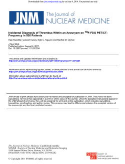
139
Olgu Sunumu
A Case of Increased F-18 FDG Uptake in Uterine
Cavity due to Diagnostic Curettage
UTERUSTA DIAGNOSTIK KÜRETAJA SEKONDER ARTMIŞ F-18 FDG TUTULUMU OLGUSU
Erdem SÜRÜCÜ, Sadet AYHAN, Gamze ÇAPA KAYA, Hatice DURAK
Dokuz Eylül University Faculty of Medicine, Department of Nuclear Medicine
Erdem SÜRÜCÜ
Dokuz Eylül University
School of Medicine
Department of Nuclear Medicine
35340 Inciralti, IZMIR
e-mail: [email protected]
phone: +90 506 9763359
fax: 90 232 4124269
SUMMARY
Endometrial cancer is one of the most common malignant tumors of the women, and if
it can be detected in the earlier stages, the curability and the prognosis of the
endometrial cancers can be better. A 52 year-old female patient, as part of ongoing
research project in our clinic on endometrial carcinoma, was referred to our clinic with
a suspicion of endometrial carcinoma with increased serum CA-125 measurement.
Increased F-18 FDG uptake in uterine cavity that was secondary to the diagnostic
curettage was demonstrated on F 18 FDG PET images. This uptake might be secondary
to benign inflammatory changes or hemorrhage arising from diagnostic curettage that
patient underwent ten days ago. According to our knowledge, this is the first case
demonstrating increased F-18 FDG uptake secondary to diagnostic curettage.
Key words: F–18 FDG, PET, uterine curettage, inflammation
ÖZET
Endometrium kanseri kadınlarda en çok görülen malign tümörlerden birisidir. Erken
teşhis edildiğinde tam tedavi şansı yüksektir ve prognozu çok iyi seyirlidir. Bölümümüzde endometrium kanseri ile ilgili olarak devam eden bir proje kapsamında, serum
CA-125 yüksekliği ile endometrium kanseri şüphesi olan 53 yaşında kadın hasta bölümümüze refere edildi. PET görüntülerinde uterusda artmış F-18 FDG tutulumu
izlenmiştir. Bu bulgunun hastanın öyküsünde 10 gün önce geçirilmiş diagnostik küretaja bağlı benign inflamatuar değişiklikler veya hemoraji ile ilgili olduğu düşünülmüştür. Bu olgu sunumunda diagnostik küretaja sekonder olduğu düşünülen F-18
FDG tutulumu muhtemelen literatürde ilk kez gösterilmektedir.
Anahtar sözcükler: F-18 FDG, PET, uterus küretajı, enflamasyon
Endometrial cancer is one of the most common malig‐
kinase for the glycolytic metabolism pathway. After the nant tumors of the women and if it can be detected in the phosphorylation, glucose keeps going along the glycolytic earlier stages, the curability and the prognosis of the en‐
pathway. However, Fluorodeoxyglucose (FDG) cannot dometrial cancer can be better (1). Tumor cells generally use glucose for aerobic glycoly‐
sis for being alive. Normally, after the transportation of glucose into the cells, they are phosphorylated by hexo‐
© 2010 DEÜ TIP FAKÜLTESİ DERGİSİ
continue the glycolytic pathway and it is trapped intracellularly. Fluorine‐18 fluorodeoxyglucose positron‐
emission tomography (F‐18 FDG PET) is an imaging tech‐
nique that shows glucose metabolism of tumor cells with CİLT 24, SAYI 3, (EYLÜL) 2010, S: 139- 142
140
A case of increased F-18 uptake in uterine cavity due to diagnostic curettage
the help of trapped F–18 FDG in the tumor cells. F‐18 FDG avidity was shown in gynecologic malignan‐
position. We obtained transmission scans with low dose CT using 50 mA and 150 kvp. cies including endometrial carcinoma. Recently, F‐18 FDG In figure 1, mildly increased F‐18 FDG uptake PET‐CT was started to use in staging, evaluation of ther‐
(SUVmax: 3.5, SUVmean: 3.2) in uterine cavity was apy response and the recurrence of the disease. PET was demonstrated on transaxial, sagittal and coronal PET/CT found as a sensitive tool for the evaluation of suspected fusion images (A), and transaxial, sagittal and coronal PET recurrence in patients with ovarian cancer, more accurate images (B) (arrows). There were no any other pathologic with increasing CA‐125 levels during follow‐up (2–8). PET findings. The patient had a history of diagnostic DESCRIPTION OF THE CASE curettage ten days before PET/CT imaging and the pathology result of diagnostic curettage was reported as a A 52 year‐old female patient was referred to our clinic benign cytology. Increased F‐18 FDG uptake in the uterus due to vaginal bleeding with a suspicion of endometrial resulting from intrauterine device and physiologic accu‐
carcinoma and increased serum CA‐125 levels. Sixty min‐
mulation of F‐18 FDG during menstruation have been utes after injection of 351.5 MBq (9.5 mCi) F‐18 FDG, im‐
reported previously. So, this increased F‐18 FDG uptake in ages were obtained using PET/CT (Gemini‐TOF‐Philips). uterine cavity might be secondary to benign inflammatory The emission scan was obtained for 1.5 minutes per bed changes or hemorrhage (9,10). Figure. Transaxial, sagittal and coronal PET/CT fusion images (A), and transaxial, sagittal and coronal PET images (B) showing mildly increased F‐18 FDG uptake in uterine cavity (SUVmax: 3.5, SUVmean: 3.2)(arrows). No any other pathological PET findings were present in the whole body scan. A case of increased F-18 uptake in uterine cavity due to diagnostic curettage
DISCUSSION 2.
fluorodeoxylucose in patients with cervical cancer under-
dictor for the extrauterine spread of clinically localized for the endometrial cancer suspicion and referral to our going radiotherapy. Gynecol Oncol 2002;84:289-295.
3.
evaluating paraaortic lymph node metastases in locally
the pathologic result of diagnostic curettage could not advanced cervical cancer before surgical staging: a sur-
reveal the reason of increased serum CA‐125 level. Be‐
FDG‐PET in the diagnosis of endometrial cancer is not gico-pathologic study. J Clin Oncol 1999;17:41-44.
4.
Reinhardt MJ, Ehritt-Braun C, Vogelgesang D, et al.
Metastatic lymph nodes in patients with cervical cancer:
clear (13‐16). Previous studies showed a mean SUV of 18.8 detection with MR imaging and FDG PET. Radiology
± 9 in five patients with endometrial cancer and a range of 3.8‐16.8 in nine patients with endometrial cancer (17,18). Rose PG, Adler LB, Rodriguez M, Faulhaber PF, AbdulKarim FW, Miraldi F. Positron emission tomography for
clinic was due to increased serum CA‐125 level. However, cause there is insufficient data in the literature, the role of Nakamoto Y, Eisbruch A, Achtes ED, et al. Prognostic
value of positron emission tomography using F-18-
Preoperative serum CA‐125 may be a potential pre‐
endometrial cancer (11‐12). In our case, one of the reasons 141
2001;218: 776-782.
5.
Grisby PW, Siegel BA, Dehdashti F. Lymph node stag-
According to our knowledge, this is the first case in the ing by positron emission tomography in patients with
literature demonstrating increased F‐18 FDG uptake sec‐
carcinoma of the cervix. J Clin Oncol 2001; 19: 27453749.
ondary to diagnostic curettage. F‐18 FDG uptake in vari‐
ous body systems has been extensively described in the 6.
Miller TR, Grisby PW. Measurement of tumor volume
literature related to surgery or inflammation (19‐21). After by PET to evaluate prognosis in patients with advanced
the diagnostic curettage, uterine tissue has developed in‐
cervical cancer treated by radiation therapy. Int J Oncol
Biol Phys 2002; 53: 353–359.
flammatory response. As a result of this inflammation, blood flow increase and subsequently plasma and in‐
7.
Zimny M, Siggelkow W, Schröder W, et al. 2-[Fluorine-
flammatory cells (neutrophils, macrophages and lympho‐
18]-fluoro-2-deoxy-Dglucose positron emission tomog-
cytes) move into the injured tissues. The increased F‐18 raphy in the diagnosis of recurrent ovarian cancer.
Gynecol Oncol 2001;83:310–315.
FDG uptake, which is due to increament of blood flow and glucose metabolism of the cells, is seen in these areas. 8.
Liu Y. Benign ovarian and endometrial uptake on FDG
Because diagnostic curettage has been frequently done PET-CT: patterns and pitfalls. Ann Nucl Med 2009; 23:
among other surgical interventions in routine clinical 107-112.
practice, uptake in uterine cavity due to diagnostic curet‐
9.
Julian A, Payoux P, Rimailho J, Paynot N, Esquerre J.
tage may have a clinical importance especially from clini‐
Uterine Uptake of F–18 FDG on Positron Emission To-
cian’s points of view in patients having a suspicious en‐
mography Induced by an Intrauterine Device: Unusual
dometrial carcinoma. Pitfall. Clin Nucl Med 2007; 32:128–129.
In conclusion, this finding may be reported as a kind of pitfall that it should be recognized during the evalua‐
tion of F‐18 FDG PET/CT images, especially in a patient with a suspicious pelvic pathology. Therefore, the patients should be asked for any symptoms that can cause false 10. Chander S, Meltzer CC, and McCook BM. Physiologic
uterine uptake of FDG during menstruation demonstrated with serial combined positron emission tomography and computed tomography. Clin Nucl Med 2002;
27:22–24.
11. Duk JM, Aalders JG, Fleuren GJ, de Bruijn HW. CA 125
positive F‐18 FDG uptake like in this case. a useful marker in endometrial carcinoma. Am J Obstet
REFERENCES Gynecol 1986; 155:1097–1102.
1.
Irvin WP, Rice LW, Berkowitz RS. Advances in the
management of endometrial adenocarcinoma. A review.
J Reprod Med 2002; 47: 173-189.
12. Dotters DJ. Preoperative CA 125 in endometrial cancer:
is it useful? Am J Obstet Gynecol 2000; 182:1328–1334.
13. Torizuka T, Kanno T, Futatsubashi M, et al. Imaging of
142
A case of increased F-18 uptake in uterine cavity due to diagnostic curettage
gynecologic tumors: comparison of 11C-choline PET with
(18) F-FDG PET in the posttherapy surveillance of
18F-FDG PET. J Nucl Med 2003; 44:1051–1056.
endometrial carcinoma. Eur J Nucl Med Mol Imaging
14. Belhocine T, de Barsy C, Hustinx R, et al. Usefulness of
2002; 29:1132–1139.
(18)F-FDG PET in the posttherapy surveillance of
18. Saga T, Higashi T, Ishimori T, et al. Clinical value of
endometrial carcinoma. Eur J Nucl Med Mol Imaging
FDG-PET in the follow up of postoperative patients with
2002; 29: 1132–1139.
endometrial cancer. Ann Nucl Med 2003; 17:197-203.
15. Horowitz NS, Dehdashti F, Herzog TJ, et al. Prospective
19. Imperiale A, Federici L, Lefebvre N, et al. F–18 FDG
evaluation of FDG-PET for detecting pelvic and para-
PET/CT as a valuable imaging tool for assessing
aortic lymph node metastasis in uterine corpus cancer.
treatment efficacy in inflammatory and infectious
Gynecol Oncol 2004; 101:164–171.
diseases. Clin Nucl Med 2010; 35: 86-90.
16. Chao A, Chang YC, Hsueh S, et al. F–18 fluorodeoxyglucose
positron
emission
tomography
in
the
management of endometrial cancer. Eur J Nucl Med
2006; 33: 36–44.
17. Belhocine T, de Barsy C, Hustinx R, et al. Usefulness of
20. Kao PF, Chou YH, Lai CW. Diffuse FDG uptake in
acute prostatitis. Clin Nucl Med 2008; 33:308-310.
21. Koff SG, Sterbis JR, Davison JM, Montilla-Soler JL. A
unique presentation of appendicitis: F-18 FDG PET/CT.
Clin Nucl Med 2006; 31:704-706.


© Copyright 2026