
Barrett’s Esophagus What is Barrett’s esophagus? National Digestive Diseases Information Clearinghouse
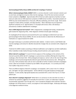
Barrett’s Esophagus National Digestive Diseases Information Clearinghouse What is Barrett’s esophagus? U.S. Department of Health and Human Services NATIONAL INSTITUTES OF HEALTH Barrett’s esophagus is a condition in which the tissue lining the esophagus—the mus cular tube that connects the mouth to the stomach—is replaced by tissue that is simi lar to the lining of the intestine. This pro cess is called intestinal metaplasia. No signs or symptoms are associated with Barrett’s esophagus, but it is commonly found in people with gastroesophageal reflux disease (GERD). A small number of people with Barrett’s esophagus develop a rare but often deadly type of cancer of the esophagus. Barrett’s esophagus affects about 1 percent1 of adults in the United States. The aver age age at diagnosis is 50, but determining when the problem started is usually difficult. Men develop Barrett’s esophagus twice as often as women, and Caucasian men are affected more frequently than men of other races. Barrett’s esophagus is uncommon in children. The Esophagus The esophagus carries food and liquids from the mouth to the stomach. The stom ach slowly pumps the food and liquids into the intestine, which then absorbs needed nutrients. This process is automatic and people are usually not aware of it. People 1 Cameron AJ. Epidemiology of Barrett’s esophagus and adenocarcinoma. Diseases of the Esophagus. 2002;15:106–108. Digestive tract. sometimes feel their esophagus when they swallow something too large, try to eat too quickly, or drink very hot or cold liquids. The muscular layers of the esophagus are normally pinched together at both the upper and lower ends by muscles called sphincters. When a person swallows, the sphincters relax to allow food or drink to pass from the mouth into the stomach. The muscles then close rapidly to prevent the food or drink from leaking out of the stom ach back into the esophagus and mouth. What is gastroesophageal reflux disease (GERD)? GERD is a more serious form of gastro esophageal reflux (GER). GER occurs when the lower esophageal sphincter opens spontaneously for varying periods of time or does not close properly and stomach contents rise into the esophagus. GER is also called acid reflux or acid regurgitation because digestive juices called acids rise with the food or fluid. When GER occurs, food or fluid can be tasted in the back of the mouth. When refluxed stomach acid touches the lining of the esophagus it may cause a burning sensa tion in the chest or throat called heartburn or acid indigestion. Occasional GER is common and does not necessarily mean one has GERD. Persistent reflux that occurs more than twice a week is considered GERD and can eventually lead to more serious health prob lems. Overall, 10 to 20 percent2 of Ameri cans experience GERD symptoms every day, making it one of the most common medical conditions. People of all ages can have GERD. GERD symptoms are often relieved by over-the-counter, acid-reducing agents called antacids. Common antacids include • Alka-Seltzer • Maalox • Mylanta • Pepto-Bismol • Riopan • Rolaids 2 El-Serag HB. Time trends of gastroesophageal reflux disease: a systematic review. Clinical Gastroenterology and Hepatology. 2007;5(1):17–26. 2 Barrett’s Esophagus Other drugs used to relieve GERD symptoms are anti-secretory drugs such as H2 blockers and proton pump inhibitors. Common H2 blockers are • cimetidine (Tagamet HB) • famotidine (Pepcid AC) • nizatidine (Axid AR) • ranitidine (Zantac 75) Common proton pump inhibitors are • esomeprazole (Nexium) • lansoprazole (Prevacid) • omeprazole (Prilosec, Zegerid) • pantoprazole (Protonix) • rabeprazole (Aciphex) People who have GERD symptoms should consult with a physician. If GERD is left untreated over a long period of time, it can lead to complications such as a bleed ing ulcer. Scars from tissue damage can lead to strictures—narrowed areas of the esophagus—that make swallowing difficult. GERD may also cause hoarseness, chronic cough, and conditions such as asthma. GERD and Barrett’s Esophagus The exact causes of Barrett’s esophagus are not known, but GERD is a risk factor for the condition. Although people who do not have GERD can have Barrett’s esophagus, the condition is found about three to five times more often in people who also have GERD. Since Barrett’s esophagus is more com monly seen in people with GERD, most physicians recommend treating GERD symptoms with acid-reducing drugs. Improvement in GERD symptoms may lower the risk of developing Barrett’s esoph agus. A surgical procedure may be recom mended if medications are not effective in treating GERD. How is Barrett’s esophagus diagnosed? Because Barrett’s esophagus does not cause any symptoms, many physicians recom mend that adults older than 40 who have had GERD for a number of years undergo an endoscopy and biopsies to check for the condition. Barrett’s esophagus can only be diagnosed using an upper gastrointestinal (GI) endos copy to obtain biopsies of the esophagus. In an upper GI endoscopy, after the patient is sedated, the doctor inserts a flexible tube called an endoscope, which has a light and a miniature camera, into the esophagus. If the tissue appears suspicious, the doc tor removes several small pieces using a pincher-like device that is passed through the endoscope. A pathologist examines the tissue with a microscope to determine the diagnosis. What is the risk of esophageal cancer with Barrett’s esophagus? People with Barrett’s esophagus have a low risk of developing a kind of cancer called esophageal adenocarcinoma. Less than 1 percent3 of people with Barrett’s esopha gus develop esophageal adenocarcinoma each year. Barrett’s esophagus may be pres ent for several years before cancer develops. Esophageal adenocarcinoma is frequently not detected until its later stages when treatments are not always effective. Surveillance for Dysplasia and Cancer Periodic endoscopic examinations with biopsies to look for early warning signs of cancer are generally recommended for people who have Barrett’s esophagus. This approach is called surveillance. Normal esophagus. Barrett’s esophagus. 3 Barrett’s Esophagus Typically, before esophageal cancer devel ops, precancerous cells appear in the Barrett’s tissue. This condition is called dysplasia and can be seen only through biopsies. Multiple biopsies must be taken because dysplasia can be missed in a single biopsy. Detecting and treating dysplasia may prevent cancer from developing. 3 Shaheen N, Ransohoff DF. Gastroesophageal reflux, Barrett’s esophagus, and esophageal cancer. Journal of the American Medical Association. 2002;287(15):1982–1986. How is Barrett’s esophagus with dysplasia or cancer treated? Endoscopic or surgical treatments can be used to treat Barrett’s esophagus with severe dysplasia or cancer. Your doctor will present the available options and help determine the best course of treatment for you. Endoscopic Treatments Several endoscopic therapies are available to treat severe dysplasia and cancer. Dur ing these therapies, the Barrett’s lining is destroyed or the portion of the lining that has dysplasia or cancer is cut out. The goal of the treatment is to encourage normal esophageal tissue to replace the destroyed Barrett’s lining. Endoscopic therapies are performed at specialty centers by physicians with expertise in these procedures. • Photodynamic Therapy (PDT). PDT uses a light-sensitizing agent called Photofrin and a laser to kill precancer ous and cancerous cells. Photofrin is injected into a vein and the patient returns 48 hours later. The laser light is then passed through the endoscope and activates the Photofrin to destroy Barrett’s tissue in the esophagus. Complications of PDT include chest pain, nausea, sun sensitivity for several weeks, and esophageal strictures. 4 Barrett’s Esophagus • Endoscopic Mucosal Resection (EMR). EMR involves lifting the Barrett’s lining and injecting a solution under it or applying suction to it and then cutting it off. The lining is then removed through the endoscope. If EMR is used to treat cancer, an endo scopic ultrasound is done first to make sure the cancer involves only the top layer of esophageal cells. The ultra sound uses sound waves that bounce off the walls of the esophagus to create a picture on a monitor. Complications of EMR can include bleeding or tear ing of the esophagus. EMR is some times used in combination with PDT. Surgery Surgical removal of most of the esophagus is recommended if a person with Barrett’s esophagus is found to have severe dyspla sia or cancer and can tolerate a surgical procedure. Many people with Barrett’s esophagus are older and have other medical problems that make surgery unwise; in these people, the less-invasive endoscopic treat ments would be considered. Surgery soon after diagnosis of severe dysplasia or cancer may provide a person with the best chance for a cure. The type of surgery varies, but it usually involves removing most of the esophagus, pulling a portion of the stomach up into the chest, and attaching it to what remains of the esophagus. Points to Remember • In Barrett’s esophagus, the tissue lining the esophagus is replaced by tissue that is similar to the lining of the intestine. • Barrett’s esophagus is associated with gastroesophageal reflux disease (GERD). • Improvement in GERD symptoms with acid-reducing drugs may decrease the risk of developing Barrett’s esophagus. • Barrett’s esophagus is diagnosed through an upper gastrointestinal endoscopy and biopsies. • People who have Barrett’s esopha gus should have periodic surveil lance endoscopies and biopsies. • Endoscopic treatments are used to destroy Barrett’s tissue, which will hopefully be replaced with normal esophageal tissue. • Removal of most of the esophagus is recommended if a person with Barrett’s esophagus is found to have severe dysplasia or cancer and can tolerate a surgical procedure. 5 Barrett’s Esophagus Hope through Research The National Institute for Diabetes and Digestive and Kidney Diseases and the National Cancer Institute sponsor research programs to investigate Barrett’s esopha gus and esophageal adenocarcinoma. Further research into Barrett’s esophagus is needed, including • establishing additional tests to identify people with Barrett’s esophagus • identifying the cause(s) of Barrett’s esophagus • studying the long-term effectiveness of treatments such as PDT and EMR • developing additional nonsurgical treatments for people who have Barrett’s esophagus and dysplasia or cancer Participants in clinical trials can play a more active role in their own health care, gain access to new research treatments before they are widely available, and help others by contributing to medical research. For information about current studies, visit www.ClinicalTrials.gov. The U.S. Government does not endorse or favor any specific commercial product or company. Trade, proprietary, or company names appearing in this document are used only because they are considered necessary in the context of the information provided. If a product is not mentioned, the omission does not mean or imply that the product is unsatisfactory. For More Information American Gastroenterological Association 4930DelRayAvenue Bethesda,MD20814 Phone:301–654–2055 Fax:301–654–5920 Email:[email protected] Internet:www.gastro.org International Foundation for Functional Gastrointestinal Disorders P.O.Box170864 Milwaukee,WI53217 Phone:1–888–964–2001or414–964–1799 Fax:414–964–7176 Email:[email protected] Internet:www.iffgd.org National Cancer Institute NationalInstitutesofHealth 6116ExecutiveBoulevard,Room3036A Bethesda,MD20892–8322 Phone:1–800–4–CANCER(422–6237) Fax:301–496–0846 Email:[email protected] Internet:www.cancer.gov Youmayalsofindadditionalinformationaboutthis topicbyvisitingMedlinePlusatwww.medlineplus.gov. Thispublicationmaycontaininformationaboutmedications.Whenprepared,thispublicationincluded themostcurrentinformationavailable.Forupdates orforquestionsaboutanymedications,contact theU.S.FoodandDrugAdministrationtoll-freeat 1–888–INFO–FDA(463–6332)orvisitwww.fda.gov. Consultyourdoctorformoreinformation. National Digestive Diseases Information Clearinghouse 2InformationWay Bethesda,MD20892–3570 Phone:1–800–891–5389 TTY:1–866–569–1162 Fax:703–738–4929 Email:[email protected] Internet:www.digestive.niddk.nih.gov TheNationalDigestiveDiseasesInformationClearinghouse(NDDIC)isaservice oftheNationalInstituteofDiabetesand DigestiveandKidneyDiseases(NIDDK). TheNIDDKispartoftheNationalInstitutesofHealthoftheU.S.Departmentof HealthandHumanServices.Establishedin 1980,theClearinghouseprovidesinformationaboutdigestivediseasestopeoplewith digestivedisordersandtotheirfamilies, healthcareprofessionals,andthepublic. TheNDDICanswersinquiries,develops anddistributespublications,andworks closelywithprofessionalandpatientorganizationsandGovernmentagenciestocoordinateresourcesaboutdigestivediseases. PublicationsproducedbytheClearinghouse arecarefullyreviewedbybothNIDDK scientistsandoutsideexperts.Thisfact sheetwasoriginallyreviewedbyG.Richard Locke,M.D.,MayoClinic,andJoelRichter, M.D.,ClevelandClinicFoundation. Thispublicationisnotcopyrighted.TheClearinghouseencouragesusersofthisfactsheettoduplicate anddistributeasmanycopiesasdesired. Thisfactsheetisalsoavailableat www.digestive.niddk.nih.gov. U.S. DEPARTMENT OF HEALTH AND HUMAN SERVICES National Institutes of Health NIHPublicationNo.08–4546 July2008


© Copyright 2026