
Allergic contact dermatitis
Review article
Pierre SAINT-MEZARD1
Aurore ROSIERES1
Maya KRASTEVA1
Frédéric BERARD1,3
Bertrand DUBOIS2
Dominique KAISERLIAN3
Jean-François NICOLAS1,3
1
INSERM U 503, IFR 128 Bioscience
Lyon-Gerland, 21, avenue Tony Garnier
69007 Lyon
2
INSERM U 404, IFR 128 Bioscience
Lyon-Gerland, 21, avenue Tony Garnier
69007 Lyon
3
Clinical Immunology and Allergy Unit,
CH Lyon-Sud, 69495 Pierre-Benite Cedex,
France.
Reprints: J.F. Nicolas,
Fax: (+33) 478 861 528.
E-mail:
[email protected]
Article accepted on 15/4/2004
Eur J Dermatol 2004; 14: 284-95
Allergic contact dermatitis
Contact dermatitis is an inflammatory skin condition induced by exposure to an environmental agent. Eczema and dermatitis are used synonymously to denote a polymorphous pattern of skin inflammation characterized at least in its acute phase by erythema, vesiculation and
pruritus. Substances responsible for contact dermatitis after single or
multiple exposures are non protein chemicals, i.e. haptens, that induce
skin inflammation through activation of innate skin immunity (irritant
contact dermatitis) or both innate and acquired specific immunity (allergic contact dermatitis). The present review will focus on allergic contact
dermatitis, a delayed-type hypersensitivity reaction, which is mediated
by hapten-specific T cells. Recent advances in the pathophysiology of
ACD have shown that the occurrence of ACD, as well as its magnitude
and duration, is controlled by the opposite functions of CD8 effector T
cells and CD4 regulatory T cells. From these studies ACD can be
considered as a breakdown of cutaneous immune tolerance to haptens.
Key words: haptens, CD8 T cells, CTL, CD4 regulatory T cells,
tolerance, contact sensitivity, contact dermatitis
C
ontact dermatitis is one of the most common skin
diseases, with a great socio-economic impact [1].
As the outermost barrier of the human body, the
skin is the first to encounter chemical and physical factors
from the environment. According to the pathophysiological
mechanisms involved, two main types of contact dermatitis
may be distinguished. Irritant contact dermatitis is due to
the pro-inflammatory and toxic effects of xenobiotics able
to activate the skin innate immunity. Allergic contact dermatitis (ACD) requires the activation of antigen specific
acquired immunity leading to the development of effector T
cells which mediate the skin inflammation.
ACD is a T-cell-mediated inflammatory reaction occurring
at the site of challenge with a contact allergen in sensitized
individuals. It is characterized by redness, papules and
vesicles, followed by scaling and dry skin [2]. Knowledge
of the pathophysiology of ACD is derived chiefly from
animal models in which the skin inflammation induced by
hapten painting of the skin is referred to as contact sensitivity (CS) or contact hypersensitivity (CHS). ACD and CS
(CHS) are thus considered as synonymous and define a
hapten-specific T cell-mediated skin inflammation [3]. The
skin and the draining lymph nodes (LN) play a central role
in the induction and triggering of a CS reaction. Three
elements are necessary for the development of a CS reaction: antigen presenting dendritic cells (DC), haptenspecific T-cells, and the hapten itself.
Haptens - contact allergens
The origin and nature of the compounds able to induce a CS
reaction are very diverse, but they share some common
features: contact allergens are low molecular weight chemicals named haptens, that are not immunogenic by themselves and need to bind to epidermal proteins. These then
284
act as carrier proteins to form the hapten-carrier complex
that finally acts as the antigen. Most haptens bear lipophilic
residues, which enable them to cross through the corneal
barrier, and electrophilic residues, which account for covalent bonds to the nucleophilic residues of cutaneous proteins [4-6].
Haptens often derive from unstable chemicals, named prohaptens, which require an additional metabolization step in
vivo in the epidermis to be converted into an electrophilic
hapten endowed with antigenic properties. This is the case
of urushiol (poison ivy) [7] and of photosensitizers, which
must be activated by UV-light in order to bind to epidermal
proteins. Metal salts do not bind covalently to cutaneous
proteins but form complexes with these proteins through
weak interactions. Some metal salts also undergo chemical
conversions in the skin, such as hexavalent chromium salts,
which are turned into trivalent chromium, a highly reactive
form binding to cutaneous proteins [8]. Evidence that the
conversion of the parent compound into a reactive metabolite is necessary for the development of CS was recently
demonstrated for the polyaromatic hydrocarbon (PAH)
dimethylbenz(a)anthracene (DMBA). CS to DMBA only
occurred in strains of mice that could metabolize the compound and inhibitors of PAH metabolism reduced the magnitude of the reaction. Furthermore, among the PAHs, only
those that could induce aryl hydrocarbon hydroxylase, the
rate-limiting enzyme in the PAH metabolic pathway, were
immunogenic [9]. The implications of these experiments
are that at least for some contact allergens, the metabolic
status of the host is a key determinant of individual susceptibility to the development of allergic contact dermatitis.
Recent studies from several laboratories have shown that T
cells recognize haptens as structural entities bound covalently to, or by complexation to, peptides anchored in the
grooves of major histocompatibility (MHC) class I and
class II molecules. Thus the contact allergen is a chemical
EJD, vol. 14, n° 5, September-October 2004
but the antigen able to activate hapten-specific T cells is a
haptenated peptide [10].
Pathophysiology of contact sensitivity
Knowledge of the mechanisms by which a xenobiotic can
induce CS responses comes from the study of strong haptens, also known as “experimental haptens”, since they do
not exist in the usual environment of human beings. The
pathophysiology of CS consists of two distinct phases (Fig.
1) which are summarized below:
Phase 1 - Sensitization phase (also referred to as afferent
phase or induction phase of CS)
This occurs at the first contact of skin with the hapten and
leads to the generation of hapten-specific T-cells in the LN
and their migration back to the skin. The ability of a hapten
to induce sensitization relies on two distinct properties.
Through their pro-inflammatory properties, haptens activate the skin innate immunity and deliver signals able to
induce the migration and maturation of cutaneous dendritic
cells (DC). Through their binding to amino-acid residues
they modify self proteins and allow the expression in the
skin of new antigenic determinants.
Haptens or haptenated proteins are loaded by cutaneous
dendritic cells (DC) and are expressed as haptenated peptides in the groove of MHC class I and class II molecules at
Sensitization site
the cell surface. Hapten-bearing DC migrate from the skin
to the regional LN where specific CD8+ and CD4+ T
lymphocytes are primed in the para-cortical area. T cells
proliferate and emigrate out of the LNs to the blood where
they recirculate between the lymphoid organs and the skin.
The sensitization step lasts 10 to 15 days in man, and 5 to
7 days in the mouse. This first step has no clinical consequence.
The theory of the first contact of a chemical being
immunogenic for the host is true for the strong haptens but
cannot be accepted for the vast majority of haptens responsible for ACD. Indeed, ACD to moderate or weak haptens
almost never occurs after the first contact but may take
years of permanent skin exposure to develop.
Phase 2 - Elicitation phase (also known as efferent
or challenge phase of CS)
Challenge of sensitized individuals with the same hapten
leads in 24/72 hours to the apparition of ACD/CS. Haptens
diffuse in the skin and are uptaken by skin cells which
express MHC I and II/haptenated peptide complexes. Specific T lymphocytes are activated in the dermis and the
epidermis, and trigger the inflammatory process responsible for the cutaneous lesions. Recent studies have demonstrated that CD8+ cytotoxic T lymphocytes are the main
effector cells of CHS to strong haptens and that they are
recruited early after challenge before the massive infiltra-
Hapten
Elicitation site
Hapten
1
Stratum corneum
Langerhans
cells
6
Epidermis
Dermis
7
5
Dermal denritic cells
2
CD4+ Regulatory
T cells
4
Efferent lymph
Afferent lymph
3
Draining Lymph Node
CD8+ Effector T cells
Figure 1. Pathophysiology of CHS Sensitization step: Haptens penetrate the stratum corneum. Hapten loading by skin dendritic cells (step
1) parallels activation and migration of DC through the afferent lymphatic vessels to the draining lymph nodes (step 2). Migrating DC are
located in the para-cortical area of the draining LN where they can present haptenated peptides on MHC class I and II molecules to CD8+
and CD4+ T cells, respectively (step 3). Specific T cells precursors expand clonally in the draining LN and diffuse to the bloodstream through
the efferent lymphatic vessels and the thoracic duct (step 4). During this process they acquire skin-specific homing antigens (CLA and CCR4)
and become memory T cells. Primed T cells preferentially diffuse in the skin after transendothelial migration. At the end of the sensitization
step everything is ready for the development of a CS reaction upon challenge with the relevant hapten. Elicitation phase: When the hapten is
painted for a second (and subsequent) time, it diffuses through the epidermis and could be loaded by LC or other skin cells expressing MHC
molecules, such as keratinocytes and dermal dendritic cells, which are then able to activate traffıcking specific T cells (step 5). CD8+ cytotoxic
T cell activation initiates the inflammatory process through keratinocyte apoptosis and cytokine/chemokine production (step 6). This is
responsible for the recruitment of leukocytes (including regulatory T cells) from the blood to the skin leading to the development of skin lesions
(step 7).
EJD, vol. 14, n° 5, September-October 2004
285
tion of leucocytes which contain the down-regulatory cells
of CHS, found in the CD4+ T cell subset.
The efferent phase of CS takes 72 hours in man, and 24 to
48 hours in the mouse. The inflammatory reaction persists
only for a few days and rapidly decreases following downregulatory mechanisms.
Primary allergic contact sensitivity
Although the development of CHS has been postulated to
require two spatially and temporally dissociated phases,
clinical evidence has demonstrated that ACD could develop
after a single skin contact with a strong hapten in previously
unsensitized patients. This phenomenon has been referred
to as “primary ACD” [11]. We have recently demonstrated,
in a murine model, that the pathophysiology of this primary
(one step) ACD is identical to the classical (two step) ACD
reaction [12]. The afferent and efferent phases of primary
ACD can be induced after a single skin contact with haptens
due to the persistence of the hapten in the skin for long
period of time, allowing the skin recruitment and the activation of T cells which have been primed in the lymphoid
organs.
The central role of cutaneous dendritic
cells
There are different subtypes of DC in the skin. Although
they are all able to uptake haptens and to present haptenated
peptides to T cells, two subsets of cutaneous DC seem
crucial in the development of CS.
The basic role of epidermal Langerhans cells (LC) has been
shown by two sets of experiments. On the one hand, animals painted with a hapten on cutaneous sites naturally or
artificially depleted in LC are unable to mount a CS response [13, 14]. On the other hand, sensitization of naive
mice can be achieved by injection of in vitro haptenized
total epidermal cells, purified LC or FSDC dendritic cell
lines, whereas injection of total epidermal cells depleted in
LC before haptenization is inefficient in inducing sensitization [15-17].
Dermal DC could also participate in the induction phase of
CS, even though their precise contribution and phenotype is
not well known [18]. Recent studies by Geissmann et al
have brought some insights into the turnover of macrophages and DC in the dermis in normal and inflammatory
skin. Two different subsets of monocytes can be defined
according to the expression of the chemokine receptor
CCR2 [19, 20]. CCR2- monocytes appear to be involved in
the physiological turnover of resident macrophages and
DC, whereas CCR2+ monocytes are recruited in inflammatory sites where they could differentiate into mature DC
able to present exogenous antigens to T cells. Since hapten
application is responsible for activation of skin innate immunity, it is possible that the CCR2+ inflammatory monocytes which are recruited in the skin could uptake the
hapten and participate in the afferent phase of CHS [21].
Cutaneous DC load haptens in the skin and migrate to
the draining LN
Activation of naive specific T cell precursors occurs in the
regional draining LN upon presentation of haptenated peptides by cutaneous migrating DC. Initial observations
286
showed that induction of a CS reaction requires an intact
draining lymphatic system [22], and that after skin painting
with the hapten fluorescein isothiocyanate (FITC), dendritic cells bearing the hapten, some of which containing
Birbeck granules, accumulate in the draining lymph nodes
[23, 24].
Cutaneous DC continuously migrate out of the skin at a low
rate which dramatically increases after hapten exposure
[25]. This phenomenon is the consequence of numerous
factors including the secretion of inflammatory cytokines
and chemokines induced by the pro-inflammatory properties of the hapten itself. Fifteen minutes after hapten painting, LC start to synthesize IL-1b mRNA and to release the
protein. Then, keratinocytes are activated and release
TFN-a and GM-CSF [26]. In the epidermis, LC and keratinocytes are firmly associated by E-cadherine/E-cadherine
junctions [27]. Binding of TNF-a and IL-1b on their cognate receptors (TNF-a RII and IL-1 RI and RII) expressed
on LC, is followed by a decreased expression of
E-cadherine on the LC membrane, allowing their disentanglement from surrounding keratinocytes [28–31]. Furthermore, IL-1b and TFN-a inhibit the expression of
chemokine receptors such as CCR1, CCR2, CCR5 and
CCR6 on the LC membrane, inducing a loss of sensitivity
to their cognate ligands, in particular to MIP-3a (CCL20), a
chemokine produced by keratinocytes [32]. In parallel,
TNF-a and IL-1b induce the expression of adhesive molecules such as CD54, a6 integrin and different isoforms of
CD44, which permit some interactions between LC and the
extracellular matrix, allowing the migration of cutaneous
DC.
In order to reach the lymphatic vessels, LC have to cross the
dermo-epidermal junction and the dermis. To this end, LC
secrete different types of enzymes, such as metalloproteinase (MMP) 3 and 9 [33], which could cleave macromolecules of the dermo-epidermal junction and of the extracellular matrix. They are also involved in the cleavage of
E-cadherin and of pro-TNF-a in its biologically active form
[34]. Once in the dermis, DC acquire sensitivity to the
chemokines MIP-3b (CCL19) and SLC (CCL21) through
the up-regulation of the chemokine receptor CCR7 [32,
35]. CCL21 is expressed on endothelial cells from lymphatic vessels and by stromal cells from the T cell area in
LN [36]. The CCL21/CCR7 interaction is crucial during
the sensitization phase of CS. Indeed, Endeman and colleagues have described a strong inhibition of CS following
injections of SLC-blocking antibodies before and during
the sensitization phase [37]. Moreover, TNF-a is able to
induce a strong up-regulation of CCL21 on the endothelial
cells of skin lymphatic vessels. The over-expression of
CCL21 is of critical importance in the migratory properties
of peripheral DC and is able to increase, by a factor of 10,
the number of cutaneous DC able to migrate from the skin
to the LN and subsequently the magnitude of antigenspecific T cell activation [38].
Maturation of cutaneous DC
The term “maturation” takes into account a group of morphological, phenotypic and functional modifications which
transform skin DC into professional antigen-presenting
cells able to prime naïve specific T cell precursors. Although the different steps of DC maturation and differentiation are well described in vitro [18], correlation with the
in vivo modifications is not yet totally achieved. Migration
EJD, vol. 14, n° 5, September-October 2004
and maturation of cutaneous DC are intimately linked.
Indeed, factors known to induce migration of DC are also
able to engage DC in a maturation program. Presence of
TNF-a and IL-1b in the skin leads to the up-regulation of
MHC class II molecules on DC membranes. In the first
3 hours following application of the hapten, expression of
MHC II molecules at the DC surface first decreases [39],
whereas their intracellular level increases, which probably
reflects an endocytosis process triggered by hapten binding
[40]. From the sixth hour after hapten painting, synthesis of
MHC II mRNA starts to increase to reach a maximum at
around 18 hours. This up-regulation of mRNA synthesis,
plus an increase in the half life of membrane MHC II
molecules [41], is responsible for the strong MHC class II
expression observed after 24 hours. In a similar way, the
expression and stability of MHC class I molecules on DC
increase.
DC maturation is also associated to the loss of capability to
internalize and process exogenous antigens. Indeed, the
expression of DC receptors such as mannose receptors or
FcR receptors decreases during DC maturation. Birbeck
granules disappear from LC and the intracellular machinery which controls macropinocytosis and phagocytosis is
blocked [42]. Although this process was clearly demonstrated in vitro, recent in vivo data suggest, however, that
mature migratory skin DC recovered from LN may still be
able, at least for a proportion of them, to internalize, process
and present some exogenous antigen [43].
In parallel, during the maturation process, DC express
costimulatory molecules such as CD80 (B7-1), CD86 (B72), CD40, CD83, adhesive molecules such as CD54
(ICAM-1) and CD58 (LFA-3) and chemokine receptors
such as CCR4, CCR7 and CXCR4, which permit them to
migrate through lymphatic vessels .
In summary, cutaneous DCs uptake haptens in the skin and
migrate to draining LNs. This migration is associated with
an up-regulation of MHC and costimulatory molecules that
confer a high efficiency in the priming of naive T cells to
skin DC.
Hapten-specific T lymphocytes
Hapten specific T cell activation
CS reactions are dependant on the priming of effector T
cells during the sensitization phase. Adoptive transfer of T
cells from sensitized mice into naive recipients results in
the transfer of sensitization. Moreover, T cell depletion of
sensitized mice totally removes the CS reaction. Finally,
patients with thymic aplasia (Di George syndrome) cannot
be sensitized [44].
T cell activation requires the combination of two distinct
signals. The first signal involves the interaction of TCR and
the MHC/peptide complex. The second signal requires
costimulatory molecules and/or the secretion of cytokines
and chemokines by DC. The absence of this second signal
may lead to anergy or the death of the T cell which has
already engaged its TCR.
Hapten determinants for T cells provide the first signal
T lymphocytes usually recognize hapten-modified peptides
in the groove of MHC molecules [45]. Most of the results
were obtained with the strong hapten TNP (trinitrophenyl)
in mouse models. In vitro experiments have shown that for
EJD, vol. 14, n° 5, September-October 2004
MHC class I [10, 46] as well as MHC class II restricted
determinants [47, 48], T cells react to MHC-associated
TNP-peptides and not to covalently TNP-modified MHC
molecules, whereas TCR would interact mainly with the
hapten TNP and parts of the MHC molecule [45]. However,
metal T cell recognition may be different from the general
scheme described above for non-metal chemicals. Indeed,
Weltzien and co-workers have recently shown that nickel
may behave like a superantigen and could directly link TCR
and MHC outside the groove of the MHC molecule, in a
peptide-independent manner [49].
The special nature of haptens may explain the different
routes of processing that they could follow in antigenpresenting cells. Haptens could bind to extracellular and
cell surface proteins which are internalized and processed
into peptides via the endosomal/lysosomal compartments
where they bind to the MHC class II groove. These haptenated peptides can eventually be recognized by class IIrestricted CD4+ T cells. In addition, since most haptens are
lipid soluble molecules they may also enter the cell, conjugate with intracytoplasmic proteins, and after processing in
the endogenous route may be presented to MHC class
I-restricted CD8+ T cells. Alternatively, direct binding of
haptens, without processing, to a peptide in the groove of
either MHC class I or class II molecules may also contribute to recognition by CD8+ or CD4+ T cells, respectively
[50]. These different routes of hapten presentation have
been demonstrated for the haptens TNP [51], urushiol [52]
and arsonate [53]. These data point to the existence of both
hapten-specific class I-restricted CD8+ T cells and class
II-restricted CD4+ T cells.
The second signal is provided by mature DC
During their maturation, DC up-regulate the expression of
CD80 and CD86, two ligands of CD28, constitutively expressed on T cells [54, 55]. CD28 ligation is mandatory for
the development of CS since mice deficient for the CD28
molecule present a strong reduction of CS [56]. CD86
seems to be the CD28 ligand involved in the second signal
[57]. Indeed, injection of anti- CD86 blocking antibodies
inhibits the activation of both CD4+ and CD8+ T cells and
the development of CS.
TCR engagement induces the activation of the nuclear
factor NF-AT which regulates the IL-2 gene transcription.
The instability of the IL-2 mRNA limits the production of
this cytokine, which is essential for the proliferation and
activation of T cells. One of the effects of CD28 engagement is to stabilize the IL-2 mRNA, leading to a 20 to
30 fold increase in IL-2 production [58]. Moreover through
the activation of AP-1 and NF-kB, CD28 engagement induces an increase in IL-2 and anti apoptotic Bcl-XL gene
transcription, which both protect T cells from the apoptotic
signals received after TCR engagement [59].
Circulating CD4+ and CD8+ T cells penetrate in the paracortical area of LNs through the post capillary high endothelial venules (HEV) in response to the chemokine DCCK1, secreted by resident DC of this zone [60]. The rare
specific T cell precursors are activated by presentation of
haptenated peptides by skin derived DC and start a program
of clonal expansion and differentiation. This process is
initiated by physical contact between T cells and DCs
which implicate cell membrane remodeling, allowing the
engagement of TCR/MHC-peptide complexes and costimulatory molecules with their cognate ligands. This
membrane juxtaposition, associated with transmission of
287
the information, presents some analogy with the neural
synapse and is called immune synapse.
The plasmic membrane of mature DC expresses a high
level of adhesive molecules which are essential for T cell
activation. It comprises integrins such as CD54 and CD58
and the lectin DC-SIGN. These molecules interact respectively with LFA-1 (CD11a/CD18), a b2 integrin, CD2 and
ICAM-3/2 molecules which are expressed on T cells [61].
In seconds following the T/DC interaction, a small number
of TCR engagements leads to cytoskeleton rearrangement
by actine polymerisation, and activation of ZAP-70 and the
adaptative protein Vav-1 [62]. These modifications generate a central zone, rich in MHC-peptide /TCR complex
surrounded by an integrin ring called SMAC (for supramolecular activation cluster). In this zone, CD4+ and CD8+
molecules are progressively replaced by CD28 and
CTLA-4 [63].
Other couplings of costimulatory molecules from the TNFTNF family receptors such as CD40/CD40-L or
RANK/RANK-L participate in T cell activation. Their interactions lead to the up-regulation of OX40-L on DC
membranes. Interaction between OX40-L and OX40, expressed on activated T cells, induces an over expression of
CD80 and CD86 and thus a better T cell activation which is
of relevance for CS because mice deficient for OX40-L
display a dramatic decrease in CS reaction to oxazolone,
DNFB, and FITC. This poor CS response was due to
deficient T cell priming, as shown by decreased T cell
proliferation induced by DC from OX40-L deficient mice
[64].
T cell polarization and constitution of CD4+ and CD8+
T cell populations
Classically, two distinct roles have been attributed to CD4+
and CD8+ T cell subsets in immunological responses.
CD4+ T cells, or T helper (Th) cells, are considered as
sources of cytokines and help the establishment of specific
B and T specific responses. CD8+ T cells are associated to
cytotoxic functions, cleaning of potentially dangerous
cells, and produce mainly IFN-c and TNF-a. However,
more recent studies on T cells subsets have revealed that
CD8+ T cells can synthesize a pattern of cytokines as large
as that produced by CD4+ T cells [65]. Moreover, CD4+ T
cell functions are not restricted to T cell help or cytokine
secretion and may comprise cytotoxicity [66].
Depending on the pattern of cytokine secretion, different
functional subpopulations of T lymphocytes serve to determine the qualitative aspects of the adaptative immune response. CD4+ Th1 cells produce IFN-c, IL-2 and TNF-a,
and CD4+ Th2 cells produce IL-4, IL-10, IL-13, and IL-5.
Similarly, Tc1 cells produce type 1 cytokines (IFN-c, IL-2
and TNF-a) and Tc2 cells type 2 cytokines (IL-4, IL-10,
IL-13, and IL-5). However, Tc2 cells could also produce
TNF-a in some conditions [65]. A simplification of the
nomenclature is the use of “type 1” and “type 2” cytokine
pattern for either CD4+ and CD8+ T cells.
For CD4+ and CD8+ T cells, orientation through one or the
other subtype is dependent on comparable environmental
factors [67]. In vitro treatment of APC/T cells mixtures
with IL-4 plus anti-IFN-c mAb polarizes T cells to a type
2 phenotype, whereas IL-12 plus anti-IL-4 mAb treatment
polarizes T cells to a type 1 phenotype. However, the
mechanisms implicated during the T cell-DC dialogue in
288
vivo which are responsible for the polarization of T cells are
still poorly understood.
Two hypotheses have been recently proposed to explain the
basis of the dichotomy of T cell responses. The first hypothesis postulates the existence of distinct DC subpopulations
(DC1 versus DC2) involved in the polarization of the immune response (type 1 versus type 2). After CD40 triggering, one subpopulation of DC, derived from monocytes in
humans or expressing CD8a in mouse (myeloid), referred
to as DC1, would provoke a type 1 response by producing
high amounts of IL-12. The other subpopulation, derived
from plasmacytoïd DC in human and CD8a neg in mouse
(lymphoid) and defined as DC2, produces few IL–12 and
might be able to induce a type 2 response [68]. However,
CD8a neg cells can prime both type 1 and type 2 responses
depending on the activation signal they received [69, 70],
demonstrating that the phenotype of a given DC subset
cannot be considered as a marker of functionality for the
activation of type 1 versus type 2 T cells. The second hypothesis considers that environmental factors will influence
the maturation process of a given DC enabling it to prime
for type 1 or type 2 T cells [71]. Along this line, Soumelis et
al have reported that human epithelial cells produce a
cytokine, thymic stromal lymphopoietin (TSLP), that binds
to specific receptors on CD11c+DCs [72]. This induces the
production of Th2-attracting chemokines and primes naïve
T cells for a Th2 phenotype.
Polarization of type 1 T cells is crucial to the development
of specific effector T cell populations and optimal CS
reaction. CS to DNFB is due to CD8+ effector type 1 T cells
and is regulated by CD4+ type 2 T cells [73, 74]. Type
1 polarization by injection of IL-12 at the time of sensitization favors CD8+ T cell differentiation and increases the CS
reaction [75]. On the contrary, type 2 polarization using
IL-4 (or anti-IFNc mAbs) leads to a diminished CS response associated with an altered CD8+ T cell priming
[75].
Need for CD4+ help in hapten-specific CD8+ responses?
CS to strong haptens is mediated by CD8+ T cells and
regulated by CD4+ T cells. More importantly, CD8+ effectors can develop in the absence of CD4+ T cells. This has
been demonstrated by different sets of experiments: i) mice
depleted in CD8+ T cells or deficient in CD8+ T cells
(MHC class I-KO mice) cannot develop CS responses; ii)
mice depleted in CD4+ T cells or genetically deficient in
CD4+ T cells (MHC class II-KO mice) develop an enhanced CS response; iii) DC recovered from MHC class
I-deficient mice cannot sensitize for CS in transfer experiments whereas DC recovered from MHC class II-deficient
mice are able to induce a normal CS reaction [73, 76–82].
That CD4+ T cell help is not necessary for development of
CHS is in keeping with recent studies showing that antigenspecific cytotoxic lymphocyte (CTL) responses can be induced in the absence of help provided that i) the immunogen has intrinsic proinflammatory properties (e.g.
endotoxins and pathogenic microorganisms) able to generate a danger signal to the DC [83]; ii) the affinity of TCR for
MHC/peptide complexes is high; iii) the frequency of specific CD8+ T cell precursors is high [85]. Contact sensitizing haptens have two important properties that may explain
their immunogenicity in the absence of CD4+ help. First,
they are proinflammatory xenobiotics through induction of
chemokine and cytokine production by skin cells [3]. SecEJD, vol. 14, n° 5, September-October 2004
ond, covalent binding of haptens, such as DNFB or TNCB,
on amino acid residues of proteins generates a high number
of haptenated peptides allowing activation of high numbers
of CTL precursors [85].
Migration of specific T cells in the blood and in the skin
Once activated, T cells emigrate from the LN through the
efferent lymphatic vessels and then circulate in the blood.
Emigration of T cells outside the LNs is associated with
modifications in the expression of chemokine and adressine
receptors. Different subsets of T cells are generated during
an immune response. A subset of activated specific T cells
down-regulates the expression of CCR7 and then loses the
ability to re-circulate into the LNs. The CCR7- T cells
constitute the peripheral memory T cell subsets able to
enter peripheral tissues and especially the skin. CCR7+ T
cells constitute the other memory subset called central
memory T cells, which has kept the ability to re-circulate
from the blood to LN, but which cannot be recruited in
peripheral tissues [86]. Upon a subsequent antigen challenge, peripheral memory T cells may act as innate cells in
respect to their quick and strong release of IFN-c and
RANTES which confer an increased efficiency in the T cell
response. The central memory T cells have a role in the
preservation of relative high frequency of hapten-specific T
cells.
CD4+ and CD8+ T cells found in the skin of sensitized
mice, express CCR4, a4b1 integrin and cutaneous lymphocyte antigen (CLA) [87]. Skin-selective homing of primed
T cells depends on tissue microenvironment and more
specifically on skin dendritic cells [88]. Migration of T cells
from the blood to the skin occurs at the site of post capillary
HEV through interactions of CLA and CCR4 with their
respective ligands, E-, P-selectine and TARC (CCL17),
constitutively expressed on endothelial cells [89–92]. The
passage of T cells in the dermis requires the sequential
interaction of VLA-4 and LFA-1 receptors on T cells with
VCAM-1 and ICAM-1 on endothelial cells [93].
Thus, at the end of the afferent phase of CHS, specific T
cells which have been activated by hapten-bearing DC are
found in the LNs (central memory cells), in the blood and in
the skin (peripheral memory cells). The skin has a normal
looking appearance. Specific T cells will be activated directly in the skin and massively recruited upon a subsequent
skin contact with the same hapten.
Figure 2. Clinical aspect of ACD.
followed by the production of cytokines and chemokines.
This first signal induces the recruitment of hapten specific T
cells from the blood to the dermis and the epidermis. One of
characteristic features of CS consists in the early recruitment of type 1 CD8+ effector T cells followed by a late
arrival of the CD4+ T cell population which contains the
regulatory T cells responsible for the resolution of the
inflammation [96].
That CD8+ and CD4+ T cells are recruited at different
times after hapten painting may be explained by the differential expression of chemokine receptors by T cell subsets
as well as by the sequential production of chemokines
during the development of the skin inflammation. Even if it
is still unclear which chemokines drive the initial influx of
T cells, the recruitment of activated CD8+ T cells seems
to be under the control of the IP-10/CXCR3 chemokine/
chemokine receptor pathway [97–99]. The contribution of
IP-10 (CXCL10) has been recently suggested by a study
showing that IP-10 deficient mice present a deficient skin
recruitment in IFN-c producing T cells and a diminished
CS reaction [100]. The recruitment of CD4+ T cells is
possibly under the control of the MDC (CCL22) and TARC
(CCL17) chemokines and their receptor CCR4 expressed
on activated CD4+ T cells [92]. These two chemokines are
up regulated in the skin around 12 hours after hapten exposure concomitantly with the infiltration of mononuclear
Epidermal spongiosis and edema
Expression of contact sensitivity reaction
Hapten skin painting in sensitized individuals induces the
skin inflammatory reaction which occurs in three steps.
First, activation of the skin innate immunity recruits
hapten-specific T cells. Second, T cells are activated, produce IFN-c and cytotoxicity which results in the activation
of skin resident cells and in the production of new mediators of the inflammatory reaction. Third, leucocytes (polymorphonuclears, monocytes, T cells) are recruited and progressively induce the morphological changes typical of
contact dermatitis (Figs. 2 and 3).
T cell recruitment
Skin contact with the hapten induces, as during the sensitization phase, the release of TNF-a and IL-1b [94, 95],
EJD, vol. 14, n° 5, September-October 2004
Epidermotropism of mononuclear cells
Dermal inflammatory cellular infiltrate
Figure 3. Histopathologic features of ACD.
289
cells. Another recently described chemokine, CTACK
(CCL27) and its receptor CCR10, is also important in the
traffic of activated T cells in the skin [101, 102]. This
chemokine is constitutively expressed by keratinocytes and
its synthesis is up-regulated following IL-1b and TNF-a
exposure.
In parallel to chemokines and chemokine receptors, adhesion molecules (CLA, VLA-4 and LFA-1) are involved in
the infiltration of T cells in the skin. Under the influence of
TNF-a, endothelial cells up-regulate their respective
ligands (E-, P-selectine, VCAM-1 and ICAM-1) allowing a
direct interaction with T cells and their extravasation in the
dermis.
Characterization of effector T cells
During the 1980s, studies from Cher and colleagues clearly
established that classical delayed-type hypersensitivity
(DTH) to nominal protein antigens was mediated by CD4+
T cells [103]. The over-representation of CD4+ T cells in
established ACD lesions and the presence of haptenspecific CD4+ T cells in the blood of sensitized patients
[104, 105], have led to the wrong conclusion that CS was
mediated by CD4+ T cells.
Animals models have contributed to elucidate the nature of
the effector T cell population in CS. Mice deficient in CD8+
T cells, following the invalidation of the MHC class I b2
microglobulin gene, or mice depleted in CD8+ T cells, are
unable to develop a CS reaction to experimental haptens
[73]. However, lack of CD8+ T cells in these mice does not
affect the classical DTH to proteins. Alternatively, mice
deficient in CD4+ T cells, following the invalidation of the
MHC class II Ab gene, or mice depleted in CD4+ T cells,
develop a stronger and sustained CS reaction, suggesting a
regulatory function for the CD4+ T cell compartment [73].
Thus CS appears as very different from classical DTH
reactions in term of T cell subset involved in the effector
pro-inflammatory functions. Recent studies on nickel ACD
have confirmed that the pathophysiology of ACD in humans was similar to that of CS in mice and involved CD8+
effector T cells and CD4+ regulatory T cells [106].
IFN-c production by CD8+ T cells
Once in the skin, type 1 CD8+ T cells recognize haptenmodified peptides presented by cutaneous cells. In part due
to their chemical properties, haptens are able to cross
through plasmic cell membranes, to bind to intracellular
proteins and are then presented in an MHC class I context
by resident skin cells. It is certainly by this mechanism, or
by a direct binding to external MHC I/peptide complexes,
that haptenated peptides are presented to CD8+ T cells [10,
51].
TCR engagement induces the release of type 1 cytokines
such as IFN-c, which in turn is responsible for the increased
production in the skin of IP-10, IP-9 and Mig, of IL-1, IL-6,
TFN-a, GM-CSF and MIP-2 (CXCL8). This complex
cytokine and chemokine production amplifies the inflammatory response initiated by the hapten (IL-1, TNF-a) and
is responsible for the massive infiltration of leukocytes.
Recruited cells comprise polynuclear neutrophils, T cells
and inflammatory monocytes able to differentiate into
macrophages and dendritic cells.
Resident mast cells have been recently proposed to be key
regulators of the amplification phase of the skin inflammation [107]. Indeed, following CD8+ T cell activation, mast
290
cells produce TNF-a and MIP-2 which are both needed for
the recruitment of neutrophils constitutively expressing
CXCR1 and 2.
Cytotoxic activity of CD8+ T cells
IFN-c release by CD8+ T cells is not sufficient for the full
development of the CS reaction, since CS is only moderately impaired in mice deficient for IFNc receptors [108].
Recent studies have shown that CD8+ T cell cytotoxicity
was mandatory for the development of CS responses since
mice deficient in the two cytotoxic pathways, i.e. Fas/Fas-L
and perforin, were unable to develop a CS reaction although IFN-c producing CD8+ T cells were detected at the
site of hapten challenge [109]. Moreover, the two CD8+
CTL pathways are redundant since abrogation of CS occurs
only when the 2 pathways are inactivated in the same animal. Thus, the CD8+ T cells are effectors of CHS through
cytotoxicity.
Keratinocytes are the main targets of the cytotoxic effect of
hapten specific CD8+ T cells [110]. Keratinocyte apoptosis
coincides with CD8+ T cell arrival in the epidermis and
increases proportionally with the number of infiltrating
CD8+ T cells [96]. These epidermal damages facilitate the
penetration of haptens present on the skin which may
increase the inflammation [111]. Finally, perforin release
by CD8+ T cells is associated to the production of
RANTES, MIP-1a and MIP-1b, which mediate the recruitment of CCR1+ and CCR5+ monocytes and granulocytes
[112].
In summary, hapten-specific type 1 T cell activation leads
to the production of a cascade of cytokines and chemokines
by skin cells which induce a massive recruitment of leukocytes responsible for the cutaneous changes typical of CS.
After a peak obtained at 24/48 hours, the inflammation
starts to resolve slowly by active down-regulatory mechanisms which limit the tissular damages and maintain the
skin integrity.
Regulation of contact sensitivity
Down-regulation of CS was initially attributed to clearance
of the hapten from the skin in the few days following hapten
painting. However, recent studies have shown that a hapten
could stay in the epidermis for as long as two weeks after a
single skin contact [12]. In parallel, several groups have
reported that the down-regulation of CS was an active
immune phenomenon mediated by a subset of
suppressor/regulatory CD4+ T cells [73, 81, 113]. Several
CD4+ T cell subsets with down-regulatory activities have
been described in murine models and in human diseases,
among which are Th2 cells, Th3 cells, Tr1 cells, CD4+25+
cells, which could be involved in the resolution of the CS
reaction [114].
The regulation of CS could be divided into two phases, a
central and peripheral phase [115, 116]. The central regulatory phase controls the expansion and differentiation of
CD8+ effector T cells in the LNs while the peripheral phase
limits the inflammatory process generated in the skin.
CD4+CD25+ natural regulatory T cells seem to be involved
in the first phase. Indeed, a recent study has shown that
CD4+CD25+ T cells are necessary in the oral tolerance
phenomenon to haptens through the total inhibition of the
clonal expansion of hapten-specific CD8+ T cells [114,
EJD, vol. 14, n° 5, September-October 2004
117]. Moreover, mice treated with an IL-2-IgG2b fusion
protein, showed a decreased CS reaction associated with an
increase in the CD4+CD25+ T cell subset [118]. Finally,
depletion of CD4+25+ T cells by in vivo treatment of mice
with an anti-CD25 mAb at the time of sensitization led to an
enhanced CS reaction and an increased CD8+ T cell priming (Dubois B and Kaiserlian D, unpublished data).
The mechanisms by which CD4+ T cells (CD4+25+ and/or
CD4+25- T cells) limit the skin inflammatory reaction are
still not understood and may involve IL-10 and other immunosuppressive cytokines [106]. The immunosuppressive
effect occurs through the inhibition of production of IFN-c,
IL-6, IL-1, GM-CSF and TNF-a. IL-10 plays a crucial role
in the down-regulation of CS reactions inasmuch as IL-10deficient mice mount an exaggerated CS reaction to oxazolone, increased in both magnitude and duration as compared to wild type mice. Moreover, IL-10 injection before
challenge totally abrogated the CS response [119]. Finally,
resolution of CS is associated with the recruitment in the
skin of a regulatory CCR8+ T cell population which produces a large amount of IL-10 around 24 hours postchallenge [120]. The production of IL-10 is not restricted to
T cells and can be supplied locally by other cells such as
keratinocytes which synthesize the cytokine by
48/72 hours after hapten painting [121].
Other regulatory mechanisms may be involved in the resolution of CS. As an example, in the presence of a large
quantity of IFN-c, endothelial cells down-regulate the expression of E and P selectins, thereby limiting the arrival of
new infiltrating leucocytes in the dermis [122].
Clinical hallmarks
In a sensitized individual, ACD appears 24 to 96 hours after
contact with the causative allergen. Its initial localization is
at the site of contact [2]. The edges of the lesions may be
well demarcated, but unlike irritant contact dermatitis it
may propagate in the immediate vicinity or to distant unrelated sites. In its acute phase, ACD is characterized by
erythema and edema, followed by the appearance of papules, closely set vesicles, oozing and crusting. In the
chronic stages, the involved skin becomes lichenified, fissured and pigmented, but new episodes of oozing and
crusting may occur, usually as a consequence of a new
exposure to the causative allergen. ACD is usually accompanied by intense pruritus. Systemically induced eczema or
hematogenous contact dermatitis is induced by oral or
parenteral application of certain contact allergens in previously sensitized individuals. The best known example is the
“flare-up” phenomenon at sites of previous eczematous
skin changes following an experimental challenge by oral
or parenteral application. Substances most often implicated
in inducing hematogenous contact eczema are metal salts
and drugs.
Histopathology of allergic contact
dermatitis
The histopathologic findings are different in acute and
chronic contact dermatitis and are dependent on the severEJD, vol. 14, n° 5, September-October 2004
ity of the inflammatory reaction. The most common histologic feature is spongiosis, which results from intercellular
edema. It is often limited to the lower epidermis but, if the
reaction is severe, it may affect the upper layers. The
clinical expression of intense fluid accumulation in the
acute stage is the formation of vesicles that may rupture at
the epidermal surface. The papillary vessels are dilated,
with perivascular lymphohistiocytic infiltrate, and the upper dermis is edematous. The lymphohistiocytic infiltrate
extends in the epidermis (exocytosis) and accumulates in
the spongiotic vesicles. In subacute and chronic ACD the
spongiotic pattern gradually fades out, the epidermis becomes hyperplastic, and parakeratosis develops.
Diagnosis
The site and clinical appearance of the lesions frequently
suggest the etiologic factor when the patient is first seen.
Thus sharply delineated geometric lesions are evocative of
sensitivity to rosin in adhesive tape [123]. Dermatitis at the
site of contact with jewelry, blue jeans buttons, wrist
watches, and other metallic objects are seen in nickel dermatitis. It is important to know the location of the initial
skin changes and to try to establish a list of possible contactants that may have caused them. If the dermatitis has
taken a chronic course, the patient’s observations about
factors causing relapses may be helpful. A search for possible sources should concentrate on occupation, hobbies,
clothing and personal objects, home environment, and past
and previous treatment. Inhalents, dust exposure and ingestion have to be considered. A family history or a past history
of atopy and psoriasis may be decisive particularly when a
diagnosis of hand eczema is discussed.
Patch testing is the universally accepted method for the
detection of the causative contact allergens. The positive
patch test reproduces an experimental contact dermatitis on
a limited area of the skin. A good patch test indicates
contact sensitization of past or present relevance and produces no false-positive reaction. Based on the principles of
evidence-based medicine, patch testing is cost-effective
only if patients are selected on the basis of a clear-cut
clinical suspicion of contact allergy and only if patients are
tested with chemicals relevant to the problem [124]. Finn
chambers and several other tape methods are currently in
use [125]. Most allergens used in patch testing are welldefined chemical substances. To save place and time, mixes
of chemically related chemicals may be used. The most
frequently encountered contact allergens have been selected by various international contact dermatitis groups
and included in standard patch test series [126]. There are
additional series aimed towards specific occupations and
other spheres of activities. Most commercially available
allergens supplied in syringes are incorporated in petrolatum. Considerable efforts have been made to standardize
the concentration of the allergens to ensure comparable
results worldwide. Great care must be taken in testing with
non standardized chemicals not found in commercially
available kits because testing with irritant concentrations
may result in false positive reactions [127].
Patch tests are usually applied for 48 hours on the upper
half of the back. Patches are read at least 20 minutes after
their removal.
291
The method of recording recommended by the European
and North American contact dermatitis groups is as follows
[128]:
+ weak positive reaction: erythema, infiltration, possibly
papules
++ strong positive reaction: erythema, infiltration, papules,
vesicles
+++ extreme positive reaction: intense erythema and infiltration, coalescing vesicles, bullous reaction.
? doubtful reaction (weak erythema only)
IR irritant reaction of different types
NR negative reaction
NT not tested
It is recommended to perform a second reading 24 or
48 hours after patch test removal. In doing only a single
reading, a large number of delayed reactions will be missed,
while others due to early irritant effects will be considered
as allergic [127]. The type of positive reaction that can
safely be interpreted as indicating allergic contact sensitivity exhibits erythema, edema, and small vesicles extending
slightly beyond the patch border. Pruritus and reactivation
of previous eczematous skin lesions at the time of testing
indicate allergy.
When a positive patch test is considered to reveal a genuine
contact sensitivity, a decision has to be taken as to its
relevance. Current relevance is related to “current clinical
symptoms, occurring in the last few days or weeks”; past
relevance refers to older clinical events [129]. The major
prerequisites for a contact allergy to be clinically relevant
are: i) exposure to the sensitizer; ii) presence of a dermatitis
which is understandable and explainable with regard to the
exposure, on the one hand, and type, localization and
course of the dermatitis, on the other hand [130].
Common causes of allergic contact
dermatitis
Metals
Nickel is the most common cause of ACD in women in
almost all countries. The greater exposure of women to
high-nickel content jewelry is a predisposing factor. Ear
piercing is considered to be the principal inducer of nickel
contact dermatitis. Hand eczema in nickel sensitive patients
is often of the dyshidrotic type and may be aggravated by
nickel ingestion. A threshold of 0.5 microgram of
nickel/cm2/week has been established to which only a small
number of nickel-sensitive patients will react [131]. The
Danish nickel exposure regulation and the nickel directive
(European union) regulating nickel content in objects
which are in direct and prolonged contact with the skin have
resulted in a significant decrease in nickel sensitization in
young patients [132, 133].
Chromate is the most common contact allergen in men and
sensitization to it is usually occupational. Occupational
exposure is most frequent in construction workers who
handle cement. Other common sources are chrome-tanned
leather, bleaching agents, paints, and printing solutions.
Cosmetics and skin care products
Compulsory ingredient labeling of cosmetic products (excluding perfumes) has greatly facilitated the diagnosis and
treatment of cosmetic contact dermatitis. Positive patch
292
tests are found most frequently to preservatives, perfumes,
active or category-specific ingredients, excipients/
emulsifiers and sunscreens [134]. The relevance of the
positive patch tests is confirmed if the contact dermatitis
disappears upon discontinuation of the use of the product.
Most allergic reactions are caused by cosmetics that remain
on the skin: “stay-on” or “leave-on” products [135].
Dermatitis from clothes and shoes
Contact dermatitis to clothes is usually located in the axillae, which is due to the release of allergens from the textile
under the action of sweat and friction. Clothing dermatitis
from formaldehyde is rare nowadays. Textile dye dermatitis
is usually related to disperse dyes [136]. Leather articles
contain several substances that may cause ACD: chrome,
adhesives (paratertiary butyl phenol formaldehyde resin),
and dyes. A number of accelerators and antioxidants used in
the production of synthetic rubber may also cause contact
dermatitis.
Drug dermatitis
Drug dermatitis may be elicited by the active ingredient of
a topical drug, by the vehicle or by a preservative. Contact
sensitization to antibiotics, antiseptics, and anesthetics is
relatively frequent, especially in leg ulcer patients. ACD
from topical corticosteroids has been reported with increasing frequency [137, 138]. Systemic application of a drug to
which an individual has been sensitized by a previous
cutaneous exposure may cause systemic contact dermatitis.
Plant dermatitis
Plant dermatitis can manifest itself in a variety of ways,
depending upon the plant and the means of exposure. Airborne contact dermatitis mimicking photodermatitis may
be caused by sesquiterpene lactones found in the Compositae family, while contact dermatitis to plants from the
Liliaceae and Alstroemeriaceae families may present as a
dry painful dermatitis of the fingers in bulb growers, called
“tulip fingers”. Urushiol, present in poison ivy and poison
oak is the most common cause of ACD in the United States,
with 50% of the adult population clinically sensitive to it.
Treatment
The only available etiologic treatment of ACD is elimination of the contact allergen. The patients should be informed about the identity of the offending agent and the
possible sources of the sensitizer. Cross-reacting substances should be listed.
Topical steroids are used in the acute stage and are gradually replaced by ointments and cold creams as the skin
lesions withdraw. If ACD is widespread and severe, systemic corticosteroids may be indicated for a short period of
time.
Reducing the total body load of nickel has been attempted
in nickel eczema by means of a nickel-restricted diet and by
treatment with disulfiram. Trials have yielded conflicting
results as regards the clinical effect of the treatment and the
application of the metal-chelator disulfiram was limited by
serious side effects [139].
Oral hyposensitization to urushiol and nickel has been
attempted but is not performed in practice.
EJD, vol. 14, n° 5, September-October 2004
Conventional immunosuppressive therapy is not appropriate in the management of ACD. New immunomodulating
macrolactams have been successfully tested in clinical trials [140–142]. Perspectives in pharmacological intervention include new classes of immunosuppressors, inhibitors
of cellular metabolic activity, inhibitors of cell adhesion
molecules, targetted skin application of regulatory cytokines and neutralization of pro-inflammatory cytokines (antisense oligonucleoides, anticytokine antibodies, soluble
cytokine receptors).
Conclusion
Recent advances in knowledge of the mechanisms by
which haptens can generate a specific T cell activation
leading to ACD have reinforced the importance of hapten
presentation by Langerhans cells to specific T cells. The
induction of ACD depends on the production by epidermal
cells, within minutes or hours following hapten application,
of a rather specific pattern of cytokines. This cytokine
milieu seems necessary for efficient hapten handling by LC
and for T cell priming in the regional draining lymph nodes.
More recently, it was demonstrated that LC have a dual
function in the pathophysiology of ACD. On the one hand,
LC activate effector cells which mediate the inflammatory
reaction aimed at eliminating the potentially harmful haptens. On the other hand, LC are able to activate regulatory
cells which limit the skin inflammation. These regulatory
cells are extremely important for the outcome of ACD,
since their absence will lead to a chronic skin inflammation
with major tissue damage. Although CD4+ T cells have
been shown to comprise regulatory cells in ACD, the molecular mechanisms by which they exert their regulatory
properties are presently unknown. Ongoing studies will
undoubtedly provide more information on how CD4+ regulatory T cells could be specifically activated and thus provide new ways of treating ACD. j
Acknowledgments . This work was supported by the
Region Rhône Alpes Grant 8HC07H and by Institutional
Grants from INSERM. Pierre Saint-Mezard is supported
by Laboratoire BIODERMA, 75 Cours Albert Thomas,
69003 Lyon, France.
References
1. Uter W, Schnuch A, Geier J, Frosch PJ. Epidemiology of contact
dermatitis. The information network of departments of dermatology
(IVDK) in Germany. Eur J Dermatol 1998; 8: 36-40.
2. Krasteva M,et al. Contact dermatitis II. Clinical aspects and diagnosis. Eur J Dermatol 1999; 9: 144-59.
3. Krasteva M, et al. Contact dermatitis I. Pathophysiology of contact sensitivity. Eur J Dermatol 1999; 9: 65-77.
4. Lepoittevin J, Leblond I. Hapten-peptide T cell receptor interactions: molecular basis for the recognition of haptens by T lymphocytes. Eur J Dermatol 1997; 7: 151-4.
5. Dupuis GB. Nature of hapten-protein interactions. Chemically
reactive function in haptens and proteins, in allergic contact dermatitis to simple chemicals. A molecular approach. New-York: Ed
Marcel Dekker, Inc.: 1982.
6. Berard F, Marty JP, Nicolas JF. Allergen penetration through the
skin. Eur J Dermatol 2003; 13: 324-30.
EJD, vol. 14, n° 5, September-October 2004
7. Dupuis G. Studies on poison ivy. In vitro lymphocyte transformation by urushiol-protein conjugates. Br J Dermatol 1979; 101:
617-24.
8. Saloga J, Knop J, Kolde G. Ultrastructural cytochemical visualization of chromium in the skin of sensitized guinea pigs. Arch Dermatol
Res 1988; 280: 214-9.
9. Anderson C, et al. Metabolic requirements for induction of contact hypersensitivity to immunotoxic polyaromatic hydrocarbons. J
Immunol 1995; 155: 3530-7.
10. Martin S, Ortmann B, Pflugfelder U, Birsner U, Weltzien HU.
Role of hapten-anchoring peptides in defining hapten-epitopes for
MHC-restricted cytotoxic T cells. Cross-reactive TNP-determinants on
different peptides. J Immunol 1992; 149: 2569-75.
11. Vigan M, Girardin P, Adessi B, Laurent R. Late reading of patch
tests. Eur J Dermatol 1997; 7: 574-6.
12. Saint-Mezard P,et al. Afferent and efferent phases of allergic
contact dermatitis (ACD) can be induced after a single skin contact
with haptens: evidence using a mouse model of primary ACD. J
Invest Dermatol 2003; 120: 641-7.
13. Toews GB, Bergstresser PR, Streilein JW. Epidermal Langerhans
cell density determines whether contact hypersensitivity or unresponsiveness follows skin painting with DNFB. J Immunol 1980; 124:
445-53.
14. Lynch DH, Gurish MF, Daynes RA. Relationship between epidermal Langerhans cell density ATPase activity and the induction of
contact hypersensitivity. J Immunol 1981; 126: 1892-7.
15. Ptak W, Rozycka D, Askenase PW, Gershon RK. Role of
antigen-presenting cells in the development and persistence of contact hypersensitivity. J Exp Med 1980; 151: 362-75.
16. Tamaki K, Fujiwara H, Levy RB, Shearer GM, Katz SI. Hapten
specific TNP-reactive cytotoxic effector cells using epidermal cells as
targets. J Invest Dermatol 1981; 77: 225-9.
17. Girolomoni G, et al. Establishment of a cell line with features of
early dendritic cell precursors from fetal mouse skin. Eur J Immunol
1995; 25: 2163-9.
18. Caux C. Pathways of development of human dendritic cells. Eur
J Dermatol 1998; 8: 375-84.
19. Kurimoto I, Grammer SF, Shimizu T, Nakamura T, Streilein JW.
Role of F4/80+ cells during induction of hapten-specific contact
hypersensitivity. Immunology 1995; 85: 621-9.
20. Geissmann F, Jung S, Littman DR. Blood monocytes consist of
two principal subsets with distinct migratory properties. Immunity
2003; 19: 71-82.
21. Taylor PR, Gordon S. Monocyte heterogeneity and innate immunity. Immunity 2003; 19: 2-4.
22. Frey JR, Wenk P. Experimental studies on the pathogenesis of
contact eczema in the guinea-pig. Int Arch Allergy Appl Immunol
1957; 11: 81-100.
23. Macatonia SE, Edwards AJ, Knight SC. Dendritic cells and the
initiation of contact sensitivity to fluorescein isothiocyanate. Immunology 1986; 59: 509-14.
24. Macatonia SE, Knight SC, Edwards AJ, Griffiths S, Fryer P. Localization of antigen on lymph node dendritic cells after exposure to
the contact sensitizer fluorescein isothiocyanate. Functional and
morphological studies. J Exp Med 1987; 166: 1654-67.
25. Kamath AT, Henri S, Battye F, Tough DF, Shortman K. Developmental kinetics and lifespan of dendritic cells in mouse lymphoid
organs. Blood 2002; 100: 1734-41.
26. Enk AH, Angeloni VL, Udey MC, Katz SI. An essential role for
Langerhans cell-derived IL-1 beta in the initiation of primary immune
responses in skin. J Immunol 1993; 150: 3698-704.
27. Borkowski TA, et al. Expression of E-cadherin by murine dendritic cells: E-cadherin as a dendritic cell differentiation antigen
characteristic of epidermal Langerhans cells and related cells. Eur J
Immunol 1994; 24: 2767-74.
28. Schwarzenberger K, Udey MC. Contact allergens and epidermal proinflammatory cytokines modulate Langerhans cell E-cadherin
expression in situ. J Invest Dermatol 1996; 106: 553-8.
29. Wang B, et al. Tumour necrosis factor receptor II (p75) signalling is required for the migration of Langerhans’ cells. Immunology
1996; 88: 284-8.
30. Wang B, et al. Depressed Langerhans cell migration and reduced contact hypersensitivity response in mice lacking TNF receptor
p75. J Immunol 1997; 159: 6148-55.
31. Tang A, Amagai M, Granger LG, Stanley JR, Udey MC. Adhesion of epidermal Langerhans cells to keratinocytes mediated by
E-cadherin. Nature 1993; 361: 82-5.
32. Dieu MC, et al. Selective recruitment of immature and mature
dendritic cells by distinct chemokines expressed in different anatomic
sites. J Exp Med 1998; 188: 373-86.
33. Kobayashi Y, Matsumoto M, Kotani M, Makino T. Possible involvement of matrix metalloproteinase-9 in Langerhans cell migration
and maturation. J Immunol 1999; 163: 5989-93.
293
34. Gearing AJ, et al. Processing of tumour necrosis factor-alpha
precursor by metalloproteinases. Nature 1994; 370: 555-7.
35. Cyster JG. Chemokines and the homing of dendritic cells to the
T cell areas of lymphoid organs. J Exp Med 1999; 189: 447-50.
36. Ngo VN, et al. Lymphotoxin alpha/beta and tumor necrosis
factor are required for stromal cell expression of homing chemokines
in B and T cell areas of the spleen. J Exp Med 1999; 189: 403-12.
37. Engeman TM,
Gorbachev AV,
Gladue RP,
Heeger PS,
Fairchild RL. Inhibition of functional T cell priming and contact
hypersensitivity responses by treatment with anti-secondary lymphoid chemokine antibody during hapten sensitization. J Immunol
2000; 164: 5207-14.
38. Martin-Fontecha A, et al. Regulation of dendritic cell migration
to the draining lymph node: impact on T lymphocyte traffic and
priming. J Exp Med 2003; 198: 615-21.
39. Becker D, Neiss U, Neis S, Reske K, Knop J. Contact allergens
modulate the expression of MHC class II molecules on murine
epidermal Langerhans cells by endocytotic mechanisms. J Invest
Dermatol 1992; 98: 700-5.
40. Becker D, Mohamadzadeh M, Reske K, Knop J. Increased level
of intracellular MHC class II molecules in murine Langerhans cells
following in vivo and in vitro administration of contact allergens. J
Invest Dermatol 1992; 99: 545-9.
41. Pierre P, Mellman I. Developmental regulation of invariant chain
proteolysis controls MHC class II trafficking in mouse dendritic cells.
Cell 1998; 93: 1135-45.
42. Garrett WS,et al. Developmental control of endocytosis in dendritic cells by Cdc42. Cell 2000; 102: 325-34.
43. Ruedl C, Koebel P, Karjalainen K. In vivo-matured Langerhans
cells continue to take up and process native proteins unlike in
vitro-matured counterparts. J Immunol 2001; 166: 7178-82.
44. Waksman BH. Cellular hypersensitivity immunity : inflammation
and cytotoxicity. Philadelphia: Saunders, 1978. p. 173-218.
45. Weltzien HU, et al. T cell immune responses to haptens. Structural models for allergic and autoimmune reactions. Toxicology
1996; 107: 141-51.
46. von Bonin A, Ortmann B, Martin S, Weltzien HU. Peptideconjugated hapten groups are the major antigenic determinants for
trinitrophenyl-specific cytotoxic T cells. Int Immunol 1992; 4: 869-74.
47. Kohler J, Hartmann U, Grimm R, Pflugfelder U, Weltzien HU.
Carrier-independent hapten recognition and promiscuous MHC restriction by CD4+ T cells induced by trinitrophenylated peptides. J
Immunol 1997; 158: 591-7.
48. Cavani A, Hackett CJ, Wilson KJ, Rothbard JB, Katz SI. Characterization of epitopes recognized by hapten-specific CD4+ T cells.
J Immunol 1995; 154: 1232-8.
49. Gamerdinger K, et al. A new type of metal recognition by
human T cells: contact residues for peptide-independent bridging of
T cell receptor and major histocompatibility complex by nickel. J Exp
Med 2003; 197: 1345-53.
50. Sinigaglia F. The molecular basis of metal recognition by T cells.
J Invest Dermatol 1994; 102: 398-401.
51. Martin S, von Bonin A, Fessler C, Pflugfelder U, Weltzien HU.
Structural complexity of antigenic determinants for class I MHCrestricted, hapten-specific T cells. Two qualitatively differing types of
H-2Kb-restricted TNP epitopes. J Immunol 1993; 151: 678-87.
52. Kalish RS, Wood JA, LaPorte A. Processing of urushiol (poison
ivy) hapten by both endogenous and exogenous pathways for
presentation to T cells in vitro. J Clin Invest 1994; 93: 2039-47.
53. Nalefski EA, Rao A. Nature of the ligand recognized by a
hapten- and carrier-specific, MHC-restricted T cell receptor. J Immunol 1993; 150: 3806-16.
54. Reiser H, Schneeberger EE. Expression and function of B7-1
and B7-2 in hapten-induced contact sensitivity. Eur J Immunol 1996;
26: 880-5.
55. Symington FW, Brady W, Linsley PS. Expression and function of
B7 on human epidermal Langerhans cells. J Immunol 1993; 150:
1286-95.
56. Kondo S, Kooshesh F, Wang B, Fujisawa H, Sauder DN. Contribution of the CD28 molecule to allergic and irritant-induced skin
reactions in CD28 -/- mice. J Immunol 1996; 157: 4822-9.
57. Xu H, Heeger PS, Fairchild RL. Distinct roles for B7-1 and B7-2
determinants during priming of effector CD8+ Tc1 and regulatory
CD4+ Th2 cells for contact hypersensitivity. J Immunol 1997; 159:
4217-26.
58. Fraser JD, Irving BA, Crabtree GR, Weiss A. Regulation of
interleukin-2 gene enhancer activity by the T cell accessory molecule
CD28. Science 1991; 251: 313-6.
59. Boise LH, et al. CD28 costimulation can promote T cell survival
by enhancing the expression of Bcl-XL. Immunity 1995; 3: 87-98.
60. Adema GJ, et al. A dendritic-cell-derived C-C chemokine that
preferentially attracts naive T cells. Nature 1997; 387: 713-7.
294
61. Geijtenbeek TB,et al. Identification of DC-SIGN, a novel dendritic cell-specific ICAM-3 receptor that supports primary immune
responses. Cell 2000; 100: 575-85.
62. Krawczyk C,et al. Vav1 controls integrin clustering and
MHC/peptide-specific cell adhesion to antigen-presenting cells. Immunity 2002; 16: 331-43.
63. Monks CR, Freiberg BA, Kupfer H, Sciaky N, Kupfer A. Threedimensional segregation of supramolecular activation clusters in T
cells. Nature 1998; 395: 82-6.
64. Chen AI, et al. Ox40-ligand has a critical costimulatory role in
dendritic cell: T cell interactions. Immunity 1999; 11: 689-98.
65. Sad S, Marcotte R, Mosmann TR. Cytokine-induced differentiation of precursor mouse CD8+ T cells into cytotoxic CD8+ T cells
secreting Th1 or Th2 cytokines. Immunity 1995; 2: 271-9.
66. Hahn S, et al. Down-modulation of CD4+ T helper type 2 and
type 0 cells by T helper type 1 cells via Fas/Fas-ligand interaction.
Eur J Immunol 1995; 25: 2679-85.
67. Li L, Sad S, Kagi D, Mosmann TR. CD8+Tc1 and Tc2 cells secrete distinct cytokine patterns in vitro and in vivo but induce similar
inflammatory reactions. J Immunol 1997; 158: 4152-61.
68. Maldonado-Lopez R, Maliszewski C, Urbain J, Moser M. Cytokines regulate the capacity of CD8+alpha(+) and CD8+alpha(-)
dendritic cells to prime Th1/Th2 cells in vivo. J Immunol 2001; 167:
4345-50.
69. Cella M, Facchetti F, Lanzavecchia A, Colonna M. Plasmacytoid dendritic cells activated by influenza virus and CD4+0L drive a
potent TH1 polarization. Nat Immunol 2000; 1: 305-10.
70. MacDonald AS, Straw AD, Bauman B, Pearce EJ. CD8+- dendritic cell activation status plays an integral role in influencing Th2
response development. J Immunol 2001; 167: 1982-8.
71. Ardavin C, et al. Origin and differentiation of dendritic cells.
Trends Immunol 2001; 22: 691-700.
72. Soumelis V, et al. Human epithelial cells trigger dendritic cell
mediated allergic inflammation by producing TSLP. Nat Immunol
2002; 3: 673-80.
73. Bour H, et al. Major histocompatibility complex class I-restricted
CD8+ T cells and class II-restricted CD4+ T cells, respectively,
mediate and regulate contact sensitivity to dinitrofluorobenzene. Eur
J Immunol 1995; 25: 3006-10.
74. Xu H, DiIulio NA, Fairchild RL. T cell populations primed by
hapten sensitization in contact sensitivity are distinguished by polarized patterns of cytokine production: interferon gamma-producing
(Tc1) effector CD8+ T cells and interleukin (Il) 4/Il-10-producing (Th2)
negative regulatory CD4+ T cells. J Exp Med 1996; 183: 1001-12.
75. Gorbachev AV, DiIulio NA, Fairchild RL. IL-12 augments CD8+
T cell development for contact hypersensitivity responses and circumvents anti-CD154 antibody-mediated inhibition. J Immunol 2001;
167: 156-62.
76. Gocinski BL, Tigelaar RE. Roles of CD4+ and CD8+ T cells in
murine contact sensitivity revealed by in vivo monoclonal antibody
depletion. J Immunol 1990; 144: 4121-8.
77. Bouloc A, Cavani A, Katz SI. Contact hypersensitivity in MHC
class II-deficient mice depends on CD8+ T lymphocytes primed by
immunostimulating Langerhans cells. J Invest Dermatol 1998; 111:
44-9.
78. Kolesaric A, Stingl G, Elbe-Burger A. MHC class I+/II- dendritic
cells induce hapten-specific immune responses in vitro and in vivo. J
Invest Dermatol 1997; 109: 580-5.
79. Krasteva M,et al. Dual role of dendritic cells in the induction and
down-regulation of antigen-specific cutaneous inflammation. J Immunol 1998; 160: 1181-90.
80. Martin S,et al. Peptide immunization indicates that CD8+ T cells
are the dominant effector cells in trinitrophenyl-specific contact hypersensitivity. J Invest Dermatol 2000; 115: 260-6.
81. Xu H, DiIulio NA, Fairchild RL. T cell populations primed by
hapten sensitization in contact sensitivity are distinguished by polarized patterns of cytokine production: interferon gamma-producing
(Tc1) effector CD8+ T cells and interleukin (Il) 4/Il-10-producing (Th2)
negative regulatory CD4+ T cells. J Exp Med 1996; 183: 1001-12.
82. Xu H, Banerjee A, Dilulio NA, Fairchild RL. Development of
effector CD8+ T cells in contact hypersensitivity occurs independently
of CD4+ T cells. J Immunol 1997; 158: 4721-8.
83. Matzinger P. The danger model: a renewed sense of self.
Science 2002; 296: 301-5.
84. Wang B,et al. Multiple paths for activation of naive CD8+ T
cells: CD4+-independent help. J Immunol 2001; 167: 1283-9.
85. Martin S,et al. A high frequency of allergen-specific CD8+ Tc1
cells is associated with the murine immune response to the contact
sensitizer trinitrophenyl. Exp Dermatol 2003; 12: 78-85.
86. Sallusto F, Lenig D, Forster R, Lipp M, Lanzavecchia A. Two
subsets of memory T lymphocytes with distinct homing potentials and
effector functions. Nature 1999; 401: 708-12.
87. Santamaria-Babi LF. CLA+ T cells in cutaneous diseases. Eur J
Dermatol 2004; 14: 13-8.
EJD, vol. 14, n° 5, September-October 2004
88. Dudda JC, Simon JC, Martin S. Dendritic cell immunization
route determines CD8+ T cell trafficking to inflamed skin: role for
tissue microenvironment and dendritic cells in establishment of T
cell-homing subsets. J Immunol 2004; 172: 857-63.
89. Berg EL,et al. The cutaneous lymphocyte antigen is a skin lymphocyte homing receptor for the vascular lectin endothelial cellleukocyte adhesion molecule 1. J Exp Med 1991; 174: 1461-6.
90. Campbell JJ,et al. The chemokine receptor CCR4 in vascular
recognition by cutaneous but not intestinal memory T cells. Nature
1999; 400: 776-80.
91. Erdmann I,et al. Fucosyltransferase VII-deficient mice with defective E-, P-, and L-selectin ligands show impaired CD4+ and CD8+ T
cell migration into the skin, but normal extravasation into visceral
organs. J Immunol 2002; 168: 2139-46.
92. Reiss Y, Proudfoot AE, Power CA, Campbell JJ, Butcher EC. CC
chemokine receptor (CCR)4 and the CCR10 ligand cutaneous T
cell-attracting chemokine (CTACK) in lymphocyte trafficking to inflamed skin. J Exp Med 2001; 194: 1541-7.
93. Santamaria LF, Perez Soler MT, Hauser C, Blaser K. Allergen
specificity and endothelial transmigration of T cells in allergic contact
dermatitis and atopic dermatitis are associated with the cutaneous
lymphocyte antigen. Int Arch Allergy Immunol 1995; 107: 359-62.
94. Enk AH, Katz SI. Early molecular events in the induction phase
of contact sensitivity. Proc Natl Acad Sci U S A 1992; 89: 1398402.
95. Heufler C, et al. Cytokine gene expression in murine epidermal
cell suspensions: interleukin 1 beta and macrophage inflammatory
protein 1 alpha are selectively expressed in Langerhans cells but are
differentially regulated in culture. J Exp Med 1992; 176: 1221-6.
96. Akiba H, et al. Skin inflammation during contact hypersensitivity
is mediated by early recruitment of CD8+ T cytotoxic 1 cells inducing
keratinocyte apoptosis. J Immunol 2002; 168: 3079-87.
97. Albanesi C,et al. A cytokine-to-chemokine axis between T lymphocytes and keratinocytes can favor Th1 cell accumulation in
chronic inflammatory skin diseases. J Leukoc Biol 2001; 70: 617-23.
98. Bonecchi R, et al. Differential expression of chemokine receptors and chemotactic responsiveness of type 1 T helper cells (Th1s)
and Th2s. J Exp Med 1998; 187: 129-34.
99. Flier J, et al. The CXCR3 activating chemokines IP-10, Mig, and
IP-9 are expressed in allergic but not in irritant patch test reactions. J
Invest Dermatol 1999; 113: 574-8.
100. Dufour JH,et al. IFN-gamma-inducible protein 10 (IP-10;
CXCL10)-deficient mice reveal a role for IP-10 in effector T cell
generation and trafficking. J Immunol 2002; 168: 3195-204.
101. Homey B, et al. Cutting edge: the orphan chemokine receptor
G protein-coupled receptor-2 (GPR-2, CCR10) binds the skinassociated chemokine CCL27 (CTACK/ALP/ILC). J Immunol 2000;
164: 3465-70.
102. Homey B,et al. CCL27-CCR10 interactions regulate T cellmediated skin inflammation. Nat Med 2002; 8: 157-65.
103. Cher DJ, Mosmann TR. Two types of murine helper T cell clone.
II. Delayed-type hypersensitivity is mediated by TH1 clones. J Immunol 1987; 138: 3688-94.
104. Gawkrodger DJ, Vestey JP, Wong WK, Buxton PK. Contact
clinic survey of nickel-sensitive subjects. Contact Dermatitis 1986;
14: 165-9.
105. Silvennoinen-Kassinen S,
Ikaheimo I,
Karvonen J,
Kauppinen M, Kallioinen M. Mononuclear cell subsets in the nickelallergic reaction in vitro and in vivo. J Allergy Clin Immunol 1992;
89: 794-800.
106. Cavani A, Albanesi C, Traidl C, Sebastiani S, Girolomoni G.
Effector and regulatory T cells in allergic contact dermatitis. Trends
Immunol 2001; 22: 118-20.
107. Biedermann T,et al. Mast cells control neutrophil recruitment
during T cell-mediated delayed-type hypersensitivity reactions
through tumor necrosis factor and macrophage inflammatory protein
2. J Exp Med 2000; 192: 1441-52.
108. Saulnier M, Huang S, Aguet M, Ryffel B. Role of interferongamma in contact hypersensitivity assessed in interferon-gamma
receptor-deficient mice. Toxicology 1995; 102: 301-12.
109. Kehren J,et al. Cytotoxicity is mandatory for CD8+(+) T cellmediated contact hypersensitivity. J Exp Med 1999; 189: 779-86.
110. Traidl C,et al. Disparate cytotoxic activity of nickel-specific
CD8+ and CD4+ T cell subsets against keratinocytes. J Immunol
2000; 165: 3058-64.
111. Trautmann A, Akdis M, Brocker EB, Blaser K, Akdis CA. New
insights into the role of T cells in atopic dermatitis and allergic contact
dermatitis. Trends Immunol 2001; 22: 530-2.
112. Wagner L,et al. Beta-chemokines are released from HIV-1specific cytolytic T-cell granules complexed to proteoglycans. Nature
1998; 391: 908-11.
EJD, vol. 14, n° 5, September-October 2004
113. Gorbachev AV, Fairchild RL. CD4+(+) T Cells Regulate
CD8+(+) T Cell-Mediated Cutaneous Immune Responses by Restricting Effector T Cell Development through a Fas Ligand-Dependent
Mechanism. J Immunol 2004; 172: 2286-95.
114. Dubois B, Chapat L, Goubier A, Kaiserlian D. CD4+CD25+ T
cells as key regulators of immune responses. Eur J Dermatol 2003;
13: 111-6.
115. Gorbachev AV, Fairchild RL. Regulatory role of CD4+ T cells
during the development of contact hypersensitivity responses. Immunol Res 2001; 24: 69-77.
116. Gorbachev AV, Fairchild RL. Induction and regulation of T-cell
priming for contact hypersensitivity. Crit Rev Immunol 2001; 21:
451-72.
117. Dubois B,et al. Innate CD4+CD25+ regulatory T cells are
required for oral tolerance and control CD8+ T cells mediating skin
inflammation. Blood 2003; 102:3295-330.
118. Ruckert R, Brandt K, Hofmann U, Bulfone-Paus S, Paus R. IL-2IgG2b fusion protein suppresses murine contact hypersensitivity in
vivo. J Invest Dermatol 2002; 119: 370-6.
119. Ferguson TA, Dube P, Griffith TS. Regulation of contact hypersensitivity by interleukin 10. J Exp Med 1994; 179: 1597-604.
120. Sebastiani S,et al. Chemokine receptor expression and function in CD4+ T lymphocytes with regulatory activity. J Immunol 2001;
166: 996-1002.
121. Enk AH, Katz SI. Identification and induction of keratinocytederived IL-10. J Immunol 1992; 149: 92-5.
122. Melrose J, Tsurushita N, Liu G, Berg EL. IFN-gamma inhibits
activation-induced expression of E- and P-selectin on endothelial
cells. J Immunol 1998; 161: 457-64.
123. Kanerva L, Alanko K. Allergic contact dermatitis from
2-hydroxyethyl methacrylate in an adhesive on an electrosurgical
earthing plate. Eur J Dermatol 1998; 8: 521-4.
124. van der Valk PG, Devos SA, Coenraads PJ. Evidence-based
diagnosis in patch testing. Contact Dermatitis 2003; 48: 121-5.
125. Suneja T, Belsito DV. Comparative study of Finn Chambers
and T.R.U.E. test methodologies in detecting the relevant allergens
inducing contact dermatitis. J Am Acad Dermatol 2001; 45: 836-9.
126. Lachapelle JM,et al. Proposal for a revised international standard series of patch tests. Contact Dermatitis 1997; 36: 121-3.
127. Loffler H, et al. Evaluation of skin susceptibility to irritancy by
routine patch testing with sodium lauryl sulfate. Eur J Dermatol 2001;
11: 416-9.
128. Fregert S. Manual of Contact Dermatitis. ed. 2. Copenhagen:
Munksgaard; 1981.
129. Lachapelle JM. A proposed relevance scoring system for positive allergic patch test reactions: practical implications and limitations. Contact Dermatitis 1997; 36: 39-43.
130. Bruze M. What is a relevant contact allergy? Contact Dermatitis 1990; 23: 224-5.
131. Menne T. Prevention of nickel allergy by regulation of specific
exposures. Ann Clin Lab Sci 1996; 26: 133-8.
132. Jensen CS, Lisby S, Baadsgaard O, Volund A, Menne T. Decrease in nickel sensitization in a Danish schoolgirl population with
ears pierced after implementation of a nickel-exposure regulation. Br
J Dermatol 2002; 146: 636-42.
133. Schnuch A, Geier J, Lessmann H, Uter W. Decrease in nickel
sensitization in young patients--successful intervention through nickel
exposure regulation? Results of IVDK, 1992-2001. Hautarzt 2003;
54: 626-32.
134. Goossens A, et al. Adverse cutaneous reactions to cosmetic
allergens. Contact Dermatitis 1999; 40: 112-3.
135. De Groot AC. Fatal attractiveness: the shady side of cosmetics.
Clin Dermatol 1998; 16: 167-79.
136. Hatch KL, Maibach HI. Textile dye allergic contact dermatitis
prevalence. Contact Dermatitis 2000; 42: 187-95.
137. Rocha N, Silva E, Horta M, Massa A. Contact allergy to topical corticosteroids 1995-2001. Contact Dermatitis 2002; 47:
362-3.
138. Corazza M, Mantovani L, Maranini C, Bacilieri S, Virgili A.
Contact sensitization to corticosteroids: increased risk in long term
dermatoses. Eur J Dermatol 2000; 10: 533-5.
139. Veien NK, Hattel T, Laurberg G. Low nickel diet: an open,
prospective trial. J Am Acad Dermatol 1993; 29: 1002-7.
140. Saripalli YV, Gadzia JE, Belsito DV. Tacrolimus ointment 0.1%
in the treatment of nickel-induced allergic contact dermatitis. J Am
Acad Dermatol 2003; 49: 477-82.
141. Ruzicka T, Assmann T, Lebwohl M. Potential future dermatological indications for tacrolimus ointment. Eur J Dermatol 2003; 13:
331-42.
142. Marsland AM, Griffiths CE. The macrolide immunosuppressants in dermatology: mechanisms of action. Eur J Dermatol 2002;
12: 618-22.
295


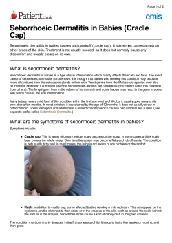


© Copyright 2026