
Managing Common Dermatoses in Skin of Color
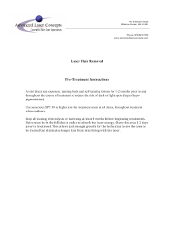
Managing Common Dermatoses in Skin of Color
Marcelyn K. Coley, MD, and Andrew F. Alexis, MD, MPH
The demographics of the United States continue to evolve, with a growing proportion of the
population consisting of non-Caucasian racial and ethnic groups. As darker skin types
become more prevalent, so will the need to better understand their skin, the conditions that
affect it, and optimal approaches for treatment. This population poses a special challenge
for practitioners in part as a result of the sequelae often associated with the conditions in
their own right—postinflammatory hyperpigmentation and scarring—and potential iatrogenic adverse effects that may occur during treatment. Through careful consideration of
cultural, clinical, and therapeutic nuances, safe and effective management of common
disorders in skin of color is achievable.
Semin Cutan Med Surg 28:63-70 © 2009 Elsevier Inc. All rights reserved.
A
s the demographics of the US population continue to
evolve, understanding racial and ethnic variations in the
presentation and management of skin disorders will be increasingly important for dermatologists. Several dermatologic concerns are more prevalent or have unique clinical
features in populations with skin of color. Moreover, treatment options often vary according to a patient’s skin type so
that safe and effective outcomes are achieved. Variations in
skin structure and function, as well as cultural practices and
cultural standards of beauty, contribute to clinical and therapeutic differences in skin of color.
Most epidemiologic studies pertaining to the prevalence of
dermatologic conditions in people of color have been practice surveys involving black patient populations. In a 1983
study by Halder et al,1 acne, eczema, pigmentary disorders,
seborrheic dermatitis, and alopecias were the most commonly diagnosed disorders in black patients in a universityaffiliated private practice in Washington, DC. Alexis et al2
showed similar results in 2007 in a New York City hospitalbased practice survey, with acne vulgaris, as the most common diagnosis documented, followed by dyschromia, eczema/dermatitis, alopecia, and seborrheic dermatitis,
respectively. In addition to the aforementioned diagnoses,
disorders such as dermatosis papulosa nigra, pseudofolliculitis barbae, and keloids are also common concerns for which
Skin of Color Center, Department of Dermatology, St. Luke’s-Roosevelt
Hospital Center, New York, NY.
Dr Andrew Alexis has served as a speaker for Sanofi-Aventis, Galderma, and
Stiefel. He has received honoraria from Medicis.
Address reprint requests to Andrew F. Alexis, MD, MPH, Skin of Color
Center, Department of Dermatology, St. Luke’s-Roosevelt Hospital Center, 1090 Amsterdam Avenue, Suite 11B, New York, NY 10025. E-mail:
[email protected].
1085-5629/09/$-see front matter © 2009 Elsevier Inc. All rights reserved.
doi:10.1016/j.sder.2009.04.006
patients of color seek dermatologic consultation. Here we
review some of the common dermatologic concerns in patients with skin of color and their management.
Acne Vulgaris
Acne vulgaris is a leading skin disorder in patient populations
with skin of color, just as it is in the general population. In
fact, acne was the most common diagnosis observed in multiple survey studies involving patients of color.1-6 Although
the etiology and treatment options are similar in all skin
types, two potential consequences of acne—postinflammatory hyperpigmentation (PIH) and keloids— occur at significantly greater rates in darkly pigmented persons; therefore,
treatment can be more challenging in this population. The
sequelae may significantly reduce quality of life through their
psychosocial impact. Discoloration and scarring, and the
stigma associated, persist long after the acne lesions themselves have resolved. Moreover, PIH is often the primary
motivation, rather than the acne itself, for dark-skinned individuals to seek medical attention (Fig. 1).7
Differences in the clinical presentation and exacerbating
factors can be seen in skin of color. The most important and
distinguishing clinical feature of acne in skin of color is the
presence of associated PIH. A survey by Taylor et al8 reported
a high incidence of PIH in black, Hispanic, and Asian patients, where 65%, 53%, and 47%, respectively, had acne and
acne-related hyperpigmented macules. The high prevalence
of PIH is attributed to the increased lability of melanocytes in
darkly pigmented skin.9 Inflammation or trauma triggers an
increase in melanogenesis or a release of melanin from labile
melanocytes.9,10 It has been reported that marked inflammation on histopathology (even in noninflammatory lesions—
63
M.K. Coley and A.F. Alexis
64
Topical Therapy
Figure 1 Acne vulgaris with PIH in a 17-year-old African-American
male patient.
ie, comedones) may also contribute to the high prevalence of
PIH.11
Common exacerbating factors in skin of color often are
culturally accepted skin or hair care practices, such as occlusive skin products (eg, cocoa butter), hair pomades (eg, “hair
grease”), and topical steroid-containing fade creams (used by
some populations, especially in Africa, to lighten the complexion of the skin). Pomade acne is a type of acne that
primarily affects individuals of African descent. To a lesser
degree, it also has been seen in Hispanic patients, particularly
men who use lubricating products on the scalp and facial
hair.12 The lesions are typically closed comedones on the
forehead and along the anterior hairline and are associated
with using pomades and oils in the hair. Pomades contain
comedogenic combinations of high-melting hydrocarbons.13
This form of acne can be remedied best if such products are
avoided. Because of a change in product formulations and
hair-care practices of African-Americans,14,15 pomade acne is
not as common today as it was in previous decades. In patients with pomade acne, less comedogenic hair emollients,
such as those containing cyclomethicone or dimethicone, can
be recommended.
Treatment
Given the high prevalence and cosmetically disfiguring nature of PIH, early and aggressive intervention is warranted.
However, because of the irritating nature of some acne treatments, it is important to balance aggressive treatment of acne
with the risk of inducing PIH secondary to irritant contact
dermatitis.
Topical agents, such as retinoids and antimicrobials, are considered the first-line treatment in skin of color, much as in
other ethnicities. In ethnic skin, topical retinoids remain the
top choice.16,17 Topical retinoids available in the United
States include tretinoin, tazarotene, and adapalene, which
have been shown to be effective in treatment of acne and the
associated PIH.17-23 With use of these agents, it is important
to consider the potential risk of irritant contact dermatitis,
which in itself may also result in PIH. To address this concern
in patients of color, it is advisable to start with low concentrations and more tolerable formulations. Typically, the use
of a cream vehicle is more tolerable than an alcohol-based gel,
especially in dry/sensitive skin types.14 However, newer
aqueous-based gels as well as microsphere and crystalline
formulations of topical retinoid gels are available and are well
tolerated, effective options in this patient population. Another approach is alternate-day dosing, usually at night (eg,
every other night), which can then be titrated up as tolerated
over 4-6 weeks.
Topical antimicrobials, such as erythromycin and clindamycin, are commonly used and are effective in reducing levels of Propionibacterium acnes. A major concern with longterm use is development of antibiotic resistance. This
resistance can be reduced by the use of benzoyl peroxide in
conjunction with topical antibiotics.24 Benzoyl peroxide formulations are important therapeutic modalities in the treatment of acne in patients with skin of color. Combination
preparations, such as 5% benzoyl peroxide along with a topical antibiotic (ie, clindamycin or erythromycin) are useful
for their antimicrobial and anti-inflammatory properties. A
primary consideration with benzoyl peroxide, like that of
topical retinoids, is minimizing irritation and drying effects
that can potentially lead to the development of hyperpigmentation. Hence, the use of concentrations of ⱕ5% along with
careful vehicle selection are strongly recommended.14 In general, aqueous gels are better tolerated than alcohol-based
gels.
Topical azelaic acid, 20% cream is also effective in managing acne in skin of color. It has been found to successfully
reduce inflammatory and noninflammatory acne lesions in
addition to decreasing hyperpigmentation via its inhibitory
effects on tyrosinase.25,26 Its efficacy has been reported to be
comparable to that of tretinoin, 0.05% cream, BPO 5% gel,
erythromycin, 2% ointment, and clindamycin, 1% gel.27 It is
generally well tolerated with a low potential for irritation;
therefore, it may be an appropriate option for patients with
sensitive skin or a history of PIH.14 Efficacy may be enhanced
by combining azelaic acid with other topical acne therapies.27
Topical dapsone 5% gel has recently been approved in the US
for the treatment of acne and may be particularly useful in
skin of color patients given its anti-inflammatory properties.
Adjunctive Therapy
The adjunctive use of depigmenting agents to improve PIH is
commonly incorporated in the management of acne in skin of
color. These agents include hydroquinone, azelaic acid, and
Managing common dermatoses in skin of color
kojic acid. Hydroquinone actively blocks melanocyte pigment production via inhibition of tyrosine conversion to melanin. Ideally, hydroquinones are introduced after acne is under control. However, in practice they are often initiated
sooner, typically as spot treatment applied with a cotton swab
once to twice daily after the application of acne medication.
Care must be taken because use could result in hypopigmentation of normal adjacent skin, producing a halo effect. This
effect gradually resolves after discontinuation of hydroquinone. Azelaic acid when used as combination therapy for
acne has also been successful. It also has effects on proliferating melanocytes and has been effective in reducing hyperpigmentation in melasma,8 and therefore, has been used in
other dyschromias.
Chemical peels, when limited to superficial peeling agents
such as salicylic acid and glycolic acid, are effective and safe
in darker skin types. Benefits of implementing chemical peels
include the ability to treat existing primary and secondary
lesions, the improvement of PIH, and the improved absorption of topical agents.28 Liquid salicylic acid solution 20-30%
applied to the face for 3-5 minutes28 or a 30-50% buffered
glycolic acid solution left in place for 2-4 minutes29 have been
effective. In one study by Grimes,30 moderate-to-significant
clearing of acne vulgaris occurred in 8 of 9 (89%) treated with
a series of salicylic acid peels. Pretreatment with 4% hydroquinone is recommended by some authors to reduce postpeel hyperpigmentation in patients with skin types V-VI. A
series of 3-6 treatments, approximately 4 weeks apart, is typically performed based on the severity of the areas to be
treated. It is strongly recommended that topical retinoids be
discontinued 1 week before the peel and restarted 5-7 days
after the peel.
An important but often overlooked adjunctive measure in
the treatment of acne is the use of sunscreen to prevent exacerbation of acne-associated PIH. Sunscreen (minimum SPF
15 with UVA and UVB protection) in combination with sun
avoidance may be a successful and necessary adjunctive
treatment in PIH.31 The selection of a noncomedogenic sunscreen is important as to not exacerbate the primary condition. The vehicles rather than the UV-absorbing compounds
themselves tend to be responsible for exacerbating acne.
Therefore, patients should be instructed to look for sunscreens labeled as noncomedogenic.
A successful outcome is largely dependent on patient compliance and the clinician’s ability to effectively choose treatment modalities that are agreeable to the patient. It is helpful
to provide instructions both verbally and in writing to reinforce the regimen prescribed. In some patients, PIH and scarring are exacerbated by self-inflicted injury (eg, picking or
scratching lesions). Counseling regarding the avoidance of
manipulating acne lesions is an important step in the management of such patients.
Pseudofolliculitis Barbae (PFB)
Epidemiology
PFB is a common inflammatory follicular disorder characterized by papules and pustules localized to the beard and/or
65
neck. Also known as “ingrown hairs” or “razor bumps,” this
condition primarily affects men who are of African ancestry
(ie, African-Americans, Afro-Caribbeans, etc.), but more specifically affects those with curly, coarse hair. Although less
common in women, it is often seen in hirsute women on the
face/neck or in young women who practice hair removal (ie,
waxing, plucking, or shaving) in the axilla or pubic regions.
Generally, associated with shaving, PFB can occur in any
hair-bearing area. Studies have identified a single nucleotide
polymorphism, a disruptive Ala12Thr substitution in the 1A
alpha-helical segment of the companion layer-specific keratin K6hf, as a genetic risk factor for the development of
pseudofolliculitis barbae.32 The prevalence ranges from 45%
to 83% and is a leading concern among black US Army recruits.33-35 A study done at the Skin of Color Center,36 which
surveyed 71 patients with PFB (41 female and 30 male subjects), showed an average age of onset of 22 years.
Pathogenesis
The etiology is related to a foreign-body reaction surrounding an ingrown hair within the dermis. It is thought that the
characteristics of the “black” hair type—a curved hair follicle
and curly shaft— contribute to the hair’s natural tendency to
coil, producing tightly curled or “kinky” hair. This predisposes cut hair to reenter the skin, causing the subsequent
inflammatory response. Cutaneous reentry of the hair shaft
occurs by 2 mechanisms, extrafollicular and transfollicular
penetration. In extrafollicular penetration, the shaved hair,
now with a sharp edge, follows its natural curvature with the
concavity towards the epidermis. As it grows out to the surface of the epidermis, it then reenters the skin. Transfollicular
penetration is similar; however, because of the hair’s natural
tendency to coil, it pierces the follicle wall and reenters the
dermis without ever exiting the epidermis.33,34,36 Transfollicular penetration occurs when the skin is pulled taut during
shaving, which causes newly cut hair to retract beneath the
skin. This may also occur with plucking, which can leave hair
fragments under the skin.34,36 In either scenario, the hair
penetrates the dermis, triggering a foreign-body inflammatory reaction resulting in the formation of papules and pustules.33,34
Clinical Features
Clinically, PFB is characterized by follicular and perifollicular
papules and pustules, often with associated hyperpigmentation. Embedded hair may be seen within papules. There may
also be grooved, linear, depressed patterns in the skin that
occur as the result of parallel hair growth.36 Most commonly
affected are the bearded area of the face, including the anterior neckline, submental and mandibular areas, chin, and
cheeks (Fig. 2). Common sites in women include the axillae
and pubis because they are the sites of most frequent hair
removal. With the exception of secondary infection, cultures
are usually sterile or contain normal flora.
In female patients with PFB secondary to hirsutism, it is
important to rule out hormonal abnormalities, ie, polycystic
ovarian syndrome or androgen-producing hormones.36,37
M.K. Coley and A.F. Alexis
66
the significant impact on quality of life, men with PFB typically seek treatment later than women.36
Treatment
Figure 2 Pseudofolliculitis barbae on the (A) chin, (B) submental,
and neck regions in a 45-year-old African-American man.
Screening for hormonal abnormalities should include serumfree and total testosterone, dehydroepiandrosterone, and luteinizing hormone/follicle-stimulating hormone.37
Psychosocial Impact
The appearance of these lesions can be very distressing to
men and women affected with this condition and may cause
a great deal of anxiety and self-consciousness about one’s
appearance. PIH may be a significant contributing factor to
associated distress. It has been documented that as much as
90% of patients with PFB report having hyperpigmentation.36 Additionally, PFB has led to significant social tension
among African-American men in the Armed Forces because
of the military grooming code requirement of a clean-shaven
face. It left many African-American men faced with the dilemma of complying with the military code and suffering
from PFB or not complying and facing discharge.35,36 Despite
Various treatment options have been employed in managing
PFB. When personal and professional circumstances allow,
avoidance of shaving altogether and growing a beard results
in resolution of the condition within 4-8 weeks in most cases.
This was demonstrated in a study by Strauss and Kligman, in
which allowing the beard to grow for approximately 1 month
resulted in spontaneous resolution of most papules.38 Beard
growth may not always be possible, however, not only because of occupational mandates for a clean-shaven appearance, but also because of personal choice, often dictated by
social acceptability.
Nevertheless, when shaving avoidance is not an option,
patient education and counseling about proper shaving techniques become important to controlling this condition. A key
concept in preventing flares of PFB is to limit the closeness of
the shave in these patients. Most men with PFB can control
the condition by maintaining an optimal beard length of
0.5-1 mm.34-36,37 Electric clippers are often recommended
because they can be set to cut hair to the desired length.34 If
the proposed length of 0.5-1 mm or “stubble” is unacceptable
and a closer shave is desired, use of a single blade razor, as
opposed to a multiblade razor, is preferred. Multiblade razors
tend to provide the closest shave but can facilitate transfollicular penetration and therefore may increase the risk for
PFB. The first blade pulls the hair out of the follicle whereas
the second blade cuts it, allowing retraction of the hair down
into skin.36 A single-blade razor, such as the Bump Fighter
(American Safety Razor Company, Staunton, VA) may be
considered.33,34,37,39 This razor includes a polymer coating
and a foil guard that keeps the blade edge slightly off the skin
to prevent hair from being trimmed too short. This blade has
been reported to significantly reduce the number of PFB lesions.36,40 However, a recent unpublished study (sponsored
by Gillette/Proctor and Gamble, Cincinnati, OH) found that
shaving with a five-blade razor did not exacerbate PFB.
Some dermatologists advocate preparing the area to be
shaved with a warm moist towel to soften hair and washing
with an antibacterial soap or benzoyl peroxide wash, along
with use of shaving gel or foam for lubrication.37 Patients
should also be advised to shave in the direction of hair growth
(ie, “with the grain”). The use of an adequately sharp blade is
recommended to prevent the need for repeat passes over a
given area with the blade. Patients should also be instructed
against stretching or pulling the skin taut while shaving because as the skin is released, cut hair may fall beneath the
skin’s surface setting the stage for transfollicular penetration.34,36,38 Another proposal is loosening of embedded hairs
the night before shaving by brushing the beard or using a
sterile needle with the aid of a magnifying mirror.37,40 The
latter requires great care to avoid inadvertently inducing
trauma or introducing infection. This step is considered unnecessary by some because the hair eventually releases itself
once the hair grows out further. This loop phenomenon,
Managing common dermatoses in skin of color
described by Strauss and Kligman, occurs with extrafollicular
penetration. A loop is formed when the hair reenters the skin.
As the hair continues to grow out, the loop enlarges, serving
as a way to release the hair. The hair will spontaneously
release after a length of 1 cm.33,34,38
Chemical Depilatories
Chemical depilatories, such as barium sulfide and calcium
thioglycolate, have been used as an alternative to shaving.
These substances, which exist in powder, paste, cream, and
lotion forms, work by weakening the disulfide bonds in keratin so that hair is easily removed from the skin’s surface. It is
applied to a moistened beard and left in place for 5 or 15
minutes (ie, barium sulfide or calcium thioglycolate, respectively) and then scraped away with a blunt instrument (ie,
wooden spatula) along with the weakened hair. This process
leaves a blunt hair tip, making extrafollicular and transfollicular penetration less likely.36 The skin is then rinsed and
emollient applied to reduce irritation. Because these chemicals, if left on for prolonged periods, may cause potential
irritant or allergic reactions, with resulting postinflammatory
hyperpigmentation, it is important to follow directions precisely. A test patch may be performed to determine irritation
potential before treatment.34
Eflornithine hydrochloride cream, 13.9% (Vaniqa, Skin
Medica, Carlsbad, CA) is used to treated unwanted facial hair
in women. It works by irreversible inhibition of skin ornithine decarboxylase, an enzyme in hair cell division, resulting in a decreased hair growth rate in the area applied.33,34,37
Although not approved by the Food and Drug Administration for use in PFB, it has been used for the same purpose of
decreasing hair growth in men with PFB. The cream is applied to the affected area of the face twice daily at least 8 hours
apart and washed off at least 4 hours after application. Because it is not a depilatory, it is used in conjunction with
other hair-removal methods. This point should be discussed
clearly with patients to avoid unrealistic expectations. Additionally, the benefit of the therapy is only maintained with its
continued use.
Medical Management
Medical management includes use of an exfoliating agent,
such as retinoids, low-potency corticosteroids, and topical
antibiotics. Often a combination of therapies is used as opposed to monotherapy. Topical retinoids have been used
with success (eg, topical 0.05% tretinoin or adapalene
nightly), which help to alleviate the hyperkeratosis associated
with repeated nicking of the follicular epithelium36 and hyperpigmentation. Some also advocate use of low-to-mid potency topical corticosteroids (eg, desonide lotion) applied in
the morning after shaving in place of commercial aftershave
products.33,37 Topical antimicrobials (clindamycin, erythromycin, benzoyl peroxide) may also be useful.41 Although PFB
is not a true folliculitis caused by bacterial infection, as implied by its name, it is thought that antimicrobials may reduce the colonization of bacteria that can aggravate the inflammation, leading to secondary infection,36 and also have
67
anti-inflammatory effects.41 As discussed in a previous section, a benzoyl peroxide wash may be added before shaving.
Depigmenting agents, such as hydroquinone 4%, azelaic
acid, and kojic acid have been effective in treating the PIH
that may result from the PFB lesions. Monthly intralesional
cortico steroids are helpful in addressing resultant hypertrophic or keloidal scars.37
Chemical Peels
Superficial chemical peels are generally well-tolerated and
beneficial adjunctive therapies in PFB. In addition to its exfoliating effect, glycolic acid is a reducing agent. As a result, it
may reduce sulfhydryl bonds in the hair shaft and lead to
straighter hair growth that reduces the chance for reentry of
the hair shaft into the epidermis.42 Salicylic acid peels can
offer comedolysis and lightening in cases complicated by
PIH.37 Reduced numbers of PFB lesions have been observed
with both glycolic acid and salicylic acid peels.33
Epilation
Electrolysis is generally not recommended in PFB. In addition to being painful and expensive, the use of electrolysis is
often unable to ablate the curved or distorted hair follicle,43
which may be distorted secondary to previous manipulation.
It has been reported by several groups that electrolysis may
actually exacerbate PFB because the needle used in electrolysis may not reach the hair bulbs of the curved follicle, perhaps promoting transfollicular penetration via relatively
sharp hair fragments left in the skin.33,34,36 Additionally, hyperpigmentation may result at the sites of needle insertion.36,43 A blend method of electrolysis using galvanic and
thermolytic currents has been effective.35,43
Surgical depilation has been described as an alternate
method of hair removal.44 This involves an incision into the
subcutaneous tissue, extensive undermining, followed by
skin inversion and removal of hair bulbs by scissor excision,
extraction or electrodessication. A possible consequence of
this procedure is keloid formation; surgical depilation is contraindicated in patients with predisposition to form keloids.40
Laser hair removal is the most recent advance in the management of PFB, with the development of lasers that can be
used safely in darker skin types. The rationale behind its use
is to reduce the amount of hair growth in affected areas, thus
eliminating the potential for developing ingrown hairs. Studies have shown safe and effective use of laser epilation in
darker skin types, including the diode (800-810),45,46 and
neodymium:yttrium aluminum garnet (Nd:YAG 1064 nm)
lasers.46-48
Ross et al48 reported the use of Nd:YAG 1064 nm coupled
with contact cooling as an effective treatment option for PFB
in skin types IV-VI, documenting significant reduction in
hair and subsequent papule formation. Histologically, posttreatment biopsies revealed evidence of severe thermal hair
bulb damage with epidermal preservation, supporting the
concept that longer wavelengths better penetrate the skin,
with less thermal damage to the epidermis.48 Weaver and
Sagaral47 also documented a decrease in the quantity of pap-
M.K. Coley and A.F. Alexis
68
ules/pustules and hairs after 2 treatments in patients with
active PFB using the long-pulse 1064 nm Nd:YAG laser on
skin types V and VI. At a 3-month follow-up period, the
mean papule/pustule percentage reduction was 75.9% as
compared to the control at 28.6%. The most common side
effects observed included transient hyperpigmentation, transient hypopigmentation, mild erythema and itching. Laser
epilation is an effective treatment for PFB in those that desire
long-term hair removal.
Dermatosis
Papulosa Nigra (DPN)
Epidemiology
DPN is a benign epithelial neoplasm, a probable variant of
seborrheic keratosis that is found in darkly pigmented populations, most commonly in those of African descent.49 The
incidence has been reported to be 35-77% in the AfricanAmerican population.50,51 DPN tends to affect women more
than men at a 2:1 ratio, usually increasing with age.51
Clinical Features
The lesions are characterized by multiple hyperpigmented
(dark brown to black) smooth papules measuring 1-5 mm in
size. They typically occur symmetrically on the face, favoring
the malar eminences, and may also involve the neck, chest,
and upper back (Fig. 3). Although the etiology is not known,
a genetic predisposition is thought to be a key factor. A positive family history is often found. However, because DPN
tend to be distributed in sun-exposed areas, some authors
have suggested that sun may play a role in the etiology—a
hypothesis that has also been reported for seborrheic keratoses in some populations.52,53 Niang et al54 further comment
on this hypothesis in a Senegalese study. In addition to observance of a prominence of lesions present in sun-exposed
Figure 3 Dermatosis papulosa nigra in a 52-year-old African-American woman.
areas, it was also documented that 8 out of 10 patients who
had profuse (100⫹) DPN had also practiced some form of
artificial depigmentation with high potency topical corticosteroids in the past. Further studies are needed to investigate
whether the sun or other nongenetic factors play any role in
the pathogenesis.
Role in Malignancy
No malignant transformation has been observed in DPN.
This situation is unlike that of seborrheic keratoses and the
rare sign of Leser-Trélat, characterized by a rapid increase in
size and number of multiple seborrheic keratoses caused by
malignancy, most commonly adenocarcinomas.55 However,
a case report from the University of Miami describes a middle-aged black woman with a history of DPN and anemia
who reported an explosive progression in the number and
size of biopsy-proven DPN lesions on the face, neck, and
upper trunk, with back lesions in a “Christmas tree” pattern.56 A subsequent colonoscopy revealed an ascending colon adenocarcinoma. It is unclear whether this could be another example of Leser-Trélat, if the DPNs observed were
simply variants of seborrheic keratoses, or if this is a new,
previously undescribed paraneoplastic entity unto itself.
Treatment
Treatment is generally performed for aesthetic reasons, although some lesions may become irritated or pruritic. Particularly bothersome may be feelings of anxiety, fear of cancer,
and/or professional concerns related to the appearance.54
Options for treatment include light electrodessication, curettage, cryotherapy, and snip excision for pedunculated lesions. Intralesional anesthetics have been useful for cases
where there are few lesions. However, topical anesthetics
may be advantageous in more widespread involvement, with
satisfactory levels of anesthesia for most patients when applied 30-60 minutes before the procedure.57
The most common adverse effect with any of the aforementioned treatments is potential for subsequent dyschromia— either hypo- or hyperpigmentation. Hypopigmentation can be particularly problematic after cryotherapy
because melanocytes are particularly sensitive to freezing
damage.58 In the present author’s (AFA) experience, light
electrodessication is the safest and most effective treatment
option for small DPNs. Care should be taken to minimize
injury to normal skin beneath and adjacent to the lesion to
prevent hyper- or hypopigmentation. For electrodessication
of lesions that are 1 mm or less, an epilating needle can be
used. In this author’s opinion, electrodessicated lesions are
best left to fall off spontaneously (rather than using curettage
post-electrodessication) to further minimize epidermal injury. Pedunculated lesions are safely and effectively removed
by Gradle scissor excision under local anesthesia.
A study by Kauh et al50 examined the efficacy of light
curettage for DPN. Twenty patients (18 women, 2 men; 16
black, 4 Asian) with clinically diagnosed DPN were treated
with “light abrasive curettage” to irritate, but not completely
remove, the lesions. Minimal bleeding controlled with pres-
Managing common dermatoses in skin of color
sure was common posttreatment. Good results were reported
in 14 of 19 patients treated, with good (only minor pigment
alteration) or excellent responses at 8 weeks. No evidence of
scarring was seen in any patients treated. A prospective study
by Niang et al54 reported success with electrosurgery. Twelve
Senegalese patients with DPN had lesions excised with fineneedle or fine-scissor electrosurgery after local anesthesia.
Aesthetic results were evaluated over 6 weeks, with 60% of
treated patients healing without sequelae at day 45. Dyschromia was the only documented adverse effect, seen in those
who had practiced artificial depigmentation.
Laser treatment of DPN has also shown promise. Good
results have been documented with use of the 532 diode
laser.49 Authors of recent reports have experienced success by
using a long-pulsed 1064 nm Nd:YAG laser for the treatment
of DPN. Schweiger et al49 reports 2 cases of middle-aged
African-American women with a long-standing history of
DPN. In both cases, each lesion was treated with a double
pulse from the Nd:YAG laser using a 3-mm spot size, fluence
range of 145-155 J/cm2 and a pulse duration of 20 milliseconds. At 2 months follow up, the treated areas had resolved
with no pigmentary changes or scarring in approximately
90% and 70% of lesions treated in the 2 cases presented,
respectively. Further studies will be necessary to corroborate
these findings.
Conclusions
Understanding the disease processes that affect patients with
skin of color and the respective treatment options available
will be increasingly important for US dermatologists. An
added challenge is being mindful of the associated complexity and variation among the skin types within this group.
However, through careful consideration of the clinical and
therapeutic nuances in skin of color, it is possible to safely
and effectively care for this population.
References
1. Halder RM, Grimes PE, McLaurin CI, et al: Incidence of common dermatoses in a predominantly black dermatologic practice. Cutis 32:378380, 1983
2. Alexis AF, Sergay AB, Taylor SC: Common dermatologic disorders in
skin of color: A comparative practice survey. Cutis 80:387-394, 2007
3. Taylor SC: Epidemiology of skin diseases in ethnic populations. Dermatol Clin 21:601-607, 2003
4. Child FJ, Fuller LC, Higgins EM, et al: A study of the spectrum of skin
disease occurring in a black population in south-east London. Br J
Dermatol 141:512-517, 1999
5. Arsouze A, Fitoussi C, Cabotin PP, et al: Presenting skin disorders in
black afro-Caribbean patients: A multicenter study conducted in the
Paris region. Ann Dermatol Venereol 135:177-182, 2008
6. Dunwell P, Rose A: Study of the skin disease spectrum occurring in an
Afro-Caribbean population. Int J Dermatol 42:287-289, 2003
7. Taylor SC: Cosmetic problems in skin of color. Skin Pharmacol Appl
Skin Physiol 12:139-143, 1999
8. Taylor SC, Cook-Bolden F, Rahman Z, et al: Acne vulgaris in skin of
color. J Am Acad Dermatol 46:S98-S106, 2002
9. Grimes PE: Pigmentary disorders in blacks. Dermatol Clin 6:271, 1988
10. Taylor SC: Skin of color: Biology, structure, function, and implications
for dermatologic disease. J Am Acad Dermatol 46:S41-S62, 2002
(suppl)
69
11. Halder RM, Holmes YC, Bridgeman-Shah S, et al: A clinicopathological
study of acne vulgaris in black females. J Invest Dermatol 106:888,
1996
12. Halder RM, Noothehi PK, Richards GM: Dermatological disorders and
cultural practices: Understanding practices that cause skin conditions
in non-Caucasian populations. Skin Aging 10:46-50, 2002
13. Laude TA: Skin disorders in black children. Curr Opin Pediatr 8:381385, 1996
14. Callender VD: Acne in ethnic skin: Special considerations for therapy.
Dermatol Ther 17:184-195, 2004
15. Halder RM, Brooks HL, Caballero JC: Common dermatological diseases
in pigmented skins, in Halder RM (ed): Dermatology And Dermatological Therapy of Pigmented Skins. Boca Raton, FL, Taylor & Francis,
2006, pp 17-39
16. Leyden JJ: Topical treatment of acne vulgaris: Retinoids and cutaneous
irritation. J Am Acad Dermatol 38:S1-S4, 1998
17. Halder RM: The role of retinoids in the management of cutaneous
conditions in blacks. J Am Acad Dermatol 39:S98-S103, 1998 (suppl 2)
18. Bulengo-Ransby SM, Griffiths C, Kimbrough-Green CK, et al: Topical
tretinoin (retinoic acid) therapy for hyperpigmented lesions caused by
inflammation of the skin in black patients. N Engl J Med 328:14381443, 1993
19. Grimes P, Callender V: Tazarotene cream for postinflammatory hyperpigmentation and acne vulgaris in darker skin: A double-blind, randomized, vehicle-controlled study. Cutis 77:45-50, 2006
20. Czernielewski J, Poncet M, Mizzi F: Efficacy and cutaneous safety of
adapalene in black patients versus white patients with acne vulgaris.
Cutis 70:243-248, 2002
21. Zhu XJ, Tu P, Zhen J, et al: Adapalene gel 0.1%: Effective and well
tolerated in the topical treatment of acne vulgaris in Chinese patients.
Cutis 68:55-59, 2001 (suppl 4)
22. Thiboutot D, Arsonnaud S, Soto P: Efficacy and tolerability of adapalene 0.3% gel compared to tazarotene 0.1% gel in the treatment of
acne vulgaris. J Drugs Dermatol 7:S3-S10, 2008 (suppl 6)
23. Jacyk WK: Adapalene in the treatment of African patients. J Eur Acad
Dermatol Venereol 15:37-42, 2001 (suppl 3)
24. Leyden JJ: Current issues in antimicrobial therapy for the treatment of
acne. J Eur Acad Dermatol Venereol 15:51-55, 2001 (suppl 3)
25. Fitton A, Goa KL: Azelaic acid: A review of its pharmacological properties and therapeutic efficacy in acne and hyperpigmentary skin disorders. Drugs 41:780-798, 1991
26. Hsu S, Quan LT: Topical antibacterial agents, in Wolverton SE (ed): Comprehensive Dermatologic Drug Therapy. Saunders, 2001, pp 473-496
27. Webster G: Combination azelaic acid therapy for acne vulgaris. J Am
Acad Dermatol 43:47-50, 2000
28. Roberts WE: Chemical peeling in ethnic/dark skin. Dermatol Ther
17:196-205, 2004
29. Burns RL, Prevost-Blank PL, Lawry MA, et al: Glycolic acid peels for
postinflammatory hyperpigmentation in black patients. A comparative
study. Dermatol Surg 23:171-174; discussion: 175, 1997
30. Grimes PE: The safety and efficacy of salicylic acid chemical peels in
darker racial-ethnic groups. Dermatol Surg 25:18-22, 1999
31. Cayce KA, McMichael AJ, Feldman SR: Hyperpigmentation: An overview of the common afflictions. Dermatol Nurs 16:401, 2004
32. Winter H, Schissel D, Parry DAD, et al: An unusual Ala12Thr polymorphism in the 1A alpha-helical segment of the companion layer-specific
keratin K6hf: Evidence for a risk factor in the etiology of the common
hair disorder psuedofolliculitis barbae. J Invest Dermatol 122:652-657,
2004
33. Bridgeman-Shah S: The medical and surgical therapy of pseudofolliculitis barbae. Dermatol Ther 17:158-163, 2004
34. Halder RM, Roberts CI, Nootheti PK, et al: Dermatologic disease in
blacks, in Halder RM (ed): Dermatology And Dermatological Therapy
of Pigmented Skins. Boca Raton, FL, Taylor & Francis, 2006, pp 331355
35. McMichael AJ: Hair and scalp disorders in ethnic populations. Dermatol Clin 21:629-644, 2003
36. Perry PK, Cook-Bolden FE, Rahman Z, et al: Defining pseudofolliculitis
M.K. Coley and A.F. Alexis
70
37.
38.
39.
40.
41.
42.
43.
44.
45.
46.
47.
barbae in 2001: A review of the literature and current trends. J Am Acad
Dermatol 46:S113-S119, 2002
Quarles FN, Brody H, Johnson BA, et al: Pseudofolliculitis barbae.
Dermatol Ther 20:133-136, 2007
Strauss JS, Kligman AM: Pseudofolliculitis of the beard. Arch Dermatol
74:533-542, 1956
Alexander MA: Evaluation of a foil-guarded shaver in the management
of pseudofolliculitis barbae. Cutis 27:534-537:540-542, 1981
Kelly AP: Pseudofolliculitis barbae and acne keloidalis nuchae. Dermatol Clin 21:645-653, 2003
Cook-Bolden FE, Barba A, Halder R, et al: Twice-daily applications of
benzoyl peroxide, 5%/clindamycin, 1% gel versus vehicle in the treatment of pseudofolliculitis barbae. Cutis 73:18-24, 2004 (suppl 6)
Perricone NV: Treatment of pseudofolliculitis barbae with topical glycolic acid: A report of two studies. Cutis 52:232-235, 1993
Richards RN, Meharg GE: Electrolysis: Observations from 13 years and
140,000 hours of experience. J Am Acad Dermatol 33:662-666, 1995
Hage JJ, Bouman FG: Surgical depilation for the treatment of pseudofolliculitis or local hirsutism of the face: Experience in the first 40
patients. Plast Reconstr Surg 88:446-451, 1991
Greppi I: Diode laser hair removal of the black patient. Lasers Surg Med
28:150-155, 2001
Battle EF, Jr, Hobbs LM: Laser-assisted hair removal for darker skin
types. Dermatol Ther 17:177-183, 2004
Weaver SM, III, Sagaral EC: Treatment of pseudofolliculitis barbae
using the long-pulse Nd:YAG laser on skin types V and VI. Dermatol
Surg 29:1187-1191, 2003
48. Ross EV, Cooke LM, Timko AL, et al: Treatment of pseudofolliculitis barbae in skin types IV, V, and VI with a long-pulsed neodymium:Yttrium
aluminum garnet laser. J Am Acad Dermatol 47:263-270, 2002
49. Schweiger ES, Kwasniak L, Aries DJ: Treatment of dermatosis papulosa
nigra with a 1064 nm Nd:YAG laser: Report of two cases. J Cosmet
Laser Ther 10:120-122, 2008
50. Kauh YC, McDonald JW, Rapaport JA, et al: A surgical approach for
dermatosis papulosa nigra. Int J Dermatol 22:590-592, 1983
51. Grimes PE, Arora S, Minus HR, et al: Dermatosis papulosa nigra. Cutis
32:385-386:392, 1983
52. Kwon OS, Hwang EJ, Bae JH, et al: Seborrheic keratosis in the Korean
males: Causative role of sunlight. Photodermatology Photoimmunol
Photomed 19:73-80, 2003
53. Yeatman JM, Kilkenny M, Marks R: The prevalence of seborrheic keratoses in an Australian population: Does exposure to sunlight play a
part in their frequency? Br J Dermatol 137:411-414, 1997
54. Niang SO, Kane A, Diallo M, et al: Dermatosis papulosa nigra in Dakar,
Senegal. Int J Dermatol 46:45-47, 2007 (suppl 1)
55. Schwartz RA: Sign of lesser-Trélat. J Am Acad Dermatol 35:88-95, 1996
56. Schwartzberg JB, Ricotti CA, Jr, Ballard CJ, et al: Eruptive dermatosis
papulosa nigra as a possible sign of internal malignancy. Int J Dermatol
46:186-187, 2007
57. Carter EL, Coppola CA, Barsanti FA: A randomized, double-blind comparison of two topical anesthetic formulations prior to electrodesiccation of dermatosis papulosa nigra. Dermatol Surg 32:1-6, 2006
58. Cockerell CJ, Larsen F: Benign epidermal tumors and proliferations, in
Bolognia JL, Lorizzo JL, Rapini RP, et al (eds): Dermatology; vol 2.
Spain, Mosby/Elsevier, 2008, pp 1661-1680


© Copyright 2026