
Report of Case
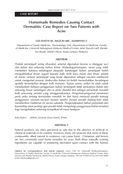
CASE REPORT Pathophysiologic Mechanisms, Diagnosis, and Management of Dapsone-Induced Methemoglobinemia John V. Ashurst, OMS IV; Megan N. Wasson, OMS IV; William Hauger, MD; and William T. Fritz, MD Dapsone is a leprostatic agent commonly prescribed for the treatment of patients with leprosy, malaria, and a variety of blistering skin diseases, including dermatitis herpetiformis. Methemoglobinemia, a potentially life-threatening condition in which the oxygen-carrying capacity of blood in body tissues is reduced, is a known adverse effect of dapsone use. The authors report a case of dapsone-induced methemoglobinemia observed in the emergency department during routine workup for contact dermatitis in a patient with celiac disease. The pathophysiologic mechanisms, diagnosis, and management of dapsone-induced methemoglobinemia are discussed. J Am Osteopath Assoc. 2010;110(1):16,19-20 D apsone, a potent anti-inflammatory and antiparasitic compound, is used worldwide for the treatment of patients with leprosy, malaria, and immunosuppressioninduced infections caused by Pneumocystis carinii and Toxoplasma gondii.1-3 In the United States, the major application of dapsone is in the treatment of patients with bullous dermatosis and dermatitis herpetiformis.1 Potentially life-threatening adverse effects of dapsone use include dapsone hypersensitivity syndrome, dose-dependent hemolytic anemia, and methemoglobinemia.1,2,4-6 In the present article, we report a case of dapsone-induced methemoglobinemia in which the diagnosis was made after a detailed history and physical examination. The diagnosis led to prompt treatment and complete recovery of the patient. From Lake Erie College of Osteopathic Medicine in Pennsylvania (Mr Ashurst, Ms Wasson) and the Department of Internal Medicine (Dr Hauger) and Department of Anesthesiology (Dr Fritz) at Conemaugh Memorial Medical Center in Johnstown, Pennsylvania. The authors have no relevant conflicts of interest or financial relationships to disclose. Address correspondence to John V. Ashurst, OMS IV, 721 Oak St, Northern Cambria, PA 15714-1438. E-mail: [email protected] Submitted April 15, 2009; revision received July 20, 2009; accepted November 11, 2009. 16 • JAOA • Vol 110 • No 1 • January 2010 Report of Case A white woman aged 68 years presented to the emergency department with a several-day history of weakness, chills, and shortness of breath, as well as a pruritic maculopapular rash bilaterally on her lower extremities. She reported recently shaving her legs with a new razor that contained an unknown lotion. However, the patient was not able to correlate the appearance of the rash with the use of the new razor. The patient’s medical history was significant for celiac disease, coronary artery disease requiring four cardiac stents, dermatitis herpetiformis, gastroesophageal reflux disease, and peripheral neuropathy. Her family history was clinically significant for deep vein thrombosis. Physical examination revealed that the patient was well nourished with no signs of physical deformity, trauma, or acute distress. Her vital signs included a blood pressure of 160/90 mm Hg; a body temperature of 98.1°F (36.7°C); a pulse of 115 beats per minute; a respiratory rate of 20 breaths per minute; and an oxygen saturation of 89% with ambient air. After supplemental oxygen of 3 L per nasal cannula was applied, her oxygen saturation increased to 93%. The patient’s physical examination and laboratory test results at presentation are shown in the Table. The patient’s lungs were clear to auscultation with decreased breath sounds bilaterally. Skin examination revealed multiple areas of excoriation, swelling, and cording of the bilateral lower extremities. Central and peripheral cyanosis was noted. Abdominal, cardiac, and neurologic examinations were unremarkable. Routine laboratory test data (Table) revealed a high white blood cell count (9900 cells/L) and a normal platelet count (313,000 cells/L), hemoglobin level (12.9 g/dL), and hematocrit concentration (39%). According to chemistry panel results, levels of chloride (106 mEq/L), potassium (3.8 mEq/L), and sodium (139 mEq/L) were also normal. The patient’s D-Dimer concentration was normal, at 1.1 mg/L, and her erythrocyte sedimentation rate was elevated, at 42 mm per hour. Results of electrocardiographic examination revealed sinus tachycardia with hyperacute T waves in leads V2 through V5. After initial evaluation, the patient was tentatively diagnosed as having an acute pulmonary embolism of unknown origin. Both a chest radiographic and spiral computed tomographic examination were performed, but neither showed Ashurst et al • Case Report CASE REPORT Table Examination and Laboratory Findings for Patient in Case at Presentation and Hospital Admission Component 䡲 Examination Findings at Presentation ▫ Blood pressure, mm Hg ▫ Body temperature, °F (°C) ▫ Pulse, beats per minute ▫ Respiratory rate, breaths per minute ▫ Oxygen saturation, % ▫ Other physical findings – Decreased breath sounds bilaterally – Excoriation, swelling, and cording of bilateral lower extremities – Central and peripheral cyanosis 䡲 Laboratory Findings at Presentation ▫ White blood cells, No. cells/L ▫ Platelets, No. cells/L ▫ Hemoglobin, g/dL ▫ Hematocrit, % ▫ Chemistry panels, mEq/L – Chloride – Potassium – Sodium ▫ D-Dimer, mg/L ▫ Erythrocyte sedimentation rate, mm per hour ▫ Electrocardiographic examination – Sinus tachycardia with hyperacute T waves in leads V2-V5 䡲 Findings After Hospital Admission and Ventilatory Support ▫ Arterial blood gasses – PaCO2, mm Hg – PaO2, torr – Oxygen saturation, % – Bicarbonates, mEq/L – Base excess, mEq/L ▫ Methemoglobin, % Value 160/90 98.1 (36.7) 115 20 89 9900 313,000 12.9 39 106 3.8 139 1.1 42 49 192 98.2 29 4.2 6.3 Abbreviations: PaCO2, partial pressure of carbon dioxide in arterial blood; PaO2, partial pressure of oxygen in arterial blood. clinically significant findings. In addition, Doppler ultrasonographic examination of the veins in the patient’s bilateral lower extremities revealed no deep vein thrombosis. The patient was admitted to the hospital for ventilatory support and further evaluation. Soon after admission, evaluations of her arterial blood gas and methemoglobin levels were performed (Table). These tests revealed elevated levels of arterial PaCO2 (49 mm Hg); arterial PaO2 (192 torr); arterial oxygen saturation (98.2%); bicarbonate (29 mEq/L); and base excess (4.2 mEq/L). The patient’s methemoglobin level was also elevated, at 6.3%. Further evaluation of the patient’s medical history revealed that she had been self-medicating with dapsone in an effort to Ashurst et al • Case Report Treatment for Patient in Case 䡺 Discontinuation of dapsone use 䡺 Application of supplemental oxygenation by nasal cannula 䡺 Use of diphenhydramine (Benadryl) cream to manage contact dermatitis Figure. Treatment successfully used for the patient in the present case, a white woman aged 68 years who was diagnosed as having dapsone-induced methemoglobinemia. Before diagnosis, the patient had been self-medicating with dapsone in an effort to manage a rash on her lower extremities, which she believed to be dermatitis herpetiformis. manage the rash on her lower extremities, which she believed to be dermatitis herpetiformis. The patient was treated with discontinuation of dapsone, application of supplemental oxygenation by nasal cannula, and use of diphenhydramine (Benadryl) cream for the rash (Figure). During the 2 days after treatment initiation, the patient’s methemoglobin level decreased to 3.8%, trending toward the normal range, and her oxygen saturation also improved. The patient was discharged from the hospital with the diagnosis of dapsone-induced methemoglobinemia and contact dermatitis. She was advised to eliminate her dapsone self-medication and to continue to use diphenhydramine for her contact dermatitis. Discussion Dapsone-induced methemoglobinemia is usually the result of acute poisoning secondary to either accidental ingestion or suicidal intent. According to recent research at several tertiary centers,7 dapsone has been shown to be the major cause of drug-induced methemoglobinemia. Dapsone (4,4’-diaminodiphenyl sulfone) is the parent compound of many sulfone medications.1,2,3,8 It is absorbed through the gut and is metabolized by the liver through the oxidation reactions of N-acetylation and N-hydroxylation.1,2,5,8 Hydroxylated amine metabolites produced in the oxidation reactions are potent oxidants that have been hypothesized to cause dapsone’s hematologic adverse effects, including hemolytic anemia and methemoglobinemia.1,8 Typically, dapsone is excreted by the kidneys. However, dapsone has been known to sometimes circulate in the enterohepatic system, resulting in a relatively long half-life of the drug in the body (ie, 24-30 hours).1 Methemoglobinemia occurs either as a congenital process in which there is a deficiency of nicotinamide adenine dinucleotide plus hydrogen (NADH)-cytochrome b5 reductase JAOA • Vol 110 • No 1 • January 2010 • 19 CASE REPORT or as an acquired disease in which there is an increase in the rate of oxidation of hemoglobin to methemoglobin.2,4-6,9 The newly formed methemoglobin is an aberrant form of hemoglobin in which the original ferrous (Fe2+) atom is oxidized to a ferric (Fe3+) atom. The ferric atom then causes an allosteric change in the heme portion of the oxidized hemoglobin molecule, resulting in an increase in its oxygen affinity but a decrease in its oxygen binding capacity.4,5,9 Diagnosis of Methemoglobinemia The diagnosis of methemoglobinemia is based on clinical symptoms and laboratory testing. Peripheral and central cyanosis is almost always present when there is a minimum methemoglobin level of 15% in the blood.2,4-6,10 These clinical signs typically result from the methemoglobin molecule causing a functional anemia, as well as from the methemoglobin molecule’s inability to bind to oxygen. As the concentration of methemoglobin in the blood increases to 45%, symptoms of dizziness, fatigue, headache, tachycardia, and weakness are expected.4-6,10 Acidosis, cardiac arrhythmia, dyspnea, seizures, and eventually coma become evident as the methemoglobin level approaches 70%.4-6,10 Arterial blood gas analysis paired with oxygen saturation analysis by pulse oximetry are now considered the definitive measures for making a correct diagnosis of methemoglobinemia. Blood gas analyses in patients with methemoglobinemia reveal normal to elevated levels of PaO2 with low oxyhemoglobin saturation.2,4-6 Management of Methemoglobinemia Traditionally, the initial management of methemoglobinemia involves ventilator support and removing the offending agent. Research has shown that a patient who has symptoms of dyspnea or a methemoglobin level of at least 30% should receive methylene blue intravenously at 1 to 2 mg per kilogram of body weight over a 5-minute period.2,5,6 Intravenously, methylene blue is oxidized into leucomethylene blue by accepting an electron from nicotinamide adenine dinucleotide phosphate (NADPH) in the presence of NADPH-methemoglobin reductase.2,9 Leukomethylene blue acts as an artificial electron acceptor to methemoglobin, resulting in methemoglobin’s conversion back to hemoglobin.2,9 Additional studies11 have shown that supplementing methylene blue with activated charcoal results in a lower dose of methylene blue being required to trigger a reversal of methemoglobinemia symptoms. In patients with celiac disease, flares of dermatitis herpetiformis can be controlled by the prophylactic use of dapsone. However, dapsone use predisposes such patients to the development of methemoglobinemia. In the present case report, the patient incorrectly believed that she had a flare of dermatitis herpetiformis, and this belief caused her to take the wrong medication—dapsone—which led to acute dapsoneinduced methemoglobinemia. 20 • JAOA • Vol 110 • No 1 • January 2010 Conclusion Based on our clinical experience—including the present case report—we believe that physicians need to remain alert to the possible association between dapsone use and methemoglobinemia. A high degree of suspicion for this association must be kept in mind when treating patients who have histories of dermatitis herpetiformis, leprosy, or malaria. For patients who do not successfully respond to supportive therapy or who have substantially higher-than-normal levels of methemoglobin, methlyene blue with adjuvant activated charcoal can be considered as a reasonable and safe treatment option. Early recognition of dapsone toxicity will allow for appropriate treatment of patients with dapsoneinduced methemoglobinemia, thereby preventing further methemoglobin production. References 1. Sener O, Doganci L, Safali M, Besirbellioglu B, Bulucu F, Pahsa A. Severe dapsone hypersensitivity syndrome. J Investig Allergol Clin Immunol. 2006;16(4):268-270. http://www.jiaci.org/issues/vol16issue04/10.pdf. Accessed November 28, 2009. 2. Turner MD, Karlis V, Glickman RS. The recognition, physiology, and treatment of medication-induced methemoglobinemia: a case report. Anesth Prog. 2007;54(3):115-117. http://www.ncbi.nlm.nih.gov/pmc/articles /PMC1993865/?tool=pubmed. Accessed November 28, 2009. 3. Coleman MD, Rhodes LE, Scott AK, Verbov JL, Friedman PS, Breckenridge AM, et al. The use of cimetidine to reduce dapsone-dependent methaemoglobinaemia in dermatitis herpetiformis patients. Br J Clin Pharmacol. 1992;34(3):244-249. http://www.ncbi.nlm.nih.gov/pmc/articles /PMC1381395/?tool=pubmed. Accessed November 28, 2009. 4. Rehman HU. Methemoglobinemia. West J Med. 2001;175(3):193-196. http://www.ncbi.nlm.nih.gov/pmc/articles/PMC1071541/?tool=pubmed. Accessed November 28, 2009. 5. Falkenhahn M, Kannan S, O’Kane M. Unexplained acute severe methaemoglobinaemia in a young adult. Br J Anaesth. 2001;86(2):278-280. 6. Bayard M, Farrow J, Tudiver F. Acute methemoglobinemia after endoscopy. J Am Board Fam Pract. 2004;17(3):227-229. http://www.jabfm.org/cgi/content/full/17/3/227. Accessed November 28, 2009. 7. Ash-Bernal R, Wise R, Wright SM. Acquired methemoglobinemia: a retrospective series of 138 cases at 2 teaching hospitals. Medicine. 2004;83(5):265273. 8. Tingle MD, Coleman MD, Park BK. An investigation of the role of metabolism in dapsone-induced methaemoglobinaemia using a two compartment in vitro test system. Br J Clin Pharmacol. 1990;30(6):829-838. http://www.ncbi.nlm .nih.gov/pmc/articles/PMC1368303/?tool=pubmed. Accessed November 28, 2009. 9. Birchem SK. Benzocaine-induced methemoglobinemia during transesophageal echocardiography. J Am Osteopath Assoc. 2005;105(8):381-384. http://www.jaoa.org/cgi/content/full/105/8/381. Accessed November 28, 2009. 10. Mayo W, Leighton K, Robertson B, Ruedy J. Intraoperative cyanosis: a case of dapsone-induced methaemoglobinaemia. Can J Anaesth. 1987;34(1):7982. 11. Hansen DG, Challoner KR, Smith DE. Dapsone intoxication: two case reports [review]. J Emerg Med. 1994;12(3):347-351. Ashurst et al • Case Report


© Copyright 2026