
oedematous malnutrition Review Article
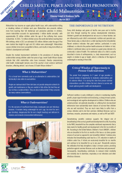
Review Article
Indian J Med Res 130, November 2009, pp 651-654
Oedematous malnutrition
Tahmeed Ahmed, Sabuktagin Rahman & Alejandro Cravioto
International Centre for Diarrhoeal Disease Research, Bangladesh, ICDDR, B, Dhaka, Bangladesh
Received April 16, 2009
Oedematous malnutrition, represented by its most severe form kwashiorkor, is rampant in many parts of
the world and is associated with a high case fatality rate. Despite being first described more than a century
ago, the pathogenesis of kwashiorkor is still not clear. The traditional thinking is that it results from a
deficiency of dietary protein and is usually associated with an infection. This has now been challenged
by the finding that there is no difference in diets of children developing marasmus or kwashiorkor.
Nutritional oedema is associated with an increased secretion of anti-diuretic substance (probably antidiuretic hormone) which prevents the normal excretory response to water administration. Experimental
studies have shown that feeding low-protein, low-calorie diets results in delayed and incomplete response
to a water load, and that the livers of the animals show a reduced capacity for inactivating anti-diuretic
hormone. There is now evidence that links generation of free radicals and depletion of anti-oxidants with
the development of oedema in kwashiorkor.
Key words Antidiuretic hormone - kwashiorkor - malnutrition - nutritional odema - oxidative changes
age, results from a deficiency of dietary protein and is
usually associated with an infection3. However in India,
Gopalan did not find any difference in diets of children
developing marasmus or kwashiorkor4. In response
to infection, amino acids are used for the production
of acute phase reactants at the expense of visceral
protein synthesis. There is a decrease in blood albumin
level, which is partly responsible for development of
oedema5. Beta-lipoprotein is not produced in adequate
amounts, resulting in impaired transport of fat from the
liver, accumulation of fat and an enlarged fatty liver6.
Kwashiorkor, the classical example of severe acute
malnutrition, is characterised by the presence of oedema.
In addition to oedema, the hallmarks of the condition
include dermatosis, diarrhoea, and fatty liver1. Typically
there are skin lesions (pigmented or depigmented areas
with or without ulceration), scanty lustreless hair, an
enlarged fatty liver, loss of interest in the surroundings,
and loss of appetite. Oedema may even progress to
generalised swelling or anasarca. Fluid homeostasis in
the body is altered resulting in excess accumulation of
fluid in the extracellular space. Cicely Willliams first
introduced the name kwashiorkor in 1935, which in
the Ga language of West Africa means “the disease of
the deposed child”2. This literally refers to the child
who develops oedema after being weaned with starchy
gruels following the birth of a sibling who is breastfed.
Kwashiorkor, which occurs mostly in children 1-3 yr of
There is no population-based data on prevalence
of oedematous malnutrition. This is due to the fact
that large-scale health and nutrition surveys do not
make any attempts to detect oedema. Case fatality,
however, is very high among children hospitalised
with oedematous malnutrition. These observations
651
652
INDIAN J MED RES, november 2009
indicate the need for better information on the global,
regional and national prevalence of kwashiorkor and
other forms of oedematous malnutrition7.
Mechanism of oedema
Oedema is facilitated by two biological processes8;
filtration is the movement of fluid out of the capillary
and, reabsorption is the movement of fluid back into
the distal end of the capillary and small venules. When
capillary fluid filtration exceeds reabsorption, fluid
accumulates within the interstitium over time if it
were not for the lymphatic system that removes excess
fluid from the interstitium and returns it back to the
intravascular compartment. Circumstances, however,
can arise where net capillary filtration exceeds the
capacity of the lymphatics to carry away the fluid
(i.e., net filtration > lymph flow). When this occurs,
the interstitium will swell with fluid and become
oedematous. Decreased plasma oncotic pressure,
as occurs in hypoproteinaemia during malnutrition,
precipitates oedema8.
How does oedema occur in kwashiorkor?
The genesis of oedema of kwashiorkor is
multifactorial, with electrolyte disturbance - potassium
deficiency and sodium retention playing an important
role9. Nevertheless, the classical theory postulates
that an inadequate intake of protein leads to a low
plasma albumin concentration which in turn causes
oedema10. Association of protein for genesis of oedema
was grounded further by indirect evidences gathered
through some of the work of Gopalan and fellow Indian
scientists, who found that diets based on protein were
associated with satisfactory clinical and biochemical
cure of kwashiorkor11,12. However, this classical
hypothesis has been challenged.
Golden et al13 in a study examined the association
between plasma albumin and nutritional oedema
by observing the changes in albumin during loss of
oedema in patients on a restricted diet. Since there was
no difference in the concentration of plasma albumin
before and after loss of oedema, the association is
not causal. In another study, Golden14 demonstrated
that loss of oedema from oedematous malnourished
children was strongly associated with dietary energy
intake but not with intake of protein. Gopalan et al4,
in a prospective study followed a group of Indian
children on poor diet. Some children developed
kwashiorkor and some marasmus, but there was no
quantitative and qualitative difference between their
diets. It was concluded that the difference lay in host’s
response and that, kwashiorkor represented the theory
of dysadaptation, i.e., adaptation failure, and good
adaptation resulting in marasmus.
Anti-diuretic factor in the urine of children with
nutritional oedema: Nutritional oedema is associated
with an increased secretion of an anti-diuretic substance
(probably anti-diuretic hormone) which prevents the
normal excretory response to water administration.
Gopalan and Venkatachalam15 in a study furnished
indirect proof of the effect of posture on the urinary
response to water load in normal subjects and in cases
of nutritional oedema. The normal subjects were found
to excrete over 100 per cent of ingested water within
4 h of ingestion in the recumbent posture, while in
the erect posture they excreted only 80 per cent. In
case of nutritional oedema, the urinary excretion was
found to be much lower than in the normal subjects
in both recumbent and erect postures. The effect of
dietary protein deficiency on the hepatic inactivation
of ADH in rats has also been investigated. It was found
that the rats maintained on low-protein, low-calorie
diets showed a delayed and incomplete response to a
water load, and that the livers of these animals showed
a reduced capacity for inactivating ADH (Gopalan &
Srikantia, unpublished).
Role of ferritin and aldosterone: Srikantia observed
presence of ferritin in children with kwashiorkor16.
With a view to reveal the precise role of ferritin in
the pathogenesis of nutritional oedema, Gopalan
and Srikantia17 investigated the sequence of changes
occurring in induced protein and calorie under-nutrition
with focus on oedema formation in monkeys. On the
basis of the findings, they suggested that calorie-protein
undernutrition leads to structural and functional changes
in the liver, further leading to defective inactivation of
ADH. Active ferritin is released from damaged liver
leading to increased secretion of ADH. The net result is
water retention. Among other factors, aldosterone, the
salt retaining hormone, which is known for influencing
water metabolism by altering renal tubular reabsorption
of sodium, is also known to be inactivated by the liver.
Altered aldosterone metabolism has been reported in
diseases of the liver. Associated hyperaldosteronism
could account for the sodium retention18. In oedematous
children aldosterone secretion becomes higher during
loss of oedema19.
Oedema of kwashiorkor - environmental and
metabolic factors: Environment, particularly diet,
AHMED et al: OEDEMATOUS MALNUTRITION
certainly has an important role in the pathogenesis of
kwashiorkor; the condition is not seen in children with
an adequate nutritional intake. Intrinsic characteristics
of the host may also be required for the development
of kwashiorkor. It is possible that variant isozymes or
variations in concentration of enzymes in the metabolic
pathways lead to the development of kwashiorkor in
children with poor diets. Differences in the pattern
of amino acid concentrations between children with
kwashiorkor and marasmus have been used in favour of
this assumption20.
The serum amino-acidogram may show distinct
differences in cases of marasmus and kwashiorkor.
Very high levels of glutamate with low or undetectable
levels of alanine are the hallmark of kwashiorkor21.
One possible explanation for this can be the low level
of transaminases in kwashiorkor. Low transaminase
levels in the liver biopsy specimens in case of
kwashiorkor have been reported earlier22. It has
also been shown that they remain low even during
recovery. It is possible that this deficiency is present
right from birth and is genetically transmitted and is
made clinically overt when the child is exposed to
stress of dietary inadequacy and may be responsible
for development of kwashiorkor. Those children who
have normal transaminase function develop marasmus
under the same dietary insult. In addition to dietary
and nutritional investigations, genetic techniques
including genome-wide association to delineate host
factors may prove useful in unscrambling the enigma
of kwashiorkor.
Evidence for free radicals/anti-oxidants
Golden and Ramdath23 proposed that kwashiorkor
results from an imbalance between the production
of free radicals and their safe disposal. This theory
is supported by the observations in other studies
where blood concentrations of vitamin E derivatives,
glutathione, and red cell antioxidant enzymes are
lower in children with kwashiorkor than in marasmic
children24-26. The study of Sive et al27 shows that,
‘free’ circulating iron may contribute to oedema
in kwashiorkor. Srikantia17, using a bioassay, had
reported that children with kwashiorkor had high
levels of circulating ferritin, which was further
confirmed by a Jamaican study when immunoassays
became available28. The study of Okunade et al29
shows that the extent of lipid peroxidation in the
erythrocytes of kwashiorkor subjects was three
times that found in erythrocytes of normal subjects.
653
This finding was supported by another study which
also demonstrated excessive lipid peroxidation in
kwashiorkor30. In a clinical trial, the administration
of N-acetylcysteine, a glutathione precursor, resulted
in more rapid resolution of oedema in kwashiorkor31.
These associations between oxidative stress and
kwashiorkor indicate that antioxidant depletion may
cause kwashiorkor which can therefore be prevented
with antioxidant supplementation.
Challenge to the oxidative stress theory?
The study by Ciliberto et al32 examined the impact of
an antioxidant cocktail containing riboflavin, vitamin E,
selenium, and N-acetyl cysteine in a dose three times of
required daily intakes, as a possible preventive treatment
for kwashiorkor in children in a highly endemic area of
Malawi but failed to find any protective effect. Although
the result may be a deterrent to the antioxidant hypothesis,
it would be premature to discard the theory altogether.
The specific antioxidants used in the Ciliberto study
are known to have a high relative antioxidant capacity,
but the amounts taken by the children may have been
insufficient to overcome high oxidative stress and
prevent kwashiorkor. Since neither antioxidant capacity
nor oxidative stress was measured, the adequacy of the
antioxidant mix or the dose can be questioned. Moreover,
the study did not assess the children’s HIV status, which
may have contributed to or affected their responses to
oxidative stress33.
Thus more than a century has elapsed but the theories
on pathogenesis of kwashiorkor oedema continue to
unfold. The age old classical theory of dietary protein
inadequacy now continues to be challenged. The
theory of dysadaptation explains a different perspective
related to differential host response leading to different
outcomes (kwashiorkor or marasmus) under nutritional
stress, and underpins a wider perspective of metabolic
and enzymatic variances leading to oedema formation.
Hormonal factors (ADH) also demonstrate a plausible
explanation of the pathology. Evidence for free radicals
/ anti-oxidants, which essentially propose an imbalance
between free radicals and their disposal, is increasingly
becoming strong. Evidence of oxidative changes in
the cell membranes of the oedematous kwashiorkor
subjects i.e., lipid peroxidation has been established
by some studies. Indirect evidences of anti-oxidant
therapy improving oedema further consolidate the
free radical/anti-oxidant theory; yet this theory is not
without question- the enigma of kwashiorkor oedema
continues!
654
INDIAN J MED RES, november 2009
References
1.
Gopalan C, Ramalingaswami V. Kwashiorkor in India. Indian
J Med Res 1955; 43 : 751-73.
2.
Williams CD. Kwashiorkor: a nutritional disease of children
associated with a maize diet. Lancet 1935; 2 : 1151-2.
3.
Suskind RM, Murthy KK, Suskind D. The malnourished
child: an overview. In: Suskind RM, Suskind LL, editors. The
malnourished child. New York: Vevey/Raven Press; 1990.
4.
5.
Gopalan C. Kwashiorkor and marasmus: evolution and
distinguishing features. In: McCance RA, Widdowson EM,
editors. Calorie deficiencies and protein deficiencies. Boston:
Little, Brown; 1968. p. 49-58.
Waterlow JC. Causes of oedema and its relation to kwashiorkor.
In: Waterlow JC, editor. Protein-energy malnutrition. London:
Edward Arnold; 1992.
6.
Truswell AS Hansen JDL. Fatty liver in protein-calorie
malnutrition. South Afr Med J 1969; 43 : 280-3.
7.
Bhutta ZA, Black RE, Cousens S, Ahmed T. Kwashiorkor
and severe acute malnutrition in childhood - Authors’ reply.
Lancet 2008; 371 : 1749.
8.
Klabunde RE. Tissue oedema and general principles of
transcapillary fluid exchange, cardiovascular physiology
concepts. Available at: www.cvphysiology.com/microcircula
tion/M010.htm.
9.
Alleyne GAO. The effect of severe protein calorie malnutrition
on the renal function of Jamaican children. Paediatrics 1967;
39 : 400-12.
10. Waterlow JC. Fatty liver disease in infants in the British West
Indies. Medical Research Council Special Report Series No.
263. London: Medical Research Council; 1948.
11. Gopalan C, Srikantia SG. Clinical trials with vegetable protein
foods in kwashiorkor. Indian J Med Res 1960; 48 : 637-44.
12. Gopalan C, Venkatachalam PS, Srikantia SG, Mehta G.
Treatment of nutritional oedema syndrome (kwashiorkor)
with vegetable protein diets. Am J Clin Nut 1962; 11 : 12733.
13. Golden MH, Golden BE, Jackson AA. Albumin and nutritional
oedema. Lancet 1980; i : 114-6.
14. Golden MHN. Protein deficiency, energy deficiency and the
oedema of malnutrition. Lancet 1982; i : 1261-5.
15. Gopalan C, Venkatachalam PS. The pathogenesis of nutritional
oedema. Indian J Med Sci 1952; 6 : 713.
18. Migeon CJ, Beitins IZ, Kowarski A, Graham CG. Plasma
aldosterone concentration and aldosterone secretion rate in
Peruvian infants with marasmus and kwashiorkor. In: Gardner
LI, Amacher P, editors. Endocrine aspects of malnutrition.
Santa Yenz: Kroc foundation; 1973. p. 399-424.
19. Lurie AO, Jackson WPU. Aldosteronuria and the oedema of
kwashiorkor. Am J Clin Nutr 1962; 11 : 115-26.
20. Phadke MA, Khedkar VA, Pashankar D, Kate SL, Mokashi
GD, Gambhir PS, et al. Serum amino acids and genesis of
protein energy malnutrition. Indian Pediatr 1995; 32 : 301-6.
21. Lunn PG, Whitehead RG, Hay RW, Baker BA. Aminoacids in
protein energy malnutrition. Br J Nutr 1973; 29 : 399-401.
22. Mclean EM. Hepatic failure in malnutrition. Lancet 1962; 2 :
1292-4.
23. Golden MHN, Ramdath D. Free radicals in the pathogenesis
of kwashiorkor. Proc Nutr Soc 1987; 46 : 53-68.
24. Jackson AA. Blood glutathione in severe malnutrition in
childhood. Trans R Soc Trop Med Hyg 1986; 80 : 911-3.
25. Sive AA, Subotzky EF, Malan H, Dempster WS, De V,
Heese H. Red blood cell antioxidant enzyme concentrations
in kwashiorkor and marasmus. Ann Trop Paediatr 1993;
13 : 33-8.
26. Becker K, Bötticher D, Leichsenring M. Antioxidant vitamins
in malnourished Nigerian children. Int J Vitam Nutr Res 1994;
64 : 306-10.
27. Sive AA, Dempster WS, Malan H, Rosseau S, Hesse HD.
Plasma free iron: a possible cause of oedema in kwashiorkor.
Arch Dis Child 1997; 76 : 54-6.
28. Ramdadi DD, Golden MH. Non-haematological aspects of
iron nutrition. Nutr Res Rev 1989; 2 : 29-49.
29. Okunade WG, Olorunsogo OO. Effect of reactive oxygen
species on the erythrocyte calcium-pump function in proteinenergy malnutrition. Pediatrics 1995; 95 : 874.
30. Lenhartz H, Ndasi R, Anninos A, Botticher D, Mayatepek E,
Tetanye E, et al. The clinical manifestation of the kwashiorkor
syndrome is related to increased lipid peroxidation. J Pediatr
1998; 132 : 879-81.
31. Badaloo A, Reid M, Forrester T, Heird WC, Jahoor F. Cysteine
supplementation improves the erythrocyte glutathione
synthesis rate in children with severe oedematous malnutrition.
Am J Clin Nutr 2002; 76 : 646-52.
16. Srikantia SG. Ferritin in nutritional oedema. Lancet 1958; i :
667-8.
32. Ciliberto H, Ciliberto M, Briend A, Ashorn P, Bier D,
Manary M. Antioxidant supplementation for the prevention
of kwashiorkor in Malawian children: randomised, double
blind, placebo controlled trial. BMJ 2005; 330 : 1109-11.
17. Srikantia SG, Gopalan C. Role of ferritin in nutritional
oedema. J Appl Physiol 1959; 14 : 829-33.
33. Fuchs GG. Antioxidants for children with Kwashiorkor. BMJ
2005; 330 : 1095-6.
Reprint requests: Dr Tahmeed Ahmed, Head, Nutrition Programme, International Centre for Diarrhoeal Disease Research, Bangladesh
(ICDDR, B), GPO Box 128, Dhaka 1000, Bangladesh
e-mail: [email protected]


© Copyright 2026