
Document 147772
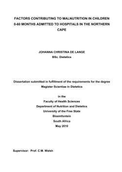
Oedematous malnutrition
Michael H N Golden
Department of Medicine and Therapeutics, University of Aberdeen, Aberdeen, UK
Correspondence to:
Prof Michael H N Golden,
Department of Medicine
and Therapeutics,
University of Aberdeen,
Foresterhill, Aberdeen
AB2S2ZD, UK
Oedema as a result of famine has been know since biblical times: the
children of Israel, during their wanderings in the desert thought that the
fungus that grows on acacia roots (mana) protected them against
oedematous malnutrition [Deuteronomy: 8-4; Nehemiah: 9-21]. Even
the typical skin changes of kwashiorkor were recognised: 'our skin was
black like an oven because of the terrible famine' [Lamentations: 5-10].
The specific nutrients involved and the pathophysiological processes
leading to the oedema and skin lesions still cause controversy.
The problems raised by nutritional oedema and the inadequacy of
current physiological explanations were raised in the classical reviews of
McCance1 and Keys2, and remain unresolved. In the early decades of this
century most work was done in Germany, where the condition in
children was know as Mehlnahrschaden3, or 'flour dystrophy', because
it was recognised in poor children weaned to a diet predominantly or
exclusively of cereal flour, although detailed dietary data were not
published. There was controversy over the cause, but protein deficiency
was most commonly cited on the basis of low plasma protein
concentrations and uncertainty over the requirements for dietary
protein. The condition was also recognised in the tropics by Guillon in
1913, where it was termed Bouffissure-d'Annam, or 'swelling [disease]
of Vietnam'; detailed studies and photographs of the patients were
published in the French literature4. In the Spanish literature, the same
condition was described in 1908 and became know as Sindrome
Policarencial Infantil, or multi-deficiency syndrome, with reports in
many local journals from most of Latin America5. From 1934, the
Spanish ascribed the disease to multiple vitamin deficiencies. There were
occasional reports in the English literature both from temperate
British Medical Bulletin 1998;54 (No. 2): 433-444
C The British Coundl 1998
Downloaded from http://bmb.oxfordjournals.org/ by guest on September 9, 2014
Oedematous malnutrition in the child or adult is not caused by protein
deficiency; such a concept can lead to fatal therapeutic error in oedematous
malnutrition treatment. On the other hand, deficiency of protein, or the other
type II nutrients, is common and causes stunting and wasting. In kwashiorkor,
the deficiency is more likely to be due to one or several type I nutrients,
particularly those involved with anti-oxidant protection.
Tropical medicine: achievements and prospects
434
Downloaded from http://bmb.oxfordjournals.org/ by guest on September 9, 2014
regions6'7 and the tropics8. However, it was only following the Williams'
report in the Lancet9, which gave the syndrome its usual name,
kwashiorkor, that this disease of poverty became widely recognised.
Following World War II, when general interest in malnutrition was
intense because of concentration camp experience, assessment missions
showed that kwashiorkor was common throughout the world. Active
research groups formed in about 15 developing countries and
international agencies formed standing committees. Most groups
concentrated upon protein metabolism in the belief that protein
deficiency was the underlying cause of kwashiorkor and energy
deficiency the cause of marasmus. Many pertinent observations were
made at this time; however, the legacy of the concepts of these early
workers is found, largely unchallenged, in most textbooks of today.
After the Great War, Denton reported the production of oedema in
rabbits by feeding them a diet of carrots10. Attempts to repeat these
experiments were unsuccessful. However, a number of American
groups11"13 produced oedema in dogs by 'plasmapheresis'. The animals
were repeatedly venesected of about one-third of their blood volume, the
cells washed, resuspended in saline and re-infused. Aseptic technique
was not used, the animals were subjected to repeated episodes of
profound hypotension and all components of plasma were removed;
nevertheless, as the plasma proteins fell the animals became salt sensitive
and oedematous. At this time the pathophysiology of kwashiorkor and
of nephrotic syndrome were thought to be the same. In the literature on
nephrotic syndrome doubts were expressed because of the frequent,
unexpected and spontaneous resolution of the oedema following a fever
with no change in plasma proteins14 and the long-lived response to
plasma infusion despite the plasma albumin returning to pretreatment
level within days15.
Williams16 ascribed kwashiorkor to weaning to a maize based diet and
showed that they responded to a high protein diet of milk and marmite.
During the Second War, Keys2 gave adult volunteers cabbage, potatoes
and salty soups to simulate the diet of prisoners of war; they developed
oedematous malnutrition, but their plasma oncotic pressure was not
particularly low; he cited numerous studies from the literature to
support the finding of non-concordance between plasma proteins and
oedema. Of special interest are the studies of Youmans et at17'18 who
investigated families of American subsistence farmers who had
'endemic' nutritional oedema, and yet their plasma proteins were
normal. The importance of these studies is that the diet of the families
was maize based: the same staple that William's patients in the Gold
Coast (Ghana) were taking. In a remarkable wartime study, Petrides19
induced kwashiorkor in several children by feeding them the diets of the
poor of Athens, he found that they did not respond to egg white but did
British Medical Bulletin 1998;54 (No. 2)
Oedematous malnutrition
British Medical Bulletin 1998;54 (No. 2)
435
Downloaded from http://bmb.oxfordjournals.org/ by guest on September 9, 2014
to egg yolk; this supports the view that the deficiency was corrected by
some other component of egg yolk apart from the protein.
After the World War II, McCance1 studied German prisoners returning
from Russian camps. Like Keys, he concluded that the underlying cause
could not be protein deficiency. When McCance visited the MRC unit in
Uganda, he saw children with gross skin lesions that were not prevalent
in the adults, very low plasma proteins and a rapid respond to milkbased diets. Unfortunately, he concluded that nutritional oedema in
adults and in children must be completely different entities20. However,
in the studies of kwashiorkor in Germany3, during the siege of
Budapest21 and in Vietnam4, skin lesions were also uncommon in
children. When the Uganda Unit moved to The Gambia, Whitehead22
found that the hormonal profiles of children with kwashiorkor in the
two countries were different and, as an extension of the concept that
adults and children were different, he then proposed that there were
different pathophysiological types of kwashiorkor. Attempts by
Widdowson and McCance and many others to reproduce kwashiorkor
in animals with low protein diets have failed. The animals develop
severe growth retardation, but not nutritional oedema. The only
convincing animal model was produced in Uganda by feeding primates
the diets of young children23; experiments that have never been repeated
or exploited.
McCance did not take into account the differences in endemic infection
between temperate and tropical studies, particularly malaria. The
geographical regions where kwashiorkor is common are generally
malarious, although eradication of malaria has not eliminated
kwashiorkor. To show the profound effect that such infection can have on
plasma albumin, Keys14 had experimentally infected syphilitics, and later
normal New York civil servants, with malaria and had shown a dramatic
fall in plasma albumin with the onset of infection.
The first serious attack on the protein deficiency hypothesis came from
Gopalan15, who examined the diets of village children in India. He could
find no antecedent dietary difference between those who developed
marasmus and those who developed kwashiorkor. This work was
unfairly criticised by those who believed in protein deficiency on the
grounds that it was not formally reported in a peer review journal.
Hansen26, in South Africa, was able to initiate cure of kwashiorkor with
entirely synthetic diets.
In an early series of experiments in Jamaica, children were kept in a
stable condition and baseline measurements of protein turnover were
made for a few hours after admission. They were given low protein diets
during this time and, surprisingly, showed a marked clinical improvement.
The period of low protein feeding was cautiously extended and the
children improved remarkably, with a lowered mortality, on a diet
Tropical medicine: achievements and prospects
Protein deficiency
Once protein deficiency had been exonerated from causing kwashiorkor,
the way was opened to examine the real effects of protein deficiency in
man. All animal species, given a low protein diet, reproducibly and
predictably fail to grow normally; and, with time, become nutritional
dwarfs. Indeed, early supplementation experiments with children showed
that provision of extra milk leads to increased growth29"31. In retrospect,
there are many examples of the average height of children and adults in
populations subsisting upon low-protein staple diets being lower than
those receiving a higher-protein staple food (for example, Nicol32).
Experiments with adults and children show that there are no acute clinical
effects of a protein-free diet, apart from weight loss, for at least several
weeks. One of the major defects of the protein-deficiency hypothesis of
kwashiorkor is that affected infants are not usually stunted in height,
whereas those with marasmus are usually much more stunted.
The concept of protein deficiency in man and animal causing stunting
has now been extended to other nutrients whose deficiency causes
similar effects33-34. A rational classification of the nutrients into those
which cause specific clinical signs (type I) and those that cause growth
failure (type II) then explains how anthropometrically normal or even
obese people can be iron or thiamine deficient, while deficiency of other
nutrients such as protein, sulphur, phosphorus, zinc or potassium leads
436
British Medical Bulletin 1998;54 (No. 2)
Downloaded from http://bmb.oxfordjournals.org/ by guest on September 9, 2014
supplying less protein than they were calculated to have had before
admission. During this time, they lost all their oedema without any
change in their serum albumin level27; the rate of loss of oedema was
entirely independent of the protein content of the diet28. The early
argument that kwashiorkor was due to protein deficiency because of the
response to diets rich in protein is fallacious: is a headache due to aspirin
deficiency because it is cured by aspirin? However, posed in a negative
form, the same argument has impeccable logic; if a headache gets better
without aspirin then it was not due to aspirin deficiency! The
demonstration that kwashiorkor could be cured with a diet lower in
protein than the diets taken by poor children in Jamaica showed that
some other cause had to be found for kwashiorkor apart from protein
deficiency. These results, which are similar to those obtained by
McCance with adults, also remove the rationale for considering adult
and childhood oedematous malnutrition to be intrinsically different and
to have a different physiological basis. The findings of Keys, McCance
and Youmans thus added force to the contention that we do not know
the aetiology of oedematous malnutrition in either adults or children.
Oedematous malnutrition
British Medial Bulletin 1998;54 (No. 2)
437
Downloaded from http://bmb.oxfordjournals.org/ by guest on September 9, 2014
to stunting and wasting. Failure to appreciate both the difference in types
of deficiency and the interdependence of the 'growth nutrients' may
underlie the very poor results of most supplementation programmes in the
developing world35 and the very high and unchanging prevalence of
stunting (about 50% of the world's population). The inadequate
convalescence and failure of children to catch-up after an episode of acute
illness, such as diarrhoea or pneumonia, can be ascribed to failure to
provide this specific portfolio of type II nutrients: the poor growth is
usually ascribed to the infection alone, although children on a normal diet
quickly regain what they have lost, whereas children on a poor diet will
become stunted even if they do not have frequent infection36.
Recently I have described 209 children with kwashiorkor who were
being breast-fed, in some cases exclusively, in African refugee camps.
The mothers were not themselves anthropometrically malnourished
(Golden and Grellety, unpublished). In this group, the amount of
oedema and mortality was the same in the breast-fed and weaned
children. The breast-fed children grew slightly more slowly than the
weaned children. The concentration of protein and the other type II
nutrients are stoutly maintained in breast milk and maternal deficiency
of these nutrients would express itself in maternal weight loss. This
again suggests that kwashiorkor is not due to deficiency of protein or
any other type II nutrient. These data, and the lack of stunting in many
of these patients, suggest that kwashiorkor is attributable to deficiency
in the type I category of nutrients.
Despite mounting evidence, it was not generally accepted that protein
deficiency dos not cause kwashiorkor, largely because there was no
replacement paradigm, and the pioneers maintained their belief in this
aetiology37. Gopalan25 had proposed that there was some intrinsic
difference between the children who developed kwashiorkor and those
who developed marasmus, and termed kwashiorkor a 'dysadaptation' to
their hostile environment. Without a more concrete hypothesis, such a
supposition could not be tested. Also rejecting protein deficiency,
Hendrikse38 proposed that kwashiorkor was due to aflatoxin poisoning
because he found higher circulating levels of aflatoxin in kwashiorkor
than marasmus. However, these results are more likely to be explained
by the liver dysfunction and failure to metabolise aflatoxin in
kwashiorkor, because the excretion of aflatoxicol in the urine is the same
in the two conditions39. Liver samples sent to Hendrikse from Jamaica
for blind analysis did not support this aetiology (Table 1).
Shrikantia40, using a bioassay, had reported that children with
kwashiorkor had high levels of circulating ferritin. When immunoassays
became available we confirmed this observation in Jamaica41 and
confirmed that there was iron overload by measuring the excretion of
iron following the administration of desferrioxamine42. Iron is a potent
Tropical medicine: achievements and prospects
Table 1 Aflatoxin concentrations in liver samples taken from children dying of
kwashiorkor or marasmus in Jamaica
Diagnosis
Aflatoxin
Type
(pg/g)
Kwashiorkor
Kwashiorkor
Kwashiorkor
Kwashiorkor
Kwashiorkor
Kwashiorkor
Kwashiorkor
Marasmic-kwashiorkor
Marasmic-kwashiorkor
Marasmic-kwashiorkor
Marasmic-kwashiorkor
Marasmic-kwashiorkor
Marasmic-kwashiorkor
Marasmic-kwashiorkor
Marasmus
Marasmus
Marasmus
0
0
0
0
0
4
90
0
0
0
0
0
1052
2500
0
698
25,608
_
B2
G2
_
B1
B1
M2
B1
free radical catalyst, that is extremely toxic if not strictly compartmentalised and firmly bound to specific proteins. The levels of transferrin in kwashiorkor are very low42 and we calculated that most of the
children would have free iron available for free-radical cycling. Iron
overload has since been confirmed in South Africa43 and free iron in the
plasma directly demonstrated44.
Oxidant stress
Jackson was pursuing the hypothesis that glycine was a limiting amino
acid in growth and measured red-cell soluble sulphydryls, mainly
glutathione, to examine glycine metabolism in malnutrition. He found the
level to be very low in kwashiorkor45 and suggested that this was due to
dietary protein deficiency. However, glutathione is a potent cellular
reductant controlling the oxo-reductive potential of the cell and is a free
radical scavenger. It is also a critical cofactor of enzymes that dispose of
the products of free-radical injury and, by maintaining cellular sulphydryls
in a reduced state, enables cellular function. Indeed, tissue glutathione
levels had been used as a measure of the degree of the imbalance between
free-radical generation and their disposal (oxidative stress)46.
Golden and Ramdath47 confirmed Jackson's observation using a
specific assay and showed that the low glutathione was not a function
of oedema per se, by finding normal values in children with nephrotic
438
British Medical Bulletin 1998;54 (No. 2)
Downloaded from http://bmb.oxfordjournals.org/ by guest on September 9, 2014
1
2
3
4
5
6
7
8
9
10
11
12
13
14
15
16
17
Oedematous malnutrition
British Medical Bulletin 1998;54 (No. 2)
439
Downloaded from http://bmb.oxfordjournals.org/ by guest on September 9, 2014
syndrome and congestive heart failure. We then investigated the enzymes
responsible for maintaining glutathione in the reduced state, hexosemonophosphate shunt (glucose-6-phosphate dehydrogenase, G6PD; 6phospho-gluconate dehydrogenase, 6PGD) and glutathione reductase,
and found them to be much more active in all forms of malnutrition
than in control children48. In response to an oxidative stress, there is
increased activity of G6PD and 6PGD to provide additional NADPH for
glutathione reduction. When this supply is adequate, NADPH levels are
maintained; when the rate of oxidation of glutathione exceeds the
capacity to supply reducing equivalents, the NADPH level can fall
despite the increased rate of its supply. We found that NADPH levels
were normal in marasmus but reduced in kwashiorkor with a
corresponding rise in NADP+. These results confirm that there is an
oxidative stress in kwashiorkor: such results could not be produced by
protein deficiency. The oxidised form of glutathione, GSSG, is toxic to
cells because it reacts with protein sulphydryls. For this reason, if the
maximum rate of GSSG reduction is less than its rate of production,
GSSG is actively exported from the cell. It is for this reason that the
glutathione level inside the cell falls and can be used as an index of
oxidative stress. During treatment it takes about 2 weeks before the
NADPH and GSH levels return to normal values.
In kwashiorkor, a relative deficiency of anti-oxidants with lipid
peroxidation also explains a previously puzzling observation by
Waterlow49. He found that hepatic mitochondria from children with
kwashiorkor had normal rates of oxidative phosphorylation immediately
after isolation, but within minutes of exposure to air they lost function;
more so with vigourous homogenisation. The lipid fraction from the
children's liver biopsies rapidly poisoned rat liver mitochondria, which
otherwise functioned normally for several hours.
Whether such oxidative stress causes all the clinical features of
kwashiorkor is uncertain. When normal red cells are incubated with
enzyme inhibitors (bis-chloronitrosourea, BiCNU) so that the level of
glutathione is reduced to the levels seen in kwashiorkor, there is an
increase in the membrane leak to electrolytes so that the cellular sodium
concentration rises, potassium concentration falls and the sodium pump
is stimulated50. These are precisely the abnormalities seen in oedematous
malnutrition51-52. There is also a very close relationship between the
amount of liver damage, as measured by gamma-glutamyl-transferase
levels, and the level of glutathione (Golden and Ramdath, unpublished),
as well as the plasma ferritin concentration. There is also a relationship
between the red-cell glutathione and the amount of liver fat measured by
ultrasound (Doherty, Ramdath and Golden, unpublished). The
appearance and evolution of the skin lesions of kwashiorkor exactly
mirror those of sun-burn, an unequivocal radical generated injury, with
Tropical medicine: achievements and prospects
an initial increase in pigmentation and then direct dermal damage. The
skin lesions do resemble those of pellagra; indeed, kwashiorkor and
pellagra were confused in early descriptions. Our findings help to explain
this on the grounds of similar mechanisms. However, in pellagra the
niacin deficiency leads to a lack of reducing equivalents for oxidised
glutathione due to a low concentration of NADPH, whereas in
kwashiorkor the total NADP(H) levels are normal48 and the oxidative
stress comes from elsewhere.
The mortality rate in hospitals, from severe malnutrition, has not
changed in about 40 years at between about 20—40%53, despite the fact
that under refugee camp conditions mortality rates of 5-10% are
achieved54. Why should this be?
First, the concept that kwashiorkor is due to protein deficiency has led
to diversion of effort and criticism of the research that is conducted55.
Secondly, the protein deficiency concept has led to treatment with high
protein diets. At the end of the war, the POWs were initially treated with
protein hydrolysates and mortality was very high56. This experience was
not applied to the disease in childhood. Several abnormal amino-acid
metabolites are excreted in kwashiorkor57"60. These metabolites are
similar to those found in inborn errors of metabolism and come from an
acquired loss of the enzymes of the catabolic pathways61'62. To give such
a patient a high-protein diet, before the enzymatic machinery has
recovered, is against all experience in treating inborn errors of
metabolism. Initially, Collins63 treated adults with kwashiorkor in the
Somalian famine of 1992/93 with the standard high protein diets
available. When he switched to a low protein diet, similar to that used
in children64, the mortality rate fell to one-quarter, the rate of loss of
oedema improved dramatically and the patients quickly regained their
appetites. The very concept that kwashiorkor was due to protein
deficiency, and that the mainstay of treatment should, therefore, be
protein replacement as one would do with any other 'deficiency', seems
to have maintained the high mortality rates for the past half century.
It is unclear how oxidative stress causes oedema. That it can is shown
by the oedema of newborn infants with vitamin E deficiency65, birds
with selenium deficiency, and the oedema of pure radical injury such as
exposure to ionising radiation. Subcutaneously injected dye dissipates
rapidly66 as if the interstitial hyaluronate and glycose-amino-glycans
were disrupted and interstitial water was in a free state. Such damage to
the interstitium, with loss of the normal negative charge, if generalised,
440
British Medical Bulletin 1998;54 (No. 2)
Downloaded from http://bmb.oxfordjournals.org/ by guest on September 9, 2014
Mortality
Oedematous malnutrition
10
n
r
KM
M
Diagnosis
would explain our findings of effacement of the podocytes onto the
glomerular basement membrane67 and invasion of normally resistant
cells, such as hepatocytes, with herpes simplex virus in kwashiorkor68.
In a therapeutic feeding centre experiencing a cholera outbreak, we have
recently observed that the attack rate in children and adults with
kwashiorkor is about 1 % whereas for those with marasmus it was 25 %
(Golden and Grellety, unpublished). Disruption of complex carbohydrates would also explain this observation.
The oxidative stress comes mainly from infection, from ingested
aflatoxin and bacterial endotoxins and from small bowel overgrowth.
Diseases such as measles are harbingers of kwashiorkor in susceptible
populations. In kwashiorkor, in comparison to marasmus, there seems
to be a specific increase in the levels of leukotrienes69, a product that
would not be expected if glutathione were limiting since glutathione is
one of its precursors. Furthermore, we have estimated the endogenous
production of nitric oxide, from arginine, by measuring the excretion of
nitrate in malnourished children on a nitrate-free diet (Fig. 1). The high
levels of nitrate production are similar to those seen in other syndromes
which resemble aspects of kwashiorkor metabolically, such as toxic
shock syndrome, multi-organ failure and adult respiratory distress
syndrome. Like these conditions, kwashiorkor is an acute disease; the
history is usually of only a few days and frequently presents with
hypovolaemia. This is due to uncontrolled vasodilatation. The second
reason for the high mortality rate from kwashiorkor in most hospitals is
that these children are diagnosed as being 'dehydrated' on the basis of
British Medical Bulletin 1998;54 (No. 2)
441
Downloaded from http://bmb.oxfordjournals.org/ by guest on September 9, 2014
Fig. 1 Nitric oxide
production, measured
as urinary nitrate
excretion whilst
taking a nitrate free
diet in children with
kwashiorkor (K),
marasmic-kwashiorkor
(MK) or marasmus (M)
over the first 24 h
after admission or as
soon as the children
were sufficiently well
to have a 24 h urine
collection performed.
r
Tropical medicine: achievements and prospects
References
1
McCance RA. The history, significance and aetiology of hunger oedema. In: Studies in Undernutntion: Wuppertal 1946-9. MRC Special Report Series No 274. London: HMSO, 1951; 21-86
2 Keys A. The edema problem. In: Keys A, Brozek J, Henschel A, Mickelsen O, Taylor HL. (eds) The
Biology of Human Starvation. Minneapolis: University of Minnesota, 1950; 921
3 Czerny A, Keller A. Des kmdes ernahrung, emabrungsstorungen und ernabrungstherapie. Leipzig:
Deuticke, 1928
4 Normet L. La bouffissure d'Annam. Bull Soc Pathol Exotiaue 1926; 3: 207-13
5 Autret M, Behar M. Sindrome policarendal infantil (kwashiorkor) and its prevention in Central
America. Rome: Food and Agriculture Organization, 1954; 181
6 Bloch C E Diseases of infants due to prolonged feeding with excess carbohydrates. BMJ1921; 1:293
7 Abt IA. Injuries produced by starch. JAMA 1913; 14: 1275-7
8 Procter RAW. Medical work in a naove reserve. Kenya Med} 1926; 3: 284-9
9 Williams CD. Kwashiorkont a nutritional disease of children associated with a maixe diet. Lancet
1935; 2: 1151-2
10 Demon MC, Kohman E. Feeding experiments with raw and boiled carrots. / Biol Chem 1918; 36:
249-63
11 Weech AA, Snelling CE, Goettsch E. The relationship between plasma protein content, plasma
specific gravity and edema in dogs maintained on a protein inadequate diet and in dogs rendered
edematous by plasmapheresis. / Cltn Invest 1933; 12: 193-216
442
British Medical Bulletin 1998;S4 (No. 2)
Downloaded from http://bmb.oxfordjournals.org/ by guest on September 9, 2014
their hypovolaemia and perhaps a history of frequent small mucoid stools
which the mother reports as diarrhoea. It is a contradiction in terms to say
that a person can be over hydrated (oedematous) and under hydrated
simultaneously. These oedematous patients, who have both a high
extracellular and high intracellular sodium content (c.f. Lot's wife who
was also a pillar of salt) are often 'rehydrated' intravenously or with ORS
(90 mM/1 Na). They die from heart failure70, which is easily misdiagnosed
as pneumonia in these children.
In kwashiorkor, because of the cell membrane leak, the intracellular
potassium is reduced as well as the sodium increased. If this defect is
corrected precipitously then there is the danger of the induced NaVK+ATPase exporting sodium from the intracellular compartment faster than
the kidney can excrete the excess, particularly if the interstitial defect is
also corrected and fluid shifts from the interstitial to the intravascular
space. Acute volume overload, hypokalaemia and death can result from
this disequilibrium syndrome71.
Until we know much more about the details of the disordered physiology and how it should be safely corrected, it would be unwise to
administer large doses of anti-oxidants to these patients. A toxin free, lowprotein, low sodium, high potassium diet with adequate amounts of phosphate and magnesium and which contains the known type 1 nutrients in
'replacement' amounts is prudent. There should be adequate easily
absorbed carbohydrate, to prevent hypoglycaemia and limited fat at this
stage. Nevertheless, using such diets and treating the inevitable infections
blindly, the mortality is still usually between 5-10%. This is a tragedy,
because the patients that recover are absolutely normal without any
known long-term sequelae.
Oedematous malnutrition
British Medical Bulletin 1998;54 (No. 2)
443
Downloaded from http://bmb.oxfordjournals.org/ by guest on September 9, 2014
12 Darrow DC, Hopper EB, Cary MK. Plasmapheresis edema. 1. The relation of reduction of serum
proteins to edema and the pathological anatomy accompanying plasmapheresis. / Clin Invest
1932; 11: 683-99
13 Maver MB. Nutritional edema and 'war dropsy'. JAMA 1920; 74: 934-41
14 Loeb RF, Atchley DW, Richards DW, Benedict EM, Driscoll ME. On the mechanism of nephrotic
edema./ Gin Invest 1932; 11: 621-39
15 Squire JR. The nephrotic syndrome. BMJ 1953; ii: 1388-99
16 Williams CD. A nutritional disease of childhood associated with a maize diet. Arch Dis Child
1933; 8: 423-33
17 Youmans JB, Bell A, Donley D, Frank H. Endemic nutritional edema. 1. Clinical findings and
dietary studies. Arch Intern Med 1932; 50: 843-54
18 Youmans JB, Bell A, Donley D, Frank H. Endemic nutritional edema. 2. Serum proteins and
nitrogen balance. Arch Intern Med 1933; 51: 45-61
19 Petrides EP. Hunger edema in children. / Pediatr 1948; 32: 333-50
20 McCance RA. Malnutrition in Uganda. Indian J Med Res 1971; 59 (Suppl): 132-42
21 Kerpel-Fronius E. The pathophystology of infantile malnutrition. Budapest Akademiai Kiado,
1983; 1-312
22 Whitehead RG, Coward WA, Lunn PG, Rutishauser ME. A comparison of the pathogenesis of
protein-energy malnutrition in Uganda and The Gambia. Trans R Soc Trap Med Hyg 1977; 71:
189-95
23 Coward DG, Whitehead RG. Experimental protein-energy maLnutrition in baby baboons.
Attempts to reproduce the pathological features of kwashiorkor as seen in Uganda. Br J Nutr
1972; 28: 223-37
24 Taylor HL, Mickelsen O, Keys A. The effect of induced malaria, acute starvation and
semistarvation on the electrophoretic diagram of the serum proteins of normal young men. /
Clin Invest 1949; 28: 273-81
25 Gopalan C. Kwashiorkor and marasmus: evolution and distinguishing features. In: McCance RA,
Widdowson EM. (eds) Calorie Deficiencies and Protein Deficiencies. London: Churchill, 1968; 48
26 Hansen JDL, Howe EE, Brock JF. Aminoacids and kwashiorkon Lancet 1956; ii: 911—3
27 Golden MH, Golden BE, Jackson AA. Albumin and nutritional oedema. Lancet 1980; i: 114-6
28 Golden MH. Protein deficiency, energy deficiency, and the oedema of malnutrition. Lancet 1982;
i: 1261-5
29 Orr JB. Milk consumption and the growth of school children. Lancet 1928; i: 202-3
30 Spies H, Dreizen S, Snodgrasse RM, Arnett CM, Webb-Peploe H. Effect of dietary supplement of
non fat milk on human growth failure. Am J Dis Child 1959; 98: 187-97
31 Malcolm LA. Growth retardation in a New Guinea boarding school and its response to
supplementary feeding. Am J Clin Nutr 1970; 24: 297-305
32 Nicol BM. The protein requirements of Nigerian peasant farmers. BrJ Nutr 1959; 13: 307-20
33 Golden MH. The role of individual nutrient deficiencies in growth retardation of children as
exemplified by zinc and protein. In: Waterlow JC. (ed) Linear Growth Retardation in Less
Developed Countries. New York, NY: Raven, 1988; 143
34 Golden MH. The nature of nutritional deficiency in relation to growth failure and poverty. Ada
Paedtatr Scand 1991; 374: 95-110
35 Beaton GH, Ghassemi H. Supplementary feeding programs for young children in developing
countries. Am J dm Nutr 1982; 35: 864-916
36 Dagnelie PC, van Staveren WA, Hautvast JG. Stunting and nutrient deficiencies in children on
alternative diets. Acta Paediatr Scand Suppl 1991; 374: 111-8
37 Waterlow JC. Protein-energy malnutrition: the nature and extent of the problem. Clin Nutr
1997; 16 (Suppl 1): 3-9
38 Hendrickse RG. Kwashiorkor: die hypothesis that incriminates aflatoxins. Pediatrics 1991 88:
376-9
39 Coulter JB, Hendrickse RG, Lamplugh SM et al. Aflatoxins and kwashiorkor: clinical studies in
Sudanese children. Trans R Soc Trop Med Hyg 1986; 80: 945-51
40 Srikantia SG. Ferrinn in nutritional oedema. Lancet 1958; i: 667—8
41 Golden MH, Golden BE, Bennett FL High ferritin values in malnourished children. In: Mills CF,
Bremner L, Chesters JK. (eds) Trace Element Metabolism in Man and Animals 5. Aberdeen:
Commonwealth Agricultural Bureau, 1985; 7-75
42 Ramdadi DD, Golden MH. Nonhaematological aspects of iron nutrition. Nutr Res Rev 1989; 2:
29-49
Tropical medicine: achievements and prospects
444
British Medical Bulletin )998;54 (No. 2)
Downloaded from http://bmb.oxfordjournals.org/ by guest on September 9, 2014
43 Dempster WS, Sive AA, Rosseau S, Malan H, Heese HV. Misplaced iron in kwashiorkor. Eur J
Gin Nutr 1995; 49: 208-10
44 Sive AA, Dempster WS, Malan H, Rosseau S, Heese HD. Plasma free iron: a possible cause of
oedema in kwashiorkor. Arch Dis Child 1997; 76: 54-6
45 Jackson AA. Blood glutathione in severe malnutrition in childhood. Trans R Soc Trop Med Hyg
1986; 80: 911-3
46 Sies H. Hydroperoxides and thiol oxidants in the study of oxidative stress in intact cells and
organs. Iru Sies H (ed). Oxidative Stress. London: Academic Press, 1985: 73-90
47 Golden MH, Ramdath DD. Free radicals in the pathogenesis of kwashiorkor. Proc Nutr Soc 1987;
46:53-68
48 Golden MH, Ramdath DD, Golden BE. Free radicals and malnutrition. In: Dreosti IE. (ed)
Trace Elements, Micronutnents and Free Radicals. Totowa, NJ: Humana Press, 1991; 199
49 Waterlow JC. Oxidative phosphorylation in the livers of normal and malnourished human infants.
Proc R Soc Lond B Biol Sci 1961: 155: 96-114
50 Forrester TE, Golden MH, Brand S, Swales J. Reduction in vitro of red cell glutathione reproduces
defects of cellular sodium transport seen in oedematous malnutrition. Eur J Clin Nutr 1990; 44:
363-9
51 Patrick J, Golden MH. Leukocyte electrolytes and sodium transport in protein energy
malnutrition. Am] Gin Nutr 1977; 30: 1478-81
52 Kaplay SS. Erythrocyte membrane Na* and K* activated adenosine triphosphatase in proteincalorie malnutrition. Am ) Clin Nutr 1978; 31: 579-84
53 Schofield C, Ashworth A. Why have mortality rates for severe malnutrition remained so high?. Bull
World Health Organ 1996; 74: 223-9
54 Prudhon C, Briend A, Laurier D, Mary JY, Golden MH. Comparison of weight and height-based
indices for assessing the risk of death in severely malnourished children. Am J Epidemiol 1996;
144: 116-23
55 Golden MH. Issues in Kwashiorkor. Lancet 1994; 343: 292
56 Vaughan J, Pitt Rivers R. The value of hydrolysates in the treatment of severe starvation. Proc R
Soc Med 1945; 38: 395
57 Whitehead RG. An unidentified compound in the serum of children with kwashiorkor (proteincalorie malnutrition). Nature 1964; 204: 389
58 Whitehead RG, Milburn TR. Metabolites of phenylalanine in the urine of children with
kwashiorkor. Nature 1962; 196: 580-1
59 Whitehead RG, Arnstein HRV. Imidazole acrylic acid excretion in kwashiorkor. Nature 1961; 190:
1105-6
60 Whitehead RG, Matthew CE. The analysis of urine of children suffering from kwashiorkor. East
AfrMedJ 1960; 37: 384-90
61 Burch HB, Arroyave G, Schwartz R et al. Biochemical changes in liver associated with
kwashiorkor. / Clin Invest 1957; 36: 1579-87
62 Waterlow JC, Patrick SJ. Enzyme activity in fatty livers in human infants. Ann NY Acad Sci 1954;
57: 750-63
63 Collins S, Myatt M, Golden BE. The dietary treatment of severe malnutrition in adults. Am ] Clin
Nutr 1998; In press
64 Golden MH. Severe malnutrition. In: Weatherall DJ, Ledington JGG, Warrell DA. (eds) Oxford
Textbook of Medicine. Oxford: Oxford University Press, 1996; 12-78
65 Hassan H, Hashim SA, Van Itallie TB, Sebrell WH. Syndrome in premature infants associated with
low plasma vitamin E levels and high polyunsaturated fatty acid diet. Am J Clin Nutr 1966; 19:
147-57
66 Winick M. Hunger Disease: Physicians of the Warsaw Ghetto. New York, NY: Wiley, 1979; 1-156
67 Golden MH, Brooks SE, Ramdath DD, Taylor E. Effacement of glomerular foot processes in
kwashiorkor. Lancet 1990; 336: 1 4 7 2 ^
68 Brooks SE, Taylor E, Golden MH, Golden BE. Electron microscopy of herpes simplex hepatitis with
hepatocyte pulmonary embolization in kwashiorkor. Arch Pathol Lab Med 1991; 115, 1247—9
69 Mayatepek E, Becker K, Gana L, Hoffmann GF, Leichsenring M. Leukotrienes in the
pathophysiology of kwashiorkor. Lancet 1993; 342: 958-60
70 Wharton BA, Howells GR, McCance RA. Cardiac failure in kwashiorkor. Lancet 1967; ii: 384-7
71 Patrick J. Death during recovery from severe malnutrition and its possible relationship to
sodium pump activity in the leucocyte. BMJ 1977; i: 1051-4


© Copyright 2026