
Production of genetically and developmentally modified seaweeds
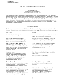
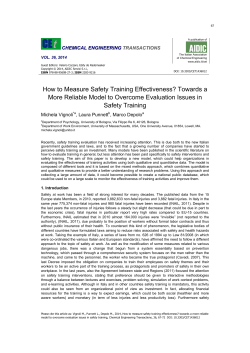
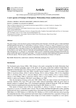
REVIEW
published: 17 March 2015
doi: 10.3389/fpls.2015.00127
Production of genetically and
developmentally modified seaweeds:
exploiting the potential of artificial
selection techniques
Bénédicte Charrier 1*, Elodie Rolland 1 , Vishal Gupta 2 and C. R. K. Reddy 2
1
Centre National de la Recherche Scientifique, Sorbonne Université, UPMC Univ Paris 06, UMR 8227, Integrative Biology of
Marine Models, Station Biologique de Roscoff, Roscoff, France, 2 Seaweed Biology and Cultivation Group, Division of Marine
Biotechnology and Ecology, CSIR-Central Salt and Marine Chemicals Research Institute, Bhavnagar, India
Edited by:
Stefan De Folter,
Centro de Investigación y de Estudios
Avanzados del Instituto Politécnico
Nacional, Mexico
Reviewed by:
Koji Mikami,
Hokkaido University, Japan
Nicholas Paul,
James Cook University, Australia
*Correspondence:
Bénédicte Charrier,
Centre National de la Recherche
Scientifique-UMR 8227, Station
Biologique, Place Georges Teissier,
Roscoff 29680, France
[email protected]
Specialty section:
This article was submitted to Plant
Evolution and Development, a section
of the journal Frontiers in
Plant Science
Received: 24 November 2014
Accepted: 17 February 2015
Published: 17 March 2015
Citation:
Charrier B, Rolland E, Gupta V and
Reddy CRK (2015) Production of
genetically and developmentally
modified seaweeds: exploiting the
potential of artificial selection
techniques. Front. Plant Sci. 6:127.
doi: 10.3389/fpls.2015.00127
Plant feedstock with specific, modified developmental features has been a quest for
centuries. Since the development and spread of agriculture, there has been a desire for
plants producing disproportionate—or more abundant and more nutritional—biomass
that meet human needs better than their native counterparts. Seaweed aquaculture,
targeted for human consumption and the production of various raw materials, is a rapidly
expanding field and its stakeholders have increasing vested interest for cost-effective
and lucrative seaweed cultivation processes. Thus, scientific research on seaweed
development is particularly timely: the potential for expansion of seaweed cultivation
depends on the sector’s capacity to produce seaweeds with modified morphological
features (e.g., thicker blades), higher growth rates or delayed (or even no) fertility.
Here, we review the various technical approaches used to modify development in
macroalgae, which have attracted little attention from developmental biologists to
date. Because seaweed (or marine macroalgae) anatomy is much less complex
than that of land plants and because seaweeds belong to three different eukaryotic
phyla, the mechanisms controlling their morphogenesis are key to understanding their
development. Here, we present efficient sources of developmentally and genetically
modified seaweeds—somatic variants, artificial hybrids and mutants—as well as the
future potential of these techniques.
Keywords: seaweed, somatic hybridization, mutagenesis, varietal improvement, genetic manipulation
Societal Importance of Seaweeds
Marine macroalgae (seaweeds) are one of the renewable resources in marine ecosystems and
carry out several key ecosystem functions that contribute to the productivity of the oceans.
Seaweeds are traditionally consumed as human food in several Asian countries, where they
are cultivated on a large scale. A wide range of seaweeds belonging to different genera have
been known as a food source since prehistoric times (Mouritsen et al., 2013). In most cases,
the entire thallus of the seaweed is consumed (e.g., species of Porphyra and Enteromorpha),
although, in some cases, only certain parts are edible (e.g., in Caulerpa lentillifera, only the
ramuli are edible). In larger seaweeds, such as Laminaria digitata and Undaria pinnatifida, the
stipe is used for preparing local dishes, such as soup. Chondrus crispus and certain species of
Frontiers in Plant Science | www.frontiersin.org
1
March 2015 | Volume 6 | Article 127
Charrier et al.
Genetic manipulation of seaweed development
variation can be transient, reversible or permanent. Temporary
changes are mainly due to either epigenetic or physiological
changes, which can be reversible, even after being heritable for
a few generations (Kaeppler et al., 2000). The underlying molecular mechanisms involved in permanent somaclonal variants are
rarely investigated and little understood (Larkin and Scowcroft,
1981). Nevertheless, most morphological variation observed during in vitro culture of seaweeds is transient and is not passed
on to progeny. There are many examples where same genotype
can produce different morphological phenotypes. For example:
in vitro tissue culture of kelp (order Laminariales) sporophytes
have a frequent developmental pattern in which outgrowths of
aposporous gametophyte-like filaments with differentiated fertile branches can give rise directly to sporophytes (Ar Gall
et al., 1996). Similarly, meristem cultures of Laminaria regenerate into one of three different body types: (1) uniseriate filaments; (2) thalloid-like structures; (3) dark green, compact calli.
Analogously, early development of embryonic germlings from 22
species of Fucaceae can show up to six different developmental
types.
Plant protoplasts are amenable to in vitro genetic manipulation techniques for developing genetically improved strains of
agronomic crops. There have been numerous studies on the isolation and regeneration of protoplasts from a wide variety of
seaweed body types ranging from simple leafy thalli to complex, cylindrical, branched thalli (see review Reddy et al., 2008).
Unlike higher plants, seaweed protoplasts regenerate and differentiate directly into a complete thallus without any addition of
phytohormones in the culture medium. Protoplasts from green
seaweeds nevertheless have different types of morphogenetic patterns (Figure 1B) and give rise to several phenotypically variable morphotypes, such as free-living sporangia, microthalli, or
saccate (or spherical), tubular (or spindle), irregular, or frondose thalli with various life spans (Reddy et al., 1989; Huang
et al., 1996; Chen, 1998; Krishna Kumar et al., 1999; Chen and
Shih, 2000; Rusig and Cosson, 2001). In the red alga Porphyra,
three different types of protoplast regeneration patterns have
been described: (1) callus form, (2) filamentous form and (3)
conchocelis form (Polne-Fuller et al., 1984; Fujita and Migita,
1985; Waaland et al., 1990; Dipakkore et al., 2005). Calluslike outgrowth (i.e., an unorganized cellular mass) has been
reported from protoplast culture of various brown macroalgal
species. Similarly, two distinct patterns of development have been
observed for protoplast-based development of Gracilaria gracilis, giving rise to plants that differ in appearance and life span.
Regenerated plants either resemble parental plants, with slender,
branched thalli, or remain small with thick, unbranched thalli,
many of which die. Although the underlying mechanisms have
been poorly studied, this variation in developmental morphological abnormalities has been attributed to a variety of factors,
including the type of donor tissue from which protoplasts are prepared and the culture conditions employed for regeneration. For
example, protoplasts isolated from the vegetative thalli of Monostroma latissimum regenerate into normal thalli, whereas protoplasts isolated from the holdfast develop into filaments (Chen,
1998). Protoplasts from Ulva fasciata develop into microthalli
when cultured in high density (Chen and Shih, 2000). For brown
Gracilaria are consumed in salad and in these cases only tender
tips are used. Seaweeds are good sources of high-quality digestible
proteins with a balanced amino acid composition, polyunsaturated fatty acids including omega-3 and omega-6 and important
vitamins and minerals, have a caloric content similar to land
crops and are high in fiber (MacArtain et al., 2007).
According to recent statistics published by the FAO (2014),
seaweed production has increased from less than 4 million wet
metric tons (t) in 1980 to almost 20 million wet t in 2012, with
more than 50% of total production is used for direct human consumption. The indirect products derived from seaweeds, polysaccharides in particular, have unique applications in processed as
well as functional foods, pet foods, feed, fertilizers, cosmetics,
and medicines. Furthermore, recent bioprocessing techniques
have also unequivocally demonstrated that seaweeds are potential feedstock for production of biofuels and commodity products
(Baghel et al., 2015).
The major seaweed species that are currently farmed include
Kappaphycus alvarezii and Eucheuma spp. both known as “cottonii” (>8 million wet t) followed by Saccharina japonica (formerly Laminaria japonica)—known as “kombu” (5.6 million wet
t), Gracilaria spp. (∼3 million wet t), U. pinnatifida—known as
“wakame” (2 million wet t) and Porphyra spp.—known as “nori”
(1.8 million wet t) (Buchholz et al., 2012). The use of Kappaphycus
enhances the texture of fish cutlets and pork patties and further
increases the nutritional value of foodstuffs by providing minerals (Senthil et al., 2005; Jeon and Choi, 2012). Other species such
as Palmaria, Chondrus, and Ulva are produced on a lower scale
(FAO, 2014). The global utilization of various seaweed species
is described in Supplementary Table 1. Seaweed cultivation is
now perceived as an excellent alternative source of revenue for
coastal fishermen, particularly in light of its advantages, including a shorter production cycle, low capital outlay and relatively
simple farming techniques. There have been some biotechnological advances to improve economically important seaweeds. The
present article briefly reviews the various research efforts that
have been made to produce morphological variants of economically important seaweeds and study the genetic basis behind the
changes in their morphology.
Genetic Manipulation and Improvement in
Seaweeds
Somaclonal Variants
Cellular biotechnology in seaweeds was initiated in the 1980s and
lags far behind that of terrestrial plants.
In macroalgae, the development of in vitro culture systems
facilitates mass proliferation of biomass all year round for the
production of valuable compounds under controlled conditions.
Culture systems are also a source of novel genetic variants with
useful traits arising from somaclonal variation (Figure 1A). The
techniques for somatic embryogenesis or dedifferentiation of
somatic cells often induce morphological variants in seaweeds.
Such morphological and developmental variation has advantages
for genetic improvement programs and also can be used as an
efficient means of germplasm storage and selection. The new
Frontiers in Plant Science | www.frontiersin.org
2
March 2015 | Volume 6 | Article 127
Charrier et al.
Genetic manipulation of seaweed development
Sub-culture of seaweed fragments and apospory; (B) Protoplast
preparation. (C) Somatic hybridization producing thalli with either novel
characters (green) or chimeric or mixed parental characters (yellow and
blue patches). (D) Parthenogenesis giving rise to morphological variants.
(E) Chemical or UV-mediated mutagenesis on gametes. Ploidy often
increases when (A–C) are employed.
FIGURE 1 | Summary of the different technological approaches for
generating morphologically modified macroalgae. Diploid sporophytes
are shown as the initial material. Change in color states indicates change in
morphology. The more the color is different from the original plant, the
more distinct the phenotype is. Dashed lines show cases that involve an
alternation of generations (e.g., from sporophyte to gametophyte). (A)
seaweeds in the order Laminariales, protoplast regeneration into
normal sporophytes can occur via different developmental processes, such as direct regeneration into plantlets (in L. digitata; Benet et al., 1997, U. pinnatifida; Matsumura et al., 2001
and L. japonica; Sawabe and Ezura, 1996; Sawabe et al., 1997;
Matsumura et al., 2000), or indirect regeneration, depending
on water temperature in some species. Indirect regeneration
occurs either after de-differentiation of the tissue into a filament (in U. pinnatifida; Matsumura et al., 2001, L. saccharina; Benet et al., 1997) or after the development of callus-like
masses [U. pinnatifida; (Matsumura et al., 2001) and L. japonica; Matsumura et al., 2000]. Gupta et al. (2012) provide the
first report of epigenetic regulation of morphology in protoplastderived germlings, with DNA methylation acting as an underlying molecular mechanism in protoplast-derived germlings in
Ulva reticulata (Figure 2). Axenic culture conditions may also
cause the development of abnormal thalli. Most studies have
shown that seaweed-associated microflora produce certain morphogenetic substances that, in turn, lead to normal thallus structure (Matsuo et al., 2005; Spoerner et al., 2012). However, most
commercial seaweed cultivation is currently based on simple vegetative propagation due to economic and farming advantages.
The in vitro culture techniques currently being developed for
Frontiers in Plant Science | www.frontiersin.org
seaweeds can create new genetic variants or promote clonal
propagation in photobioreactors for high end applications.
Inducing Morphological Variation through
Somatic Hybridization
In addition to morphological variation induced by in vitro culture, hybridization is an important process that combines phylogenetically distinct genetic lineages and results in morphotypes
that are either intermediate to the parental species or completely
novel (Figure 1C). In seaweeds, species are not always accurately
defined and are constantly being delineated through molecular genotyping or sequencing (Maggs et al., 2007). Through the
crossing of two genetically distinct species, hybridization can
bring together divergent genetic lineages (Hodge et al., 2010).
The resulting hybrid either combines parental phenotypes or
results in a new phenotype quite distinct from the parental type.
In addition to natural hybridization events that lead to speciation, somatic hybridization via protoplast fusion offers great
promise for achieving wide crosses between species that are difficult or impossible to hybridize conventionally (Davey et al.,
2005). Fusing protoplasts of different origins harnesses natural genetic diversity and provides novel genetic combinations
resulting in the potential improvement of various functional
3
March 2015 | Volume 6 | Article 127
Charrier et al.
Genetic manipulation of seaweed development
increase in ploidy itself can improve agronomic traits. Most
studies use color differences to distinguish heterokaryons from
homokaryons and from unfused parental protoplasts. Somatic
hybridization has shown unprecedented success in terrestrial
plants but few attempts have been made in seaweeds (Reddy
et al., 2008). Furthermore, a detailed description of cell division
and developmental stages is still lacking in almost all the protoplast fusion studies published to date. Below, we provide a brief
overview of the regeneration of fusion products so far reported
from different seaweed species.
Intrageneric Hybridization
The first report of protoplast fusion and fusion product regeneration between two color morphs of Porphyra was that of Fujita
and Migita (1987) using the polyethylene glycol (PEG) method.
Protoplast fusion from two different color types (green and reddish purple) in Porphyra yezoensis UEDA gave rise to calluslike masses that were composed of greenish and reddish-purple
cells of various diameters. The young thalli grown from these
cell masses attained a length of about 60 cm and were composed of microscopically chimeral tissues (as observed in the
calli), irregularly variegated with greenish and reddish purple
cell groups and possibly due to the independent segregation of
two chloroplast types from the fusion partners. Further studies on sexual crosses between green mutant and wild-type red
P. yezoensis demonstrate the production of uniform wild-type
color and green mutants along with mutants consisting both of
wild-type patches and green mutant patches (Niwa et al., 2002).
The mechanisms behind the formation of chimeric thalli from
protoplast fusion clearly differ from those involved in natural
crosses. Protoplast fusions between other Porphyra species such
as P. yezoensis and P. pseudolinearis give rise to plants resembling
either one or the other parent or occasionally mosaic chimeric
plants with different color contours (Fujita and Saito, 1990). Similar mechanisms of independent segregation of chloroplasts have
been reported in various studies on higher plants (Cocking, 1983;
Davey et al., 2005). These results reveal that crossing two species
by protoplast fusion or normal sexual crosses shows different
developmental processes. The possible reason for the development of chimeric thalli may be chromosomal complementation
from both fusion partners, whereas in natural crosses, only one
set of chromosomes from each fusion partner is found in the
resulting genotype.
Protoplast fusion between two Gracilaria species, i.e., between
G. tikvahiae (green pigmented) and G. chilensis (red pigmented)
result in bicolor, red and green chimeric plants (Cheney, 1990).
The hybrids exhibit several unusual morphological features, such
as branching morphology unlike either of the parental plants.
Limitations in the regeneration of somatic hybrids have led some
researchers to fuse newly released spores. Kapraun (1989, 1990)
developed parasexual hybrids from the fusion of zoospores from
Enteromorpha (now Ulva) and Ulvaria. Most hybrid germlings
showed morphology similar to Enteromorpha, although a few
showed multinucleate thalli with “giga” characteristics, including
larger cells with greatly enlarged vacuoles (nuclei in “gigas” vegetative cells are 8–10 µm diameter compared with 5–6 µm diameters in control plants). Mizukami et al. (1995) demonstrated
FIGURE 2 | Illustration of developmental variation in marine
macroalgae upon protoplast generation. Developmental variants among
protoplast-derived germlings of Ulva reticulata Forsskål C (Source: Gupta
et al., 2012). U. reticulata protoplasts (A) regenerate either into a callus (D–F)
due partly to symmetrical cell divisions (B,C) or into an organism displaying a
developmental pattern similar to that of native organisms (G–K). Note the
asymmetrical division taking place in (G), better seen in (H) after cell
enlargement.
traits. However, protoplast fusion leads to either heterokaryon
(fusion of nuclei of different species) or homokaryon (fusion
of nuclei of same species) recombination. In both cases, the
Frontiers in Plant Science | www.frontiersin.org
4
March 2015 | Volume 6 | Article 127
Charrier et al.
Genetic manipulation of seaweed development
for continued research in the development of macroalgae with
improved functional traits.
protoplast fusion between P. suborbiculata and P. yezoensis. The
hybrids initially showed repeated cell divisions and formed a
multicellular body, which, upon subsequent culturing, produced
rhizoids. The germinated hybrids showed thalli morphologically
different from each other as well as from the fusion partners.
The thallus of P. suborbiculata is brownish and roundish, and
possesses spike-like serrations on the margin, whereas the thallus of P. yezoensis is greenish and slender and has smooth margins. Hybrid thalli were usually brownish and had a roundish,
long and slender shape—thus combining features of the parental
thalli—and wrinkled margins.
Inducing Morphological Variation through
Parthenogenesis
Parthenogenesis can occur either through apospory (Figure 1A)
or apogamy (Figure 1D) without any ploidy changes. In the former, diploid gametophytes are produced directly by sporophytic
cells whereas, in the latter, haploid sporophytes produced directly
from gametophytic cells. In most cases, parthenogenetic plants
are identical to wild types but occasionally give rise to phenotypic variants. Parthenogenetic proliferation (asexual reproduction) has been extensively reported for several taxa of brown
and green seaweeds cultured in vitro, but are rarely reported in
red algae. Tatarenkov et al. (2005) observed dwarf morphs of
Fucus vesiculosus along with common morphs in wild populations itself in the brackish water Baltic Sea. The dwarf morphs
may have evolved from common morphs in response to the prevailing low salinity habitat. Shan et al. (2013) reported induction
of larger parthenogenetic sporophytes from gametophytic clones
of U. pinnatifida. Furthermore, the blades of parthenogenetic
sporophytes were uniformly smooth and without wrinkles on
either side of the mid-rib. The genetic features of parthenogenetic phenotypes have also been investigated for Laminaria
and Undaria. Parthenogenetic plants with morphological variation can lead to new insights into the genetics of the relationship
between ploidy level and morphology. These parthenogenetic
plants can also be used as a potential resource for breeding studies
for the genetic improvement of macroalgal strains of aquaculture
importance.
Intergeneric Hybridization
Subsequent studies on intergeneric protoplast fusion have been
carried out between morphologically different strains of Ulva
pertusa and Enteromorpha prolifera (Reddy et al., 1992). Presumptive heteroplasmic fusion products were identified based on
their larger size and the presence of twin chloroplasts. Subsequent
analysis on regeneration patterns of fused protoplasts showed
that they were similar to normal (unfused) protoplast development. Most of the regenerated plants from fusion products had
a thallus similar to either U. pertusa or E. prolifera. However, the
thalli of some plants had a characteristic irregular and dentate
margin, which has never been observed in the parental type.
Transdivisional Hybridization
Kito et al. (1998) published the first report of successful transdivisional protoplast fusion between Monostroma and Porphyra.
Protoplast fusion products of these two species showed different
regeneration processes and characteristics. Although the fusion
partners displayed distinct monostromatic and distromatic thalli,
the regenerated hybrids were green with a distromatic structure.
Initial heterokaryons were identified based on clearly distinguishable chloroplast colors, but were indistinguishable after 5 days of
culture. Of the hybrids generated, one of the heterofusant plants
grew into a multicellular body followed by the development of
rhizoid-like and bud-like organs. Finally, the plant grew into a
long stringy plant (1.5 m in length and 1.5 cm in width) and was
named “cyojo.” Another hybrid grew a multicellular body, which,
upon subsequent culturing, separated into individual cells, each
one growing into a long stringy plant; this mutant was named
“kattsunbo.” A third fusion product grew into a bud. This budlike plant became a thallus (7.5 cm in length and 6.5 cm in width)
and was named “nigo.”
These few examples illustrate the range of morphological
alterations that can be generated by somatic hybridization in
seaweeds. Furthermore, procedures have been developed to isolate seaweed protoplasts and bring them to the full thallus
regeneration stage on a wide range of seaweeds including the
most anatomically complex taxa such as Laminaria, Undaria,
Gracilaria, and Kappaphycus—which are some of the most
commercially important seaweed genera. Nevertheless, somatic
hybrids have not been widely developed and have not yet produced “cultivars” for field cultivation. However, the progress
made thus far with the development of homo- or heterokaryons via somatic hybridization provides useful groundwork
Frontiers in Plant Science | www.frontiersin.org
Mutagenesis-Mediated Morphological Variations
Macroalgal mutants have been little studied. In 1958, Ralph
Lewin emphasized the importance of macroalgal genetics (Lewin,
1958); now, more than 50 years later, still very few mutants
have been isolated and analyzed. In addition to the difficulties
of growing macroalgae in laboratory conditions and their complicated and relatively long life cycles, there is a surprising lack
of interest for this field of study. As a result—and despite the
amenability of haploid organisms for genetic and molecular analyses of underlying mechanisms and pathways—the identification
of genes involved in the control of marine macroalgal growth and
development lags far behind that of other studied multicellular
organisms (Maluszynski et al., 1995; Howell, 1998).
The most advanced genetic characterization of developmental mutants in a multicellular alga has been carried out in the
Chlorophyceae taxon Volvox carteri (sub-division Chlorophyta;
Leliaert et al., 2012). This microscopic freshwater alga is composed of about 2000 bi-flagellated cells stuck together within a
thick extracellular gelatinous matrix, forming a moving spherical body. While most cells remain flagellated and vegetative,
16 cells differentiate into non-flagellated, larger asexual reproductive cells (gonidia) from asymmetric cell divisions (Starr,
1969). Subsequent developmental steps lead to embryo inversion,
externalizing the flagellated somatic cells and internalizing the
gonidia.
5
March 2015 | Volume 6 | Article 127
Charrier et al.
Genetic manipulation of seaweed development
Chemical mutagenesis and transposon-tagging in V. carteri
produced several morphologically impaired mutants (Sessoms
and Huskey, 1973). The glsA gene, coding for a chaperone protein
involved in both protein translation and transcriptional regulation (Miller and Kirk, 1999; Pappas and Miller, 2009), controls
the asymmetric cell division giving rise to the gonidia cells. Additional mutants led to the identification of transcription factors
controlling genes specific to either the gonidial cells (lag gene) or
the somatic cells (regA gene) (Kirk, 2003). In addition, the invB
mutant, impaired in the inversion process, has been shown to
code for a nucleotide-sugar transporter (Ueki and Nishii, 2009)
necessary for the expansion of the glycoprotein-rich gonidia
vesicle, which tightly surrounds the multicellular sphere before
inversion.
The use of temperature-sensitive transposons to generate both
tagged mutants and revertants, and the use of genetic transformation to complement the mutants and further analyze them
at the molecular level are important assets of Volvox genetics
because they allow rapid molecular identification of the causal
genes (Ueki and Nishii, 2008). A similar approach can be used
in macroscopic and marine macroalgae. To date, only a very
few morphological mutants have been characterized—primarily
at the phenotypical level. In compensation for their complex
life cycles and sometimes challenging culture conditions, these
mutants often display similar or increased reproductive capabilities in lab conditions compared to the wild types (Ulva mutabilis, Fjeld and Løvlie, 1976). Mutants were obtained either from
sampling natural stocks, or among offspring of mutagenized
populations.
Naturally-Induced Mutations in Marine Macroalgae
Some macroalgal lines are particularly prone to spontaneous
mutations. The resulting phenotypes often revert to the wild
type, indicating that the mutations are unstable, with most being
attributed to a particularly unstable locus. However, there have
been some stable mutants that have lent themselves to genetic
analyses (Föyn, 1961). One laboratory strain of the green marine
macroalga U. mutabilis has been shown to be particularly inclined
to spontaneous morphological changes with a rate 10 times
higher than other strains (Fjeld and Løvlie, 1976). Some generated mutants displaying growth and morphological features different from the wild type parent have been reported (Föyn, 1959).
The slender mutant (Figure 3) displays a higher growth rate and
impairment in cell differentiation and cell enlargement, because
only small and undifferentiated cells are observed (Løvlie, 1969).
Similarly, cells show altered polarized division and cell differentiation in the lumpy mutant, which develops as a loose aggregate of undifferentiated cells with no specific organs (Bryhni,
1973, Figure 3). In this mutant, the composition of the cell wall
is modified, with an increased proportion of water-extractable
polysaccharides, resulting in higher plasticity of the cell wall
(Bryhni, 1978). It is not clear whether the inhibition of cell differentiation is a result of increased cell wall plasticity. In contrast, the globose mutant is small and seems to have a more
active differentiation process, developing many more rhizoids
and a darker thallus than the wild type (Föyn, 1961, not shown).
There are other distinct phenotypes, less well described (e.g.,
non-sporulating mutant, Figure 3). Løvlie (1978) also showed
developmental variations in U. mutabilis temperature-sensitive
FIGURE 3 | Morphology of some marine macroalgal mutants. Examples
of some morphological mutants of the green macroalga Ulva mutabilis, the red
macroalga Gracilaria tikvahiae at mature stages, and the brown macroalga
Ectocarpus siliculosus at early stages. Unless otherwise specified, the scale
Frontiers in Plant Science | www.frontiersin.org
bar is the same as for the wild-type individual. Permission to reproduce the
images of Ulva mutabilis (Fjeld and Løvlie, 1976) and Gracilaria tikvahiae
(Patwary and van der Meer, 1982) was granted by John Wiley & Sons, Inc. and
the journal Botany, respectively. Ectocarpus: personal photos (B. Charrier).
6
March 2015 | Volume 6 | Article 127
Charrier et al.
Genetic manipulation of seaweed development
transition to the reproductive phase is altered in some mutants.
Interestingly, most mutants are affected in several of these developmental features, indicating that the function of the wild-type
gene is not constrained to a single, precise developmental stage,
but spans different developmental phases. This is consistent with
the low level of architectural complexity of the Ectocarpus body,
which is mainly composed of filaments (Le Bail et al., 2008).
Because these mutations were generated randomly and
were not tagged, their identification requires forward genetic
approaches and molecular tools, unavailable at the time. Today
however, the advent of cost-effective and improved molecular
and sequencing techniques can help tackle the genetic characterization of these mutants, and even identify the causal mutation, and the gene directly responsible for the morphological
alteration. The recently described next-generation sequencingbased mapping approach developed for E. siliculosus (Billoud
et al., 2015) should pave the way to the other macroalgal
mutants generated from seaweeds with preferably small genome
sequences.
mutants. The mutants develop normal phenotypes, forming a filament consisting of a row of cells at 22◦ C, whereas at 15◦ C they
produce abnormal phenotypes.
In the brown alga Ectocarpus siliculosus, the natural immediate upright mutant shows a flaw in the early morphogenesis of
the filamentous sporophyte, in which the prostrate basal body is
reduced, thereby resembling a gametophyte with mainly upright
filaments (Peters et al., 2008). This phenotype is due to a singlelocus mutation, emerging in the offspring population (gametophytes) from a wild-type sporophyte, probably by spontaneous
random mutation.
Chemically or UV-Light Induced Mutations in Marine
Macroalgae
Various mutagenic agents have been used in marine macroalgae
(Figure 1E). Chemical agents, such as N-methyl-N’-nitro-Nnitrosoguanidine and ethyl-methane sulfonate (EMS) are efficient. EMS has been used to produce red macroalga G. tikvahiae
mutants (van der Meer, 1979), which display altered branching
patterns and thallus thickness, potentially due to the modification of the size of the medullary and sub-cortical cells of the
algal thallus (Patwary and van der Meer, 1982). These morphological alterations are accompanied by a modification in the agar
composition: agar strength is higher in the MP-40 mutant, but
lower in the MP-61 mutant (Patwary and van der Meer, 1983a)
(Figure 3). Interesting biological issues therefore lie in the potential functional link between the biophysical characteristics of agar
and the morphological alterations observed in these mutants. The
demonstration of this functional link requires adequate segregation analyses of these characters in the progeny of the mutants,
which, unfortunately, were not carried out at that time. Nevertheless, and of interest for aquaculture purposes, the MP-40 mutant
displays a faster growth rate than the wild-type even at high densities. It also fixes ammonium better from seawater and is more
resistant to epiphytic colonization (Patwary and van der Meer,
1983b). In contrast to most generated mutations that were single
locus and recessive, the MP-40 mutant displayed a partly dominant mutation (Patwary and van der Meer, 1982). Unavailable in
the 1980s, modern technologies can now use this genetic feature
to identify the mutated gene.
The use of chemical mutagens in liquid media represents a
major drawback due to the risk of contamination during waste
elimination. UV irradiation is safer and as efficient, and has
been used on the green macroalga U. mutabilis and the brown
macroalga E. siliculosus. Half-life curves related to the mutagen dose have been established (Le Bail and Charrier, 2013).
Exposition of gametes to UV-irradiation doses corresponding
to 50% survival produce only single-locus mutations in Ectocarpus (étoile mutant, Le Bail et al., 2011, orobourous mutant,
Coelho et al., 2011; mut mutant, Billoud et al., 2015), making
them amenable to relatively simple genetic analyses. A library
of 60 morphological mutants has been constructed for E. siliculosus, which all show a stable phenotype after five rounds of
sub-culturing or five parthenogenetic generations (Le Bail and
Charrier, 2013). The observed morphological defaults cover all
the developmental steps in this alga, ranging from alterations
in cell differentiation and filament growth polarity, to positional
and stage modification in the branching process (Figure 3). The
Frontiers in Plant Science | www.frontiersin.org
Conclusion and Future Prospects
Evidence that intensive cultivation of inbred seaweed lines results
in both an impoverishment of wild biodiversity (possibly by
gene swamping) and an outbreak of infections and epiphytism
in culture plants (Loureiro et al., 2015), has raised awareness
in aquaculture stakeholders and policy-makers that the inventory, the maintenance and the exploitation of seaweed genetic
resources should be fully deployed (Pullin, 2006; Pullin and
White, 2011; FAO, 2013). Hence, initiation and development of
long-term seaweed genetic improvement programs are recommended, which include continuous selection processes targeting
the maintenance of genetic diversity throughout the production
of improved lines (Robinson et al., 2013). Macroalgal stocks with
altered morphological traits can either be continuously collected
from the wild or generated on demand in culture facilities using
a combination of techniques likely to modify the developmental
traits in a stable way. Although somatic hybridization frequently
gives unpredictable results, generating hybrids between macroalgal partners displaying interesting and stable morphological
features—obtained for example by induced mutagenesis—can
combine both traits in a single organism. Using this approach,
Mizukami et al. (1995) generated hybrids between a wild type and
a mutant strain of P. yezoensis.
In addition to the interest of these techniques for aquaculture,
production of morphologically altered macroalgae is a very valuable tool for studying the genetics of basic developmental mechanisms. In comparison with the wild-type organism, a mutant
can provide information about cell types, specific developmental
steps and gene expression patterns that play a role in the impaired
developmental process, and may lead to the identification of the
causal gene itself. In this regard, the development of genetic studies should be undertaken to address some biological issues that
can only otherwise just be touched upon. Genetic transformation, of little use in the aquaculture sector (Robinson et al., 2013),
can target specific cellular mechanisms by selectively modifying the expression of key genes, as it has been performed for
decades in land plants. Genetic transformation of macroalgae is
7
March 2015 | Volume 6 | Article 127
Charrier et al.
Genetic manipulation of seaweed development
now possible in red and green seaweeds (Mikami, 2014; Wichard
et al., 2015) and should be further developed, especially in brown
algae, so that genetics and transgenesis can be used to their full
potential in the field of macroalgal developmental biology.
Altogether, the current stage of technological knowledge now
allows access to new and more powerful ways of generating morphologically altered macroalgae, using both traditional methods,
such as the production of somaclonal variants (whose phenotype
cannot be predicted and controlled), and newly developed genetargeted methods, such as creating macroalgal variants with specific, precise and predictable phenotypes. The combined use of
these methods will help bring the field of macroalgal development
up to speed, to match that of land plants.
Acknowledgments
References
FAO. (2014). The State of World Fisheries and Aquaculture: opportunities and
challenges. Rome.
Fjeld, A., and Løvlie, A. (1976). “Genetics of multicellular marine algae,” in The
Genetics of Algae Botanical Monographs, eds R. A. Lewin (Oxford; London;
Edinburgh; Melbourne, VIC: Blackwell Scientific Publication), 219–235.
Föyn, B. (1959). Geschlechtskontrollierte Vererbung bei der marinen Grünalga
Ulva mutabilis. Arch. Protistenkd. 104, 236–253.
Föyn, B. (1961). Globose, a recessive mutant in Ulva mutabilis. Bot. Mar. 3, 60–64.
Fujita, Y., and Migita, S. (1985). Isolation and culture of protoplasts from seaweeds.
Bull. Fac. Fish. Nagasaki Univ. 57, 39–45.
Fujita, Y., and Migita, S. (1987). Fusion of protoplasts from thalli of two different
color types in Porphyra yezoensis Ueda and development of fusion products.
Jpn. J. Phycol. 35, 201–208.
Fujita, Y., and Saito, M. (1990). Protoplasts isolation and fusion in Porphyra (Bangiales, Rhodophyta). Hydrobiologia 204/205, 161–166. doi:
10.1007/BF00040228
Gupta, V., Bijo, A. J., Kumar, M., Reddy, C. R. K., and Jha, B. (2012). Detection
of epigenetic variations in the protoplast-derived germlings of Ulva reticulata using methylation sensitive amplification polymorphism (MSAP). Mar.
Biotechnol. 14, 692–700. doi: 10.1007/s10126-012-9434-7
Hodge, F. J., Buchanan, J., and Zuccarello, G. C. (2010). Hybridization between
the endemic brown algae Carpophyllum maschalocarpum and Carpophyllum
angustifolium (Fucales): genetic and morphological evidence. Phycol. Res. 58,
239–247. doi: 10.1111/j.1440-1835.2010.00583.x
Howell, S. H. (1998). Molecular Genetics of Plant Development. Cambridge, UK:
Cambridge University Press.
Huang, X., Weber, J. C., Hinson, T. K., Mathieson, A. C., and Minocha, S. C. (1996).
Transient expression of the GUS reporter gene in the protoplasts and partially
digested cells of Ulva lactuca L. (Chlorophyta). Bot. Mar. 39, 467–474.
Jeon, M. R., and Choi, S. H. (2012). Quality characteristics of pork patties added
with seaweed powder. Korean J. Food Sci. Anim. Resour. 32, 77–83. doi:
10.5851/kosfa.2012.32.1.71
Kaeppler, S. M., Kaeppler, H. F., and Rhee, Y. (2000). Epigenetic aspects
of somaclonal variation in plants. Plant Mol. Biol. 43, 179–188. doi:
10.1023/A:1006423110134
Kapraun, D. F. (1989). Recovery and development of parasexual fusion products in
Enteromorpha linza (L.) J Ag (Ulvales, Chlorophyta). J. App. Phycol. 1, 239–246.
doi: 10.1007/BF00003649
Kapraun, D. F. (1990). Parasexual fusion products in green algae: Enteromorpha and Ulvaria (Ulvales, Chlorophyta). Hydrobiologia 204/205, 151–159. doi:
10.1007/BF00040227
Kirk, D. L. (2003). Seeking the ultimate and proximate causes of volvox multicellularity and cellular differentiation. Integr. Comp. Biol. 43, 247–253. doi:
10.1093/icb/43.2.247
Kito, H., Kunimoto, M., Kamanishi, Y., and Mizukami, Y. (1998). Protoplasts fusion between Monostroma nitidum and Porphyra yezoensis and
subsequent growth of hybrid plants. J. Appl. Phycol. 10, 15–21. doi:
10.1023/A:1008063415548
CSIR, New Delhi, Department of Biotechnology, New Delhi
and Department of Science and Technology, New Delhi
are gratefully acknowledged for their long term funding support that led to advancement of Indian seaweed
research.
Supplementary Material
The Supplementary Material for this article can be found
online at: http://www.frontiersin.org/journal/10.3389/fpls.2015.
00127/abstract
Ar Gall, E., Asensi, A., Marie, D., and Kloareg, B. (1996). Parthenogenesis and
apospory in the Laminariales: a flow cytometry analysis. Eur. J. Phycol. 31,
369–380.
Baghel, R. S., Trivedi, N., Vishal, G., Neori, A., Reddy, C. R. K., Lali, A., et al.
(2015). Biorefining of marine macroalgal biomass for production of biofuel and
commodity chemicals. Green Chem. doi: 10.1039/C4GC02532F
Benet, H., Gall, A. E., Asensi, A., and Kloareg, B. (1997). Protoplast regeneration
from gametophytes and sporophytes of some species in the order Laminariales
(Phaeophyceae). Protoplasma 199, 39–48. doi: 10.1007/BF02539804
Billoud, B., Jouanno, É., Nehr, Z., Carton, B., Rolland, É., Chenivesse, S., et al.
(2015). Localisation of causal locus in the genome of the brown macroalga Ectocarpus: NGS-based mapping and positional cloning approaches. Front. Plant
Sci. 6:68. doi: 10.3389/fpls.2015.00068
Bryhni, E. (1973). Differences in cell-walls of Ulva mutabilis wild-type and its
disorganized mutant lumpy. J. Ultrastruct. Res. 42, 412.
Bryhni, E. (1978). Quantitative differences between polysaccharide compositions
in normal differentiated Ulva mutabilis and the undifferentiated mutant lumpy.
Phycologia 17, 119–124. doi: 10.2216/i0031-8884-17-2-119.1
Buchholz, C. M., Krause, G., and Buck, B. H. (2012). “Seaweed and man,” in Seaweed Biology: Novel Insights into Ecophysiology, Ecology, and Utilization. Ecological Studies, Vol. 219, eds C. Wiencke and K. Bischof (Berlin; Heidelberg:
Springer-Verlag), 471–493.
Chen, Y. C. (1998). Development of protoplasts from holdfasts and vegetative
thalli of Monostroma latissimum (Chlorophyta, Monostromatacae) for algal
seed stock. J. Phycol. 34, 1075–1081. doi: 10.1046/j.1529-8817.1998.341075.x
Chen, Y. C., and Shih, H. C. (2000). Development of protoplasts of Ulva fasciata
(Chlorophyta) for algal seed stock. J. Phycol. 36, 608–615. doi: 10.1046/j.15298817.2000.99128.x
Cheney, D. (1990). “Genetic improvement of seaweeds through protoplasts
fusion,” in Economically Important Marine Plants of the Atlantic: their Biology
and Cultivation, eds C. Yarish, C. Penniman, and P. van Patten (Storrs, CT: Univ
Conn Sea Grant Program), 15–25.
Cocking, E. C. (1983). The enzymatic isolation of plant protoplasts: historical
perspective. Int. Rev. Cytol. 16, 1–3.
Coelho, S. M., Godfroy, O., Arun, A., Le Corguillé, G., Peters, A. F., and Cock, J. M.
(2011). OUROBOROS is a master regulator of the gametophyte to sporophyte
life cycle transition in the brown alga Ectocarpus. Proc. Natl. Acad. Sci. U.S.A.
108, 11518–11523. doi: 10.1073/pnas.1102274108
Davey, M. R., Anthony, P., Power, J. B., and Lowe, K. C. (2005). Plant protoplasts: status and biotechnological perspectives. Biotechnol. Adv. 23, 131–171.
doi: 10.1016/j.biotechadv.2004.09.008
Dipakkore, S., Reddy, C. R. K., and Jha, B. (2005). Production and seeding of
protoplast of Porphyra okhaensis (Bangials, Rhodophyta) in laboratory culture.
J. Appl. Phycol. 17, 331–337. doi: 10.1007/s10811-005-7291-8
FAO. (2013). CGRFA-14/13/Report- Report of the Fourteenth Regular Session of the
Commission on Genetic Resources for Food and Agriculture Rome, Italy, 15–19.
Frontiers in Plant Science | www.frontiersin.org
8
March 2015 | Volume 6 | Article 127
Charrier et al.
Genetic manipulation of seaweed development
Niwa, K., Mizuta, A., and Aruga, Y. (2002). Genetic characterization of a spontaneous green-type pigmentation mutant of Porphyra yezoensis and the significance of using heterozygous conchocelis in nori farming. Fish. Sci. 68, 729–735.
doi: 10.1046/j.1444-2906.2002.00486.x
Pappas, V., and Miller, S. M. (2009). Functional analysis of the Volvox carteri asymmetric division protein GlsA. Mech. Dev. 126, 842–851. doi:
10.1016/j.mod.2009.07.007
Patwary, M. U., and van der Meer, J. P. (1982). Genetics of Gracilaria tikvahiae (Rhodophyceae). VIII: phenotypic and genetic characterization of some
selected morphological mutants. Can. J. Bot. 60, 2556–2564. doi: 10.1139/b
82-310
Patwary, M. U., and van der Meer, J. P. (1983a). Genetics of Gracilaria tikvahiae
(Rhodophyceae). IX: some properties of agars extracted from morphological
mutants. Bot. Mar. 26, 295–299.
Patwary, M. U., and van der Meer, J. P. (1983b). Growth experiments on morphological mutants of Gracilaria tikvahiae (Rhodophyceae). Can. J. Bot. 61,
1654–1659.
Peters, A. F., Scornet, D., Ratin, M., Charrier, B., Monnier, A., Merrien, Y., et al.
(2008). Life-cycle-generation-specific developmental processes are modified in
the immediate upright mutant of the brown alga Ectocarpus siliculosus. Dev.
Camb. Engl. 135, 1503–1512.
Polne-Fuller, M., Biniaminov, M., and Gibor, A. (1984). Vegitative propagation of
Porphyra perforata. Proc. Int. Seaweed Symp. 11, 308–313. doi: 10.1007/978-94009-6560-7_60
Pullin, R. S. V. (2006). “Genetic resources for aquaculture: status and trends,”
in Workshop on Status and Trends in Aquatic Genetic Resources. A Basis for
International Policy, eds D. M. Bartley, B. J. Harvey, and R. S. V. Pullin (FAO
Fisheries Proceedings), 109–143.
Pullin, R., and White, P. (2011). Climate Change and Aquatic Genetic Resources for
Food and Agriculture: State of Knowledge, Risks and Opportunities. Background
Study Paper No. 55. Rome: FAO Commission on Genetic Resources for Food
and Agriculture.
Reddy, C. R. K., Gupta, M. K., Mantri, V. A., and Jha, B. (2008). Seaweed protoplasts: status, biotechnological perspectives and needs. J. Appl. Phycol. 20,
619–632. doi: 10.1007/s10811-007-9237-9
Reddy, C. R. K., Iima, M., and Fujita, Y. (1992). Induction of fast growing and
morphologically different strains through intergeneric protoplasts fusion of
Ulva and Enteromorpha (Ulvales, chlorophyta). J. Appl. Phycol. 4, 57–65. doi:
10.1007/BF00003961
Reddy, C. R. K., Migita, S., and Fujita, Y. (1989). Protoplasts isolation and regeneration of three species of Ulva in axenic culture. Bot. Mar. 32, 483–490. doi:
10.1515/botm.1989.32.5.483
Robinson, N., Winberg, P., and Kirkendale, L. (2013). Genetic improvement of
macroalgae: status to date and needs for the future. J. Appl. Phycol. 25, 703–716.
doi: 10.1007/s10811-012-9950-x
Rusig, A., and Cosson, J. (2001). Plant regeneration from protoplasts of
Enteromorpha intestinalis (Chlorophyta, Ulvophyceae) as seed stock for
macroalgal culture. J. Appl. Phycol. 13, 103–108. doi: 10.1023/A:101113
6712938
Sawabe, T., and Ezura, Y. (1996). Regeneration from Laminaria japonica
Areschoug (Laminariales, Phaeophyceae) protoplasts isolated with bacterial
alginase. Plant Cell Rep. 15, 892–895.
Sawabe, T., Ezura, Y., and Yamamoto, H. (1997). Plant regeneration from protoplasts of Laminaria japonica Areschoug (Laminariales, Phaeophyceae) in a
continuous-flow culture system. Plant Cell Rep. 17, 109–112.
Senthil, A., Mamatha, B. S., and Mahadevaswamy, M. (2005). Effect of using seaweed (Euchema) powder on the quality of fish cutlet. J. Food Sci. Nutr. 56,
327–335. doi: 10.1080/09637480500224205
Sessoms, A. H., and Huskey, R. J. (1973). Genetic control of development in
Volvox: isolation and characterization of morphogenetic mutants. Proc. Natl.
Acad. Sci. U.S.A. 70, 1335–1338. doi: 10.1073/pnas.70.5.1335
Shan, T. S., Pang, S. J., and Gao, S. Q. (2013). Novel means for variety breeding and sporeling production in the brown seaweed Undaria pinnatifida
(Phaeophyceae): crossing female gametophytes from parthenosporophytes
with male gametophyte clones. Phycol. Res. 61, 154–161. doi: 10.1111/pre.
12014
Spoerner, M., Wichard, T., Bachhuber, T., Stratmann, J., and Kendal, W. S.
(2012). Ulva mutabilis (Chlorophyta) depends on a combination of two
Krishna Kumar, G. R., Addepalli, M. K., and Reddy, C. R. K. (1999). Regeneration
of the thallus of Monostroma oxyspermum (Chlorophyta) from protoplasts in
axenic culture. Phycologia 38, 503–507. doi: 10.2216/i0031-8884-38-6-503.1
Larkin, P. J., and Scowcroft, W. R. (1981). Somaclonal variation—a novel source
of variability from cell cultures for plant improvement. Theor. Appl. Genet. 60,
197–214.
Le Bail, A., Billoud, B., Le Panse, S., Chenivesse, S., and Charrier, B. (2011). ETOILE
regulates developmental patterning in the filamentous brown alga Ectocarpus
siliculosus. Plant Cell 23, 1666–1678. doi: 10.1105/tpc.110.081919
Le Bail, A., Billoud, B., Maisonneuve, C., Peters, A., Cock, J. M., and Charrier,
B. (2008). Initial pattern of development of the brown alga Ectocarpus siliculosus (Ectocarpales, Phaeophyceae) sporophyte. J. Phycol. 44, 1269–1281. doi:
10.1111/j.1529-8817.2008.00582.x
Le Bail, A., and Charrier, B. (2013). “Culture methods and mutant generation in the
filamentous brown algae Ectocarpus siliculosus,” in Plant Organogenesis Methods in Molecular Biology, ed I. De Smet (Humana Press), 323–332. Available at:
http://dx.doi.org/10.1007/978-1-62703-221-6_22.
Leliaert, F., Smith, D. R., Moreau, H., Herron, M. D., Verbruggen, H., Delwiche,
C. F., et al. (2012). Phylogeny and molecular evolution of the green algae. Crit.
Rev. Plant Sci. 31, 1–46. doi: 10.1080/07352689.2011.615705
Lewin, R. A. (1958). “Genetics and marine algae,” in Perspectives in Marine Biology, eds L. A. Berkeley and A. A. Buzzati-Traverso (Berkeley, CA: University of
California Press), 547–557.
Loureiro, R., Gachon, C. M. M., and Rebours, C. (2015). Seaweed cultivation:
potential and challenges of crop domestication at an unprecedented pace. New
Phytol. doi: 10.1111/nph.13278. [Epub ahead of print].
Løvlie, A. (1969). Cell size, nucleic acids and synthetic efficiency in the wild type
and a growth mutant of the multicellular alga Ulva mutabilis Föyn. Dev. Biol.
20, 349–367. doi: 10.1016/0012-1606(69)90019-0
Løvlie, A. (1978). On the genetic control of cell cycles during morphogenesis in Ulva mutabilis. Dev. Biol. 64, 164–177. doi: 10.1016/0012-1606(78)9
0068-4
MacArtain, P., Gill, C. I., Brooks, M., Campbell, R., and Rowland, I. R.
(2007). Nutritional value of edible seaweeds. Nutr. Rev. 65, 535–543. doi:
10.1111/j.1753-4887.2007.tb00278.x
Maggs, C. A., Verbruggen, H., and De Clerck, O. (2007). “Molecular systematics
of red algae: building future structures on firm foundations,” in Unravalling the
Algae: The Past, Present and Future of Algal Systematics, eds J. Brodie and J.
M. Lewis (Boca Raton, FL; London, UK; New York, NY: Taylor and Francis),
103–121.
Maluszynski, M., Ahloowalia, B. S., and Sigurbjörnsson, B. (1995). “Application of
in vivo and in vitro mutation techniques for crop improvement,” in The Methodology of Plant Genetic Manipulation: Criteria for Decision Making. Developments in Plant Breeding, eds A. C. Cassells and P. W. Jones (Dordrecht:
Springer), 303–315.
Matsumura, W., Yasui, H., and Yamamoto, H. (2000). Mariculture of Laminaria
japonica (Laminariales, Phaeophyceae) using protoplast regeneration. Phycol.
Res. 48, 169–176. doi: 10.1111/j.1440-1835.2000.tb00213.x
Matsumura, W., Yasuj, H., and Yamamoto, H. (2001). Successful sporophyte regeneration from protoplasts of Undaria pinnatifida (laminariales, phaeophyceae).
Phycologia 40, 10–20. doi: 10.2216/i0031-8884-40-1-10.1
Matsuo, Y., Imagawa, H., Nishizawa, M., and Shizuri, Y. (2005). Isolation of an
algal morphogenesis inducer from a marine bacterium. Science 307, 1598. doi:
10.1126/science.1105486
Mikami, K. (2014). A technical breakthrough close at hand: feasible approaches
toward establishing a gene-targeting genetic transformation system in seaweeds. Front. Plant Sci. 5:498. doi: 10.3389/fpls.2014.00498
Miller, S. M., and Kirk, D. L. (1999). glsA, a Volvox gene required for asymmetric division and germ cell specification, encodes a chaperone-like protein. Dev.
Camb. Engl. 126, 649–658.
Mizukami, Y., Okauchi, M., Kito, H., Ishimoto, S., Ishida, T., and Fuseya,
M. (1995). Culture and development of electrically fused protoplasts from
red marine algae, Porphyra yezoensis and P. suborbiculata. Aquaculture 132,
361–367. doi: 10.1016/0044-8486(95)93662-T
Mouritsen, O. G., Dawczynski, C., Duelund, L., Jahreis, G., Vetter, W., and
Schröder, M. (2013). On the human consumption of the red seaweed dulse
(Palmaria palmate (L.) Weber & Mohr). J. Appl. Phycol. 25, 1777–1791. doi:
10.1007/s10811-013-0014-7
Frontiers in Plant Science | www.frontiersin.org
9
March 2015 | Volume 6 | Article 127
Charrier et al.
Genetic manipulation of seaweed development
Waaland, J. R., Dickson, L. G., and Watson, B. A. (1990). Protoplasts isolation and
regeneration in the marine red alga Porphyra nereocystis. Planta 181, 522–528.
doi: 10.1007/BF00193005
Wichard, T., Charrier, B., Mineur, F., Bothwell, J. H., De Clerck, O., and Coates,
J. C. (2015). The green seaweed Ulva: a model system to study morphogenesis.
Front. Plant Sci. 6:72. doi: 10.3389/fpls.2015.00072
bacterial species excreting regulatory factors. J. Phycol. 48, 1433–1447. doi:
10.1111/j.1529-8817.2012.01231.x
Starr, R. C. (1969). Structure, reproduction and differentiation in Volvox carteri f.
nagariensis Iyengar, HK9 and 10. Arch. Protistenkd. 11, 204–222.
Tatarenkov, A., Bergstrom, L., Jonsson, R. B., Serrao, E. A., Kautsky, L., and
Johannesson, K. (2005). Intriguing asexual life in marginal populations of the
brown seaweed Fucus vesiculosus. Mol. Ecol. 14, 647–651. doi: 10.1111/j.1365294X.2005.02425.x
Ueki, N., and Nishii, I. (2008). Idaten is a new cold-inducible transposon of Volvox
carteri that can be used for tagging developmentally important genes. Genetics
180, 1343–1353. doi: 10.1534/genetics.108.094672
Ueki, N., and Nishii, I. (2009). Controlled enlargement of the glycoprotein vesicle surrounding a Volvox embryo requires the InvB nucleotide-sugar transporter and is required for normal morphogenesis. Plant Cell 21, 1166–1181.
doi: 10.1105/tpc.109.066159
van der Meer, J. P. (1979). Genetics of Gracilaria sp. (Rhodophyceae, Gigartinales).
V. Isolation and characterization of mutant strains. Phycologia 18, 47–54. doi:
10.2216/i0031-8884-18-1-47.1
Frontiers in Plant Science | www.frontiersin.org
Conflict of Interest Statement: The authors declare that the research was conducted in the absence of any commercial or financial relationships that could be
construed as a potential conflict of interest.
Copyright © 2015 Charrier, Rolland, Gupta and Reddy. This is an open-access
article distributed under the terms of the Creative Commons Attribution License
(CC BY). The use, distribution or reproduction in other forums is permitted, provided the original author(s) or licensor are credited and that the original publication in this journal is cited, in accordance with accepted academic practice. No
use, distribution or reproduction is permitted which does not comply with these
terms.
10
March 2015 | Volume 6 | Article 127


© Copyright 2026