
Long-term Consequences of Repetitive Brain Trauma: Chronic Traumatic Encephalopathy Concussion Supplement
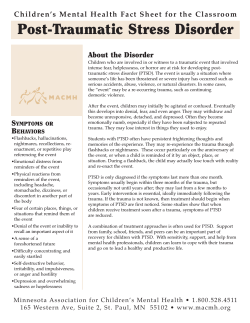
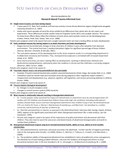
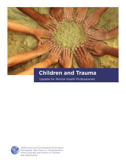
Concussion Supplement
Long-term Consequences of Repetitive Brain Trauma:
Chronic Traumatic Encephalopathy
Robert A. Stern, PhD, David O. Riley, BS, Daniel H. Daneshvar, MA,
Christopher J. Nowinski, BA, Robert C. Cantu, MD, Ann C. McKee, MD
Abstract: Chronic traumatic encephalopathy (CTE) has been linked to participation in
contact sports such as boxing and American football. CTE results in a progressive decline of
memory and cognition, as well as depression, suicidal behavior, poor impulse control,
aggressiveness, parkinsonism, and, eventually, dementia. In some individuals, it is associated with motor neuron disease, referred to as chronic traumatic encephalomyelopathy,
which appears clinically similar to amyotrophic lateral sclerosis. Results of neuropathologic
research has shown that CTE may be more common in former contact sports athletes than
previously believed. It is believed that repetitive brain trauma, with or possibly without
symptomatic concussion, is responsible for neurodegenerative changes highlighted by
accumulations of hyperphosphorylated tau and TDP-43 proteins. Given the millions of
youth, high school, collegiate, and professional athletes participating in contact sports that
involve repetitive brain trauma, as well as military personnel exposed to repeated brain
trauma from blast and other injuries in the military, CTE represents an important public
health issue. Focused and intensive study of the risk factors and in vivo diagnosis of CTE will
potentially allow for methods to prevent and treat these diseases. Research also will provide
policy makers with the scientific knowledge to make appropriate guidelines regarding the
prevention and treatment of brain trauma in all levels of athletic involvement as well as the
military theater.
PM R 2011;3:S460-S467
INTRODUCTION
There has been increased attention given to the recognition, diagnosis, and management of
sports-related concussion. Participants in popular sports such as American football, hockey,
wrestling, rugby, soccer, and lacrosse all risk exposure to brain injury that range from
asymptomatic subconcussive blows to symptomatic concussion to more moderate or severe
traumatic brain injury (TBI). In addition, military service and many other activities,
including, but not limited to, downhill skiing, martial arts, horse riding, parachuting, and
other adventure sports have been associated with TBI [1-3]. An estimated 1.6-3.8 million
sports- and recreation-related concussions occur in the United States each year, although
the true figure is unknown because most concussions are not recognized and reported [4-6].
In addition to symptomatic concussions, players in collision sports such as American
football may experience many more subconcussive impacts throughout a season and career.
Athletes at certain positions (eg, linemen) may sustain up to 1400 impacts per season, and
high school players who play both offense and defense potentially sustain closer to 2000
impacts [7-10].
A mild TBI (mTBI) or concussion is a brain injury that results from a force transmitted to
the head that leads to a collision between the brain and skull or to a strain on the tissue and
vasculature of the brain [11,12]. This injury can lead to a variety of physical, psychosocial,
and cognitive symptoms, and, when these deficits do not resolve, can result in postconcussion syndrome (PCS) [13]. Common symptoms include fatigue, dizziness, headache, light
and noise sensitivity, mental fogginess, depression, irritability, and impairments of executive functioning and concentration. Because a concussion and its symptoms result from
temporary, reversible cytoskeletal and metabolic derangements that involve shifts in ion
PM&R
S460
1934-1482/11/$36.00
Printed in U.S.A.
R.A.S. Center for the Study of Traumatic Encephalopathy, Boston University School of
Medicine, Boston University, 72 East Concord
St, 7380, Boston, MA 02118; Department of
Neurology and Neurosurgery; Alzheimer’s Disease Center Clinical Core, Boston University
School of Medicine, Boston, MA. Address
correspondence to: R.A.S.; e-mail: bobstern@
bu.edu
Disclosure: nothing to disclose
D.O.R. Center for the Study of Traumatic Encephalopathy, Department of Neurology, Boston University School of Medicine, Boston, MA
Disclosure: nothing to disclose
D.H.D. Department of Neurology, Boston University School of Medicine, Boston, MA
Disclosure: nothing to disclose
C.J.N. Center for the Study of Traumatic Encephalopathy, Boston University School of
Medicine, Boston, MA, Sports Legacy Institute,
Waltham, MA
Disclosure: nothing to disclose
R.C.C. Center for the Study of Traumatic Encephalopathy, and the Department of Neurosurgery, Boston University School of Medicine,
Boston, MA; Sports Legacy Institute, Waltham,
MA; Neurosurgery Service, and the Department of Surgery, Emerson Hospital, Concord,
MA; and Sports Medicine, Emerson Hospital,
Concord, MA
Disclosure: nothing to disclose
A.C.M. Center for the Study of Traumatic
Encephalopathy; Neurology and Pathology,
Boston University School of Medicine, Boston,
MA
Disclosure: nothing to disclose
Disclosure Key can be found on the Table of
Contents and at www.pmrjournal.org
Research support: This work was supported
by the Boston University Alzheimer’s Disease
Center NIA P30 AG13846, supplement
0572063345-5; NIH R01NS078337; a grant
from the National Operating Committee on
Standards for Athletic Equipment; the Sports
Legacy Institute; and an unrestricted gift from
the National Football League
© 2011 by the American Academy of Physical Medicine and Rehabilitation
Vol. 3, S460-S467, October 2011
DOI: 10.1016/j.pmrj.2011.08.008
PM&R
channels and energy imbalance, the majority of deficits associated with a concussive injury resolve within a matter of
days, weeks, or months [13-16]. In some instances, PCS may
persist for months or years after the initial injury. However, it
is believed that no more than 15% of individuals with a
history of mTBI still experience symptoms 1 year after injury
[13,17,18].
To date, the resulting progressive tauopathy, known as
chronic traumatic encephalopathy (CTE), has only been
found in individuals with a history of repetitive brain trauma
[3,7]. Despite the recent increase in attention on the longterm effects of repetitive brain trauma, including CTE, it has
been known for some time that contact sports may be associated with neurodegenerative disease. In 1928, Martland
[19] described a symptom spectrum in boxers, which he
termed “punch drunk,” that appeared to result from the
repeated blows experienced in the sport, particularly in slugging boxers who took significant head punishment as part of
their fighting style. In 1937, Millspaugh [20] introduced the
term dementia pugilistica to describe the syndrome characterized by motor deficits and mental confusion in boxers. By
1973, a neuropathologic report, by Corsellis et al [21], of
dementia pugilistica in 15 boxers concluded that, although
similar to other neurodegenerative diseases, dementia pugilistica is a neuropathologically distinct disorder.
After its initial description, evidence emerged that the
clinical symptoms and neuropathology associated with dementia pugilistica were not solely limited to the boxing
population. As a result, the term CTE surfaced in the 1960s
and is now the term used to describe the neurologic deterioration that results from repetitive brain trauma [3,22]. Recent
research results have demonstrated neuropathologic evidence of CTE in participants of many sports outside of
boxing, including American football, professional hockey,
and professional wrestling. CTE also has been found in those
with a history of repetitive brain trauma aside from athletics,
including a victim of physical abuse, a person who is a
self-injurer, a person with epilepsy, and a circus clown
[3,7,23-28].
Although CTE is associated with a history of repetitive
brain trauma, the exact relationship between the acute traumatic injury and CTE is unclear. It has been hypothesized
that a neurodegenerative cascade is triggered by repetitive
axonal stretching and deformation induced by trauma, particularly in individuals with previous unresolved concussive
and/or subconcussive injuries [14,29]. It also is unknown
whether CTE is more likely to occur after extended exposure
to repetitive brain trauma or whether a single traumatic
injury can initiate this neurodegenerative cascade in susceptible individuals. Given the current understanding of CTE, it
seems likely that trauma type and frequency play a role in
CTE development [30]. An athlete’s specific sport, level of
competition (eg, professional versus collegiate), position,
and playing career duration may all confer different degrees
Vol. 3, Iss. 10S2, 2011
S461
of CTE risk [30]. Other factors, such as age, gender, and
genetic predisposition, may contribute to CTE’s development in susceptible individuals, although these variables
require further investigation [3].
To date, there has been no randomized neuropathologic
study of CTE, and, as a result, there is a selection bias in the
reported cases. Fourteen of the 15 professional American
football players examined neuropathologically at the Veterans Affairs Center for the Study of Traumatic Encephalopathy
(VA CSTE) Brain Bank have been diagnosed with CTE. This is
a biased sample, which overrepresents the actual incidence of
CTE in professional American football players, because families are more likely to consider neuropathologic examination
if they suspect that their loved one has symptoms related to
CTE or another neurodegenerative disease. Future research,
perhaps in vivo studies that use biologic markers of disease
and new clinical diagnostic criteria for CTE, will lead to
improved understanding of the incidence, prevalence, and
risk factors for CTE.
In the sections below, we will provide (1) an overview of
the neuropathologic findings of CTE (including gross and
microscopic pathology) and a related variant, chronic traumatic encephalomyelopathy that is associated with motor
neuron disease; (2) descriptions of the early and later clinical
presentations and course of CTE; and (3) a description of the
possible risk factors for CTE, in addition to the necessary
repetitive brain trauma.
GROSS NEUROPATHOLOGY
The gross changes of CTE are typically observed in late-stage
disease (Table 1). Advanced cases of CTE demonstrate generalized atrophy, most prominent in the frontal and medial
temporal lobes; enlargement of the lateral and third ventricles; cavum septum pellucidum; and septal fenestrations
(Figure 1). There also may be thinning of the hypothalamic
Table 1. Gross neuropathology of chronic traumatic encephalopathy
Overall
Decrease in brain mass
Cavum septum pellucidum
Septal fenestrations
Ventricles
Enlarged lateral ventricles
Enlarged third ventricle
Atrophy
Generalized atrophy, particularly of the frontal and
temporal lobes
Atrophy of the medial temporal lobes
Mammillary body atrophy
Thalamic atrophy
Pallor
Locus coeruleus
Substantia nigra
S462
Stern et al
CHRONIC TRAUMATIC ENCEPHALOPATHY
matter. The specific soluble and insoluble tau isoforms found
in CTE are indistinguishable from those found in AD, and the
ratio of tau isoforms with 4 versus 3 microtubule binding
repeats is approximately 1 in both diseases [31]. Importantly,
these changes seen in CTE usually occur in the relative
absence of beta-amyloid (A!) deposits [3,7].
In addition to the widespread tau immunoreactivity, the
majority of CTE cases also are marked by TDP-43 proteinopathy. The TDP-43 inclusions can be widespread and
are typically found in the brainstem; basal ganglia; diencephalon; medial temporal lobe; frontal, temporal, and insular
cortices; and subcortical white matter. In a subset of individuals with CTE, abundant TDP-43 immunoreactive inclusions
and neurites are found in the anterior horns of the spinal cord
and motor cortex, combined with corticospinal-tract degeneration, loss of anterior horn cells of the spinal cord, and
ventral root atrophy. These individuals develop a progressive
motor neuron disease that appears very similar to amyotrophic lateral sclerosis (ALS) and is characterized by profound weakness, muscular atrophy, spasticity, and fasciculations [32]. The fact that an ALS-like condition is found in
some individuals with CTE suggests that some forms of
clinical ALS may be associated with TBI [33,34].
Table 2. Microscopic neuropathology of chronic traumatic
encephalopathy
Figure 1. Gross pathology of chronic traumatic encephalopathy (CTE). The coronal section of normal brain (top), showing
the expected size and relationship of the cerebral cortex and
ventricles. The brain from a retired professional football
player (bottom), showing the characteristic gross pathology
of CTE with severe dilatation of ventricles II (1) and III (2),
cavum septum pellucidum (3), marked atrophy of the medial temporal lobe structures (4), and shrinkage of the
mammillary bodies (5).
floor, shrinkage of the mammillary bodies, and atrophy of the
hippocampus, entorhinal cortex, and amygdala [3,7].
MICROSCOPIC NEUROPATHOLOGY:
GENERAL DESCRIPTION
CTE is characterized by a unique pattern of microscopic
changes (Table 2). There are extensive tau-immunoreactive
neurofibrillary tangles (NFT), neuropil neurites (NT), and
glial tangles (GT) in the frontal and temporal cortices (Figure
2). Unlike Alzheimer disease (AD) or many other tauopathies, the tau immunoreactive abnormalities tend to cluster at
the depths of sulci, around small blood vessels, and in
superficial cortical layers [3,7]. In advanced cases, there are
tau-immunoreactive inclusions in the limbic and paralimbic
regions, diencephalon, brainstem, and subcortical white
Neurofibrillary tangles, frequent
Olfactory bulb
Dorsolateral frontal cortex
Orbital frontal cortex
Subcallosal frontal cortex
Insular cortex
Superior and/or middle temporal gyri
Inferior temporal gyrus
Entorhinal cortex
Hippocampus
Amygdala
Mammillary bodies
Substantia nigra
Locus coeruleus
Neurofibrillary tangles, common
Hypothalamus
Substantia innominata
Medulla
Thalamus
Neurofibrillary tangles, rare
Cingulate gyrus
Inferior parietal cortex
Occipital lobe
!-Amyloid deposits
Diffuse plaques in 45%
Sparse neuritic plaques
White matter
Loss of myelinated fibers
Perivascular macrophages
Cribriform state
PM&R
Vol. 3, Iss. 10S2, 2011
S463
CLINICAL PRESENTATION
Figure 2. Microscopic pathology of chronic traumatic encephalopathy (CTE). Top panel: Phosphorylated tau (AT8)
immunostained coronal hemisections of a normal brain (left)
and a brain from a retired professional football player with
CTE (right). The brain with CTE, showing severe neurofibrillary
degeneration of the amygdala (a), entorhinal cortex (ec),
temporal cortex, insular cortex (ins), nucleus basalis of
Meynert (nbM), and frontal cortex. The cortical changes are
most severe at the depths of the sulci. Lower panels: (A) Tau
neurofibrillary tangles (NFT) are often prominent at depths of
the sulci (AT8 immunostain, original magnification !60). (B)
Subpial tau immunoreactive tangles are found in both
neurons and astrocytes (double immunostained section for
GFAP [red] and AT8 [brown], showing colocalization of tau
and GFAP; original magnification !350). (C) Extremely
dense NFTs are found in the medial temporal lobe structures,
including CA1 of the hippocampus, shown here. Senile
plaques are absent (AT8 immunostain, original magnification !150). (D) NFTs and astrocytic tangles tend to be
centered around small blood vessels and in subpial patches
(AT8 immunostain, original magnification !150). (E) NFTs
characteristically involve cortical layers II and III (AT8 immunostain, original magnification !150). (F) NFT in a Betz cell of
primary motor cortex (AT8 immunostain, original magnification !350). (G) Perivascular tau immunoreactive NFTs are a
characteristic feature of CTE (original magnification !150).
To date, we have found more than 50 neuropathologicconfirmed cases of CTE, with patients ranging in age from
teens to their 80s, and occurring in individuals who have
played contact sports as well as military personnel exposed to
blast injuries. During diagnosis, the neuropathologist
(A.C.M.) remained blinded to the patient’s clinical history
(eg, medical, behavioral, cognitive, brain trauma exposure)
until after the neuropathologic examination was completed
and the pathologic diagnosis was made. This clinical history
was obtained from semistructured interviews with next of kin
and by review of medical records by the neuropsychologist
(R.A.S.) who remained blinded to the neuropathologic results until after all aspects of the clinical history were completed. The results of this experience, along with our previously published review of the literature of CTE and/or
dementia pugilistica as of 2009, resulted in a surprisingly
consistent description of the clinical course and presentation
of CTE [3].
It is important to note that the clinical presentation of CTE
is distinct from the long-term sequelae of a concussion or
from PCS. CTE is not the accumulation of symptoms from
the earlier injuries. Rather, the symptoms of CTE, like other
neurodegenerative diseases, results from the progressive decline in functioning of neurons or of the progressive neuronal
death. That is, when there is sufficient disruption of normal
neuronal functioning, symptoms specific to the area(s) of that
disruption will begin to exhibit. Based on our currently
unpublished observations from our series of more than 50
cases, there may have been no earlier symptoms of concussion or PCS, and, therefore, the symptoms of CTE begin
insidiously and are apparently unrelated to earlier impairment. In other cases, PCS symptoms may completely abate
months or years before the onset of CTE symptoms. In still
other cases, there may be overlap; that is, the PCS symptoms
may begin to abate but CTE symptoms gradually worsen at
the same time.
Typically, CTE symptoms present in midlife, usually years
or decades after the end of exposure to repetitive brain
trauma (ie, retirement from sports). Although we have seen
the earliest stages of neuropathologic changes of CTE in the
brains of individuals in their teens or early 20s, it is unclear if
any cognitive, mood, or behavioral symptoms at that time
Table 3. Early clinical presentation of chronic traumatic encephalopathy
Short-term memory problems
Executive dysfunction (eg, planning, organization,
multitasking)
Depression and/or apathy
Emotional instability
Impulse control problems (eg, disinhibition, having a “short
fuse”)
Suicidal behavior
S464
Stern et al
Table 4. Later clinical presentation of chronic traumatic encephalopathy
Worsening memory impairment
Worsening executive dysfunction
Language difficulties
Aggressive and irritable behavior
Apathy
Motor disturbance, including parkinsonism
Dementia (ie, memory and cognitive impairment severe
enough to impair social and/or occupational functioning)
were directly the result of the mild neuronal disruption
caused by the disease. When symptoms begin, the onset is
earlier than that of sporadic AD and usually earlier than that
of frontotemporal lobar degeneration or frontotemporal dementia (FTD). Symptom progression is slow and gradual,
often over several decades. Early symptoms (Table 3) are
consistent with those expected from the neuropathologic
changes observed at autopsy. These include impairments in
cognition (eg, from medial temporal and dorsolateral frontal
degeneration), mood (eg, amygdala degeneration), and behavior (eg, amygdala and orbitofrontal degeneration). Although the cognitive difficulties (including short-term memory problems and executive dysfunction) are similar to those
seen in other neurodegenerative diseases, the mood and
behavioral symptoms may be the most concerning, especially
to family members and coworkers [35]. These include depressed mood and/or apathy, emotional instability, suicidal
ideation and behavior, and problems with impulse control,
especially having a “short fuse.” Substance abuse (sometimes
fatal) and suicide are not uncommon.
As the disease progresses, symptoms become more severe
and their scope broadens. The primary clinical features of
worsening CTE are listed in Table 4, including worsening
memory impairment (although it may not resemble the severe
rapid forgetting typical of the hippocampal episodic memory
impairment of AD), worsening executive dysfunction (eg, poor
planning, organization, multitasking, judgment), language difficulties (including speech output), aggressive and irritable behavior (including physical aggression), apathy (sometimes profound and in contrast to the outward aggressive behavior), and
motor disturbance (most frequently parkinsonism, as well as
difficulties with gait and falling). As the condition progresses,
instrumental activities of daily living worsen and the symptoms
are severe enough to impair social and/or occupational functioning, with eventual dementia.
RISK FACTORS
Repetitive brain trauma appears to be a necessary variable for
the development of CTE but may not be sufficient. All neuropathologically confirmed cases of CTE have had a history
of brain trauma exposure but not all individuals with exposure to brain trauma develop CTE. A major goal of CTE
research must be epidemiologic and prospective studies to
CHRONIC TRAUMATIC ENCEPHALOPATHY
identify the specific risk factors for the development of this
neurodegenerative disease.
An important potential risk factor for CTE may be genetic
predisposition. There have been preliminary studies that
linked the apolipoprotein E (APOE) gene, specifically the
APOE "4 allele, to worse cognitive functioning in boxers and
professional football players, and to prolonged recovery after
a single TBI [36-38]. In our case series of 12 professional
football players and boxers with neuropathologically confirmed CTE, 5 were APOE "4 carriers, and 2 of those were
homozygous for the "4 allele [32]. Although these findings
taken together suggest that APOE may be a susceptibility
gene for CTE, much more research is required. Moreover,
because of the presence of TDP-43 in CTE brains and the
similar clinical presentations between CTE and FTD, additional genes associated with familial FTD, and perhaps ALS,
may prove to play a role in CTE risk.
There are many other nongenetic variables to consider
when evaluating an individual’s risk of developing CTE (Table 5). As stated above, all confirmed cases of CTE have had
a history of repetitive brain trauma. However, the specific
nature of the brain trauma exposure necessary for the development of the disease is not yet known. For example, it is
unknown if CTE is any more likely to manifest after a few
severe TBIs versus numerous repetitive subconcussive impacts. Further complications arise when comparing impact
exposure and type both between and within sports. For
example, although boxing and American football both have a
high incidence of head impacts, results of a study have shown
that boxers are exposed to a greater amount of rotational
forces, whereas American football players receive more linear
blows [39]. More recently, a study that used an accelerometer-based system in the helmets of 3 college American football teams throughout a season found that head-impact exposure differed significantly based on position [9]. Linemen
(both offensive and defensive) and linebackers received more
impacts per practice and game than other positions. Lineman, linebackers, and defensive backs received more impacts
to the front of the head than the back, whereas quarterbacks
had a higher percentage of impacts to the back of the head
compared with the front [9]. Future implementation of similar technology will help clarify the type and volume of
impacts in American football and other sports, which can be
extrapolated to estimate total impacts received over the career duration and may translate to information on the risk of
developing CTE based on the sport and position.
Age at the time of head injury also may affect an individual’s risk of developing CTE later in life, although the relationship is not yet understood. It has been suggested that the
increased plasticity of a younger brain may allow a younger
individual to better compensate and recover after brain injury [40]. However, current literature indicates that a
younger brain may be more susceptible to diffuse brain
injury, which leads to more pronounced and prolonged
PM&R
Vol. 3, Iss. 10S2, 2011
S465
Table 5. Potential additional risk factors for chronic traumatic encephalopathy
Potential General Risk Factor
Genetics
Family history
Type of brain trauma exposure
Age and duration of brain trauma
exposure
Frequency of brain trauma exposure
Chronic inflammation
Cognitive reserve
Gender
Race
Specific Examples and/or Questions
APOE "4; MAPT; GRN; TARDP
First- and/or second-degree relatives with history of dementia
Symptomatic concussions; asymptomatic subconcussive blows; blast wave; minimum
gravitational force; degree of axonal injury and/or microhemorrhages
Susceptibility period during youth; years of overall exposure
Minimum number of injuries (eg, can one moderate-severe TBI lead to CTE, without
any additional repetitive concussions or subconcussive exposure history?); amount
of “rest” (and overall time interval) between injuries
Obesity, hypertension, diabetes, and heart disease may exacerbate
neurodegeneration and NFT formation
Greater cognitive reserve (or brain reserve capacity) may be less likely to display the
clinical symptoms associated with the neurodegeneration or exhibit them later in
the neuropathologic process
Are women at greater risk if they had the same exposure as men?
Are there racial differences in risk?
CTE " chronic traumatic encephalopathy; NFT " neurofibrillary tangles; TBI " traumatic brain injury.
cognitive deficits [41,42]. Therefore, it is possible that exposure to repetitive brain trauma at an early age may increase
the risk of CTE more than exposure later in life, although this
has yet to be proven. Gender also may play a role, as girls and
women appear to be at greater risk for concussion and
PCS-related symptoms, although this may be due in part to
girls and women being more forthcoming when reporting
their symptoms [43,44]. Other health-related variables may
affect the neurodegeneration and clinical symptom spectrum
associated with CTE. For example, chronic inflammation
associated with obesity, hypertension, diabetes, and heart
disease may exacerbate neurodegeneration and NFT formation in AD [45-48]. Cognitive reserve (ie, differential resilience to the clinical presentation of underlying neuropathologic disease) also may play a role in the clinical manifestation
of CTE neuropathology. That is, given identical neuropathologic severity, individuals with greater cognitive reserve may
be less likely to display the clinical symptoms of the disease
than individuals with less cognitive reserve [49].
ONGOING AND FUTURE RESEARCH
As stated above, CTE is a neuropathologically distinct disorder, different in many ways from AD, FTD, sporadic ALS,
Parkinson disease, or other neurodegenerative diseases. Nevertheless, its clinical presentation can be similar to these
diseases, especially in the later stages when subtleties in
presentation are less likely to be delineated. In recent years,
research to investigate other neurodegenerative diseases, for
example, AD, has been moving toward the integration of
clinical (eg, neurologic, neuropsychological), biologic (eg,
cerebrospinal fluid [CSF] and/or functional neuroimaging
measurements of proteins), anatomical (eg, structural neuroimaging), biochemical (eg, magnetic resonance spectroscopy), and genetic (eg, APOE genotype) information for the
purposes of early detection, differential diagnosis, treatment,
and prevention [50]. In addition, recent advances in CSF
biomarkers have resulted in a “signature” biomarker for AD
of low CSF A! and elevated CSF tau [51]. Our group has
recently begun investigations aimed at developing biomarkers for CTE through funding from the National Institute of
Neurological Disorders and Stroke, the National Institute on
Aging, and the National Institute of Child Health and Human
Development. Through this research on biomarker development for CTE, accurate diagnostic criteria will be able to be
proposed and validated by using similar approaches to the
recently published revised diagnostic criteria for Alzheimer
disease [52-55]. This approach includes clinical symptoms
and history combined with objective biomarker evidence of
the disease. This method will allow for the detection and
diagnosis of the CTE in the early symptomatic stages and,
possibly, in the preclinical stages of the disease. Theoretically, the earlier in the disease process one can intervene, the
more effective disease-modifying agents will likely be. If a
disease-modifying agent (eg, a tau antagonist) can be developed, proven effective, and has an adequate risk profile, then
preclinical intervention with the drug could result in such a
long delay in symptom presentation that there would be de
facto prevention of the clinical disorder or CTE.
DISCUSSION
There has been a tremendous growth in the awareness of CTE
in both scientific and lay circles in recent years. What was
believed to be a very rare disease only seen in boxers is now
commonly discussed as a potential consequence of repetitive
brain trauma seen in multiple different sports and at all levels
of play. In contrast to the rapid increase in media coverage,
new policies, and culture change, the rate of new published
research on CTE has been relatively slow. Although we know
much more about certain aspects of the disease now compared with just 5 years ago, especially the neuropathology
S466
Stern et al
and clinical history associated with CTE, we remain in infancy in the study of CTE. Because it has been well over 100
years since AD was first described, and there remains no
consensus as to the underlying causal mechanism of AD,
no highly accurate method of in vivo AD diagnosis, and no
available Alzheimer disease–modifying intervention, it
should not be surprising that many critical questions remain
regarding CTE. These questions include the following: What
is the underlying mechanism of disease? What are the risk
factors for disease (including susceptibility genes, brain
trauma exposure variables, and others)? What is the population prevalence of CTE? What is the incidence of CTE? How
common is the disease among individuals with specific athletic and/or military histories? How can CTE be detected and
diagnosed accurately during life? How can CTE be prevented? What interventions can result in successful disease
modification?
Only through further study (including animal modeling,
basic and translational investigations, and clinical and epidemiologic research) can these questions be answered and can
CTE move from a disease only diagnosed postmortem to one
that can be identified in life and eventually be treated, prevented, and cured. In addition, by addressing these critical
questions, new research findings will provide policy makers
with urgently needed scientific knowledge to make appropriate guidelines regarding the prevention and required treatment of brain trauma in all levels of athletic involvement as
well as the military theater.
CONCLUSION
CTE has been linked to participation in contact sports such as
boxing, American football, and hockey, and has been seen in
individuals with non–sports-related histories of repetitive
head injuries. It is believed that repetitive brain trauma, with
or without symptomatic concussion, sets off a cascade of
events that results in neurodegenerative changes marked by a
unique tauopathy and TDP-43 proteinopathy. Symptoms
may begin years or decades after the cessation of brain trauma
exposure although earlier than most other neurodegenerative
diseases. Early symptoms include a decline of memory and
executive functioning, depression, suicidal ideation and/or behavior, and poor impulse control. Disease progression is
relatively slow and eventually leads to dementia. In some
individuals, CTE may lead to a motor neuron disease, similar
to ALS. Recent neuropathologic research suggests that CTE
may be more widespread than previously believed. Given the
millions of athletes participating in contact sports that involve repetitive brain trauma, as well as military troops
exposed to repetitive brain trauma from blast and other
injuries and others in society who experience repetitive head
injuries, CTE represents an important public health issue.
CHRONIC TRAUMATIC ENCEPHALOPATHY
REFERENCES
1. Aotsuka A, Kojima S, Furumoto H, Hattori T, Hirayama K. Punch
drunk syndrome due to repeated karate kicks and punches [in Japanese
with English abstract]. Rinsho Shinkeigaku 1990;30:1243-1246.
2. McCrory P, Turner M, Murray J. A punch drunk jockey? Br J Sports Med
2004;38:e3.
3. McKee AC, Cantu RC, Nowinski CJ, et al. Chronic traumatic encephalopathy in athletes: Progressive tauopathy after repetitive head injury.
J Neuropathol Exp Neurol 2009;68:709-735.
4. Langlois JA, Rutland-Brown W, Wald MM. The epidemiology and
impact of traumatic brain injury: A brief overview. J Head Trauma
Rehabil 2006;21:375-378.
5. Thurman DJ, Branche CM, Sniezek JE. The epidemiology of sportsrelated traumatic brain injuries in the United States: Recent developments. J Head Trauma Rehabil 1998;13:1-8.
6. Daneshvar DH, Nowinski CJ, McKee AC, Cantu RC. The epidemiology
of sport-related concussion. Clin Sports Med 2011;30:1-17, vii.
7. Gavett BE, Stern RA, McKee AC. Chronic traumatic encephalopathy: A
potential late effect of sport-related concussive and subconcussive head
trauma. Clin Sports Med 2011;30:179-188, xi.
8. Greenwald RM, Gwin JT, Chu JJ, Crisco JJ. Head impact severity
measures for evaluating mild traumatic brain injury risk exposure.
Neurosurgery 2008;62:789-798; discussion, 798.
9. Crisco JJ, Fiore R, Beckwith JG, et al. Frequency and location of head
impact exposures in individual collegiate football players. J Athl Train
2010;45:549-559.
10. Talavage TM, Nauman E, Breedlove EL, et al. Functionally-detected
cognitive impairment in high school football players without clinicallydiagnosed concussion. J Neurotrauma 2010 Oct 1 [Epub ahead of
print].
11. Concussion (mild traumatic brain injury) and the team physician: A
consensus statement. Med Sci Sports Exerc 2006;38:395-399.
12. Barkhoudarian G, Hovda DA, Giza CC. The molecular pathophysiology of concussive brain injury. Clin Sports Med 2011;30:33-48, vii-iii.
13. Hall RC, Chapman MJ. Definition, diagnosis, and forensic implications
of postconcussional syndrome. Psychosomatics 2005;46:195-202.
14. Giza CC, Hovda DA. The neurometabolic cascade of concussion. J Athl
Train 2001;36:228-235.
15. Sojka P, Stalnacke BM, Bjornstig U, Karlsson K. One-year follow-up of
patients with mild traumatic brain injury: Occurrence of post-traumatic stress-related symptoms at follow-up and serum levels of cortisol,
S-100B and neuron-specific enolase in acute phase. Brain Inj 2006;20:
613-620.
16. Sterr A, Herron KA, Hayward C, Montaldi D. Are mild head injuries as
mild as we think? Neurobehavioral concomitants of chronic postconcussion syndrome. BMC Neurol 2006;6:7.
17. Carroll LJ, Cassidy JD, Peloso PM, et al. Prognosis for mild traumatic
brain injury: Results of the WHO Collaborating Centre Task Force on
Mild Traumatic Brain Injury. J Rehabil Med 2004(Suppl):84-105.
18. Rees PM. Contemporary issues in mild traumatic brain injury. Arch
Phys Med Rehabil 2003;84:1885-1894.
19. Martland HS. Punch drunk. JAMA 1928;91:1103-1107.
20. Millspaugh JA. Dementia pugilistica. US Naval Med Bull 1937;35:297303.
21. Corsellis JA, Bruton CJ, Freeman-Browne D. The aftermath of boxing.
Psychol Med 1973;3:270-303.
22. Miller H. Mental after-effects of head injury. Proc R Soc Med 1966;59:
257-261.
23. Geddes JF, Vowles GH, Nicoll JA, Revesz T. Neuronal cytoskeletal
changes are an early consequence of repetitive head injury. Acta Neuropathol 1999;98:171-178.
24. Hof PR, Knabe R, Bovier P, Bouras C. Neuropathological observations
in a case of autism presenting with self-injury behavior. Acta Neuropathol 1991;82:321-326.
PM&R
25. Omalu BI, DeKosky ST, Hamilton RL, et al. Chronic traumatic encephalopathy in a national football league player: Part II. Neurosurgery
2006;59:1086-1092; discussion, 1092-1093.
26. Omalu BI, DeKosky ST, Minster RL, Kamboh MI, Hamilton RL, Wecht
CH. Chronic traumatic encephalopathy in a National Football League
player. Neurosurgery 2005;57:128-134; discussion, 128-134.
27. Omalu BI, Fitzsimmons RP, Hammers J, Bailes J. Chronic traumatic
encephalopathy in a professional American wrestler. J Forensic Nurs
2010;6:130-136.
28. Roberts GW, Whitwell HL, Acland PR, Bruton CJ. Dementia in a
punch-drunk wife. Lancet 1990;335:918-919.
29. Yuen TJ, Browne KD, Iwata A, Smith DH. Sodium channelopathy
induced by mild axonal trauma worsens outcome after a repeat injury.
J Neurosci Res 2009;87:3620-3625.
30. Gavett BE, Stern RA, Cantu RC, Nowinski CJ, McKee AC. Mild traumatic brain injury: A risk factor for neurodegeneration. Alzheimers Res
Ther 2010;2:18.
31. Schmidt ML, Zhukareva V, Newell KL, Lee VM, Trojanowski JQ. Tau
isoform profile and phosphorylation state in dementia pugilistica recapitulate Alzheimer’s disease. Acta Neuropathol 2001;101:518-524.
32. McKee AC, Gavett BE, Stern RA, et al. TDP-43 proteinopathy and
motor neuron disease in chronic traumatic encephalopathy. J Neuropathol Exp Neurol 2010;69:918-929.
33. Chen H, Richard M, Sandler DP, Umbach DM, Kamel F. Head injury
and amyotrophic lateral sclerosis. Am J Epidemiol 2007;166:810-816.
34. Schmidt S, Kwee LC, Allen KD, Oddone EZ. Association of ALS with
head injury, cigarette smoking and APOE genotypes. J Neurol Sci
2010;291:22-29.
35. Stern RA, Andersen SL, Gavett BE. Executive functioning in dementia.
In: Kowall NW, Budson AE, eds. The Handbook of Alzheimer’s Disease
and Other Dementias. Chichester, West Sussex, UK: Wiley-Blackwell;
2011.
36. Jordan BD, Relkin NR, Ravdin LD, Jacobs AR, Bennett A, Gandy S.
Apolipoprotein E epsilon4 associated with chronic traumatic brain
injury in boxing. JAMA 1997;278:136-140.
37. Kutner KC, Erlanger DM, Tsai J, Jordan B, Relkin NR. Lower cognitive
performance of older football players possessing apolipoprotein E
epsilon4. Neurosurgery 2000;47:651-657; discussion 657-658.
38. Teasdale GM, Nicoll JA, Murray G, Fiddes M. Association of apolipoprotein E polymorphism with outcome after head injury. Lancet 1997;
350:1069-1071.
39. Viano DC, Casson IR, Pellman EJ, et al. Concussion in professional
football: Comparison with boxing head impacts—Part 10. Neurosurgery 2005;57:1154-1172; discussion 1154-1172.
40. Schneider GE. Is it really better to have your brain lesion early? A
revision of the “Kennard principle”. Neuropsychologia 1979;17:557583.
Vol. 3, Iss. 10S2, 2011
S467
41. Field M, Collins MW, Lovell MR, Maroon J. Does age play a role in
recovery from sports related concussion? A comparison of high school
and collegiate athletes. J Pediatr 2003;142:546-553.
42. Pullela R, Raber J, Pfankuch T, et al. Traumatic injury to the immature
brain results in progressive neuronal loss, hyperactivity and delayed
cognitive impairments. Dev Neurosci 2006;28:396-409.
43. Covassin T, Elbin RJ. The female athlete: The role of gender in the
assessment and management of sport-related concussion. Clin Sports
Med 2011;30:125-131, x.
44. Dick RW. Is there a gender difference in concussion incidence and
outcomes? Br J Sports Med 2009;43(Suppl 1):i46-50.
45. Arnaud L, Robakis NK, Figueiredo-Pereira ME. It may take inflammation, phosphorylation and ubiquitination to “tangle” in Alzheimer’s
disease. Neurodegener Dis 2006;3:313-319.
46. Arnaud LT, Myeku N, Figueiredo-Pereira ME. Proteasome-caspasecathepsin sequence leading to tau pathology induced by prostaglandin
J2 in neuronal cells. J Neurochem 2009;110:328-342.
47. Duong T, Nikolaeva M, Acton PJ. C-reactive protein-like immunoreactivity in the neurofibrillary tangles of Alzheimer’s disease. Brain Res
1997;749:152-156.
48. Ke YD, Delerue F, Gladbach A, Gotz J, Ittner LM. Experimental diabetes
mellitus exacerbates tau pathology in a transgenic mouse model of
Alzheimer’s disease. PLoS One 2009;4:e7917.
49. Stern Y. What is cognitive reserve? Theory and research application of
the reserve concept. J Int Neuropsychol Soc 2002;8:448-460.
50. Dubois B, Feldman HH, Jacova C, et al. Research criteria for the
diagnosis of Alzheimer’s disease: Revising the NINCDS-ADRDA criteria. Lancet Neurol 2007;6:734-746.
51. De Meyer G, Shapiro F, Vanderstichele H, et al. Diagnosis-independent
Alzheimer disease biomarker signature in cognitively normal elderly
people. Arch Neurol 2010;67:949-956.
52. Jack CR Jr, Albert MS, Knopman DS, et al. Introduction to the recommendations from the National Institute on Aging-Alzheimer’s Association workgroups on diagnostic guidelines for Alzheimer’s disease.
Alzheimers Dement 2011;7:257-262.
53. McKhann GM, Knopman DS, Chertkow H, et al. The diagnosis of
dementia due to Alzheimer’s disease: Recommendations from the National Institute on Aging-Alzheimer’s Association workgroups on diagnostic guidelines for Alzheimer’s disease. Alzheimers Dement 2011;7:
263-269.
54. Sperling RA, Aisen PS, Beckett LA, et al. Toward defining the preclinical
stages of Alzheimer’s disease: Recommendations from the National
Institute on Aging-Alzheimer’s Association workgroups on diagnostic
guidelines for Alzheimer’s disease. Alzheimers Dement 2011;7:280292.
55. Smith EE, Salat DH, Jeng J, et al. Correlations between MRI white
matter lesion location and executive function and episodic memory.
Neurology 2011;76:1492-1499.


© Copyright 2026