
Document 15251
REVIEWS
How antibiotics kill bacteria:
from targets to networks
Michael A. Kohanski*‡, Daniel J. Dwyer* and James J. Collins*‡§
Abstract | Antibiotic drug–target interactions, and their respective direct effects, are
generally well characterized. By contrast, the bacterial responses to antibiotic drug
treatments that contribute to cell death are not as well understood and have proven to be
complex as they involve many genetic and biochemical pathways. In this Review, we discuss
the multilayered effects of drug–target interactions, including the essential cellular
processes that are inhibited by bactericidal antibiotics and the associated cellular response
mechanisms that contribute to killing. We also discuss new insights into these mechanisms
that have been revealed through the study of biological networks, and describe how these
insights, together with related developments in synthetic biology, could be exploited to
create new antibacterial therapies.
Bactericidal
Antimicrobial exposure that
leads to bacterial cell death.
Bacteriostatic
Antimicrobial exposure that
inhibits growth with no loss of
viability.
Cell envelope
Layers of the cell surrounding
the cytoplasm that include lipid
membranes and peptidoglycans.
*Howard Hughes Medical
Institute and the Department
of Biomedical Engineering,
Center for BioDynamics,
and Center for Advanced
Biotechnology, Boston
University, 44 Cummington
Street, Boston,
Massachusetts 02215, USA.
‡
Boston University School of
Medicine, 715 Albany Street,
Boston, Massachusetts
02118, USA.
§
Wyss Institute for
Biologically Inspired
Engineering, Harvard
University, Boston,
Massachusetts 02115, USA.
Correspondence to J.J.C.
e‑mail: [email protected]
doi:10.1038/nrmicro2333
Published online 4 May 2010
Our understanding of how antibiotics induce bacterial cell death is centred on the essential bacterial cell
function that is inhibited by the primary drug–target
interaction. Antibiotics can be classified based on the
cellular component or system they affect, in addition to
whether they induce cell death (bactericidal drugs) or
merely inhibit cell growth (bacteriostatic drugs). Most
current bactericidal antimicrobials — which are the
focus of this Review — inhibit DNA, RNA, cell wall or
protein synthesis1.
Since the discovery of penicillin in 1929 (Ref. 2), other,
more effective antimicrobials have been discovered and
developed by elucidation of drug–target interactions and by
drug molecule modification. These efforts have greatly
enhanced our clinical armamentarium. Antibioticmediated cell death, however, is a complex process that
begins with the physical interaction between a drug
molecule and its specific target in bacteria, and involves
alterations to the affected bacterium at the biochemical,
molecular and ultrastructural levels. The increasing prevalence of drug-resistant bacteria3, as well as the increased
means of gaining resistance, has made it crucial to better understand the multilayered mechanisms by which
currently available antibiotics kill bacteria, as well as to
explore and find alternative antibacterial therapies.
Antibiotic-induced cell death has been associated
with the formation of double-stranded DNA breaks
following treatment with inhibitors of topoisomerase II (also known as DNA gyrase)4, with the arrest of
DNA-dependent RNA synthesis following treatment
with rifamycins5, with cell envelope damage and loss of
structural integrity following treatment with inhibitors of
cell wall synthesis6 , and with cellular energetics, ribosome
binding and protein mistranslation following treatment
with inhibitors of protein synthesis7. In addition, recent
evidence points towards a common mechanism of cell
death involving disadvantageous cell responses to druginduced stresses that are shared by all classes of bactericidal antibiotics, which ultimately contributes to killing
by these drugs8. Specifically, treatment with lethal concentrations of bactericidal antibiotics results in the production of harmful hydroxyl radicals through a common
oxidative damage cell death pathway that involves alterations in central metabolism (that is, in the tricarboxylic
acid (TCA) cycle) and iron metabolism8–10.
In this Review we describe our current knowledge of
the drug–target interactions and the associated mechanisms by which the main classes of bactericidal antibiotics kill bacteria. We also describe recent efforts in network
biology that have yielded new mechanistic insights into
how bacteria respond to lethal antibiotic treatments, and
discuss how these insights and related developments in
synthetic biology could be used to develop new, effective
means to combat bacterial infections.
Inhibition of DNA replication by quinolones
DNA synthesis, mRNA transcription and cell division
require the modulation of chromosomal supercoiling
through topoisomerase-catalysed strand breakage and
rejoining reactions11–13. These reactions are exploited by
the synthetic quinolone class of antimicrobials, including the clinically relevant fluoroquinolones, which target
NATuRe RevIeWS | Microbiology
vOluMe 8 | juNe 2010 | 423
© 2010 Macmillan Publishers Limited. All rights reserved
REVIEWS
Table 1 | Antibiotic targets and pathways
Drug type
Drug
Fluoroquinolones*
DNA synthesis Nalidixic acid,
inhibitor
ciprofloxacin,
levofloxacin and
gemifloxacin
Trimethoprim–sulfamethoxazole
DNA synthesis Co-trimoxazole
inhibitor
(a combination of
trimethoprim and
sulfamethoxazole
in a 1:5 ratio)
Rifamycins
RNA synthesis Rifamycins, rifampin
inhibitor
and rifapentine
Derivation
Species range
Primary target
Pathways affected
Synthetic
Aerobic Gram-positive Topoisomerase II
and Gram-negative
(DNA gyrase),
species, some
topoisomerase IV
anaerobic
Gram-negative species
(C. perfringes) and
M. tuberculosis
Synthetic
Aerobic Gram-positive
and Gram-negative
species
Natural and semi-synthetic
forms of ansamycins (derived
from S. mediterranei)
Gram-positive and
DNA-dependent
Gram-negative species, RNA polymerase
and M. tuberculosis
RNA transcription, DNA
replication and SOS
response
Natural and semi-synthetic
forms of carbonyl lactam
ring-containing azetidinone
molecules (from P. notatum,
C. acremonium and S. cattleya)
Aerobic and anaerobic Penicillin-binding
Gram-positive and
proteins
Gram-negative species
Cell wall synthesis, cell
division, autolysin activity
(regulated by LytSR–VncRS
two-component system),
SOS response, TCA cycle,
Fe–S cluster synthesis,
ROS formation,
and envelope and
redox-responsive
two-component systems
Gram-positive species
Cell wall synthesis,
transglycosylation,
transpeptidation and
autolysin activation (VncRS
two-component system)
DNA replication, SOS
response, cell division,
ATP generation, TCA
cycle, Fe–S cluster
synthesis, ROS formation,
and envelope and
redox-responsive
two-component systems
Tetrahydrofolic acid Nucleotide biosynthesis
synthesis inhibitors and DNA replication
β-lactams*
Cell wall
synthesis
inhibitors
Penicillins (penicillin,
ampicillin, oxacillin),
cephalosporins
(cefazolin, cefoxitin
ceftriaxone,
cefepime) and
carbapenems
(imipenem)
Glycopeptides and glycolipopeptides
Cell wall
Vancomycin;
Natural and semi-synthetic forms
synthesis
teicoplanin
of amino sugar-linked peptide
inhibitor
chains (for glycopeptides) or
of fatty acid-bearing, amino
sugar-linked peptide chains (for
glycolipopetides) derived from
actinobacteria
Lipopeptides
Cell wall
Daptomycin and
Natural and semi-synthetic
synthesis
polymixin B
forms of fatty acid-linked
inhibitors
peptide chains (from
S. roseosporus and B. polymyxa)
Aminoglycosides
Protein
Gentamicin,
Natural and semi-synthetic
synthesis
tobramycin,
forms of amino sugars (-mycins
inhibitors
streptomycin and
from Streptomyces spp. and
kanamycin
-micins from Micromonospora
spp.)
Tetracyclines
Protein
synthesis
inhibitors
Peptidoglycan
units (terminal
d-Ala-d-Ala
dipeptide)
Gram-positive
Cell membrane
species (daptomycin),
Gram-negative species
(polymixins)
Cell wall synthesis and
envelope two-component
systems
Aerobic Gram-positive
and Gram-negative
species, and
M. tuberculosis
30S ribosome
Protein translation
(mistranslation by tRNA
mismatching), ETC, SOS
response, TCA cycle, Fe–S
cluster synthesis, ROS
formation, and envelope
and redox-responsive
two-component systems
Aerobic Gram-positive
and Gram-negative
species
Tetracycline and
doxycycline
Natural and semi-synthetic
forms of four-ringed
polyketides (from
S. aureofaciens and S. rimosus)
30S ribosome
Protein translation
(through inhibition of
aminoacyl tRNA binding
to ribosome)
Erythromycin and
azythromycin
Natural and semi-synthetic
Aerobic and anaerobic 50S ribosome
forms of 14- and 16-membered Gram-positive and
lactone rings (from S. erythraea Gram-negative species
and S. ambofaciens)
Protein translation
(through inhibition
of elongation and
translocation steps) and
free tRNA depletion
Macrolides
Protein
synthesis
inhibitors
424 | juNe 2010 | vOluMe 8
www.nature.com/reviews/micro
© 2010 Macmillan Publishers Limited. All rights reserved
REVIEWS
Table 1 (cont.) | Antibiotic targets and pathways
Drug type
Drug
Derivation
Species range
Primary target
Pathways affected
50S ribosome
Protein translation
(through inhibition of
initiation, elongation
and translocation
steps) and free tRNA
depletion
50S ribosome
Protein translation
(through inhibition of
elongation step)
Streptogramins
Protein
synthesis
inhibitors
Pristinamycin,
dalfopristin and
quinupristin
Natural and semi-synthetic
Aerobic and anaerobic
forms of pristinamycin I (group B,
Gram-positive and
macrolactone ringed-peptides) and
Gram-negative species‡
pristinamycin II (group A,
endolactone oxazole nucleus-bearing
depsipeptides) (from Streptomyces
spp.)
Phenicols
Protein
synthesis
inhibitor
Chloramphenicol Natural and semi-synthetic forms of
dichloroacetic acid with an aromatic
nucleus and aminopropanediol chain
(from S. venezuelae)
Some Gram-positive and
Gram-negative species,
including B. fragilis,
N. meningitidis, H. influenzae
and S. pneumoniae
* Drug efficacy can vary across species range based on drug generation. ‡When used as a combination of pristinamycin I and pristinamycin II. B. fragilis, Bacillus fragilis;
B. polymyxa, Bacillus polymyxa; C. acremonium, Cephalosporium acremonium; ETC: electron transport chain; H. influenzae, Haemophilus influenzae; M. tuberculosis,
Mycobacterium tuberculosis; N. meningitidis, Neisseria meningitidis; P. notatum, Penicillum notatum; ROS, reactive oxygen species; S. ambofaciens, Streptomyces ambofaciens;
S. aureofaciens, Streptomyces aureofaciens; S. cattleya, Streptomyces cattleya; S. erythraea, Saccharopolyspora erythraea; S. mediterranei, Streptomyces mediterranei;
S. pneumoniae, Streptococcus pneumoniae; S. rimosus, Streptomyces rimosus; S. roseosporus, Streptomyces roseosporus; S. venezuelae, Streptomyces venezuelae; TCA,
tricarboxylic acid.
DNA–topoisomerase complexes4,14,15. Quinolones are
derivatives of nalidixic acid, which was discovered as a
byproduct of the synthesis of chloroquine (a quinine)
and was introduced in the 1960s to treat urinary tract
infections16. Nalidixic acid and other first generation
quinolones (for example, oxolinic acid) are rarely used
today owing to their toxicity 17. Second (ciprofloxacin),
third (levofloxacin) and fourth (gemifloxacin) generation quinolone antibiotics (TABLe 1) can be classified on
the basis of their chemical structure and of qualitative
differences between the killing mechanisms they
use16,18.
The quinolone class of antimicrobials interferes with
the maintenance of chromosomal topology by targeting topoisomerase II and topoisomerase Iv, trapping
these enzymes at the DNA cleavage stage and preventing strand rejoining 4,19,20 (fIG. 1). Despite the general
functional similarities between topoisomerase II and
topoisomerase Iv, their susceptibility to quinolones
varies across bacterial species20 (TABLe 1). For example, several studies have shown that topoisomerase Iv
is the primary target of quinolones in Gram-positive
bacteria (for example, Streptococcus pneumoniae 21),
whereas in Gram-negative bacteria (for example,
Escherichia coli 13 and Neisseria gonorrhoea22) their primary target is topoisomerase II (and topoisomerase Iv
is the secondary target).
SOS response
The DNA stress response
pathway in E. coli, the
prototypical network of genes
of which is regulated by the
transcriptional repressor LexA,
and is commonly activated by
the co-regulatory protein RecA,
which promotes LexA
self-cleavage when activated.
Introduction of DNA breaks and replication fork arrest.
The ability of quinolone antibiotics to kill bacteria is
a function of the stable interaction complex that is
formed between drug-bound topoisomerases and
cleaved DNA4. On the basis of studies using DNA cleavage mutants of topoisomerase II23 and topoisomerase
Iv24 that do not prevent quinolone binding, and studies that have shown that strand breakage can occur in
the presence of quinolones25, it is accepted that DNA
strand breakage occurs after the drug has bound to the
enzyme. Therefore, the net effect of quinolone treatment
is to generate double-stranded DNA breaks that are
trapped by covalently (but reversibly) linked topoisomerases, the function of which is compromised26–28.
As a result of quinolone–topoisomerase–DNA complex
formation, the DNA replication machinery becomes
arrested at blocked replication forks, leading to inhibition of DNA synthesis, which immediately leads
to bacteriostasis and eventually cell death4 (fIG. 1). It
should be noted that the effects on DNA replication
that correlate with bacteriostatic concentrations of quinolones are thought to be reversible4,29. Nonetheless,
considering that topoisomerase II has been found to
be distributed approximately every 100 kb along the
chromosome30, inhibition of topoisomerase function
by quinolone antibiotics and the resulting formation of
stable complexes with DNA have substantial negative
consequences for the cell in terms of its ability to deal
with drug-induced DNA damage31.
The role of protein expression in quinolone-mediated
cell death. The introduction of double-stranded
DNA breaks following topoisomerase inhibition by
quinolones induces the DNA stress response ( SOS
response), in which RecA is activated by DNA damage and promotes self-cleavage of the lexA repressor protein, inducing the expression of SOS response
genes such as DNA repair enzymes32. Notably, several
studies have shown that preventing the induction of
the SOS response enhances killing by quinolones
(except in the case of nalidixic acid)8,33. Preventing
the activation of the SOS response has also been
shown to reduce the formation of drug-resistant
mutants by blocking the induction of error-prone
DNA polymerases34, homologous recombination20 and
horizontal transfer of drug-resistance elements35,36.
Together with studies revealing that co-treatment with
quinolones and the protein synthesis inhibitor chloramphenicol inhibits the ability of quinolones to kill bacteria19,37, there seems to be a clear relationship between the
NATuRe RevIeWS | Microbiology
vOluMe 8 | juNe 2010 | 425
© 2010 Macmillan Publishers Limited. All rights reserved
REVIEWS
Quinolones
DNA polymerase
complex
Protein independent
Quinolones
Replication
fork
SOS
Cell death
DNA repair
Topoisomerase
Protein dependent
β-lactams
Gram negative
Gram positive
Outer
membrane
PBP
β-lactam
Autolysin
Lysis and
cell death
Inner
membrane
Periplasmic
membrane
Aminoglycosides
Aminoglycoside
mRNA
Outer
membrane
30s
Cell death
Ribosome
50s
Inner
membrane
Misfolded
protein
Increased
aminoglycoside
uptake
Figure 1 | Drug-target interactions and associated cell death mechanisms. Quinolone antibiotics interfere
with changes in DNA supercoiling by binding to topoisomerase II or topoisomerase IV. This leads
to the
formation
of
Nature
Reviews
| Microbiology
double-stranded DNA breaks and cell death in either a protein synthesis-dependent or protein synthesis-independent
manner. β-lactams inhibit transpeptidation by binding to penicillin-binding proteins (PBPs) on maturing peptidoglycan
strands. The decrease in peptidoglycan synthesis and increase in autolysins leads to lysis and cell death. Aminoglycosides
bind to the 30S subunit of the ribosome and cause misincorporation of amino acids into elongating peptides. These
mistranslated proteins can misfold, and incorporation of misfolded membrane proteins into the cell envelope leads
to increased drug uptake. This, together with an increase in ribosome binding, has been associated with cell death.
primary effects of quinolone–topoisomerase–DNA complex formation and the response of the bacteria (through
the stress-induced expression of proteins) to these effects
in the bactericidal activity of quinolone antibiotics. For
example, the contribution of reactive oxygen species
(ROS) to quinolone-mediated cell death has recently been
shown to occur in a protein synthesis-dependent manner38. Also, the chromosome-encoded toxin MazF has
been shown to be required under certain conditions for
efficient killing by quinolones owing to its ability to alter
protein carbonylation39, a form of oxidative stress40.
Inhibition of RNA synthesis by rifamycins
The inhibition of RNA synthesis by the rifamycin class
of semi-synthetic bactericidal antibiotics, similarly to the
inhibition of DNA replication by quinolones, has a catastrophic effect on prokaryotic nucleic acid metabolism
and is a potent means of inducing bacterial cell death5.
Rifamycins inhibit DNA-dependent transcription by stably binding with high affinity to the β-subunit (encoded
by rpoB) of a DNA-bound and actively transcribing
RNA polymerase41–43 (TABLe 1). The β-subunit is located
in the channel that is formed by the RNA polymerase–
DNA complex, from which the newly synthesized RNA
strand emerges44. Rifamycins uniquely require RNA
synthesis to not have progressed beyond the addition
of two ribonucleotides; this is attributed to the ability of
the drug molecule to sterically inhibit nascent RNA
strand initialization45. It is worth noting that rifamycins
are not thought to act by blocking the elongation step of
426 | juNe 2010 | vOluMe 8
www.nature.com/reviews/micro
© 2010 Macmillan Publishers Limited. All rights reserved
REVIEWS
RNA synthesis, although a recently discovered class
of RNA polymerase inhibitors (based on the compound
CBR703) could inhibit elongation by allosterically
modifying the enzyme46.
Rifamycins were first isolated47 from the Gram-positive
bacterium Amycolatopsis mediterranei (originally known
as Streptomyces mediterranei) in the 1950s. Mutagenesis of
this organism has led to the isolation and characterization
of more potent rifamycin forms48, including the clinically
relevant rifamycin Sv and rifampicin. Rifamycins are
considered bactericidal against Gram-positive bacteria
and bacteriostatic against Gram-negative bacteria, a difference that has been attributed to drug uptake and not to
affinity of the drug with the RNA polymerase β-subunit 49.
Notably, rifamycins are among the first-line therapies
used against mycobacteria because they efficiently induce
mycobacterial cell death50, although rifamycins are often
used in combinatorial therapies owing to the rapid nature
of resistance development49,51.
Interestingly, an interaction between DNA and
the hydroquinone moiety of RNA polymerase-bound
rifamycin has been observed52, and this interaction has
been attributed to the location of the rifamycin molecule in relation to DNA in the DNA–RNA polymerase
complex 42. This proximity, coupled with the reported
ability of rifamycin to cycle between a radical and nonradical form (rifamycin Sv and rifamycin S52,53), may
damage DNA through a direct drug–DNA interaction.
This hypothesis could account for the observation that
rifamycin Sv can induce the SOS DNA damage response
in E. coli and that treatment of recA-mutant E. coli results in
cell death whereas treatment of wild-type E. coli leads
to bacteriostasis8.
Lysis
Rupture of the cell envelope
leading to the expulsion of
intracellular contents into the
surrounding environment with
eventual disintegration of the
cell envelope.
Peptidoglycan hydrolase
An enzyme that introduces
cuts between carbon–nitrogen
non-peptide bonds while
pruning the peptidoglycan
layer. It is important for
homeostatic peptidoglycan
turnover.
Autolysin
An enzyme that hydrolyses the
β-linkage between the
monosaccharide monomers in
peptidoglycan units and can
induce lysis when in excess.
Inhibition of cell wall synthesis
Lytic cell death. The bacterial cell is encased by layers of
peptidoglycan (also known as murein), a covalently crosslinked polymer matrix that is composed of peptide-linked
β-(1-4)-N-acetyl hexosamine54. The mechanical strength
afforded by this layer of the cell wall is crucial to a bacterium’s ability to survive environmental conditions that can
alter prevailing osmotic pressures; of note, the degree of
peptidoglycan cross-linking correlates with the structural
integrity of the cell55. Maintenance of the peptidoglycan
layer is accomplished by the activity of transglycosylases
and penicillin-binding proteins (PBPs; also known as
transpeptidases), which add disaccharide pentapeptides
to extend the glycan strands of existing peptidoglycan
molecules and cross-link adjacent peptide strands of
immature peptidoglycan units, respectively 56.
β-lactams and glycopeptides are among the classes of
antibiotics that interfere with specific steps in homeostatic
cell wall biosynthesis. Successful treatment with a cell wall
synthesis inhibitor can result in changes to cell shape and
size, induction of cell stress responses and ultimately cell
lysis6 (fIG. 1). For example, β-lactams (including penicillins, carbapenems and cephalosporins) block the crosslinking of peptidoglycan units by inhibiting the peptide
bond formation reaction that is catalysed by PBPs55,57,58.
This inhibition is achieved by penicilloylation of the PBP
active site — the β-lactam (containing a cyclic amide
ring) is an analogue of the terminal d-alanyl-d-alanine
dipeptide of peptidoglycan and acts as a substrate for the
PBP during the acylation phase of cross link formation.
Penicilloylation of the PBP active site blocks the hydrolysis of the bond created with the now ring-opened drug,
thereby disabling the enzyme59,60.
By contrast, most actinobacterium-derived glycopeptide antibiotics (for example, vancomycin) inhibit
peptidoglycan synthesis by binding peptidoglycan units
(at the d-alanyl-d-alanine dipeptide) and by blocking
transglycosylase and PBP activity 61. In this way, glycopeptides (whether free in the periplasm like vancomycin or membrane-anchored like teicoplanin62) generally
act as steric inhibitors of peptidoglycan maturation and
reduce the mechanical strength of the cell, although some
chemically modified glycopeptides have been shown to
directly interact with the transglycosylase63. It is worth
noting that β-lactams can be used to treat Gram-positive
and Gram-negative bacteria, whereas glycopeptides are
effective against only Gram-positive bacteria owing to
low permeability (TABLe 1). In addition, antibiotics that
inhibit the synthesis (for example, fosfomycin) and transport (for example, bacitracin) of individual peptidoglycan units are also currently in use, as are lipopeptides (for
example, daptomycin), which affect structural integrity by
inserting themselves into the cell membrane and inducing
membrane depolarization.
Research into the mechanism of killing by peptidoglycan synthesis inhibitors has centred on the lysis event.
Initially, it was thought that inhibition of cell wall synthesis by β-lactams caused cell death when internal pressure built up owing to cell growth outpacing cell wall
expansion, resulting in lysis6. This unbalanced growth
hypothesis was based in part on the notion that active protein synthesis is required for lysis to occur following the
addition of β-lactams.
The lysis-dependent cell death mechanism, however,
has proven to be much more complex, involving many
active cellular processes. Seminal work showed that
S. pneumoniae deficient in amidase activity (possessed by
peptidoglycan hydrolase or autolysins) did not grow or die
following treatment with a lysis-inducing concentration
of a β-lactam, an effect known as antibiotic tolerance64.
Autolysins are membrane-associated enzymes that break
down bonds between and within peptidoglycan strands,
making them important during normal cell wall turnover
and maintenance of cell shape55. Autolysins have also been
shown to play a part in lytic cell death in bacterial species
that contain numerous peptidoglycan hydrolases, such as
E. coli 65. In E. coli, a set of putative peptidoglycan hydrolases (lytM domain factors) were shown to be important
for rapid ampicillin-mediated lysis66. The discovery that
autolysins contribute to cell death expanded our understanding of lysis and showed that active degradation of
the peptidoglycan layer by peptidoglycan hydrolases, in
conjunction with inhibition of peptidoglycan synthesis by
a β-lactam antibiotic, triggers lysis64 (fIG. 1).
Non-lytic cell death. S. pneumoniae lacking peptidoglycan
hydrolase activity can still be killed by β-lactams, but at
a slower rate than autolysin-active cells, indicating that
NATuRe RevIeWS | Microbiology
vOluMe 8 | juNe 2010 | 427
© 2010 Macmillan Publishers Limited. All rights reserved
REVIEWS
there is a lysis-independent mode of killing induced by
β-lactams64,67. evidence suggests that some of these nonlytic pathways are regulated by bacterial two-component
systems68. For example, in S. pneumoniae, the vncSR twocomponent system controls the expression of the autolysin
lytA and regulates tolerance to vancomycin and penicillin through lysis-dependent69 and lysis-independent70 cell
death pathways.
In Staphylococcus aureus, the lytSR two-component
system can similarly affect cell lysis by regulating autolysin
activity 71. lytR activates the expression of lrgAB72, which
was found to inhibit autolysin activity and thereby lead
to antibiotic tolerance73. lrgA is similar to bacteriophage
holin proteins73, which regulate the access of autolysins
to the peptidoglycan layer. Based on this information,
an additional holin-like system, cidAB, was uncovered
in S. aureus and found to activate autolysins, rendering S. aureus more susceptible to β-lactam-mediated
killing 74,75. Complementation of cidA into a cidA-null
strain reversed the loss of autolysin activity but did not
completely restore sensitivity to β-lactams74.
Two-component system
A two-protein signal relay
system composed of a sensor
histidine kinase and a cognate
receiver protein, which is
typically a transcription factor.
Role of the SOS response in cell death by β-lactams.
Treatment with β-lactams leads to changes in cell morphology that are associated with the primary drug–PBP
interaction. Generally speaking, PBP1 inhibitors cause
cell elongation and are potent triggers of lysis, PBP2
inhibitors alter cell shape but do not cause lysis and
PBP3 inhibitors influence cell division and can induce
filamentation76. Interestingly, β-lactam subtypes have distinct affinities for certain PBPs, which correlate with the
ability of these drugs to stimulate autolysin activity and
induce lysis76,77. Accordingly, PBP1-binding β-lactams are
also the most effective inducers of peptidoglycan hydrolase activity, and PBP2 inhibitors are the least proficient
autolysin activators77.
Filamentation can occur following the activation of the
DNA damage-responsive SOS network of genes78 owing
to expression of SulA, a key component of the SOS network that inhibits septation and leads to cell elongation
by binding to and inhibiting polymerization of septationtriggering FtsZ monomers79,80. Interestingly, β-lactams
that inhibit PBP3 and induce filamentation have been
shown to stimulate the DpiAB two-component system,
which can activate the SOS response81. β-lactam lethality
can be enhanced by disrupting DpiAB signalling or by
knocking out sulA. This indicates that SulA may protect
against β-lactam killing by shielding FtsZ and limiting a
division ring interaction among PBPs and peptidoglycan
hydrolases. In support of this idea, SulA expression limits the lysis observed in a strain of E. coli that expresses
FtsZ84 (a mutant of FtsZ that is active only under certain
temperatures and media conditions) and lacks PBP4 and
PBP7 (Ref. 82).
DNA-damaging antimicrobials that do not directly
disrupt peptidoglycan turnover, such as quinolones, also
cause filamentation by activating the SOS response4.
Interestingly, a mutant strain of E. coli that is deficient
in diaminopimelic acid synthesis (E. coli W7), a key
building block of peptidoglycan, undergoes lysis following treatment with the fluoroquinolone antimicrobials
ofloxacin or pefloxacin83. This suggests that peptidoglycan turnover and the SOS response could have a role
in antibiotic-mediated lytic killing responses.
Inhibition of protein synthesis
The process of mRNA translation occurs over three
sequential phases (initiation, elongation and termination) that involve the ribosome and a range of cytoplasmic accessory factors84. The ribosome is composed of
two ribonucleoprotein subunits, the 50S and 30S, which
assemble (during the initiation phase) following the
formation of a complex between an mRNA transcript,
N-formylmethionine-charged aminoacyl tRNA, several
initiation factors and a free 30S subunit 85. Drugs that
inhibit protein synthesis are among the broadest classes
of antibiotics and can be divided into two subclasses: the
50S inhibitors and 30S inhibitors (TABLe 1).
50S ribosome inhibitors include macrolides (for
example, erythromycin), lincosamides (for example,
clindamycin), streptogramins (for example, dalfopristin–
quinupristin), amphenicols (for example, chloramphenicol) and oxazolidinones (for example, linezolid)86,87.
50S ribosome inhibitors work by physically blocking
either initiation of protein translation (as is the case for
oxazolidinones88) or translocation of peptidyl tRNAs,
which serves to inhibit the peptidyltransferase reaction that elongates the nascent peptide chain. A model
for the mechanism by which these drugs act has been
formulated by studies of macrolides, lincosamides and
streptogramins. The model involves blocking the access
of peptidyl tRNAs to the ribosome (to varying degrees),
subsequent blockage of the peptidyltransferase elongation reaction by steric inhibition and eventually triggering dissociation of the peptidyl tRNA89,90. This model
also accounts for the phenomenon that these classes of
drugs lose their antibacterial activity when elongation has
progressed beyond a crucial length91.
30S ribosome inhibitors include tetracyclines and
aminocyclitols. Tetracyclines work by blocking the access
of aminoacyl tRNAs to the ribosome92. The aminocyclitol class comprises spectinomycin and aminoglycosides
(for example, streptomycin, kanamycin and gentamicin),
which bind the 16S rRNA component of the 30S ribosome subunit. Spectinomycin interferes with the stability
of peptidyl tRNA binding to the ribosome by inhibiting
elongation factor-catalysed translocation, but does not
cause protein mistranslation93–95. By contrast, the interaction between aminoglycosides and the 16S rRNA can
induce an alteration in the conformation of the complex formed between an mRNA codon and its cognate
charged aminoacyl tRNA at the ribosome. This promotes tRNA mismatching, which can result in protein
mistranslation96–99.
Among ribosome inhibitors, naturally derived
aminoglycosides are the only class that is broadly bactericidal. Macrolides, streptogramins, spectinomycin,
tetracyclines, chloramphenicol and macrolides are typically bacteriostatic; however, they can be bactericidal in a
species- or treatment-specific manner. For example, chloramphenicol has been shown to kill S. pneumoniae and
Neisseria meningitidis effectively 100, and chloramphenicol
428 | juNe 2010 | vOluMe 8
www.nature.com/reviews/micro
© 2010 Macmillan Publishers Limited. All rights reserved
REVIEWS
Box 1 | Drug synergy
Combinatorial antibiotic treatments can have diverse effects on bacterial survival.
Antibiotics can be more effective as a combination treatment displaying either an
additive effect (an effect equal to the sum of the treatments) or a synergistic effect
(an effect greater than the sum of the treatments). The combination can also be
antagonistic — that is, the effect of the combination treatment is less than the
effect of the respective single-drug treatments136. Technological advances have
allowed high-throughput quantification of drug–drug interactions at the level of
cell survival and target binding, thereby opening the door for the systematic study
of synergistic and antagonistic drug combinations137.
The exploration of the survival fitness landscape between drug combinations has
allowed the study of the mechanisms by which antibiotics work against bacteria138 and
has also allowed a study of the evolution of drug resistance137. Further study of the synergy
or antagonism between antibiotics will provide additional insight into the underlying cell
death mechanisms for the individual classes of antibiotics. For example, the suppressive
interaction between protein synthesis inhibitors and DNA synthesis inhibitors has been
shown to be due to non-optimal ribosomal RNA regulation by DNA-inhibiting drugs139.
The synergy between aminoglycosides and β-lactams has been attributed to
β-lactam-mediated membrane damage leading to increased uptake of aminoglycosides140.
It will be interesting to see whether the synergy between these two drugs is also related
to the induction of the envelope stress response that has been observed following
treatment with aminoglycosides10.
and the macrolide azythromycin have exhibited bactericidal activity against Haemophilus influenzae100,101. This
species-specific variability in ribosome inhibitor-mediated cell death probably has to do with sequence differences among bacterial species in the variable regions of
the highly conserved ribosomal proteins and RNAs102. In
addition, high concentrations of macrolides and combinations of streptogramin group A and group B can behave in
a bactericidal manner. For the rest of this section, however,
we focus on aminoglycosides, which have the best-studied
mechanism of killing by ribosome inhibition.
Quinone pool
Membrane-associated cyclic
aromatic-based compounds
that shuttle electrons along the
electron transport chain.
Aminoglycoside uptake and cell death. Binding of
aminoglycosides to the ribosome does not bring translation to an immediate standstill. Instead, as noted
above, this class of drugs promotes protein mistranslation through the incorporation of inappropriate amino
acids into elongating peptide strands96; this phenotype
is specific for aminoglycosides and contributes to cell
killing (fIG. 1).
Respiration also has a crucial role in aminoglycoside
uptake and lethality 103. Following the initial step of drug
molecule adsorption (in Gram-negative species such
as E. coli) through electrostatic interaction, changes in
membrane potential allow aminoglycosides to access the
cell. Respiration-dependent uptake relies on the activity
of membrane-associated cytochromes and maintenance of
the electrochemical potential through the quinone pool104,105.
Accordingly, under anaerobic conditions aminoglycoside uptake is severely limited in both Gram-positive and
Gram-negative bacteria106,107, although there is evidence
that aminoglycoside uptake can occur under certain
anaerobic conditions by a mechanism that is sensitive
to nitrate levels. In E. coli and Pseudomonas aeruginosa,
aminoglycoside uptake can take place when nitrate is
used as an electron acceptor in place of oxygen, and
anaerobic bacteria that have quinones and cytochromes
can take up aminoglycosides if sufficient anaerobic
electron transport occurs108.
In E. coli aminoglycoside-mediated killing has
been linked with alterations to the cell membrane
ultrastructure that ultimately increase drug uptake109,110.
Aminoglycosides can affect membrane composition
through the incorporation of mistranslated membrane
proteins into the cytoplasmic membrane, thereby increasing cell permeability, which allows increased access of the
drug 103 (fIG. 1). Sufficient aminoglycoside uptake resulting
in increased ribosome inhibition and cell death could also
occur as a function of the changes in membrane integrity
owing to the incorporation of mistranslated membrane
proteins103. An alteration in membrane permeability
owing to aminoglycoside-induced membrane damage is
thought to be one of the mechanisms by which aminoglycosides cooperate with β-lactams (see BOX 1 for more
on drug synergy and antagonism).
Another consequence of mistranslated protein incorporation into the bacterial membrane is the activation of envelope (Cpx) and redox-responsive (Arc) two-component
systems. These intracellular signal relay systems regulate
the expression of genes that are important for the maintenance of membrane integrity and composition111, and
membrane-coupled energy generation112,113, respectively.
Disruption of Cpx or Arc two-component system signalling (through a series of single-gene knockouts) has
recently been shown to reduce the killing efficacy of
aminoglycosides, a result associated with findings linking bactericidal antibiotic-induced cell death with drug
stress-induced changes in metabolism. Interestingly, disruption of Cpx or Arc two-component system signalling
was also shown to reduce the lethality of β-lactam and
quinolone antibiotics10. Together, these findings point
towards a broad role for the envelope stress-responsive
and redox-responsive two-component systems in killing
by bactericidal drugs (fIG. 2).
Antibiotic network biology
As noted above, antibiotic-mediated cell death is a complex
process that only begins with the drug–target interaction
and the primary effects of these respective interactions.
The development of new antibiotics and the improvement
of current antibacterial drug therapies would benefit from
a better understanding of the specific sequences of events
beginning with the binding of a bactericidal drug to its
target and ending in bacterial cell death.
Bioinformatics approaches that use high-throughput
genetic screening or gene expression profiling have
proven to be valuable tools to explore the response layers
of bacteria to different antibiotic treatments114. For example, recent screens for antibiotic susceptibility in a singlegene deletion library of non-essential genes in E. coli 115
and a transposon mutagenesis library in P. aeruginosa116
have provided important insights into the numbers and
types of genes that affect treatment efficiency (bactericidal
versus bacteriostatic effects), including those related to
drug molecule efflux, uptake or degradation. In addition,
monitoring global changes in gene expression patterns,
or signatures, resulting from antibiotic treatment over
a range of conditions, has advanced our understanding
of the off-target effects elicited by primary drug–target
interactions114.
NATuRe RevIeWS | Microbiology
vOluMe 8 | juNe 2010 | 429
© 2010 Macmillan Publishers Limited. All rights reserved
REVIEWS
Aminoglycoside
β-lactam
Quinolone
PBP
Topoisomerase
NADH
mdh
NAD+
Metabolic
feedback
TCA cycle
Cpx and Arc two
component systems
acnB
[Fe–S]
NADP+
NADH
II
IV
Hyperactivation of
electron transport chain
S
HS
HS
Disulphide
bond
formation
NADPH
NAD+
I
S
V
Uq
O2–
NADH
NAD+
ADP + Pi
Hydroxyl radical
production
•OH
Fenton
reaction
Superoxide
formation
ATP
Damage
Fe–S cluster
damage
Fe2+
Fe3+
Damage to DNA,
lipids and proteins
Cell death
Figure 2 | common mechanism of cell death induced by bactericidal antibiotics. The primary drug–target interactions
(aminoglycoside with the ribosome, quinolone with topoisomerase, and β-lactam with penicillin-binding proteins (PBPs))
Reviews | Microbiology
stimulate the oxidation of NADH through the electron transport chain, which is dependent onNature
the tricarboxylic
acid (TCA)
cycle. Hyperactivation of the electron transport chain stimulates superoxide (O2–) formation. Superoxide damages Fe–S
clusters, making ferrous iron available for oxidation by the Fenton reaction. The Fenton reaction leads to the formation of
hydroxyl radicals (•OH), which damage DNA, lipids and proteins. This contributes to antibiotic-induced cell death.
Quinolones, β-lactams and aminoglycosides also trigger hydroxyl radical formation and cell death through the envelope
(Cpx) and redox-responsive (Arc) two-component systems. It is also possible that redox-sensitive proteins, such as those
containing disulphides, contribute in undetermined way to the common mechanism (dashed lines). Figure modified, with
permission, from Ref. 8 © (2007) Elsevier Science. acnb, aconitase b; mdh, malate dehydrogenase; uq, ubiquinone.
A need also exists for the application of network biology methods to discern and resolve the potential interplay
between genes and proteins coordinating bacterial stress
response pathways. Typically, such methods incorporate
gene expression profiling data and the results of highthroughput genetic screens, along with the contents of
databases detailing experimentally identified regulatory
connections and biochemical pathway classifications,
to functionally enrich datasets and predict relationships
that exist among genes under tested conditions. As such,
biological network studies of drug-treated bacteria can
be used to advance our understanding of how groups of
genes interact functionally, rather than in isolation, when
cells react to antibiotic stress117.
430 | juNe 2010 | vOluMe 8
www.nature.com/reviews/micro
© 2010 Macmillan Publishers Limited. All rights reserved
REVIEWS
Outer membrane
Protein
degradation
DegP
ArcA
SecA
PP
ArcB
SecA
CpxA
P
CpxA
Uq
I
Inner
membrane
NO3–
O2
II
Uq IV
V
P
ADP + Pi
ATP
ArcA
CpxR
Aminoglycoside
CpxR
Mistranslation
P
ArcA
Envelope stress response
TCA cycle
P
Superoxide
formation
and cell death
Figure 3 | Aminoglycosides trigger hydroxyl radical-mediated cell death. The interaction between aminoglycosides and
the ribosome causes mistranslation and misfolding of membrane proteins. Incorporation of mistranslated, misfolded proteins
Nature Reviews | Microbiology
into the cell membrane stimulates the envelope (Cpx) and redox-responsive (Arc) two-component systems. Activation of these
systems perturbs cell metabolism and the membrane potential, resulting in the formation of lethal hydroxyl radicals. Figure
modified, with permission, from Ref. 10 © (2008) Elsevier Science. TCA, tricarboxylic acid; uq, ubiquinone.
To help address this problem, researchers have
developed methods to construct quantitative models of
regulatory networks118–122 and have recently used these
reconstructed network models to identify the sets of
genes, associated functional groups and biochemical pathways that act in concert to mediate bacterial responses
to antibiotics8–10,119. Below we highlight some mechanistic
insights that have been obtained from antibiotic network
biology, and discuss some opportunities and challenges
for this emerging area of research.
Fenton reaction
Reaction of ferrous iron (fe2+)
with hydrogen peroxide to
produce ferric iron (fe3+) and
a hydroxyl radical.
A common mechanism for antibiotic-mediated cell
death. As an example of the utility of studying bacterial
stress responses at the systems level, biological network
analysis methods were recently employed to identify
new mechanisms that contribute to bacterial cell death
following topoisomerase II inhibition by the fluoroquinolone antibiotic norfloxacin9. As noted above,
quinolones are known to induce cell death through
the introduction of double-stranded DNA breaks following arrest of topoisomerase function4. To identify
additional contributions to cell death resulting from
topoisomerase II poisoning, reconstruction of stress
response networks was carried out following treatment
of E. coli with lethal concentrations of norfloxacin. This
work identified an oxidative damage-mediated cell
death pathway, which involves ROS generation and
a breakdown in iron regulatory dynamics following
norfloxacin-induced DNA damage. More specifically,
norfloxacin treatment was found to promote superoxide generation soon after topoisomerase II poisoning and to ultimately result in the generation of highly
destructive hydroxyl radicals through the fenton
reaction123.
under these conditions, the Fenton reaction was found to be fuelled by superoxide-mediated
destabilization of Fe–S cluster catalytic sites, repair
of these damaged Fe–S clusters and related changes
in iron-related gene expression9.
Building on this work, it was later shown that all
major classes of bactericidal antibiotics (including
β-lactams, aminoglycosides and quinolones) promote the generation of lethal hydroxyl radicals in
both Gram-negative and Gram-positive bacteria,
despite the stark differences in their primary drug–
target interactions8. Stress response network analysis
methods used in this study suggested that antibioticinduced hydroxyl radical formation is the end product of a common mechanism, in which alterations in
central metabolism related to NADH consumption
(increased TCA cycle and respiratory activity) are
crucial to superoxide-mediated iron–sulphur cluster
destabilization and stimulation of the Fenton reaction.
These predictions were validated by the results of additional phenotypic experiments, biochemical assays and
gene expression measurements, confirming that lethal
levels of bactericidal antibacterials trigger a common oxidative damage cellular death pathway, which
contributes to killing by these drugs (fIG. 2).
Most recently, the study of antibiotic-induced stress
response networks has been aimed at determining
exactly how the primary effect of a given bactericidal
triggers aspects of cell death that are common to all
bactericidal drugs. For example, a comparative analysis of stress response networks, reconstructed using
gene expression data from E. coli treated with aminocyclitols (spectinomycin, gentamicin and kanamycin),
NATuRe RevIeWS | Microbiology
vOluMe 8 | juNe 2010 | 431
© 2010 Macmillan Publishers Limited. All rights reserved
REVIEWS
Box 2 | Synthetic biology for antibacterial applications
The study of complex antibiotic-related cell death systems
can be aided by synthetic biology. Delivery of engineered
gene circuits that alter response network behaviour can serve
as a tool to experimentally examine antibiotic-mediated
cell death pathways, as well as a means to enhance killing by
an antibiotic (see the figure).
Bacteriophages, which are bacterium-specific viruses,
show promise as an effective means to deliver network
perturbations to bacteria to improve antibiotic
lethality141,142. Bacteriophages have been used to enhance
killing of Escherichia coli by bactericidal antibiotics through
the delivery of proteins that modify the oxidative stress
response or inhibit DNA damage repair systems142.
Bacteriophages are species specific, so it may be possible
to use engineered bacteriophages to deliver antibioticenhancing synthetic gene networks, therapeutic proteins
or antimicrobial peptides that are highly specific for an
infecting organism. This would allow efficient treatment of
a bacterial infection, while sparing the typical commensal
body flora (see the figure).
was used to identify the incorporation of mistranslated proteins into the cell membrane as the trigger
for aminoglycoside-induced oxidative stress10 (fIG. 3).
Interestingly, mistranslated membrane proteins were
shown to stimulate radical formation by activating
the Cpx and Arc two-component systems, ultimately
altering TCA cycle metabolism; the TCA cycle had
previously been implicated in bacterial susceptibility
to aminoglycosides8,124.
The discovery of the common oxidative damage
cellular death pathway has important implications for
the development of more effective antibacterial therapies. Specifically, it indicates that all major classes of
bactericidal drugs can be potentiated by inhibition
of the DNA stress response network (that is, the SOS
response), which plays a key part in the repair of
hydroxyl radical-induced DNA damage. This may be
accomplished through the development of small molecules (for example, RecA inhibitors125) or synthetic
biology approaches (BOX 2).
ROS, such as superoxide and hydroxyl radicals, are
highly toxic and have deleterious effects on bacterial
physiology 123,126,127, even under steady-state conditions.
There is still much to be learned about how oxidative
stress-related changes in bacterial physiology affect
antibiotic-mediated cell death and the emergence of
resistance128,129. For example, it was recently discovered that endogenous nitric oxide produced by bacteria with nitric oxide synthases can protect against
ROS-mediated cell death130. In addition, considering
bacteria have developed mechanisms to avoid ROS
produced by phagocytes of the immune system 131, it
will be interesting to explore, from a systems-level perspective, the relationship between immune-mediated
and drug-mediated cell death.
Opportunities and challenges for antibiotic network
biology. One of the more intriguing aspects of antibacterial therapies is that not all bacterial species respond
Synthetic
perturbation
Antibiotic
perturbed network
Novel
Bacteriophage
drug
targets
Network
reconstruction
algorithms
Increased antibiotic
susceptibility
Antibiotics
Nature Reviews | Microbiology
in the same way to antibiotic treatment. Network biology approaches, which provide the field of antibiotic
research with an opportunity to view response mechanisms of different bacterial species to various classes of
antibiotics, could be extended to the context of particular infectious species, persistent infections or disease
settings. As an example, it is generally accepted that
Gram-negative bacteria are not susceptible to the glycopeptide vancomycin or the depolarizing lipopeptide
daptomycin; however, a single gene, yfgL, was recently
found that can make E. coli susceptible to glycolipid
derivatives of vancomycin132. Gene expression profiling of daptomycin-treated S. aureus has revealed that
daptomycin perturbs peptidoglycan synthesis through
a mechanism involving the activation of cell wall stress
systems and membrane depolarization133. Given these
findings, we might be able to combine our knowledge of
β-lactam- and aminoglycoside-induced gene signatures
with the results of high-throughput screens at various
drug doses to reconstruct drug-specific cell death networks that use Ygfl as a network anchor. Predicted functional and regulatory relationships between enriched
genes could then be used to determine the secondary
effects of lipopeptide antibiotics and gain insight into
the different properties of this drug in Gram-negative
and Gram-positive bacteria.
Moreover, the development of comparative network biology techniques will be essential to further
our understanding of how species-specific differences manifest themselves in divergent drug-specific
cell death networks and variations in physiological
responses. These methods could be particularly useful when examining pathogenic bacteria with sparse
systems-level data (such as Shigella or Salmonella spp.)
that are closely related to well-studied bacteria (such
as E. coli). Through a greater understanding of the
biological networks that are related to an individual
drug target, we eventually might be able to search for
meaningful network homologues among species in
432 | juNe 2010 | vOluMe 8
www.nature.com/reviews/micro
© 2010 Macmillan Publishers Limited. All rights reserved
REVIEWS
Antimicrobial peptide
A short, naturally occurring
cationic peptide that has
antibacterial properties
through its ability to interfere
with bacterial membranes.
the same spirit as we currently search for gene homologues. Network-based efforts could also lead to the
development of species-specific treatments, including synthetic biology-derived therapies (BOX 2), which
could be useful in killing off harmful, invasive bacteria, while leaving our normal bacterial flora intact.
Finally, bacterial network analyses will also be useful in the study of non-classical antibacterial agents
that induce cell death. Antimicrobial peptides are short
cationic peptides that are thought to kill through
interactions with the membrane that result in pore
formation134,135. However, the mode of action of many
antimicrobial peptides could, in fact, be more complex,
and cell death networks uncovered for existing antibiotics could be used as mechanistic templates to study
cellular responses induced by antimicrobial peptides.
Walsh, C. Antibiotics: actions, origins, resistance (ASM
Press, Washington, D.C., 2003).
2.
Fleming, A. On antibacterial action of culture of
penicillium, with special reference to their use in
isolation of B. influenzae. Br. J. Exp. Pathol. 10,
226–236 (1929).
3.
Taubes, G. The bacteria fight back. Science 321,
356–361 (2008).
4.
Drlica, K., Malik, M., Kerns, R. J. & Zhao, X.
Quinolone-mediated bacterial death. Antimicrob.
Agents Chemother. 52, 385–392 (2008).
5.
Floss, H. G. & Yu, T. W. Rifamycin-mode of action,
resistance, and biosynthesis. Chem. Rev. 105,
621–632 (2005).
6.
Tomasz, A. The mechanism of the irreversible
antimicrobial effects of penicillins: how the betalactam antibiotics kill and lyse bacteria. Annu. Rev.
Microbiol. 33, 113–137 (1979).
This seminal review of β‑lactam‑mediated cell death
discusses the intricacies of killing by various
members of this antibiotic class in terms of the
specific drug‑inhibited protein targets and their
related cell wall maintenance functions.
7.
Vakulenko, S. B. & Mobashery, S. Versatility of
aminoglycosides and prospects for their future. Clin.
Microbiol. Rev. 16, 430–450 (2003).
8.
Kohanski, M. A., Dwyer, D. J., Hayete, B., Lawrence,
C. A. & Collins, J. J. A common mechanism of cellular
death induced by bactericidal antibiotics. Cell 130,
797–810 (2007).
Reveals that treatment of Gram‑positive and
Gram‑negative bacteria with lethal levels of
bactericidal antibiotics induces the formation of
hydroxyl radicals through a common mechanism
involving drug‑induced changes in NADH
consumption and central metabolism, notably the
TCA cycle.
9.
Dwyer, D. J., Kohanski, M. A., Hayete, B. & Collins,
J. J. Gyrase inhibitors induce an oxidative damage
cellular death pathway in Escherichia coli. Mol. Syst.
Biol. 3, 91 (2007).
Describes the physiological responses of E. coli
following inhibition of topoisomerase by a
fluoroquinolone and a peptide toxin, which include
activation of the superoxide stress response and
increased Fe–S cluster synthesis. These
physiological changes result in hydroxyl radical
production, which contributes to cell death.
10. Kohanski, M. A., Dwyer, D. J., Wierzbowski, J.,
Cottarel, G. & Collins, J. J. Mistranslation of
membrane proteins and two-component system
activation trigger antibiotic-mediated cell death. Cell
135, 679–690 (2008).
Shows that systems which facilitate membrane
protein trafficking are central to aminoglycoside‑
induced oxidative stress and cell death. This occurs
by signalling through the redox‑ and the envelope
stress‑responsive two‑component systems.
11. Espeli, O. & Marians, K. J. Untangling intracellular
DNA topology. Mol. Microbiol. 52, 925–931
(2004).
12. Drlica, K. & Snyder, M. Superhelical Escherichia coli
DNA: relaxation by coumermycin. J. Mol. Biol. 120,
145–154 (1978).
1.
Concluding remarks
Drug-resistant bacterial infections are becoming more
prevalent and are a major health issue facing us today.
This rise in resistance has limited our repertoire of
effective antimicrobials, creating a problematic situation that has been exacerbated by the small number of
new antibiotics introduced in recent years. The complex effects of bactericidal antibiotics discussed in this
Review provide a large playing field for the development of new antibacterial compounds, as well as adjuvant molecules and synthetic biology constructs, that
could enhance the potency of current antibiotics. It
will be important to translate our growing understanding of antibiotic mechanisms into new clinical treatments and approaches so that we can effectively fight
the growing threat from resistant pathogens.
13. Gellert, M., Mizuuchi, K., O’Dea, M. H. & Nash, H. A.
DNA gyrase: an enzyme that introduces superhelical
turns into D.NA. Proc. Natl Acad. Sci. USA 73,
3872–3876 (1976).
14. Sugino, A., Peebles, C. L., Kreuzer, K. N. & Cozzarelli,
N. R. Mechanism of action of nalidixic acid:
purification of Escherichia coli nalA gene product and
its relationship to DNA gyrase and a novel nickingclosing enzyme. Proc. Natl Acad. Sci. USA 74,
4767–4771 (1977).
15. Gellert, M., Mizuuchi, K., O’Dea, M. H., Itoh, T. &
Tomizawa, J. I. Nalidixic acid resistance: a second
genetic character involved in DNA gyrase activity.
Proc. Natl Acad. Sci. USA 74, 4772–4776 (1977).
References 14 and 15 discuss the results of
complementary in vivo and in vitro studies that
characterized the genetic locus (nalA, later gyrA)
and the basic mechanism of quinolone antibiotic
action (prevention of DNA duplex strand
rejoining yielding double‑stranded DNA breaks),
while postulating on the composition and
energetic requirements of DNA gyrase activity.
16. Hooper, D. C. & Rubinstein, E. Quinolone
antimicrobial agents (ASM Press, Washington, D.C.,
2003).
17. Rubinstein, E. History of quinolones and their side
effects. Chemotherapy 47 (Suppl. 3), 3–8 (2001).
18. Lu, T. et al. Enhancement of fluoroquinolone activity
by C-8 halogen and methoxy moieties: action against
a gyrase resistance mutant of Mycobacterium
smegmatis and a gyrase-topoisomerase IV double
mutant of Staphylococcus aureus. Antimicrob. Agents
Chemother. 45, 2703–2709 (2001).
19. Chen, C. R., Malik, M., Snyder, M. & Drlica, K. DNA
gyrase and topoisomerase IV on the bacterial
chromosome: quinolone-induced DNA cleavage.
J. Mol. Biol. 258, 627–637 (1996).
Identifies topoisomerase IV as a second target of
fluoroquinolone antibiotics in Gram‑negative
bacteria and characterizes subtle but crucial
differences in the mechanism of killing by various
quinolone drugs.
20. Drlica, K. & Zhao, X. DNA gyrase, topoisomerase IV,
and the 4-quinolones. Microbiol. Mol. Biol. Rev. 61,
377–392 (1997).
21. Munoz, R. & De La Campa, A. G. ParC subunit of DNA
topoisomerase IV of Streptococcus pneumoniae is a
primary target of fluoroquinolones and cooperates
with DNA gyrase A subunit in forming resistance
phenotype. Antimicrob. Agents Chemother. 40,
2252–2257 (1996).
22. Belland, R. J., Morrison, S. G., Ison, C. & Huang,
W. M. Neisseria gonorrhoeae acquires mutations in
analogous regions of gyrA and parC in
fluoroquinolone-resistant isolates. Mol. Microbiol. 14,
371–380 (1994).
23. Critchlow, S. E. & Maxwell, A. DNA cleavage is not
required for the binding of quinolone drugs to the
DNA gyrase-DNA complex. Biochemistry 35,
7387–7393 (1996).
24. Marians, K. J. & Hiasa, H. Mechanism of quinolone
action. A drug-induced structural perturbation of the
DNA precedes strand cleavage by topoisomerase, IV.
J. Biol. Chem. 272, 9401–9409 (1997).
NATuRe RevIeWS | Microbiology
25. Kampranis, S. C. & Maxwell, A. The DNA gyrasequinolone complex. ATP hydrolysis and the mechanism
of DNA cleavage. J. Biol. Chem. 273, 22615–22626
(1998).
Reveals that quinolone antibiotic binding to the
topoisomerase II–DNA complex occurs before DNA
strand breakage and that DNA cleavage can occur,
albeit at a slower rate, in the presence of the drug
based on the results of ATP hydrolysis and DNA
cleavage assays.
26. Yoshida, H., Bogaki, M., Nakamura, M. & Nakamura, S.
Quinolone resistance-determining region in the DNA
gyrase gyrA gene of Escherichia coli. Antimicrob.
Agents Chemother. 34, 1271–1272 (1990).
27. Morais Cabral, J. H. et al. Crystal structure of the
breakage-reunion domain of DNA gyrase. Nature 388,
903–906 (1997).
28. Heddle, J. & Maxwell, A. Quinolone-binding pocket of
DNA gyrase: role of GyrB. Antimicrob. Agents
Chemother. 46, 1805–1815 (2002).
29. Goss, W. A., Deitz, W. H. & Cook, T. M. Mechanism of
action of nalidixic acid on Escherichia coli.II. Inhibition
of deoxyribonucleic acid synthesis. J. Bacteriol. 89,
1068–1074 (1965).
30. Snyder, M. & Drlica, K. DNA gyrase on the bacterial
chromosome: DNA cleavage induced by oxolinic acid.
J. Mol. Biol. 131, 287–302 (1979).
31. Cox, M. M. et al. The importance of repairing stalled
replication forks. Nature 404, 37–41 (2000).
32. Courcelle, J. & Hanawalt, P. C. RecA-dependent
recovery of arrested DNA replication forks. Annu. Rev.
Genet. 37, 611–646 (2003).
33. Howard, B. M., Pinney, R. J. & Smith, J. T. Function of
the SOS process in repair of DNA damage induced by
modern 4-quinolones. J. Pharm. Pharmacol. 45,
658–662 (1993).
34. Cirz, R. T. et al. Inhibition of mutation and combating
the evolution of antibiotic resistance. PLoS Biol. 3,
e176 (2005).
35. Guerin, E. et al. The SOS response controls integron
recombination. Science 324, 1034 (2009).
36. Beaber, J. W., Hochhut, B. & Waldor, M. K. SOS
response promotes horizontal dissemination of
antibiotic resistance genes. Nature 427, 72–74
(2004).
37. Lewin, C. S., Howard, B. M. & Smith, J. T. Protein- and
RNA-synthesis independent bactericidal activity of
ciprofloxacin that involves the A subunit of DNA
gyrase. J. Med. Microbiol 34, 19–22 (1991).
38. Wang, X., Zhao, X., Malik, M., & Drlica, K.
Contribution of reactive oxygen species to pathways of
quinolone-mediated bacterial cell death. J. Antimicrob.
Chemother. 65, 520–524 (2010).
39. Kolodkin-Gal, I., Sat, B., Keshet, A. & Engelberg-Kulka,
H. The communication factor EDF and the toxinantitoxin module mazEF determine the mode of action
of antibiotics. PLoS Biol. 6, e319 (2008).
40. Dukan, S. et al. Protein oxidation in response to
increased transcriptional or translational errors. Proc.
Natl Acad. Sci. USA 97, 5746–5749 (2000).
41. Hartmann, G., Honikel, K. O., Knusel, F. & Nuesch, J.
The specific inhibition of the DNA-directed RNA
synthesis by rifamycin. Biochim. Biophys. Acta 145,
843–844 (1967).
vOluMe 8 | juNe 2010 | 433
© 2010 Macmillan Publishers Limited. All rights reserved
REVIEWS
42. Campbell, E. A. et al. Structural mechanism for
rifampicin inhibition of bacterial RNA polymerase. Cell
104, 901–912 (2001).
Describes the intricacies of binding between
rifamycin, rifampicin and a DNA‑engaged RNA
polymerase, and provides a detailed mechanism for
rifamycin action.
43. Naryshkina, T., Mustaev, A., Darst, S. A. & Severinov, K.
The β‘ subunit of Escherichia coli RNA polymerase is
not required for interaction with initiating nucleotide
but is necessary for interaction with rifampicin. J. Biol.
Chem. 276, 13308–13313 (2001).
44. Chamberlin, M. & Losick, R. (eds) RNA polymerase
(Cold Spring Harbor, New York, 1976).
45. McClure, W. R. & Cech, C. L. On the mechanism of
rifampicin inhibition of RNA synthesis. J. Biol. Chem.
253, 8949–8956 (1978).
46. Artsimovitch, I., Chu, C., Lynch, A. S. & Landick, R. A
new class of bacterial RNA polymerase inhibitor
affects nucleotide addition. Science 302, 650–654
(2003).
47. Sensi, P., Margalith, P. & Timbal, M. T. Rifomycin, a
new antibiotic; preliminary report. Farmaco Sci. 14,
146–147 (1959).
48. Sensi, P. History of the development of rifampin. Rev.
Infect. Dis. 5 (Suppl. 3), 402–406 (1983).
49. Wehrli, W. Rifampin: mechanisms of action and
resistance. Rev. Infect. Dis. 5 (Suppl. 3), 407–411
(1983).
50. Burman, W. J., Gallicano, K. & Peloquin, C.
Comparative pharmacokinetics and
pharmacodynamics of the rifamycin antibacterials.
Clin. Pharmacokinet 40, 327–341 (2001).
51. Hobby, G. L. & Lenert, T. F. The action of rifampin
alone and in combination with other antituberculous
drugs. Am. Rev. Respir. Dis. 102, 462–465 (1970).
52. Kono, Y. Oxygen enhancement of bactericidal activity
of rifamycin SV on Escherichia coli and aerobic
oxidation of rifamycin SV to rifamycin S catalyzed by
manganous ions: the role of superoxide. J. Biochem.
(Tokyo) 91, 381–395 (1982).
Reveals that redox cycling of rifamycin results in
the formation of ROS, which contributes to the
bactericidal activity of the antibiotic.
53. Scrutton, M. C. Divalent metal ion catalysis of the
oxidation of rifamycin SV to rifamycin S. FEBS Lett. 78,
216–220 (1977).
54. Bugg, T. D. & Walsh, C. T. Intracellular steps of
bacterial cell wall peptidoglycan biosynthesis:
enzymology, antibiotics, and antibiotic resistance. Nat.
Prod. Rep. 9, 199–215 (1992).
55. Holtje, J. V. Growth of the stress-bearing and shapemaintaining murein sacculus of Escherichia coli.
Microbiol. Mol. Biol. Rev. 62, 181–203 (1998).
56. Park, J. T. & Uehara, T. How bacteria consume their
own exoskeletons (turnover and recycling of cell wall
peptidoglycan). Microbiol. Mol. Biol. Rev. 72,
211–227 (2008).
57. Wise, E. M. Jr & Park, J. T. Penicillin: its basic site of
action as an inhibitor of a peptide cross-linking
reaction in cell wall mucopeptide synthesis. Proc. Natl
Acad. Sci. USA 54, 75–81 (1965).
58. Tipper, D. J. & Strominger, J. L. Mechanism of action
of penicillins: a proposal based on their structural
similarity to acyl-D-alanyl-D-alanine. Proc. Natl Acad.
Sci. USA 54, 1133–1141 (1965).
References 57 and 58 describe the results of
complementary studies first revealing that
inhibition of cell wall biosynthesis by β‑lactams is
due to catalytic site modification of transpeptidase
and carboxypeptidase enzymes (later PBPs), which
misrecognize the drug as a peptidoglycan substrate
mimic.
59. Waxman, D. J., Yocum, R. R. & Strominger, J. L.
Penicillins and cephalosporins are active site-directed
acylating agents: evidence in support of the substrate
analogue hypothesis. Philos. Trans. R. Soc. Lond. B
Biol. Sci. 289, 257–271 (1980).
60. Josephine, H. R., Kumar, I. & Pratt, R. F. The perfect
penicillin? Inhibition of a bacterial DD-peptidase by
peptidoglycan-mimetic β-lactams. J. Am. Chem. Soc.
126, 8122–8123 (2004).
61. Kahne, D., Leimkuhler, C., Lu, W. & Walsh, C.
Glycopeptide and lipoglycopeptide antibiotics. Chem.
Rev. 105, 425–448 (2005).
62. Cooper, M. A. & Williams, D. H. Binding of
glycopeptide antibiotics to a model of a vancomycinresistant bacterium. Chem. Biol. 6, 891–899 (1999).
63. Ge, M. et al. Vancomycin derivatives that inhibit
peptidoglycan biosynthesis without binding D-Ala-DAla. Science 284, 507–511 (1999).
64. Tomasz, A., Albino, A. & Zanati, E. Multiple antibiotic
resistance in a bacterium with suppressed autolytic
system. Nature 227, 138–140 (1970).
Shows for the first time that β‑lactam‑induced cell
lysis is regulated by the activity of peptidoglycan
hydrolases. Also reveals that wild‑type
pneumococci and lysis‑defective, peptidoglycan
hydrolase activity‑deficient pneumococci are
equally sensitive to β‑lactam treatment despite
starkly different phenotypic effects.
65. Heidrich, C., Ursinus, A., Berger, J., Schwarz, H. &
Holtje, J. V. Effects of multiple deletions of murein
hydrolases on viability, septum cleavage, and
sensitivity to large toxic molecules in Escherichia coli.
J. Bacteriol. 184, 6093–6099 (2002).
Reveals that peptidoglycan hydrolases in E. coli are
important for cell separation following cell division
and shows that the deletion of multiple
peptidoglycan hydrolase enzymes delays
β‑lactam‑induced lysis.
66. Uehara, T., Dinh, T. & Bernhardt, T. G. LytM-domain
factors are required for daughter cell separation and
rapid ampicillin-induced lysis in Escherichia coli.
J. Bacteriol. 191, 5094–5107 (2009).
67. Moreillon, P., Markiewicz, Z., Nachman, S. & Tomasz, A.
Two bactericidal targets for penicillin in pneumococci:
autolysis-dependent and autolysis-independent killing
mechanisms. Antimicrob. Agents Chemother. 34,
33–39 (1990).
Describes the characterization of the cid system in
pneumococci, which contributes to killing by
β‑lactams independently of peptidoglycan
hydrolase (autolysin) activity.
68. Hoch, J. A. Two-component and phosphorelay signal
transduction. Curr. Opin. Microbiol. 3, 165–170
(2000).
69. Novak, R., Henriques, B., Charpentier, E., Normark, S.
& Tuomanen, E. Emergence of vancomycin tolerance in
Streptococcus pneumoniae. Nature 399, 590–593
(1999).
70. Novak, R., Charpentier, E., Braun, J. S. & Tuomanen, E.
Signal transduction by a death signal peptide:
uncovering the mechanism of bacterial killing by
penicillin. Mol. Cell 5, 49–57 (2000).
71. Brunskill, E. W. & Bayles, K. W. Identification and
molecular characterization of a putative regulatory
locus that affects autolysis in Staphylococcus aureus.
J. Bacteriol. 178, 611–618 (1996).
72. Brunskill, E. W. & Bayles, K. W. Identification of LytSRregulated genes from Staphylococcus aureus.
J. Bacteriol. 178, 5810–5812 (1996).
73. Groicher, K. H., Firek, B. A., Fujimoto, D. F. & Bayles,
K. W. The Staphylococcus aureus lrgAB operon
modulates murein hydrolase activity and penicillin
tolerance. J. Bacteriol. 182, 1794–1801 (2000).
74. Rice, K. C. et al. The Staphylococcus aureus cidAB
operon: evaluation of its role in regulation of murein
hydrolase activity and penicillin tolerance. J. Bacteriol.
185, 2635–2643 (2003).
Suggests that CidAB and LrgAB function as holin–
anti‑holin‑like systems that regulates that activity
of peptidoglycan hydrolases, and subsequently,
tolerance to β‑lactam treatment.
75. Bayles, K. W. The biological role of death and lysis in
biofilm development. Nature Rev. Microbiol. 5,
721–726 (2007).
76. Spratt, B. G. Distinct penicillin binding proteins
involved in the division, elongation, and shape of
Escherichia coli K12. Proc. Natl Acad. Sci. USA 72,
2999–3003 (1975).
77. Kitano, K. & Tomasz, A. Triggering of autolytic cell wall
degradation in Escherichia coli by beta-lactam
antibiotics. Antimicrob. Agents Chemother. 16,
838–848 (1979).
78. Lewin, C. S., Howard, B. M., Ratcliffe, N. T. & Smith,
J. T. 4-quinolones and the SOS response. J. Med.
Microbiol. 29, 139–144 (1989).
79. Bi, E. & Lutkenhaus, J. Cell division inhibitors SulA and
MinCD prevent formation of the FtsZ ring. J. Bacteriol.
175, 1118–1125 (1993).
80. Goehring, N. W. & Beckwith, J. Diverse paths to
midcell: assembly of the bacterial cell division
machinery. Curr. Biol. 15, R514–R526 (2005).
81. Miller, C. et al. SOS response induction by β-lactams
and bacterial defense against antibiotic lethality.
Science 305, 1629–1631 (2004).
Describes observations made in E. coli that
β‑lactam antibiotics can uniquely stimulate the
expression of the SOS stress response through
activation of the DpiBA two‑component signal
transduction system, and suggests that
434 | juNe 2010 | vOluMe 8
82.
83.
84.
85.
86.
87.
88.
89.
90.
91.
92.
93.
94.
95.
96.
97.
98.
99.
100.
101.
102.
SOS‑mediated arrest of cell division may be a
protective reaction to transpeptidase inactivation
by these drugs.
Varma, A. & Young, K. D. FtsZ collaborates with
penicillin binding proteins to generate bacterial cell
shape in Escherichia coli. J. Bacteriol. 186,
6768–6774 (2004).
Vincent, S., Glauner, B. & Gutmann, L. Lytic effect of
two fluoroquinolones, ofloxacin and pefloxacin, on
Escherichia coli W7 and its consequences on
peptidoglycan composition. Antimicrob. Agents
Chemother. 35, 1381–1385 (1991).
Garrett, R. A. The Ribosome: Structure, Function,
Antibiotics, and Cellular Interactions (ASM Press,
Washington, D.C., 2000).
Nissen, P., Hansen, J., Ban, N., Moore, P. B. & Steitz,
T. A. The structural basis of ribosome activity in peptide
bond synthesis. Science 289, 920–930 (2000).
Katz, L. & Ashley, G. W. Translation and protein
synthesis: macrolides. Chem. Rev. 105, 499–528
(2005).
Mukhtar, T. A. & Wright, G. D. Streptogramins,
oxazolidinones, and other inhibitors of bacterial
protein synthesis. Chem. Rev. 105, 529–542 (2005).
Patel, U. et al. Oxazolidinones mechanism of action:
inhibition of the first peptide bond formation. J. Biol.
Chem. 276, 37199–37205 (2001).
Vannuffel, P. & Cocito, C. Mechanism of action of
streptogramins and macrolides. Drugs 51 (Suppl. 1),
20–30 (1996).
Menninger, J. R. & Otto, D. P. Erythromycin,
carbomycin, and spiramycin inhibit protein synthesis
by stimulating the dissociation of peptidyl-tRNA from
ribosomes. Antimicrob. Agents Chemother. 21,
811–818 (1982).
Tenson, T., Lovmar, M. & Ehrenberg, M. The mechanism
of action of macrolides, lincosamides and streptogramin
B reveals the nascent peptide exit path in the ribosome.
J. Mol. Biol. 330, 1005–1014 (2003).
Reveals that 50S ribosomal subunit binding to
macrolides, lincosamides and streptogramin B
allow elongation of distinct amino acid chain
lengths during translation, which are determined
by the fit between drug molecule and the peptidyl‑
transferase centre of the ribosome before forcing
dissociation of the nacent peptidyl tRNA.
Chopra, I. & Roberts, M. Tetracycline antibiotics:
mode of action, applications, molecular biology, and
epidemiology of bacterial resistance. Microbiol Mol.
Biol. Rev. 65, 232–260 (2001).
Davis, B. D. Mechanism of bactericidal action of
aminoglycosides. Microbiol. Rev. 51, 341–350
(1987).
Weisblum, B. & Davies, J. Antibiotic inhibitors of the
bacterial ribosome. Bacteriol. Rev. 32, 493–528
(1968).
Hancock, R. E. Aminoglycoside uptake and mode of
action—with special reference to streptomycin and
gentamicin. I. Antagonists and mutants. J. Antimicrob.
Chemother. 8, 249–276 (1981).
Davies, J., Gorini, L. & Davis, B. D. Misreading of RNA
codewords induced by aminoglycoside antibiotics.
Mol. Pharmacol. 1, 93–106 (1965).
Describes the results of detailed studies that
determine the degree of mistranslation and types
of mutagenesis induced by various aminoglycosides
while the genetic code was first being deciphered.
Karimi, R. & Ehrenberg, M. Dissociation rate of
cognate peptidyl-tRNA from the A-site of hyperaccurate and error-prone ribosomes. Eur. J. Biochem.
226, 355–360 (1994).
Fourmy, D., Recht, M. I., Blanchard, S. C. & Puglisi,
J. D. Structure of the A site of Escherichia coli 16S
ribosomal RNA complexed with an aminoglycoside
antibiotic. Science 274, 1367–1371 (1996).
Pape, T., Wintermeyer, W. & Rodnina, M. V.
Conformational switch in the decoding region of 16S
rRNA during aminoacyl-tRNA selection on the
ribosome. Nature Struct. Biol. 7, 104–107 (2000).
Rahal, J. J. Jr & Simberkoff, M. S. Bactericidal and
bacteriostatic action of chloramphenicol against
memingeal pathogens. Antimicrob. Agents Chemother.
16, 13–18 (1979).
Goldstein, F. W., Emirian, M. F., Coutrot, A. & Acar, J. F.
Bacteriostatic and bactericidal activity of azithromycin
against Haemophilus influenzae. J. Antimicrob.
Chemother. 25 (Suppl. A), 25–28 (1990).
Roberts, E., Sethi, A., Montoya, J., Woese, C. R. &
Luthey-Schulten, Z. Molecular signatures of ribosomal
evolution. Proc. Natl Acad. Sci. USA 105,
13953–13958 (2008).
www.nature.com/reviews/micro
© 2010 Macmillan Publishers Limited. All rights reserved
REVIEWS
103. Davis, B. D., Chen, L. L. & Tai, P. C. Misread protein
creates membrane channels: an essential step in the
bactericidal action of aminoglycosides. Proc. Natl
Acad. Sci. USA 83, 6164–6168 (1986).
104. Arrow, A. S. & Taber, H. W. Streptomycin accumulation
by Bacillus subtilis requires both a membrane
potential and cytochrome aa3. Antimicrob. Agents
Chemother. 29, 141–146 (1986).
105. Bryan, L. E. & Kwan, S. Roles of ribosomal binding,
membrane potential, and electron transport in
bacterial uptake of streptomycin and gentamicin.
Antimicrob. Agents Chemother. 23, 835–845 (1983).
Discusses the role of respiration in the uptake of
aminoglycosides and the effects of feedback on
respiratory activity on initial drug molecule–target
binding.
106. Hancock, R. Uptake of 14C-streptomycin by some
microorganisms and its relation to their streptomycin
sensitivity. J. Gen. Microbiol. 28, 493–501 (1962).
107. Kogut, M., Lightbrown, J. W. & Isaacson, P.
Streptomycin action and anaerobiosis. J. Gen.
Microbiol. 39, 155–164 (1965).
108. Bryan, L. E., Kowand, S. K. & Van Den Elzen, H. M.
Mechanism of aminoglycoside antibiotic resistance in
anaerobic bacteria: Clostridium perfringens and Bacteroides
fragilis. Antimicrob. Agents Chemother. 15, 7–13 (1979).
109. Anand, N. & Davis, B. D. Damage by streptomycin to
the cell membrane of Escherichia coli. Nature 185,
22–23 (1960).
110. Anand, N., Davis, B. D. & Armitage, A. K. Uptake of
streptomycin by Escherichia coli. Nature 185, 23–24
(1960).
111. Ruiz, N. & Silhavy, T. J. Sensing external stress:
watchdogs of the Escherichia coli cell envelope. Curr.
Opin. Microbiol. 8, 122–126 (2005).
112. Liu, X. & De Wulf, P. Probing the ArcA-P modulon of
Escherichia coli by whole genome transcriptional
analysis and sequence recognition profiling. J. Biol.
Chem. 279, 12588–12597 (2004).
113. Malpica, R., Franco, B., Rodriguez, C., Kwon, O. &
Georgellis, D. Identification of a quinone-sensitive
redox switch in the ArcB sensor kinase. Proc. Natl
Acad. Sci. USA 101, 13318–13323 (2004).
114. Brazas, M. D. & Hancock, R. E. Using microarray gene
signatures to elucidate mechanisms of antibiotic
action and resistance. Drug Discov. Today 10,
1245–1252 (2005).
Discusses the utility of studying gene expression
signatures (or patterns in gene expression), derived
from microarray‑based studies of antibiotic‑treated
bacteria, in efforts to uncover new drug targets and
off‑target effects that contribute to drug‑induced cell
death.
115. Tamae, C. et al. Determination of antibiotic
hypersensitivity among 4,000 single-gene-knockout
mutants of Escherichia coli. J. Bacteriol. 190,
5981–5988 (2008).
116. Breidenstein, E. B., Khaira, B. K., Wiegand, I.,
Overhage, J. & Hancock, R. E. Complex ciprofloxacin
resistome revealed by screening a Pseudomonas
aeruginosa mutant library for altered susceptibility.
Antimicrob. Agents Chemother. 52, 4486–4491
(2008).
117. Dwyer, D. J., Kohanski, M. A. & Collins, J. J.
Networking opportunities for bacteria. Cell 135,
1153–1156 (2008).
118. Faith, J. J. et al. Large-scale mapping and validation
of Escherichia coli transcriptional regulation from a
compendium of expression profiles. PLoS Biol. 5, e8
(2007).
119. Gardner, T. S., di Bernardo, D., Lorenz, D. & Collins,
J. J. Inferring genetic networks and identifying
compound mode of action via expression profiling.
Science 301, 102–105 (2003).
120. Bonneau, R. et al. A predictive model for
transcriptional control of physiology in a free living
cell. Cell 131, 1354–1365 (2007).
121. Ronen, M., Rosenberg, R., Shraiman, B. I. & Alon, U.
Assigning numbers to the arrows: parameterizing a
gene regulation network by using accurate
expression kinetics. Proc. Natl Acad. Sci. USA 99,
10555–10560 (2002).
122. Feist, A. M., Herrgard, M. J., Thiele, I., Reed, J. L. &
Palsson, B. O. Reconstruction of biochemical
networks in microorganisms. Nature Rev. Microbiol.
7, 129–143 (2009).
123. Imlay, J. A. Pathways of oxidative damage. Annu.
Rev. Microbiol. 57, 395–418 (2003).
124. Schurek, K. N. et al. Novel genetic determinants of
low-level aminoglycoside resistance in Pseudomonas
aeruginosa. Antimicrob. Agents Chemother. 52,
4213–4219 (2008).
125. Wigle, T. J. et al. Inhibitors of RecA activity
discovered by high-throughput screening: cellpermeable small molecules attenuate the SOS
response in Escherichia coli. J. Biomol. Screen 14,
1092–1101 (2009).
126. Imlay, J. A. How oxygen damages microbes: oxygen
tolerance and obligate anaerobiosis. Adv. Microb.
Physiol. 46, 111–153 (2002).
127. Davies, B. W. et al. Hydroxyurea induces hydroxyl
radical-mediated cell death in Escherichia coli. Mol.
Cell 36, 845–860 (2009).
128. Dwyer, D. J., Kohanski, M. A. & Collins, J. J. Role of
reactive oxygen species in antibiotic action and
resistance. Curr. Opin. Microbiol 12, 482–489
(2009).
129. Kohanski, M. A., DePristo, M. A. & Collins, J. J. Sublethal antibiotic treatment leads to multidrug
resistance via radical-induced mutagenesis. Mol. Cell
37, 311–320 (2010).
130. Gusarov, I., Shatalin, K., Starodubtseva, M., &
Nudler, E. Endogenous nitric oxide protects bacteria
against a wide spectrum of antibiotics. Science 325,
1380–1384 (2009).
131. Vazquez-Torres, A. et al. Salmonella pathogenicity
island 2-dependent evasion of the phagocyte
NADPH oxidase. Science 287, 1655–1658
(2000).
132. Eggert, U. S. et al. Genetic basis for activity
differences between vancomycin and glycolipid
derivatives of vancomycin. Science 294, 361–364
(2001).
133. Muthaiyan, A., Silverman, J. A., Jayaswal, R. K. &
Wilkinson, B. J. Transcriptional profiling reveals that
daptomycin induces the Staphylococcus aureus cell
NATuRe RevIeWS | Microbiology
wall stress stimulon and genes responsive to
membrane depolarization. Antimicrob. Agents
Chemother. 52, 980–990 (2008).
134. Hancock, R. E. & Rozek, A. Role of membranes in the
activities of antimicrobial cationic peptides. FEMS
Microbiol. Lett. 206, 143–149 (2002).
135. Hancock, R. E. & Sahl, H. G. Antimicrobial and hostdefense peptides as new anti-infective therapeutic
strategies. Nature Biotechnol. 24, 1551–1557
(2006).
136. Keith, C. T., Borisy, A. A. & Stockwell, B. R.
Multicomponent therapeutics for networked
systems. Nature Rev. Drug Discov. 4, 71–78
(2005).
137. Yeh, P. J., Hegreness, M. J., Aiden, A. P. & Kishony, R.
Drug interactions and the evolution of antibiotic
resistance. Nature Rev. Microbiol. 7, 460–466
(2009).
138. Yeh, P., Tschumi, A. I. & Kishony, R. Functional
classification of drugs by properties of their pairwise
interactions. Nature Genet. 38, 489–494 (2006).
Shows a quantitative, network‑based approach
for studying drug–drug interactions that also
allows the elucidation of the functional
mechanisms underlying drug mode of action and
affected cellular targets.
139. Bollenbach, T., Quan, S., Chait, R. & Kishony, R.
Nonoptimal microbial response to antibiotics
underlies suppressive drug interactions. Cell 139,
707–718 (2009).
140. Plotz, P. H. & Davis, B. D. Synergism between
streptomycin and penicillin: a proposed mechanism.
Science 135, 1067–1068 (1962).
141. Lu, T. K. & Collins, J. J. Dispersing biofilms with
engineered enzymatic bacteriophage. Proc. Natl
Acad. Sci. USA 104, 11197–11202 (2007).
142. Lu, T. K. & Collins, J. J. Engineered bacteriophage
targeting gene networks as adjuvants for antibiotic
therapy. Proc. Natl Acad. Sci. USA 106, 4629–4634
(2009).
Acknowledgments
We thank the anonymous reviewers for their helpful comments and suggestions. This work was supported by the
National Institutes of Health (NIH) through the NIH Director’s
Pioneer Award Program, grant number DP1 OD003644, and
the Howard Hughes Medical Institute.
Competing interests statement
The authors declare no competing financial interests.
DATABASES
Entrez Genone Project: http://www.ncbi.nlm.nih.gov/sites/
entrez?db=genomeprj
Escherichia coli | Neisseria meningitidis | Pseudomonas
aeruginosa | Staphylococcus aureus
FURTHER INFORMATION
James J. Collins’ homepage: http://www.bu.edu/abl
All linkS Are Active in the online PDF
vOluMe 8 | juNe 2010 | 435
© 2010 Macmillan Publishers Limited. All rights reserved


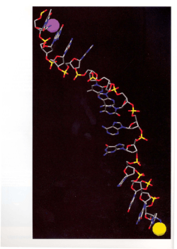


© Copyright 2026