
Lazaroid U-74389G Administration in Pancreatic Ischemia
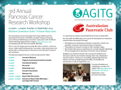
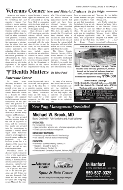
JOP. J Pancreas (Online) 2015 Mar 20; 16(2):176-184.
RESEARCH ARTICLE
Lazaroid U-74389G Administration in Pancreatic Ischemia-Reperfusion
Injury: A Swine Model Encompassing Ischemic Preconditioning
Dimosthenis T Chrysikos1, Theodoros N Sergentanis2, Flora Zagouri1, Theodora Psaltopoulou2,
George Theodoropoulos1, Ioannis Flessas1, George Agrogiannis3, Nikolaos Alexakis1,
Maria Lymperi1, Ageliki I Katsarou4, Efstratios S Patsouris3, Constantine G Zografos1,
Apostolos E Papalois1,5
Departments of 1Propaedeutic Surgery, Hippokratio Hospital, 2Hygiene, Epidemiology and Medical
Statistics and 3Pathology, School of Medicine, University of Athens, 75 Mikras Asias str., Athens 115 27,
GreeceDepartment of , 4Nutrition and Dietetics, Harokopio University, Athens, Greece
5
Experimental–Research Center, Elpen Pharmaceuticals, Athens. 95 Marathonos Av. 19009, Pikermi,
Athens, Greece
ABSTRACT
Context The potential of lazaroid U-74389G in attenuating injury after ischemia and reperfusion has been reported in various organs.
Objective The present study focuses specifically on the pancreas and aims to examine any effects of U-74389G in a swine model of pancreatic ischemia and reperfusion, encompassing ischemic preconditioning. Methods Twelve pigs, weighing 28–35 kg, were randomized into
two experimental groups. Group A (control group, n=6): Two periods of ischemic preconditioning (5 min each) separated by a 5- min rest
interval; then ischemia time 30 min and reperfusion for 120 min. Group B (n=6): the same as above, with U-74389G intravenous injection
in the inferior vena cava immediately prior to the initiation of reperfusion. Blood sampling and pancreatic biopsies were conducted at 0,
30, 60, 90 and 120 min after reperfusion. Results Repeated-measures ANOVA was undertaken to evaluate differences between the two
study groups. No statistically significant differences were noted concerning the histopathological parameters in the control and therapy
groups (P=0.563 for edema, P=0.241 for hemorrhage, P=0.256 for leukocyte infiltration, P=0.231 for acinar necrosis and P=0.438 for vacuolization). In accordance with the above, serum metabolic data (glucose, creatinine, urea, total and direct bilirubin, total calcium, amylase,
lipase, SGOT/AST, SGPT/ALT, ALP, GGT, LDH, CRP, insulin) were not significantly different between the two groups; similarly, tumor necrosis factor-α values (P=0.705) and tissue malondialdehyde levels (P=0.628) did not differ between the two groups. Conclusion This swine
model of pancreatic ischemia and reperfusion, encompassing preconditioning, indicates that U-74389G lazaroid does not seem to exert
protective effects from pancreatic damage.
CORE TIP
Oxidative stress is a crucial factor in the pathophysiology of acute pancreatitis and its systemic complications. Lazaroids are a novel class
of antioxidants that potently protect pancreatic acinar cells against oxidant attack. This study encompasses ischemic preconditioning with
lazaroid U-74389G in a swine model of pancreatic ischemia and reperfusion injury.
INTRODUCTION
Acute pancreatitis remains an important surgical problem
with high morbidity and mortality [1, 2]. It does not
merely represent an injury caused through the process
of autodigestion by activated pancreatic enzymes, as
Received January 24th, 2015 – Accepted February 28th, 2015
Keywords Ischemia; Ischemic Preconditioning;
Malondialdehyde; Pancreas; Reperfusion; Tumor Necrosis
Factor-alpha; U 74389F
Correspondence Dimosthenis T Chrysikos
1st Department of Propaedeutic Surgery
School of Medicine
Hippokratio Hospital,
University of Athens
114, Vas. Sofias Ave
Athens 11527, Greece
Phone +30-697.306.3590
Fax +30-210.770.7574
E-mail [email protected]
complicated mechanisms intervene, often involving
pancreatic ischemia. In the clinical practice, ischemia can
induce or aggravate acute pancreatitis, as observed in
several clinical situations, such as selective embolization
of the primary vessels supplying the pancreas,
hypoperfusion of the pancreas during cardiac surgery,
visceral hypoperfusion due to hemorrhagic shock and
pancreas transplantation [3].
Evidence in basic research suggests that disturbance of
pancreatic microcirculation plays an important role in
the underlying pathophysiological processes [4-14]; for
instance, microvascular perfusion failure is essential for
the development of acute pancreatitis after cardiac or
aortic surgery, hypovolemic shock, hypothermia, and
transplantation of the pancreas [15].
Pancreatic changes in perfusion and hypoxia have
a significant impact not only on the early stages of
JOP. Journal of the Pancreas - http://www.serena.unina.it/index.php/jop - Vol. 16 No. 2 – Mar 2015. [ISSN 1590-8577]
176
JOP. J Pancreas (Online) 2015 Mar 20; 16(2):176-184.
acute pancreatitis but also in its progression and the
pathogenesis of necrosis [4, 5]. It has been also suggested
that reperfusion injury is a major mechanism of acinar
damage, which then progresses to intracellular activation of
proteases and tissue destruction [4, 16]. The postischemic
breakdown of the microvascular perfusion is considered
to be one of the key factors in ischemia/reperfusion injury.
At the microcirculatory level two distinct phenomena
occur: the “no-reflow” phenomenon [17, 18], i.e., primary
capillary perfusion failure after onset of reperfusion and
the “reflow paradox”, i.e., accumulation and adherence of
leukocytes with enhanced microvascular permeability for
macromolecules [19, 20].
Leukocyte infiltration into pancreatic tissue subsequently
causes release, within the pancreas and systematically,
of tumor necrosis factor-α (TNF-α), proinflammatory
cytokines (such as IL-1, IL-6) and anti-inflammatory
cytokine IL-10 [21, 22]; all the above mentioned molecules
along with CRP could potentially be evaluated as markers
or indexes for severity and outcome of acute pancreatitis
[23, 24]. Free oxygen radicals are also produced, attacking
cellular lipids, a reaction that results in the formation of
aldehydic lipid hydroperoxide decomposition products,
such as malondialdehyde (MDA).
Experimental efforts to attenuate tissue reperfusion injury
have spanned different animal models and various organs,
including the heart, brain, kidney, liver, skeletal muscle and
stomach. In these models the above mentioned tissues have
responded to brief exposures to ischemia with increased
resistance to severe ischemia, a phenomenon called
ischemic preconditioning. The mechanisms suggested to
explain the protective effects of ischemic preconditioning
include activation of adenosine, adrenergic, bradykinin, and
opioid receptors, modulation of protein kinase C, tyrosine
kinases, mitogen activated protein kinases, adenosine
triphosphate sensitive potassium channel (KATP channel)
in the mitochondrial membrane, expression of heat shock
proteins and synthesis of antioxidant enzymes [25, 26].
In an attempt to evaluate or attenuate events in the
postischemic pancreas, various experimental studies
have been conducted to modify factors of pancreatic
microcirculatory impairment [2] along with several
pharmacological agents with therapeutic effect, that
could potentially inhibit pancreas reperfusion injury;
lazaroids are among these pharmacological agents.
Lazaroids belong to a novel class of antioxidants
(synthetic 21-aminosteroids) with radical-scavenging,
iron-chelating, and membrane-stabilizing potential [2729]. Although derived from glucocorticosteroids, lazaroid
compounds are devoid of glucocorticoid receptor activity
and, therefore, lack the side-effects of the glucocorticoids.
Owing to their low molecular mass, lazaroids can leave the
circulation and readily penetrate tissues. In vitro evidence
has shown that lazaroids may potently protect pancreatic
acinar cells from oxidant attack [27, 30]. U-74389G is an
agent belonging to the 21-aminosteroid group.
Our previous study in the swine model highlighted that the
administration of U-74389G did not seem to exert a sizable
therapeutic effect in attenuating pancreatic damage during
ischemia/reperfusion of the pancreas; rather impressively,
edema seemed more pronounced in the U-74389G group
[31]. Nevertheless, the aforementioned experimental
design had not included ischemic preconditioning; as
mentioned above, preconditioning may denote a modified
context, with reduced oxidative burden, that could
potentially enhance the effects of U-74389G.
Precisely, in the present study, we interjected two brief
(5 min) ischemic periods (separated by a 5- min rest
interval), which could protect the pancreas against severe
ischemia/reperfusion-induced pancreatitis. Similarly
to our previous design the lazaroid was administered at
the end of ischemia and before reperfusion injury of the
pancreas [31].
Summarizing the above, this study aims to assess the
therapeutic potential of 21-aminosteroid U-74389G in a
swine model of pancreatic ischemia/reperfusion injury
encompassing ischemic preconditioning. To this end,
histopathological parameters, serum biochemical indices,
as well as TNF-α and MDA levels were evaluated. To the
best of our knowledge, there are no reports on the use
of U-74389G in pancreatic ischemia-reperfusion injury,
adopting a swine model and encompassing ischemic
preconditioning.
MATERIALS AND METHODS
Animals
Twelve male and female pigs, weighting 28–35 kg, were
used in the study. The animals were randomized into the
following two experimental groups, using a computer
random number generator: Group A (control group, n=6):
Two times of ischemic preconditioning for 5 min with 5min interval and then pancreatic ischemia for 30 min and
reperfusion for 120 min. Group B (U-74389G group, n=6):
the same as above, with U-74389G intravenous injection in
the inferior vena cava immediately prior to the initiation of
reperfusion.
The used dose of U-74389G (by Calbiochem® Company, Cat.
No. 153190-29-5, only for experimental use) was 10 mg/
kg animal body weight (ABW). This dose of the lazaroid
was determined based on previous experience in the same
laboratory with the use of U-74389G [31-33], as well as on
data stemming from other research groups [32, 33].
The pigs were acclimatized to laboratory conditions for 7
days prior to use in experiments. All animals were fasted
for 24 h before the procedures, which were performed
under general anesthesia.
The pigs were premeditated with ketamine 15 mg/
kg animal body weight and midazolam 0.5 mg/kg
ABW administered intramuscularly and were placed
on the operating table. Anesthesia was induced with
bolus intravenous injection of propofol 3 mg/kg ABW,
pancuronium 0.15 mg/kg ABW, and fentanyl 17.5 μg/kg
JOP. Journal of the Pancreas - http://www.serena.unina.it/index.php/jop - Vol. 16 No. 2 – Mar 2015. [ISSN 1590-8577]
177
JOP. J Pancreas (Online) 2015 Mar 20; 16(2):176-184.
ABW. During operation, anesthesia was maintained with
propofol 0.15 mg/kg ABW, pancuronium 0.07 mg/kg
ABW every 20 min, and fentanyl 15 μg/kg ABW per hour.
Polyethylene catheters were inserted into a peripheral vein
and into the jugular vein for infusion of anesthetic drugs,
infusion of crystalloid solutions, and measurement of the
vein pressure. Catheter was also inserted in the femoral
artery and blood samples were collected. Monitoring of the
animals was continuous with electrocardiography, pulse
oximetry, and measurements of arterial blood pressure.
The surgical preparation of the abdomen was performed
through a midline laparotomy using aseptic techniques.
It is worth to mention that the swine pancreas consists of
two parts that are crossed by the portal vein. The blood
supply of the right part is provided mainly by the common
hepatic artery and rarely from the superior mesenteric
artery, while the left one from the splenic artery and a
small non stable branch that may arise from the splenic or
the common hepatic artery or even from the celiac trunk
[34, 35]. The pancreas was prepared and the splenic artery,
the common hepatic artery and the pancreaticoduodenal
arteries (superior and inferior) that provide blood supply
to the swine pancreas, were identified and then were
isolated and prepared for occlusion.
The experiment was short-term, and the animals underwent
euthanasia at the end of each operation. Afterwards, tissue
samples were taken from the liver, kidney, lungs, and sent
to the pathologist for histopathological evaluation [32].
Histopathological Assessment
Tissue specimens were fixed in formalin (4%) and
embedded in paraffin according to standard histological
procedures. Four-micron sections of paraffin-embedded
tissue samples from each case were sectioned,
deparaffinized, rehydrated, and finally subjected to
conventional hematoxylin and eosin staining (H&E).
Histopathological assessment was based on the research
work of Dembrinski et al. [36] and graded a number of
parameters [32]. First, histological grading of edema
was conducted using a scale ranging from 0 to 3: 0 = no
edema, 1 = interlobular edema, 2 = interlobular and
moderate intralobular edema and 3 = severe interlobular
and intralobular edema. Second, grading of hemorrhage
was performed as follows: 0 = absent, 1 = from one to two
foci per slide, 2 = from three to five foci per slide, 3 = more
than five foci per slide. Third, leukocyte infiltration was
graded: 0 = absent, 1 = scarce perivascular infiltration, 2
= moderate perivascular and scarce diffuse infiltration, 3
= abundant diffuse infiltration. Fourth, findings of acinar
necrosis were graded: 0 = absent, 1 = less than 15% of cells
involved, 2 = from 15% to 35% of cells involved, 3 = more
than 35% of cells involved. Fifth, grading of vacuolization
was based on the percentage of cells involved: 0 = absent,
1 = less than 25%, 2 = 25–50% and 3 = more than 50%.
Specimens were examined under a Nikon Eclipse 50i
microscope. Histomorphological analysis was performed
independently by two experienced pathologists (G.A. and
E.S.P.), who were blinded to the current experimental
protocol.
Biochemical
Parameters,
Measurements
MDA
and
TNF-Α
MDA measurement was based on the reaction of a
chromogenic reagent, N-methyl-2-phenylindole (MPI),
with MDA at 45°C. One molecule of MDA reacted with
two molecules of MPI to yield a stable chromophore with
maximum absorbance at 586 nm. The reagents used
included Reagent MPI, 10.3 mmol/L MPI in acetonitrile,
MDA standard, 10 mmol/L 1,1,3,3-tetramethoxypropane
in 20 mmol/L Tris-HCl, 500 mmol/L butylatedhydroxytoluene, in acetonitrile, 20 mmol/L Tris buffer
pH 7.4, 0.9% NaCl, 37% (12 mol/L) HCl, methanol, highpressure liquid chromatography grade, and acetonitrile,
high-pressure liquid chromatography grade. Before the
procedure, three volumes of the MPI reagent were diluted
with one volume of 100% methanol. Tissue samples were
rinsed with ice-cold isotonic saline before homogenization,
which was carried out using Tris-buffered 20 mmol/L
pH 7.4 and an Ultra-Turrax (Ika– Labortecnik) blender.
One milliliter of buffer was used for 0.1 g of tissue. Ten
milliliters of 500 mmol/L butylated hydroxy toluene was
added to 1 mL of tissue homogenate to prevent sample
oxidation. The homogenate was centrifuged at 3000 rpm
at 4°C for 10 min. Then 0.2 mL of sample (supernatant of
tissue homogenate) and 0.65 mL of diluted MPI reagent
were added to a polypropylene microcentrifuge tube. The
mixture was vortexes and then 0.15 mL of 12 mol/L HCl
was added. Tubes were incubated at 45°C for 60 min and
centrifuged at 6000 rpm for 15 min. Then 0.8 mL of the
supernatant was measured at 586 nm. MDA standards
for the standard curve were made by dilutions of the
stock 10 mm tetramethoxypropane solution. The final
concentrations were 2.08, 4.16, 8.33, 12.5, and 16.66
μmol/L and the assay procedure was followed as for the
samples. The absorbance was 0.059, 0.124, 0.264, 0.4 and
0.545, respectively [31, 32].
TNF-α was determined after tissue homogenization by
enzyme-linked immunosorbent assay. To avoid errors in
the interpretation of the results, a specific swine antibody
was used (anti-swine; Bio Source Co., Carlsbad, CA, USA)
instead of human antibody against TNF- α [31, 32].
In addition at 0, 30, 60, 90 and 120 min after reperfusion,
complete blood cell count, as well as measurements of
serum biochemical parameters (glucose, creatinine, urea,
total bilirubin, direct bilirubin, total calcium, amylase,
lipase, SGOT/AST, SGPT/ALT, gamma-GT (GGT), LDH, CRP
and insulin) were performed [31, 32].
ETHICS
All experiments described herein were approved by
ELPEN Laboratories and the veterinary authorities of East
Attica Region, in accordance with Greek Law No. 160, A-64,
May 1991, European Union regulations, and the principles
of the Helsinki declaration. All animals received humane
care according to the criteria outlined in the "Guide for the
Care and Use of Laboratory Animals (1996)" prepared by
the National Academy of Sciences.
JOP. Journal of the Pancreas - http://www.serena.unina.it/index.php/jop - Vol. 16 No. 2 – Mar 2015. [ISSN 1590-8577]
178
JOP. J Pancreas (Online) 2015 Mar 20; 16(2):176-184.
STATISTICAL ANALYSIS
Repeated-measures analysis of variance (ANOVA) was
performed to assess the differences between groups,
given the longitudinal nature of the dataset. Regarding
the main outcome TNF-α, power calculation indicated that
six animals per group were sufficient for the detection
of a 35% increase in its tissue levels, to achieve a power
equal to 80%. Mean and standard error were calculated
for each variable and are presented in the manuscript for
descriptive reasons. The statistical software STATA 11.1
(Stata Corp., College Station, TX, USA) was used for random
number generation, as well as for all statistical analyses.
Values of p<0.05 were considered statistically significant.
RESULTS
The results of the histopathological evaluation are
presented in Table 1. No statistically significant differences
were noted concerning edema (P=0.563), hemorrhage
(P=0.241), leukocyte infiltration (P=0.256), acinar
necrosis (P=0.231) and vacuolization (P=0.438) between
the two groups. Figures 1 and 2 present representative
pathological cases.
Blood cell count and serum biochemical parameters are
summarized in Table 2. None of the evaluated blood cell
count parameters (P=0.403 for white blood cells, P=0.222
for hemoglobin, P=0.900 for hematocrit, P=0.684 for
platelets) pointed to statistically significant differences
between control and therapeutic group. In accordance
with the above, all studied serum biochemical parameters
(P=0.966 for glucose, P=0.661 for creatinine, P=0.848 for
urea, P=0.915 for total bilirubin and P=0.742 for direct
bilirubin, P=0.477 for total calcium, P=0.509 for amylase,
P=0.912 for lipase, P=0.876 for SGOT/AST, P=0.387 for
SGPT/ALT, P=0.938 for ALP, P=0.581 for GGT, P=0.666
for LDH, P=0.823 for CRP and P=0.926 for insulin) did not
differ between control and therapeutic group.
In addition to the pancreas samples, whose
histopathological evaluation has been presented above,
after euthanasia of the animals, other organ samples
(liver, kidney and lung) were sent to the pathologist for
evaluation. Normal histological findings were observed
in all cases; however, this might be attributed to the acute
phase of the experiment and due to the short survival
of the laboratory animals, as detailed in our respective
manuscript examining the effects of U-74389G upon liver
ischemia in the swine model [32]. Table 3 summarizes
the results regarding tissue MDA and TNF-α level. No
significant differences were noted for either TNF-α
(P=0.705) or MDA (P=0.628).
DISCUSSION
The present study suggests that the lazaroid U-74389G
does not seem to possess protective potential in the context
of ischemia-reperfusion injury of the swine pancreas.
All examined parameters, namely histopathologic
pancreatitis severity, serum biochemical parameters and
indicators of inflammation and oxidative stress (TNF-α
and MDA) pointed to the same null direction. This pattern
further confirms the lack of protective potential that was
demonstrated in our previous work regarding U-74389G
in ischemia-reperfusion injury of the swine pancreas
without preconditioning [31].
In the present setting, ischemic preconditioning
involved repeated short episodes of warm ischemia and
reperfusion before the long-term I/R injury. Compared
to our previous work [31], a completely altered free
radical and inflammatory context should be declared and
stressed, as preconditioning is associated with milder
ischemia-reperfusion pancreatitis, as a rule. Specifically,
studies conducted by Dembinski et al. [36] have showed
that ischemic preconditioning of the rat pancreas seems
to be associated with decreased biochemical signs of
pancreatitis, improved pancreatic blood flow, as well as
reduced histological scores of pancreatic damage, together
with reduced plasma lipase activity and decreases in
plasma concentrations of proinflammatory interleukin-1
[36]; nevertheless, the findings regarding plasma
concentrations of anti-inflammatory interleukin-10 (IL10) remain controversial [36, 37]. Moreover, in the context
of pancreas transplantation there are data indicating
that ischemic preconditioning attenuates apoptosis in
different models of pancreatic warm ischemia [7, 19, 20].
Of note, our time frame for ischemic preconditioning was
based on the studies by Dembrinski et al. [36] where a
preconditioning ischemia of 10 min (2 times of 5 min
ischemic preconditioning with 5 min interval), followed by
intervening reperfusion was found to have superior effects
when compared with other procedures [36].
In our previous study conducted in the absence of ischemic
preconditioning [31], we have shown that interlobular
and intralobular edema was more pronounced in the
lazaroid U-74389G group. Given that lazaroids at high
concentrations may accelerate cell injury, the absence of
this finding in our present study might be attributed to (or
ameliorated by) ischemic preconditioning. Nevertheless,
the remaining pathological parameters aiming to assess the
effect of post-reperfusion tissue damage i.e., hemorrhage,
leukocyte infiltration, acinar necrosis and vacuolization,
did not point to significant differences between the two
groups either in the present or the previous study.
Regarding the interpretation of therapeutic failure a
variety of notions merit highlighting. First, the application
of U-74389G was initiated 30 minutes after pancreatitis
induction and therefore the short time interval may
not have allowed the agent to effectively reduce lipid
peroxidation. For instance, antioxidant agents have
occasionally been administered before ischemia [31, 3739]; nevertheless, in clinical practice, it is nearly impossible
to begin treatment before the onset of ischemia [14, 31, 32],
rendering our current setting closer to the inherent clinical
limitations of pancreatitis. Second, despite the relative
attenuation mediated by preconditioning, ischemiareperfusion pancreatitis is a fulminant disease, whose
JOP. Journal of the Pancreas - http://www.serena.unina.it/index.php/jop - Vol. 16 No. 2 – Mar 2015. [ISSN 1590-8577]
179
JOP. J Pancreas (Online) 2015 Mar 20; 16(2):176-184.
Table 1. Histopathological parameters in the two groups.
Parameter
Edema
Hemorrhage
Leukocyte infiltration
Acinar necrosis
Vacuolization
a
Time (min)
0
30
60
90
120
0
30
60
90
120
0
30
60
90
120
0
30
60
90
120
0
30
60
90
120
all values were equal to the mean; hence standard error equaled zero
Group A
Mean (SE)
1.33 (0.21)
1.50 (0.22)
1.67 (0.21)
1.50 (0.22)
1.67 (0.21)
0.83 (0.17)
1.00 (0.26)
0.83 (0.17)
1.00 (0.00)a
1.17 (0.17)
0.33 (0.21)
0.50 (0.22)
0.50 (0.34)
0.33 (0.21)
0.67 (0.21)
0.00 (0.00)a
0.00 (0.00)a
0.17 (0.17)
0.00 (0.00)a
0.00 (0.00)a
0.17 (0.17)
0.33 (0.21)
0.33 (0.21)
0.17 (0.17)
0.50 (0.22)
Figure 1. Effects of acute ischemia and reperfusion on pancreatic
histology. This field is characterized by the complete distortion of
the pancreatic parenchyma caused by the presence of conspicuous
hemorrhage, moderate interlobular and intralobular edema and some
small necrotic foci (approx. 20%). H&E x400.
rapid development of necrosis and early death may limit
the anticipated efficacy of any pharmacologic intervention.
Despite its original conclusions, the present study bears
certain limitations that need to be discussed. Evaluation
of lazaroids at higher doses, adoption of earlier time
points for the administration of treatment may allow the
emergence of therapeutic effects; indeed, this effort did not
encompass the measurement of intrapancreatic U-74389G
concentrations. In addition, our study adopted two periods
of preconditioning, similarly to the previous literature
[15, 36]; a larger number of preconditioning periods
Group B
Mean (SE)
1.17 (0.17)
1.33 (0.21)
1.67 (0.42)
1.33 (0.21)
1.50 (0.22)
0.67 (0.21)
0.83 (0.17)
0.83 (0.31)
0.67 (0.33)
0.50 (0.22)
0.33 (0.21)
0.83 (0.31)
0.83 (0.31)
0.83 (0.17)
0.83 (0.17)
0.00 (0.00)a
0.00 (0.00)a
0.33 (0.21)
0.33 (0.21)
0.33 (0.21)
0.17 (0.17)
0.17 (0.17)
0.17 (0.17)
0.17 (0.17)
0.33 (0.21)
P value
0.563
0.241
0.256
0.231
0.438
Figure 2. Effects of acute ischemia and reperfusion on pancreatic
histology. The main feature in this field is the distortion of pancreatic
histology. Note the significant hemorrhage, the moderate interlobular
and intralobular edema and the presence of some necrotic foci (15-35%).
H&E x400.
(e.g. four) may modify the results, representing reduced
oxidative burden. Furthermore, the rather small sample size
(six subjects in each arm) should be declared; nevertheless, all
performed tests were far from yielding even trends of borderline
significance, underlining the true lack of associations, whereas
the power calculation indicated that a sufficient power of 80%
was achieved. Moreover, the examination of other lazaroid
compounds may point to similarities or differences in their
effectiveness compared to U-74389G. Finally, it should always
be declared that conditions in the experimental setting do not
obligatorily reflect the actual clinical conditions in the human
context [36].
JOP. Journal of the Pancreas - http://www.serena.unina.it/index.php/jop - Vol. 16 No. 2 – Mar 2015. [ISSN 1590-8577]
180
JOP. J Pancreas (Online) 2015 Mar 20; 16(2):176-184.
Table 2. Blood cell count and biochemical serum parameters in the two study groups.
Parameter
Time (min)
White blood cells (10 cells/mL)
3
Hemoglobin (g/dL)
Hematocrit (%)
Platelets (103cells/mL)
Glucose (mg/dL)
Creatinine (mg/dL)
Urea (mg/dL)
Total bilirubin (mg/dL)
Direct bilirubin (mg/dL)
Total calcium (mg/dL)
Amylase (U/L)
Lipase (U/L)
0
30
60
90
120
0
30
60
90
120
0
30
60
90
120
0
30
60
90
120
0
30
60
90
120
0
30
60
90
120
0
30
60
90
120
0
30
60
90
120
0
30
60
90
120
0
30
60
90
120
0
30
60
90
120
0
30
60
Group A
Mean (SE)
13.86 (1.24)
13.24 (0.78)
14.83 (1.89)
14.71 (1.99)
15.65 (1.96)
9.93 (0.10)
9.63 (0.08)
9.77 (0.09)
9.80 (0.12)
9.82 (0.12)
31.90 (0.63)
31.17 (0.64)
31.48 (0.85)
31.57 (0.92)
31.67 (0.95)
282.7 (55.8)
283.0 (55.8)
281.7 (52.8)
276.8 (59.4)
291.8 (67.1)
85.1 (5.3)
89.6 (6.7)
92.1 (7.7)
90.6 (7.9)
91.3 (8.7)
1.29 (0.06)
1.27 (0.06)
1.36 (0.08)
1.44 (0.09)
1.49 (0.08)
27.69 (2.12)
29.37 (2.40)
30.27 (2.07)
31.81 (2.44)
32.07 (2.24)
0.56 (0.08)
0.56 (0.11)
0.54 (0.11)
0.60 (0.13)
0.60 (0.15)
0.32 (0.06)
0.28 (0.05)
0.31 (0.08)
0.32 (0.10)
0.35 (0.09)
9.53 (0.29)
9.31 (0.30)
9.34 (0.39)
9.39 (0.43)
9.29 (0.42)
1622.3 (171.0)
1614.9 (158.8)
1671.5 (161.7)
1715.1 (171.6)
1753.6 (204.9)
30.67 (3.03)
32.50 (4.13)
33.07 (4.78)
Group B
Mean (SE)
13.09 (1.23)
12.26 (1.06)
13.03 (1.23)
13.31 (1.13)
13.17 (0.70)
10.35 (0.27)
10.02 (0.32)
10.05 (0.29)
10.20 (0.30)
10.32 (0.30)
32.38 (0.83)
31.28 (1.05)
31.42 (0.85)
31.55 (0.72)
31.88 (0.80)
316.3 (35.0)
322.5 (39.2)
312.2 (41.6)
301.7 (38.3)
308.2 (45.8)
79.9 (6.3)
89.6 (8.0)
93.9 (10.0)
92.4 (11.2)
95.5 (13.6)
1.39 (0.08)
1.40 (0.07)
1.47 (0.05)
1.41 (0.05)
1.36 (0.04)
27.07 (2.61)
29.90 (3.41)
31.79 (2.98)
33.84 (2.44)
31.87 (2.09)
0.58 (0.10)
0.57 (0.06)
0.61 (0.08)
0.62 (0.06)
0.55 (0.05)
0.33 (0.08)
0.33 (0.03)
0.37 (0.08)
0.40 (0.05)
0.28 (0.05)
9.65 (0.14)
9.66 (0.15)
9.46 (0.28)
9.88 (0.40)
9.79 (0.37)
1626.8 (160.0)
1574.0 (151.1)
1560.1 (136.1)
1479.4 (110.7)
1399.8 (118.4)
33.21 (3.63)
35.56 (4.94)
34.46 (5.08)
JOP. Journal of the Pancreas - http://www.serena.unina.it/index.php/jop - Vol. 16 No. 2 – Mar 2015. [ISSN 1590-8577]
P value
0.403
0.222
0.900
0.684
0.966
0.661
0.848
0.915
0.742
0.477
0.509
0.912
181
JOP. J Pancreas (Online) 2015 Mar 20; 16(2):176-184.
SGOT (AST) (U/L)
SGPT (ALT) (U/L)
ALP (U/L)
GGT (U/L)
LDH (U/L)
CRP (mg/L)
Insulin (μU/mL)
90
120
0
30
60
90
120
0
30
60
90
120
0
30
60
90
120
0
30
60
90
120
0
30
60
90
120
0
30
60
90
120
0
30
60
90
120
Table 3. Tissue TNF-α and MDA levels in the two study groups.
Parameter
TNF-α (pg/mL of total protein)
MDA (pmol/mg of total protein)
CONCLUSION
35.29 (7.16)
43.77 (10.69)
60.94 (4.79)
67.59 (7.60)
66.14 (5.97)
66.92 (7.37)
68.24 (7.94)
43.54 (3.64)
42.16 (3.62)
42.97 (3.30)
43.23 (2.92)
43.73 (3.74)
133.17 (13.66)
133.33 (12.22)
136.17 (12.31)
138.83 (12.69)
140.50 (13.64)
29.76 (3.15)
30.58 (4.09)
30.00 (3.66)
30.71 (4.67)
30.88 (4.59)
1148.5 (197.0)
1137.5 (200.0)
1171.2 (192.6)
1150.3 (198.7)
1162.0 (204.7)
55.02 (8.48)
58.82 (9.45)
58.62 (9.52)
58.57 (9.97)
53.23 (9.22)
12.30 (3.47)
12.82 (3.57)
12.27 (3.44)
12.13 (3.39)
11.65 (3.48)
Time (min)
0
30
60
90
120
0
30
60
90
120
In summary, our study shows that lazaroid U-74389G does
not seem capable of protecting the swine pancreas from
ischemia-reperfusion injury in the experimental model
encompassing ischemic preconditioning. Protective effects
derived from lazaroid compounds might strongly depend
on the type of organ, the time frame and the experimental
setting.
Group A
Mean (SE)
1.87 (0.55)
2.50 (0.93)
2.81 (1.11)
3.11 (1.12)
3.39 (1.60)
52.2 (10.1)
50.1 (12.0)
65.8 (7.5)
64.7 (9.6)
74.4 (10.4)
37.70 (5.19)
38.37 (4.08)
66.43 (11.69)
64.02 (9.78)
64.68 (8.37)
65.02 (7.40)
61.07 (7.32)
46.90 (6.38)
48.06 (4.78)
49.69 (3.77)
48.97 (3.85)
46.49 (4.21)
138.17 (16.78)
140.00 (14.64)
139.33 (13.32)
143.33 (10.58)
128.33 (10.32)
31.22 (3.64)
31.05 (2.21)
32.72 (2.07)
33.25 (2.15)
35.68 (2.62)
1295.5 (278.7)
1377.5 (312.9)
1342.7 (338.2)
1309.2 (318.4)
1262.7 (306.1)
52.84 (10.61)
53.32 (11.69)
53.78 (12.85)
56.43 (12.13)
50.81 (12.12)
12.83 (3.65)
12.67 (3.71)
12.58 (3.39)
12.97 (2.94)
12.40 (3.12)
Group B
Mean (SE)
2.08 (0.57)
2.06 (0.41)
2.66 (0.80)
2.30 (0.60)
2.52 (0.66)
55.2 (11.3)
65.3 (9.7)
74.6 (10.6)
71.3 (14.6)
73.6 (17.8)
0.876
0.387
0.938
0.581
0.666
0.823
0.926
P value
0.705
0.628
Acknowledgements
We are grateful to Apostolos E Papalois who conceived,
designed and acted as a guarantor of the study
Conflict of Interest
Authors declare to have no conflict of interest.
JOP. Journal of the Pancreas - http://www.serena.unina.it/index.php/jop - Vol. 16 No. 2 – Mar 2015. [ISSN 1590-8577]
182
JOP. J Pancreas (Online) 2015 Mar 20; 16(2):176-184.
References
1. Slavin J, Ghaneh P, Sutton R, Hartley M, Rowlands P, Garvey C,Hughes
M, Neoptolemos J.Management of necrotizing pancreatitis. World J
Gastroenterol 2001; 7:476-481 [PMID: 11819813]
2. Zhou ZG, Chen YD. Influencing factors of pancreatic microcirculatory
impairment in acute panceatitis. World J Gastroenterol 2002; 8:406-412.
[PMID:12046059]
3. Drognitz O, Liu X, Obermaier R, Neeff H, von Dobschuetz E, Hopt UT,
Benz S. Ischemic preconditioning fails to improve microcirculation but
increases apoptotic cell death in experimental pancreas transplantation.
Transpl Int 2004; 17:317-324. [PMID: 15221122]
4. Cuthbertson CM, Christophi C. Disturbances of the microcirculation
in acute pancreatitis. Br J Surg 2006; 93:518-530. [PMID: 16607683]
5. Kinnala PJ, Kuttila KT, Gronroos JM, Havia TV, Nevalainen TJ,
Niinikoski JH. Pancreatic tissue perfusion in experimental acute
pancreatitis. Eur J Surg 2001; 167:689–694. [PMID: 11759740]
6. Plusczyk T, Witzel B, Menger MD, Schilling M. ETA and ETB
receptor function in pancreatitis-associated microcirculatory failure,
inflammation, and parenchymal injury. Am J Physiol Gastrointest Liver
Physiol 2003; 285:G145-153. [PMID: 12799311]
7. Yaginuma N, Takahashi T, Saito K, Kyoguku M. The microvasculature
of the human pancreas and its relation to Langerhans islets and lobules.
Pathol Res Pract 1986; 181:77–84. [PMID: 3517840]
8. Zhou ZG, Chen YD, Sun W, Chen Z. Pancreatic microcirculatory
impairment in experimental acute pancreatitis in rats. World J
Gastroenterol 2002; 8:933-936. [PMID: 12378645]
9. Schiller WR, Anderson MC. Microcirculation of the normal and
inflamed canine pancreas. Ann Surg 1975; 181:466–470. [PMID:
1130866]
10. Zhou ZG, Gao XH. Morphology of pancreatic microcirculation in the
monkey: light and scanning electron microscopic study. Clin Anat 1995;
8:190–201.[PMID: 7606592]
11. Weidenbach H, Lerch MM, Gress TM, Pfaff D, Turi S, Adler G.
Vasoactive mediators and the progression from oedematous to
necrotising experimental acute pancreatitis. Gut 1995; 37:434–440.
[PMID: 7590444]
12. Sunamura M, Yamauchi J, Shibuya K, Chen HM, Ding L, Takeda Κ,
Kobari M, Matsuno S. Pancreatic microcirculation in acute pancreatitis. J
Hepatobiliary Pancreat Surg 1998; 5:62–68. [PMID: 9683756]
13. Henderson JR, Moss MC. A morphometric study of the endocrine and
exocrine capillaries of the pancreas. Q J Exp Physiol 1985; 70:347–356.
[PMID: 3898188]
14. Bockman DE. Microvasculature of the pancreas. Relation to
pancreatitis. Int J Pancreatol 1992; 12:11–21. [PMID:1527446]
15. Warzecha Z, Dembiński A, Ceranowicz P, Dembiński M, Cieszkowski
J, Kuśnierz-Cabala B, Naskalski JW, Jaworek J, Konturek SJ, Pawlik
WW, Tomaszewska R. Influence of ischemic preconditioning on blood
coagulation, fibrinolytic activity and pancreatic repair in the course of
caerulein-induced acute pancreatitis in rats. J Physiol Pharmacol 2007;
58:303-319. [PMID: 17622699]
16. Vollmar B, Menger MD. Microcirculatory dysfunction in acute
pancreatitis. A new concept of pathogenesis involving vasomotionassociated arteriolar constriction and dilation. Pancreatology 2003;
3:181–190. [PMID: 12817573]
17. Menger MD, Rücker M, Vollmar B. Capillary dysfunction in striated
muscle ischemia/reperfusion: on the mechanisms of capillary ‘‘noreflow’’. Shock 1997; 8:2–7. [PMID: 9249906]
18. Nolte D, Zeintl H, Steinbauer M, Pickelmann S, Messmer K. Functional
capillary density: an indicator of tissue perfusion? Int J Microcirc Clin Exp
1995; 15:244–249. [PMID: 8852622]
19. Hoffmann TF, Leiderer R, Waldner H, Arbogast S, Messmer K. Ischemia
reperfusion of the pancreas: a new in vivo model for acute pancreatitis in
rats. Res Exp Med (Berl) 1995; 195:125–144 [PMID: 8570908]
20. Hoffmann TF, Leiderer R, Waldner H, Messmer K. Bradykinin
antagonists HOE-140 and CP-0597 diminish microcirculatory injury after
ischaemia– reperfusion of the pancreas in rats. Br J Surg 1996; 83:189195 [PMID: 8689161]
21. Ishimaru K, Mitsuoka H, Unno N, Inuzuka K, Nakamura S, SchmidSchönbein GW. Pancreatic proteases and inflammatory mediators in
peritoneal fluid during splanchnic arterial occlusion and reperfusion.
Shock 2004; 22:467-471. [PMID: 15489640]
22. Leindler L, Morschl E, László F, Mándi Y, Takács T, Jármai K, Farkas
G. Importance of cytokines, nitric oxide, and apoptosis in the pathological
process of necrotizing pancreatitis in rats. Pancreas 2004; 29:157-161.
[PMID: 15257108]
23. Alhan E, Türkyilmaz S, Erçin C, Kural BV. Effects of lazaroid U-74389G
on acute necrotizing pancreatitis in rats. Eur Surg Res 2006; 38:70-75.
[PMID: 16557023]
24. Keck T, Werner J, Banafsche R, Stalmann A, Schneider L, Gebhard
MM, Herfarth C, Klar E. Oxygen radicals promote ICAM-1 expression
and microcirculatory disturbances in experimental acute pancreatitis.
Pancreatology 2003; 3:156–163. [PMID: 12748425]
25. Obermaier R, von Dobschuetz E,Drognitz O, Hopt UT, Benz S. Ischemic
preconditioning attenuates capillary no-reflow and leukocyte adherence
in postischemic pancreatitis. Langenbecks Arch Surg 2004; 389:511-516.
[PMID: 14716491]
26. Cleveland JC Jr, Raeburn C, Harken AH. Clinical applications of
ischemic preconditioning: from head to toe. Surgery 2001; 129:664–667.
[PMID: 11391362]
27. Schulz HU, Hoenl H, Schrader T, Kropf S,Halangk W,Ochmann
C,Matthias R, Letko G, Roessner A, Lippert H, Niederau C. Randomized,
placebo-controlled trial of lazaroid effects on severe acute pancreatitis in
rats. Crit Care Med 2001; 29:861-869. [PMID: 11373483]
28. Braughler JM, Pregenzer JF, Chase RL, Duncan LA, Jacobsen EJ, McCall
JM. Novel 21-amino steroids as potent inhibitors of iron-dependent lipid
peroxidation. J Biol Chem 1987; 262:10438–10440. [PMID: 3611075]
29. Clark WM, Hazel JS, Coull BM. Lazaroids: CNS pharmacology and
current research. Drugs 1995; 50:971-983 [PMID: 8612475]
30. Niederau C, Schulz HU, Klonowski H. Lazaroids protect isolated rat
pancreatic acinar cells against damage induced by free radicals. Pancreas
1995; 11:107-121. [PMID: 7479666]
31. Chrysikos DT, Sergentanis TN, Zagouri F, Psaltopoulou T, Flessas I,
Agrogiannis G, Alexakis N, Bramis I, Patsouri EE , Patsouris ES, Korontzi
M, Katsarou A, Zografos GC, Papalois AE. The Effect of U-74389G on
Pancreas Ischemia-Reperfusion Injury in a Swine Model. J Surg Res 2014;
187:450-457.[PMID: 24332939]
32. Tsaroucha AK, Papalois A, Vernadakis S, Adamopoulos S,
Papadopoulos K, Lambropoulou M, Constadinidis T, Kyriazi A,
Papadopoulos N, Simopoulos C. The effect of U-74389G on liver recovery
after acute liver ischemia-reperfusion injury in a swine model. J Surg Res
2009; 151:10-4. [PMID: 18468628]
33. Cosenza CA, Cramer DV, Cunneen SA, Tuso PJ, Wang HK, Makowka L.
Protective effect of the lazaroid U74006F in cold ischaemia-reperfusion
injury of the liver. Hepatology 1994; 19:418-425. [PMID: 8294099]
34. Troisi R, Jacobs B, Berrevoet F, Vereycken R, De Hemptinne B, Hesse
UJ. The role of hepato-coeliac arterial reconstruction in porcine segmental
pancreatic autotransplantation. Transplant Proc 1997; 29:3625-3626.
[PMID: 9414864]
35. Troisi R, Maene L, Jacobs B, Berrevoet F, Claus H, De Hemptinne
B, Hesse UJ. Haemodynamic profiles of hepato-coeliac arterial
reconstruction in porcine segmental pancreatic autotransplantation.
Transplant Proc 1998; 30:582-583. [PMID: 9532185]
JOP. Journal of the Pancreas - http://www.serena.unina.it/index.php/jop - Vol. 16 No. 2 – Mar 2015. [ISSN 1590-8577]
183
JOP. J Pancreas (Online) 2015 Mar 20; 16(2):176-184.
36. Dembiński A, Warzecha Z, Ceranowicz P, Tomaszewska R, Dembiński
M, Pabiańczyk M, Stachura J, Konturek SJ. Ischemic preconditioning
reduces the severity of ischemia/reperfusion-induced pancreatitis. Eur J
Pharmacol 2003; 473:207-216. [PMID:12892840]
37. Warzecha Z, Dembinski A, Ceranowicz P, Dembinski M, Sendur R,
Cieszkowski J, Sendur P, Tomaszewska R. Heparin inhibits protective
effect of ischemic preconditioning in ischemia/reperfusion-induced acute
pancreatitis. J Physiol Pharmacol 2012; 63:355-365. [PMID: 23070084]
38. Andreadou I, Poussios D, Papalois A, Gavalakis N, Aroni K, Gazouli M,
Gorgoulis VG, Fotiadis C. Effect of U-74389G (21-lazaroid) on intestinal
recovery after acute mesenteric ischaemia and reperfusion in rats. In
Vivo 2003; 17:463-468. [PMID: 14598610]
39. Leventis I, Andreadou I, Papalois A, Sfiniadakis I, Gorgoulis VG,
Korkolis DP, Hadjipavlou-Litina D, Kourounakis PN, Fotiadis C. A
novel antioxidant non-steroidal anti-inflammatory agent protects rat
liver against ischaemia-reperfusion injury. In Vivo 2004; 18:161-169.
[PMID: 15113043]
JOP. Journal of the Pancreas - http://www.serena.unina.it/index.php/jop - Vol. 16 No. 2 – Mar 2015. [ISSN 1590-8577]
184


© Copyright 2026