
Gestating for 22 months: luteal development and pregnancy maintenance in elephants
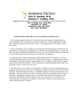
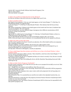
Downloaded from rspb.royalsocietypublishing.org on September 11, 2014
Gestating for 22 months: luteal development and pregnancy
maintenance in elephants
Imke Lueders, Cheryl Niemuller, Peter Rich, Charlie Gray, Robert Hermes, Frank Goeritz and
Thomas B. Hildebrandt
Proc. R. Soc. B published online 20 June 2012
Supplementary data
"Data Supplement"
http://rspb.royalsocietypublishing.org/content/suppl/2012/06/18/rspb.2012.1038.DC1.h
tml
References
This article cites 55 articles, 23 of which can be accessed free
P<P
Published online 20 June 2012 in advance of the print journal.
Subject collections
Articles on similar topics can be found in the following collections
http://rspb.royalsocietypublishing.org/content/early/2012/06/18/rspb.2012.1038.full.ht
ml#ref-list-1
developmental biology (132 articles)
evolution (1880 articles)
physiology (135 articles)
Email alerting service
Receive free email alerts when new articles cite this article - sign up in the box at the top
right-hand corner of the article or click here
Advance online articles have been peer reviewed and accepted for publication but have not yet appeared in
the paper journal (edited, typeset versions may be posted when available prior to final publication). Advance
online articles are citable and establish publication priority; they are indexed by PubMed from initial publication.
Citations to Advance online articles must include the digital object identifier (DOIs) and date of initial
publication.
To subscribe to Proc. R. Soc. B go to: http://rspb.royalsocietypublishing.org/subscriptions
Downloaded from rspb.royalsocietypublishing.org on September 11, 2014
Proc. R. Soc. B
doi:10.1098/rspb.2012.1038
Published online
Gestating for 22 months: luteal development
and pregnancy maintenance in elephants
Imke Lueders1, *, Cheryl Niemuller2, Peter Rich3, Charlie Gray3,
Robert Hermes1, Frank Goeritz1 and Thomas B. Hildebrandt1
1
Leibniz-Institute of Zoo- und Wildlife Research (IZW), Reproduction Management, Alfred-Kowalke-Strasse 17,
10315 Berlin, Germany
2
Kingfisher International Inc. (KFI), Conservation Biology Laboratories, 165 Mostar Street, Unit 8, Stouffville,
Ontario, Canada L4A 1E3
3
African Lion Safari and Game Farm Ltd. (ALS), R.R#1, Cambridge, Ontario, Canada N1R 5S2
The corpus luteum, a temporally established endocrine gland, formed on the ovary from remaining cells of
the ovulated follicle, plays a key role in maintaining the early mammalian pregnancy by secreting progesterone. Despite being a monovular species, 2–12 corpora lutea (CLs) were found on the elephant ovaries
during their long pregnancy lasting on average 640 days. However, the function and the formation of
the additional CLs and their meaning remain unexplained. Here, we show from the example of the elephant, the close relationship between the maternally determined luteal phase length, the formation of
multiple luteal structures and their progestagen secretion, the timespan of early embryonic development
until implantation and maternal recognition. Through three-dimensional and Colour Flow ultrasonography of the ovaries and the uterus, we conclude that pregnant elephants maintain active CL throughout
gestation that appear as main source of progestagens. Two LH peaks during the follicular phase ensure
the development of a set of 5.4 + 2.7 CLs. Accessory CLs (acCLs) form prior to ovulation after the
first luteinizing hormone (LH) peak, while the ovulatory CL (ovCL) forms after the second LH peak.
After five to six weeks (the normal luteal phase lifespan), all existing CLs begin to regress. However,
they resume growing as soon as an embryo becomes ultrasonographically apparent on day 49 + 2. After
this time, all pregnancy CLs grow significantly larger than in a non-conceptive luteal phase and are maintained until after parturition. The long luteal phase is congruent with a slow early embryonic development
and luteal rescue only starts ‘last minute’, with presumed implantation of the embryo. Our findings demonstrate a highly successful reproductive solution, different from currently described mammalian models.
Keywords: corpus luteum; luteinized follicles; luteal rescue; embryo; Paenungulata
1. INTRODUCTION
The establishment and maintenance of pregnancy are
among the most fascinating physiological events occurring
in nature. Several models explain the diverse mammalian
mechanisms of luteal rescue, maternal recognition of
pregnancy and pregnancy maintenance [1,2]. Despite
general comparability, e.g. in hormones, anatomical and
functional structures, mammals exhibit an amazing variety
of functional detail in comparative reproductive physiology.
The elephant species (Asian elephant: Elephas maximus;
African savannah and forest elephants: Loxodonta africana
africana and Loxodonta africana cyclotis), represent a
fantastic example for an exceptional reproductive anatomy
and physiology.
The special reproductive features of the female elephant
became known during post-mortem dissections of African
elephants derived from culling procedures in Southern and
East Africa during the 1950s and has continued into the
1990s. Since then, the anatomy of the reproductive tract
with an 1.5 m long vestibulum (canalis urogenitalis) preceding the vagina [3,4], and ovaries showing multiple
corpora lutea (CLs) [5–8], as well as the secretion of
reproduction-associated hormones have been the focus
of much research. Elephants show a double luteinizing
hormone (LH)-peak during the follicular phase and a
12–16 weeks long oestrous cycle [9–11]. The pregnancy
lasts from 620 to 680 days, which led to research interest
on placentation and embryonic development [4,12–14].
However, several findings have remained without adequate
explanation. The role of accessory CLs (acCLs) in the elephant pregnancy as well as timing of implantation and
maternal recognition have remained unclear [8,10].
In polyestric, spontaneously ovulating mammals, ovulation is followed by the luteal phase; a species-specific
predetermined period of elevated progesterone (P4)
levels secreted by the temporarily formed corpus luteum
(CL). The CL-derived P4 initially aids in ensuring an
adequate uterine surrounding for early embryonic
development and implantation [2]. Typically, one CL
develops on the mammalian ovary from each ovulated
follicle, so that the number should correspond with the
number of embryos. In the elephant, the luteal phase duration is between six and 10 weeks, if no conception occurs
[11,15]. Elephant cows normally carry only one foetus—
the incidence of twinning was calculated to be less than
1 per cent [16]—yet show up to 30 and more [17], but
usually between two and 12 pregnancy CLs on their
* Author for correspondence ([email protected]).
Electronic supplementary material is available at http://dx.doi.org/
10.1098/rspb.2012.1038 or via http://rspb.royalsocietypublishing.org.
Received 8 May 2012
Accepted 28 May 2012
1
This journal is q 2012 The Royal Society
Downloaded from rspb.royalsocietypublishing.org on September 11, 2014
2 I. Lueders et al.
Elephant pregnancy maintenance
ovaries [5– 8]. Different hypotheses have been put forward over the years to account for this phenomenon
[8,18,19]. However final proof is lacking, especially
since in post-mortem studies only a single stage of gestation may be evaluated per female, not accounting for
the ongoing ovarian process.
Initially, the mammalian CL is maintained if conception occurs, a process called luteal rescue [20]. In
many species, there is a shift from CL to placentally
derived P4 at a certain stage of gestation (luteoplacental-shift). However, the elephant placenta has been
shown to be steroidogenically inert [10,12,21,22]. Also,
no chorionic gonadotropin and no endometrial cup-like
structures could be identified in the gravid elephant
uterus [4], suggesting that elephants pursue a different
strategy compared with the mechanism of acCL formation in the pregnant mare. There is evidence that the
foetal gonads of both genera may contribute to circulatory
progestagen levels of the female elephant in late gestation,
because, similar as in the foetal horse, the gonads enlarge
from mid gestation and stain positive for 5a-reduced
pregnanes [23,24].
Recent application of transrectal ultrasonography in
trained elephants made observation of reproductive processes possible over time [14,19,25 – 27]. We showed
previously that during the six-week-long follicular phase
of the oestrous cycle, the acCLs are derived from luteinized unruptured follicles [26]. Luteinization of the first
wave of follicles is induced by the first, anovulatory LH
peak. Three weeks later, a second LH peak occurs
which induces ovulation of a single-dominant follicle
[26]. However, some intriguing questions remain unanswered: are there additional CLs forming during early
pregnancy? Why is the oestrous cycle and gestation so
much longer when compared with other large mammals,
and how is the elephant able to maintain this enormously
long gestation if there is no contribution of progestagen
from the placenta?
Here, we address the long-standing debate on the
function and formation of multiple CL during pregnancy in elephants through direct ultrasonic observation
and discuss costs and benefits of the extraordinary
pregnancy length.
conceptive cycles prior to the fertile mating in five females.
Here, examinations were performed once a week during the
luteal phase, three times per week during the follicular
phase and daily around ovulation, and in one female
occasionally throughout both phases. In addition, five
Asian and two African elephants were examined by ultrasound between three and 10 times during gestation.
Another five Asian elephants were sonographically examined
once during pregnancy.
Measurements were taken from the embryo (diameter of
the embryonic vesicle (EV), length of the embryo) as well as
from the largest cross-sectional diameter of the CL (height
and length in the two-dimensional image). Colour Flow
Doppler (CFD) or Power Doppler (PD) mode were applied
during beginning and the end of gestation in four Asian elephant females to depict the blood flow within the CL and
the ovaries. Ultrasounds were performed by three different
operators. For consistency, images and videos were evaluated
and structures measured by the same researcher.
(c) Hormone determination
Elephants were housed in different countries, thus hormone
determinations were performed in different laboratories by
described standard methods. As in a previous study, for statistical analysis only the serum data was used from elephants
in Canada (n ¼ 3) and Britain (n ¼ 3), which derived from
two different enzyme immunoassays (EIA) producing comparable results [26]. The first and second LH peaks were
determined from daily blood samples collected during the
follicular phase in five animals (starting two weeks after the
progestagen metabolite (Pm) drop to non-luteal levels) from
the ear vein into serum separator tubes (Corvac; Tyco
Healthcare Group, Mansfield, MA, USA). Blood was centrifuged at 200g for 30 min at room temperature. The serum
and urine samples were stored at 2208C until assayed.
The previously established EIA methodologies for progesterone and its metabolites (Pm) in elephants were applied to
determine hormone concentrations from serum [28] or urine
[9]. The protocols for serum Pm analysis were slightly adapted
as previously published [27]. Progestagen assay sensitivity was
0.06 ng ml21 at 90 per cent binding. The intra- and inter-assay
coefficients of variation were less than 15 per cent. Serum LH
was quantified by a double-antibody EIA [29]. Assay sensitivity
was 0.08 ng ml21 at 90 per cent binding. Intra- and inter-assay
coefficients of variation were less than 15 per cent.
2. MATERIAL AND METHODS
(a) Elephants
Fifteen Asian and two African elephant females housed in
zoological facilities in Canada, the USA, Britain, Germany
and Australia were examined during this study between 2006
and 2011. Animals were aged between 7 and 28 years (mean
age: 16.9 years). Ten Asian and the two African elephants conceived naturally while another five Asian elephants became
pregnant via artificial insemination. Nine elephants were nulliparous, five had produced one calf previously while four
females had given birth twice or more. In one Asian elephant,
pregnancy loss occurred around day 120.
(b) Ultrasound
A total of 336 transrectal ultrasound examinations were performed using previously reported approaches [13,25,26].
Five Asian elephants were ultrasonographically examined
on random days during the entire pregnancy and in one
case until day 459. Ultrasound was performed during nonProc. R. Soc. B
(d) Data analysis
The first day of pregnancy was set as the day of the ultrasonographically observed ovulation which typically occurred the
day after the second (ovulatory) LH peak [26]. Both, ovulatory
LH peak and ovulation were determined for artificial insemination procedures, but not necessarily for natural mating. In
case LH was not determined or ultrasound was not performed
on the day of ovulation, day 1 of pregnancy was set as the day
of last observed mating, as breeding usually stops with follicle
rupture [26]. Not all parameters could be retrieved for all animals, depending on time and number of investigations.
Furthermore, it was not possible to perform ultrasound examinations at the same day of pregnancy for all individuals.
Hence, values measured in the same week of pregnancy were
taken to calculate the overall mean when presented in the
graphs and to perform the statistical analysis. For five females,
CL measurements from a previous non-conceptive oestrous
cycle were available. Statistics were performed using GRAPHPAD
Downloaded from rspb.royalsocietypublishing.org on September 11, 2014
Elephant pregnancy maintenance
I. Lueders et al.
3
PRISM v. 4.0 (GraphPad Software, La Jolla, CA, USA). Assumption for Gaussian distribution was tested using the method of
Kolmogorov and Smirnov (KS). Owing to the problem of multiple measurement of the same individual, we used an additive
generalized mixed model to explain variance in CL size in R
(R 2.14.0, the R Foundation for Statistical Computing,
Lucent Technologies, New Jersey, USA). Fixed factors were
defined as time (weeks), P4 concentration (ng ml21), number
of CLs in the ovulatory ovary, as well if the elephant was pregnant and if the CL was derived from ovulation (ovCL) or not,
both as factor. As a random factor, we defined the individual
animal. We used only a complete dataset with 163 entries comprising data of five elephants up until week 19 of pregnancy
(table 1). The significance level was set to a ¼ 0.05. Values
are given as mean + s.d. and as mean + s.e. of the mean (see
the electronic supplementary material) in the graphs.
Table 1. Results of the mixed model calculations testing the
influence of gestation time (weeks) until week 19 on Pm
concentration and number of CLs as well as the effect if the
CL derived from ovulation (ovCL yes) and if it occurs
during pregnancy. (In result, the ovCL is the largest, and
pregnancy has a strong effect on ovCL size, but not on the
number of CLs. The level of Pm increases with advance of
the pregnancy.)
3. RESULTS
Mean gestation length recorded in this study was 647 +
23 days (n ¼ 16). A set of 5.4 + 2.7 CLs (range: 2– 11;
n ¼ 17), including multiple acCLs and a single ovCL,
which had formed originally during the previous fertile
oestrous cycle, was maintained over the entire course of
the pregnancy in number and size, and no additional
follicular growth or CL development was observed on
the ovaries of 11 females that were examined multiple
times at the beginning and the end of the gestational
period. There was no significant difference in the total
number of CLs between cycling (mean + s.d.: 4.9 + 1.5
CLs, range 2– 7) and pregnant females (mean + s.d.:
5.5 + 2.9 CLs, range: 3 –11; paired t-test, p ¼ 0.45,
t13 ¼ 0.78, KS ¼ 0.15, n ¼ 14). Different phases of
functional structural development were distinguishable
throughout the oestrous cycle and the pregnancy as
described below.
(b) The luteal phase of the non-conceptive cycle
A clear ovCL was only ultrasonographically recognizable
a week after ovulation. It became distinctly visible initially
as a hyperechoic, irregular-shaped mass measuring
17.1 + 1.5 mm. The previously formed acCLs had also
grown considerably in size at that stage and measured
26.0 + 4.3 mm. The anechoic centre remained present
in most of the accessory luteal structures until week
three after ovulation. Pm concentrations started rising
anywhere from 1 to 3 days post ovulation.
The ovCL always became the largest of all luteal structures. The acCLs and the ovCL reached their maximum
diameter after week three, and after week four post ovulation, respectively. A regression of all CLs was noticeable
around week five to six and matched the Pm-level decrease.
While the luteal phase ended when Pm concentration
reached baseline, typically between weeks eight to 10 post
ovulation, the CLs remained visible throughout the next
follicular phase, although they continued to regress.
(a) Ovarian and hormonal observations until
ovulation (n 5 5)
During the follicular phase, two follicular waves occurred,
both ending with an LH peak. After the first LH peak,
luteinization of 2 –10 larger, non-ovulatory follicles
(diameter: 7.0 – 19.0 mm) occurred on both ovaries, as
seen by a distinct hyperechoic wall. In contrast to other
antral, non-luteinizing follicles, as soon as signs of luteinization appeared, blood supply towards the luteal wall
could be depicted when the CFD was applied. At the
second LH peak, only one large follicle dominated alongside one to five subordinate follicles. The time span
between the two LH peaks was between 19 and 22
days. Within 24 h after the second LH peak, ovulation
occurred. The ovulatory follicle on the day of ovulation
measured 21.0 + 2. mm. At the second LH peak, the follicles which previously luteinized without ovulation
(LUF) had grown to diameters averaging 22.9 +
4.0 mm. The diameter of the luteal wall increased,
while a fluid-filled centre was maintained. Based on the
continuous observation of the same structures, the LUF
are the source of acCLs that form beside the main CL
derived from ovulation. While in an LUF the hypoechoic,
fluid-filled centre is prominent, later on, the acCL had
only little or no fluid content and luteal tissue clearly
predominated.
(c) Ovulation and early pregnancy
The development of the ovCL and acCLs as well as the
Pm secretion during early pregnancy was identical to a
typical luteal phase until week seven post ovulation
(figure 1a,b,d). In week four post ovulation, no significant
difference was measurable in size between the ovCL
of early pregnant females (33.7 + 4.0 mm) and their
non-conceptive luteal phase (32.1 + 2.9 mm). Similarly,
the variation in mean size of the largest acCLs in week
four during the cycle (21.2 + 1.2 mm) when compared
with the pregnancy (25.1 + 6.7 mm) was insignificant (repeated measure ANOVA, t ¼ 0.8860, t ¼ 2.137
p . 0.05, n ¼ 6). The now mature CL appeared hypoechoic compared with the freshly formed luteal tissues
(figure 2a,b). CFD ultrasonography showed a marginal
vascularization with smaller colour echoes branching
into the luteal stroma (figure 2b – d). Even small blood
vessels within the CLs were clearly visible when the PD
was applied and when this mode was combined with a
three-dimensional scan (figure 2d ).
Like in a non-gravid luteal phase, Pm concentrations as
well as all luteal structures started declining significantly
after week five (figure 1a,b,d ). In week seven, CL diameters (mean diameter of all CLs: 22.4 + 5.3 mm) were
significantly smaller than in week four of pregnancy
(25.1 + 6.6 mm; paired t-test, p ¼ 0.0185, t4 ¼ 3.836;
Proc. R. Soc. B
intercept
time (weeks)
Pm (ng ml21)
number of CLs
pregnancy (yes)
ovCL (yes)
mean
s.e.
d.f.
t-value
p-value
12.621
0.438
0.404
0.677
5.296
8.119
1.738
0.076
0.110
0.399
0.997
0.703
153
153
153
153
153
153
7.26
5.73
3.67
1.70
5.31
11.55
,0.001
,0.001
,0.001
0.091
,0.001
,0.001
Downloaded from rspb.royalsocietypublishing.org on September 11, 2014
4 I. Lueders et al.
(a)
Figure 1. (Opposite.) Weekly measurements depicted as
mean + s.e.m. for (a) ovulatory CL (ovCL) diameter,
(b) diameter of the largest accessory CL (acCL), (c) embryonic
vesicle (EV) diameter, and (d) progestagen (Pm) concentration
during the first 19 weeks of gestation. Note the different
growth curves for ovCL and acCL, the parallel decline in Pm
concentrations and CL diameter from week five to seven or
eight, as well as the second growth phase of CLs and increase
of Pm simultaneously with ultrasonic appearance of the EV
around week seven, which presumably equals implantation
(impl). While the ovCL is relatively preserved in size across
individuals (ovulatory follicle reaches 20 mm), the largest
acCL (depicted representatively for development of all acCL)
may vary strongly in diameter, depending on the size of the
luteinized unruptured follicle it derived from. (a,b,d) show
measurements derived from the same five Asian elephants,
(c) shows EV measurements from four out of the five Asian elephants from above as well as of three additional Asian and two
African elephants. ov, ovulation. Asterisks (*) denote the
significant points in time during early gestation for CL
diameter and Pm concentration changes.
impl ?
50
40
ovCL diameter (mm)
Elephant pregnancy maintenance
*
*
*
*
*
*
30
20
10
0
(b)
acCL diameter (mm)
40
30
20
10
0
100
EV diameter (mm)
(c)
80
60
40
20
Pm conc. (ng ml–1)
(d )
0
16
12
*
8
*
*
4
0
0
2
4
6
8 10 12 14
week of gestation
16
18
20
Figure 1. (Caption opposite.)
KS ¼ 0.26, p . 0.05, n ¼ 5). This matched the onset of
luteolysis in a non-gravid luteal phase and was reflected
by the Pm concentrations (figure 1d ). The Pm levels
also reached a transient minimum of 1.7 + 0.7 ng ml21
during week seven of pregnancy. However, in contrast
Proc. R. Soc. B
to a normal, non-gravid luteal phase, concentrations did
not continue to fall, but increased again during the following week with significant higher levels attained from
week 11 onwards (figure 1d). During temporary hormone
depression in week seven, the serum Pm concentration
was significantly lower than during week 11 (8.1 +
5.5 ng ml21; paired t-test, p ¼ 0.0315, t4 ¼ 3.246; KS ¼
0.17, p . 0.05, n ¼ 5).
The existence of a pregnancy was confirmed through a
small, circumscript anechoic spot (fluid) within the uterine
horn ipsilateral to the ovulatory ovary. In two females, a
small amount of fluid accumulation (0.9 and 4.8 mm)
became visible on day 38 and day 42, respectively
(figure 2e). A clear EV was not evident before week seven
of pregnancy (figure 1c). Typically, the EVappeared between
days 48 and 52 (mean 49.4 + 1.7, n ¼ 5) and measured
12.7 + 1.7 mm within the uterine lumen (figure 2f ).
In contrast to a non-conceptive cycle, the decrease in
Pm level and regression of the CL size ended after week
seven or eight of pregnancy and all structures resumed
growing. Consequently, Pm rose again and reached concentrations on average higher than during the luteal
phase (figures 1 and 3). With more than 11 weeks of gestation, the ovCL measured 39.9 + 3.3 mm and the mean
of all acCLs measured 25.6 + 5.5 mm (n ¼ 17). The pregnancy ovCL was now significantly larger than the cyclical
ovCL at its maximum dimension during week four of the
luteal phase (pregnancy ovCL: 38.5 + 2.0 mm versus.
cycle ovCL: 32.1 + 3.0 mm; repeated measure ANOVA,
t5 ¼ 3.489, p , 0.05, n ¼ 6). Also compared with week
four of the pregnancy (33.7 + 4.0 mm), which represents
the time of the first maximum diameter, the ovCL was
significantly larger in week 11 (t5 ¼ 4.344, p , 0.05).
Similarly, the biggest acCLs of week 11 (28.4 + 5.0 mm)
of the pregnancy was significantly larger than the same
structure in week four of the oestrus cycle (21.2 +
1.2 mm; t5 ¼ 3.596, p , 0.05). This shows a significant
gain in size of all CLs, most likely associated with establishment of pregnancy.
The mixed model analysis confirmed the observations
(table 1): all significant effects were positive. As expected,
the fact that the measured CL was an ovCL had the strongest influence on CL size, because ovCL grow larger than
Downloaded from rspb.royalsocietypublishing.org on September 11, 2014
I. Lueders et al.
Elephant pregnancy maintenance
(a)
(b)
5
(c)
ii
ii
ii
i
ii
i
(d )
ii
(f)
(e)
(g)
EV
em
ut
ut
Figure 2. Ultrasonographic images from the ovary (a –d) and the uterus (e –f ) of pregnant elephants. (a) Ovulatory ovary showing multiple hypoechoic corpora lutea (CLs), (i), ovulatory CL, (ii), acCLS, arrow heads, ovarian border (day 162); (b) Power
Doppler (PD) mode depicting minimal blood streams of two CL ((i) ovCL, (ii) acCL, arrow, ovarian boarder) of the same
ovary as in (a); (c) Colour Flow Doppler applied on the ovCL shortly before parturition (day 603); (d) three-dimensional
PD ultrasound image (glass mode) depicting the vascularization of the ovulatory CL on day 74; (e) first ultrsonographic
appearance of a conceptus (arrow) as a tiny fluid accumulation within the uterine horn (ut) on day 42; ( f ) clear EV with typical
hyperechoic enhancement lines on day 55; (g) three-dimensional ultrasound image looking into the uterine horn (ut) and the
EV revealing the embryo (arrow) close to the endometrium (em) on day 72.
(a)
?
?
cycle CLs
pregnancy CLs
impl
placental lactogen
(b)
LH peaks
Pm
Pm
PRL
ov
oestrous cycle
week
–6
ov
0
ev
4
7
birth
anoestrus
pregnancy
11
16
60
89
Figure 3. Schematic of (a) the growth behaviour of CL during the oestrous cycle (cycle CLs) and the pregnancy (pregnancy
CLs) and (b) hormone secretion during the pregnancy and a previous oestrous cycle in relation to ovulation (ov), presumed
implantation period (impl.) and placental lactogen production by the trophoblast. Pm, progesterone metabolites; PRL,
prolactin; EV, embryonic vesicle becomes ultrasonographically visible for the first time.
acCLs. Also in line with expectation was that pregnancy
had a strong positive effect on CL size. Compared with
those factors, time and P4 concentration had a lower,
but nevertheless also highly significant effect on CL
size. This is in accordance with our observations of
Proc. R. Soc. B
increased CL size and Pm output from week 11 of pregnancy. The number of CLs in the ovulatory ovary
showed no significant effect on CL sizes, suggesting that
higher numbers of CLs do not compensate for smaller
CL diameters.
Downloaded from rspb.royalsocietypublishing.org on September 11, 2014
6 I. Lueders et al.
Elephant pregnancy maintenance
(d) The course of pregnancy and parturition
Beyond day 200 post ovulation, the uterus descended
deep into the abdominal cavity and it was no longer possible to visualize the foetus through transrectal ultrasound
on a regular base. However, the ovaries remained accessible during the entire pregnancy. After week 11 and after
reaching their final diameter, all CLs on both ovaries
were maintained at the same number and size until parturition (figure 3). Apart from two exceptions, the
ovulatory (ov ovary) ovary contained more CLs, which
were significantly larger in size (mean CL diameter ov
ovary: 31.3 + 7.2 mm; contralateral ovary (ctrla ovary):
21.6 + 11.0; paired t-test, p ¼ 0.0025, t16 ¼ 3.573;
KS ¼ 0.18, p . 0.05, n ¼ 17). Between two to seven
CLs (mean: 3.4 + 1.7) were present on the ov ovary,
including the ovCL. The ctrla ovary contained either
none or between one to six acCLs (mean: 2.0 + 1.7).
The application of the CFD mode revealed a constant
blood supply of all CLs until the day before parturition
in two animals (figure 2c). In four females, where daily
Pm measurement took place around the expected time
of parturition, Pm dropped to nadir 1 – 3 days prior to
giving birth. A noticeable diameter decrease was not
seen before day 5 after parturition in two females.
Thereafter, all CLs regressed slowly within two to three
months.
4. DISCUSSION
Known extra-ovarian sources for progesterone during the
mammalian pregnancy are the placenta or the gonads of
the foetus. Because no placental contribution was detectable in elephants, it has been speculated in the past that
additional CLs form during the early elephant pregnancy
[17,25], or that foetal ovaries or testicles, which enlarge
during the second half of gestation may contribute to
peripheral progestagen concentration [23,24]. Although,
contribution of the latter cannot be categorically excluded
and is possible in late gestation, we could not observe an
additional CL formation after ovulation. Instead, all
acCLs were formed prior to ovulation in response to the
first, anovulatory LH peak during the follicular phase of
the oestrous cycle (figure 3). A single ovCL formed from
the rupture of the dominant follicle after the second, ovulatory LH peak. While the ovulatory follicle usually does
not exceed 20.0–23.0 mm, which is about the same size
as in women or cattle (as reviewed by Evans [30]), the
resulting ovCL is twice the size in elephants. The set of
1–11 acCLs and the additional ovCLs was maintained
for the entire gestational period. The maintenance of
number, diameter and blood supply of all luteal structures
suggests their functionality throughout the entire gestational period of on average 640 days. Ovarian blood flow
has been shown to be correlated to luteal progesterone
secretion in cows, ewes and pigs [31]. Similarly, the constant blood supply that we could visualize via CFD
sonography may be employed as a marker for luteal activity
in elephants. Because steroidogenical activity of all pregnancy CLs has been reported previously [22,32] and
onset of structural regression was only seen after parturition, the CLs are likely to be the main source of Pm in
elephants. A recent examination of ovaries obtained from
pregnant African elephant females supports the idea of
active CL throughout the pregnancy [24]. Although
Proc. R. Soc. B
degeneration of large luteal cells was observed in late compared with earlier pregnancy stages, they still stained
positive for 3b hydroxysteroid dehydrogenase (an enzyme
important for the biosynthesis of progesterone from pregnenolon) in CL even in near term pregnancy. However,
compared with the first half of gestation, the staining
was more patchy and not as uniform, implying reduced
luteal secretory capacity [24]. This may be reflected in
flattening serum progestagen concentrations during the
second half of gestation as denoted in other studies
[32,33].
Our observations on the oestrous cycle prior to conception agree with previous findings [26] and show that
it is impossible to distinguish between a non-conceptive
luteal phase and an early pregnancy during the first six
to seven weeks post ovulation by means of ovarian ultrasonography or Pm concentration (figures 1 and 3).
A normal pattern of a (non-fertile) luteal phase CL development occurs until the embryo becomes visible in the
uterus around day 48 –50 of gestation. Although onset
of structural CL regression and decreasing Pm levels
were noticed after week five, this decline was only temporary and by week 11 all CLs reached larger diameters and
Pm concentrations rose higher than non-pregnancy levels.
The transient functional luteolysis (Pm level decline),
resembling a normal non-conceptive luteal phase, has
been noted previously in early pregnancy of elephants
[33], but does not appear in other domestic species
[20]. In the pregnant mare, progesterone fluctuation is a
result of the secretory shift from ovulatory CL to newly
forming secondary CLs (see [34] and electronic supplementary material, file S1). However, we clearly show
that in elephants, acCLs form already during the follicular
phase of the oestrous cycle and that in contrast to many
other species, antral follicle formation ceases after ovulation. Interestingly, even though LUF (acCLs) become
distinct already prior to ovulation in elephants, measureable Pm concentration only occurs after the second LH
peak. It was speculated that the luteal cells of acCLs
only start secreting Pm in pregnant elephants after five
to six weeks [24] or after induction by the second LH
peak [10,19]. The observation of a rapid Pm increase
after the second LH peak despite the slow ovCL formation [26] may support the latter theory. The LUF
rather seems to secrete inhibin in order to enable selection
of a single-dominant follicle [27]. We observed that the
set of all CLs grew to a significantly larger size after ultrasonic recognition of the embryo and was maintained until
after parturition. Therefore, it is likely the gain in size of
pregnancy CLs beginning in week 9– 11 which explains
the sudden increase of Pm concentrations [33] as well as
the fact that pregnancy Pm levels are on average higher
than luteal phase levels [22].
It remains remarkable that final rescue of luteal structures occurs so late in elephants, even after a measureable
onset of structural and functional luteolysis (figures 1
and 3). In most domestic species, this signal for luteal
rescue occurs between day 10 and 15 post ovulation,
prior to any onset of luteolysis [20,35]. It appears that
in elephants, the maternal organism gives just enough
time for the early embryo to induce luteal rescue.
Taking our observations together with previous findings
[13,14], we speculate that implantation takes place not
earlier than 40 – 45 days after ovulation.
Downloaded from rspb.royalsocietypublishing.org on September 11, 2014
Elephant pregnancy maintenance
One wonders why the elephant developed this almost
risky ‘last minute’ strategy of luteal rescue? Maternal
investment into offspring is enormous for elephant
mothers, with pregnancy lasting 22 months, a lactational
anoestrus of up to 2 years and a resulting inter-birth interval of 3– 7 years [36]. In respect to these long periods, it is
even more relevant for the elephant to ensure that it
invests in a healthy conceptus. Thus, only with full establishment of a maternofoetal interface (attachment to the
endometrium) the pregnancy may be maintained. Different from other species, where a free embryo may signal for
maternal recognition, we propose, that only with the
attachment of the hatched, healthy and viable blastocyst
a signal is transmitted that stops further CL regression
and induces additional diameter growth. A recent study
supports this idea: elephant placental lactogen (elPL)
was suggested to be this luteotrophic agent [37]. It
appeared only with the attachment of the embryo to the
endometrium. The increased Pm concentration and CL
size, observed in the present study after week seven or
eight of gestation, coincides with the ultrasonographic
appearance of the EV (figure 1). Prolactin and placental
lactogen are known to exert luteotropic actions in several
species [38]. Because elPL was located in the trophoblast in African elephants through virtually all stages of
pregnancy [37], ultimately, elPL may trigger enhanced
luteal Pm output as well as maintenance of all CLs from
implantation onwards through the course of the pregnancy. Prolactin appears in the circulation after four to
six months of pregnancy (see [33,39]; figure 3; electronic
supplementary material, file S1). Even though PRL may
not be responsible for initiating luteal rescue in the elephant, as it appears too late, it may still be an important
factor for amplifying and sustaining progestagen output
from luteal cells throughout gestation [39]. Speculations
on a possible pregnancy gonadotropin-like agent could
not yet been proven either in Asian or in African elephants [10,12,21], highlighting again the difference in
acCL development between the horse and the elephant
(see the electronic supplementary material, file S1).
Similar to previous studies [12,18,26,27], the current
investigations showed that there were usually significantly
more CLs on the ovary of ovulation with a significantly greater
diameter compared with the contralateral ovary. Interestingly, close relatives, the sirenia, seem to also possess
additional CL. Dugongs (Dugong dugon) and manatees
(Tricheus inuguis) are uniparous, but show the same pattern
of multiple CLs clustering on the ovary of the gravid uterine
horn [40,41]. Since sirenia also show a zonary, endotheliochorial placenta very similar to elephants [42], it is possible
that the function and the formation of acCLs are the same.
Reproductive processes usually appear relatively conserved
across a genus and the special reproductive features found
in elephants and sirenia may consolidate their close relationship. However, LH concentrations have not yet been
measured either in the dugong or in the manatee. Another
recognized close relative of the elephant, the hyracoideae,
also show a zonary placenta and multiple CL. Hyracoideae
may give birth to up to four young [43] and the number of
conceptus match the number of CLs [44]. Thus, CLs form
from ovulated follicles rather than from LUF. Regarding
lifestyle and body size, in contrast to the larger manatee/
dugong and the elephant, the hyrax may afford to raise
more than one young. All three Orders, sirenia, hyracoideae
Proc. R. Soc. B
I. Lueders et al.
7
and proboscidea, form together the Clade of Paenungulata
[45]. The Paenungulata are assigned to the Superorder of
Afrotheria, which also include mammals such as tenreks,
aardvark and elephant shrews. The formerly isolation of
Africa from Eurasia probably gave rise to these mammals
[46]. Although not obvious, from the molecular evidence,
it can be anticipated that there are shared morphological
characteristics that link together this rather strange assortment of mammals [46]. Some ancient reproductive
features, such as intra-abdominal testicles, are still preserved
in the recent species and suggest that the connection may be
found in the reproduction physiology. Another similarity in
Paenungulata is the relatively long pregnancy. It lasts about
12 months in sirenia [40] and up to eight months in the
small hyracoidea [43]. Yet, the reason for the extraordinary
length of the entire pregnancy in elephants needs to be determined. An important factor in the slow development of
reproductive functional structures is the large body size of
the elephant. Metabolic rates decrease with body mass
[47,48], thus parameters such as cell division, rates of
DNA substitution and molecular evolution are also reduced
in larger animals [49]. Similarly, there is a positive allometric
relationship between body size and pregnancy length for
most mammals [50]. However, this may not be the main
reason, because in much heavier whales, shorter pregnancies
than in the elephant may be found [50]. In addition, the
Hyracoidea represent a remarkable exception with their comparatively long pregnancy for such a small mammal. Other
factors may be more relevant for the pregnancy length. The
placenta may be a limiting factor for foetal growth, because
the placental hilus, measured in near term elephant pregnancies, is relatively narrow. While the total placental band may
be 150.0 cm in length and as wide as 35.0 cm, the hilus, as
the only connection between endometrium and conceptus,
is only 2.0–4.0 cm wide [4]. Supply of the foetus may be
restricted particularly in later gestation.
Elephants are long-lived mammals, living up to 65
years in the wild [51], and slow embryonic development
has been shown to be an adaptation to delay ageing
[52,53]. Thus, a prolonged intrauterine development in
the elephant might be advantageous for its longer lifespan. From all possible factors, central nervous system
development may play the most important role. Elephants
are classified at the same intelligence level as great apes
and dolphins [54] and also show self-recognition [55].
Neonate brain size and pregnancy length in mammals
were shown to be strongly related [56]. New born elephants show an advanced level of brain and cognitive
capacity, which is important to recognize the complex
social structure of the herd from the first day in order to
survive. Furthermore, the use of the dextrous trunk, a
sophisticated organ with a unique muscle structure and
sensory innervation [57], requires advanced development. Very young elephants are capable of grabbing,
stopping and pushing objects with their trunks [58],
implicating an established cross-linking of motory and
sensory neurons at birth. Therefore, a longer intrauterine
development may enhance competency and subsequently
reduce neonatal mortality. Thus, the elephant is an interesting example of the links between body size and brain
size in neonates and adults with respect to gestation
period, basal metabolic rate and lifespan [59].
In summary, to ensure pregnancy maintenance over a
period of 22 months, elephants have evolved an unusual
Downloaded from rspb.royalsocietypublishing.org on September 11, 2014
8 I. Lueders et al.
Elephant pregnancy maintenance
reproductive strategy: Pm secretion appears to be secured
and supported throughout the entire gestation by forming
acCLs in each oestrous cycle. The multitude of luteal
structures, which enlarge only after foetomaternal interaction (implantation), allow the elephant to invest only
into a viable embryo and probably to abandon an extraovarian progestagen source. With this distinct reproductive
pattern, elephants add an interesting, yet effective model
for luteal capacity building and pregnancy maintenance
compared with other described species. This special reproductive strategy makes the elephant enormously successful,
despite investing 4–6 years, a large proportion of the
reproductive lifespan, into a single calf.
The Canadian elephant keeping facility is a registered
research institution under the Animals for Research Act of
the Ontario Ministry of Agriculture, Food and Rural
Affairs. All other examinations were conducted in the
frame of routine reproductive monitoring and pregnancy
health checks as recommended by the European
Endangered Species Programme for elephant breeding
facilities. All examinations were conducted in accord with
accepted and legally mandated Canadian standards of
humane animal care.
We thank the zoos and their staff for supporting this research,
namely African Lion Safari (Canada), Whipsnade Zoo and
Twycross Zoo (UK) as well Hannover and Wuppertal Zoo
(Germany). Furthermore, we wish to thank Dr Walter
Elger as well as Dr Barbara Drews, Dr Jan Axtner and Dr
Kathleen Roellig for input and discussion on this subject.
This work has been funded by the German Academic
Exchange Service (DAAD, 2007) and the International
Elephant Foundation (IEF, 2008).
9
10
11
12
13
14
15
REFERENCES
1 Niswender, G. D., Juengel, J. L., McGuire, W. J.,
Belfiore, C. J. & Wiltbank, M. C. 1994 Luteal function:
the estrous cycle and early pregnancy. Biol. Reprod. 50,
239– 247. (doi:10.1095/biolreprod50.2.239)
2 Spencer, T. E. & Bazer, F. W. 2004 Conceptus signals
for establishment and maintenance of pregnancy.
Reprod. Biol. Endocrinol. 2, 49. (doi:10.1186/14777827-2-49)
3 Balke, J. M. E., Boever, W. J., Ellersieck, M. R., Seal,
U. S. & Smith, D. A. 1988 Anatomy of the reproductive
tract of the female African elephant (Loxodonta africana)
with reference to development of techniques for artificial
breeding. J. Reprod. Fertil. 84, 485 –492. (doi:10.1530/jrf.
0.0840485)
4 Allen, W. R., Mathias, S., Wooding, F. B. P. & van Aarde,
R. J. 2003 Placentation in the African elephant
(Loxodonta Africana). II. Morphological changes in the
uterus and placenta throughout gestation. Placenta 24,
598– 617. (doi:10.1016/S0143-4004(03)00102-4)
5 Perry, J. S. 1953 The reproduction of the African
elephant, Loxodonta africana. Phil. Trans. R. Soc. Lond.
B 237, 93– 149. (doi:10.1098/rstb.1953.0001)
6 Short, R. V. & Buss, I. O. 1965 Biochemical and histological observations on the corpora lutea of the African
elephant (L. africana). J. Reprod. Fertil. 9, 61– 67.
(doi:10.1530/jrf.0.0090061)
7 Hanks, J. & Short, R. V. 1972 The formation and the
function of the corpus luteum in the African elephant
(L. africana). J. Reprod. Fertil. 29, 79–89. (doi:10.1530/
jrf.0.0290079)
8 Allen, W. R. 2006 Ovulation, pregnancy, placentation
and husbandry in the African elephant (L. Africana).
Proc. R. Soc. B
16
17
18
19
20
21
22
Phil. Trans. R. Soc. B 361, 821 –834. (doi:10.1098/rstb.
2006.1831)
Niemuller, C. A., Shaw, H. J. & Hodges, J. K. 1993 Noninvasive monitoring of ovarian function in Asian
elephants (Elephas maximus) by measurement of urinary
5a-pregnanetriol. J. Reprod. Fertil. 99, 617 –625.
(doi:10.1530/jrf.0.0990617)
Hodges, J. K. 1998 Endocrinology of the ovarian
cycle and pregnancy in the Asian (Elephas maximus)
and African (Loxodonta africana) elephant. Anim.
Reprod. Sci. 53, 3– 18. (doi:10.1016/S0378-4320
(98)00123-7)
Brown, J. L., Schmitt, D. J., Bellem, A., Graham, L. H. &
Lehnhardt, J. 1999 Hormone secretion in the Asian
elephant (Elephas maximus): characterization of
ovulatory and anovulatory luteinizing hormone surges.
Biol. Reprod. 61, 1294–1299. (doi:10.1095/biol
reprod61.5.1294)
Allen, W. R., Mathias, S. S., Wooding, F. B. P., Skidmore,
J. A. & van Aarde, R. J. 2002 Placentation in the African
elephant (Loxodonta Africana). I. Endocrinological
aspects. Reprod. Suppl. 60, 105–116. (doi:10.1530/rep.1.
00696)
Hildebrandt, T. B., Drews, B., Gaeth, A. P., Goeritz, F.,
Hermes, R. & Schmitt, D. 2007 Foetal age determination
and development in elephants. Proc. R. Soc. B 274,
323 –331. (doi:10.1098/rspb.2006.3738)
Drews, B., Hermes, R., Goeritz, F., Gray, C., Kurz, J.,
Lueders, I. & Hildebrandt, T. B. 2008 Early embryo
development in the elephant assessed by serial ultrasound
examinations. Theriogenology 69, 1120– 1128. (doi:10.
1016/j.theriogenology.2008.01.026)
Plotka, E. D., Sael, U. S., Zarembka, F. R., Simmons,
L. G., Teare, A., Phillips, L. G. & Hinshaw, K. C.
1988 Ovarian function in the elephant: luteinizing
hormone and progesterone cycles in African and Asian
elephants. Biol. Reprod. 38, 309 –314. (doi:10.1095/biol
reprod38.2.309)
Niemuller, C., Brown, J. L. & Hodges, J. K. 1998 Reproduction in elephants. In Encyclopedia of reproduction 1 (eds
E. Knobil & J. Neill), pp. 1018–1029. New York:
Academic Press.
Laws, R. M. 1969 Aspects of reproduction in the African
elephant, Loxodonta africana. J. Reprod. Fertil. 6,
193 –217.
Smith, N. S. & Buss, I. O. 1975 Formation, function and
persistence of the corpora lutea of the African elephant
(Loxodonta africana). J. Mammal. 56, 30–43. (doi:10.
2307/1379604)
Hermes, R., Olson, D., Go¨ritz, F., Brown, J. L., Schmitt,
D. L., Hagan, D., Peterson, J. S., Fritsch, G. &
Hildebrandt, T. B. 2000 Ultrasonography of the estrous
cycle in female African elephants (Loxodonta africana).
Zoo Biol. 19, 369– 382. (doi:10.1002/1098-2361(2000)
19:5,369::AID-ZOO7.3.0.CO;2-K)
Thatcher, W. W., Bazer, F. W., Sharp, D. C. & Roberts,
R. M. 1986 Interrelationshop between uterus and
conceptus to maintain corpus luteum function in early
pregnancy: sheep, cattle, pigs and horses. J. Anim. Sci.
Suppl. 62, 25–46.
Allen, W. R., Matthias, S. S., Skidmore, J. A., Wooding,
F. B. P. & van Aarde, R. J. 1996 Fetoplacental function in
the African elephant. In Proc. 13th Int. Congress on Animal
Reproduction (eds G. Stone & G. Evans), pp. 9–10.
Amsterdam, The Netherlands: Elsevier.
Brown, J. L. 2000 Reproductive endocrine monitoring of elephants: an essential tool for assisting
captive management. Zoo Biol. 19, 347 –367. (doi:10.
1002/1098-2361(2000)19:5,347::AID-ZOO6.3.0.
CO;2-V)
Downloaded from rspb.royalsocietypublishing.org on September 11, 2014
Elephant pregnancy maintenance
23 Allen, W. R., Stout, S. & Ford, M. 2005 Placentation in
the African elephant, Loxodonta africana. IV. Growth and
organisation of the fetal gonads. Reproduction 130,
713 –720. (doi:10.1530/rep.1.00696)
24 Stansfield, F. & Allen, W. R. 2012 Luteal maintenance
of pregnancy in the African elephant (Loxodonta africana). Reproduction 143, 845 –854. (doi:10.1530/REP12-0032)
25 Hildebrandt, T. B., Go¨ritz, F., Pratt, N. C., Brown, J. L.,
Montali, R. J., Schmitt, D. L., Fritsch, G. & Hermes, R.
2000 Ultrasonography of the urogenital tract in elephants
(Loxodonta africana and Elephas maximus): an important
tool for assessing female reproductive function. Zoo
Biol. 19, 321–332. (doi:10.1002/1098-2361(2000)
19:5,321::AID-ZOO4.3.0.CO;2-K)
26 Lueders, I., Niemuller, C., Gray, C., Rich, P. &
Hildebrandt, T. B. 2010 Luteogenesis during the estrous
cycle in Asian elephants (Elephas maximus). Reproduction
140, 777 –786. (doi:10.1530/REP-10-0022)
27 Lueders, I., Taya, K., Watanabe, G., Niemuller, C.,
Gray, C., Yamamoto, Y., Yamamoto, T., Kaewmanee,
S. & Hildebrandt, T. B. 2011 Role of the double LH
peak, luteinizing follicles and the secretion of inhibin
for dominant follicle selection in Asian elephants (Elephas
maximus). Biol. Reprod. 85, 714–720. (doi:10.1095/biol
reprod.110.090167)
28 Graham, L., Schwarzenberger, F., Moestl, E.,
Galama, W. & Savage, A. 2001 A versatile enzyme
immunoassay for the determination of progestogens
in feces and serum. Zoo Biol. 20, 227 –236. (doi:10.
1002/zoo.1022)
29 Dahl, N. J., Olson, D., Schmitt, D. L., Blasko, D. R.,
Kristipati, R. S. & Roser, J. F. 2004 Development of an
enzyme-linked immunosorbent assay (ELISA) for
Luteinizing Hormone (LH) in the elephant (Loxodonta
africana and Elephas maximus). Zoo Biol. 23, 65–78.
(doi:10.1002/zoo.10129)
30 Evans, A. C. O. 2003 Characteristics of ovarian
follicle development in domestic animals. Reprod.
Dom. Anim. 38, 240 –246. (doi:10.1046/j.1439-0531.
2003.00439.x)
31 Ford, S. P. 1985 Maternal recognition of pregnancy in
the ewe, cow and sow: vascular and immunological
aspects. Theriogenology 23, 145. (doi:10.1016/0093691X(85)90079-2)
32 Hodges, J. K., Heistermann, M., Beard, A. & van Aarde,
R. J. 1997 Concentrations of progesterone and the 5
alpha-reduced progestins, 5 alpha-pregnane-3,20-dione
and 3 alpha-hydroxy-5 alpha-pregnan-20-one, in luteal
tissue and circulating blood and their relationship to
luteal function in the African elephant, Loxodonta
africana. Biol. Reprod. 56, 640 –646. (doi:10.1095/biolreprod56.3.640)
33 Meyer, J. M., Walker, S. L., Freeman, E. W., Steinetz, B.
G. & Brown, J. L. 2004 Species and fetal gender effects
on the endocrinology of pregnancy in elephants. Gen.
Comp. Endocrinol. 138, 263– 270. (doi:10.1016/j.ygcen.
2004.06.010)
34 Allen, W. R. 2001 Fetomaternal interactions and
influences during equine pregnancy. Reproduction 121,
513 –527. (doi:10.1530/rep.0.1210513)
35 McCracken, J. A., Custer, E. E. & Lamsa, J. C. 1999
Luteolysis: a neuroendocrine-mediated event. Physiol.
Rev. 79, 263 –324.
36 Hildebrandt, T. B., Goeritz, F. & Hermes, R. 2006
Aspects of the reproductive biology and breeding
management of Asian and African elephants
(Elephas maximus and Loxodonta africana). Int. Zoo
Yearb. 40, 20–40. (doi:10.1111/j.1748-1090.2006.
00020.x)
Proc. R. Soc. B
I. Lueders et al.
9
37 Yamamoto, T., Yamamoto, Y., Taya, K., Watanabe, G.,
Stansfield, F. & Allen, T. 2011 Placentation in the
African elephant (Loxodonta africana). V. The trophoblast
secretes placental lactogen. Placenta 32, 506 –510.
(doi:10.1016/j.placenta.2011.04.012)
38 Telleria, C. M., Zhong, L., Deb, S., Srivastava, R. K.,
Park, K. S., Sugino, N., Park-Sarge, O.-K. &
Gibori, G. 1998 Differential expression of the estrogen
receptors a and b in the rat corpus luteum of pregnancy:
regulation by prolactin and placental lactogens. Endocrinology 139, 2432– 2442. (doi:10.1210/en.139.5.2432)
39 Carden, M., Schmitt, D., Tomasi, T., Bradford, J.,
Moll, D. & Brown, J. 1998 Utility of serum progesterone
and prolactin analysis for assessing reproductive status in
the Asian elephant (Elephas maximus). Anim. Reprod. Sci.
53, 133–142. (doi:10.1016/S0378-4320(98)00109-2)
40 Marsh, H., Heinsohn, G. E. & Channells, P. W. 1984
Changes in the ovaries and uterus of the dugong,
Dugong dugon (Sirenia: Dugongidae), with age and reproductive activity. Aust. J. Zool. 32, 743 –766. (doi:10.1071/
ZO9840721)
41 Rodrigues, F. R., Da Silva, V. M. F., Barcellos, J. F. M. &
Lazzarini, S. M. 2008 Reproductive anatomy of the
female Amazonian manatee Trichechus inunguis Natterer,
1883 (Mammalia: Sirenia). Anat. Rec. 291, 557–564.
(doi:10.1002/ar.20688)
42 Carter, A. M., Miglino, M. A., Ambrosio, C. E., Santos,
T. C., Rosas, F. C. W., d’Affonseca Neto, J. A.,
Lazzaroni, S. M., Carvalho, A. F. & Da Silva, V. M. F.
2008 Placentation in the Amazonian manatee (Trichechus
inunguis). Reprod. Fertil. Dev. 20, 537–545. (doi:10.1071/
RD08009)
43 Shoshani, J. 2002 Hyracoidea (Hyraxes) eLS. New York,
NY: JohnWiley & Sons.
44 Kayanja, F. I. B. & Sale, J. B. 1973 The ovary of rock
hyrax of the genus Procavia. J. Reprod. Fertil. 33,
223 –230. (doi:10.1530/jrf.0.0330223)
45 Kellogg, M. E., Burkett, S., Dennis, T. R., Stone, G.,
Gray, B. A., McGuire, P. M., Zori, R. T. & Stanyon, R.
2007 Chromosome painting in the manatee supports
Afrotheria and Paenungulata. BMC Evol. Biol. 7, 6.
(doi:10.1186/1471-2148-7-6)
46 Hedges, S. B. 2001 Afrotheria: plate tectonics meets
genomics. Proc. Natl Acad. Sci. USA 98, 1–2. (doi:10.
1073/pnas.98.1.1)
47 Kleiber, M. 1961 The fire of life: an introduction to animal
energetics. New York, NY: John Wiley and Sons.
48 McNab, B. K. 1983 Energetics, body size, and the limits
to endothermy. J. Zool. 199, 1 –29. (doi:10.1111/j.14697998.1983.tb06114.x)
49 Martin, A. O. & Palumbi, S. R. 1993 Body size, metabolic rate, generation time, and the molecular clock.
Proc. Natl Acad. Sci. USA 90, 4087–4091. (doi:10.
1073/pnas.90.9.4087)
50 Atanasov, A. T. 2005 Allometric relationship between the
length of pregnancy and body weight in mammals.
Bulgarian J. Vet. Med. 8, 13–22.
51 Moss, C. J. 2001 The demography of an African elephant
(Loxodonta africana) population in Amboseli, Kenya.
J. Zool. Soc. Lond. 255, 145– 156. (doi:10.1017/
S0952836901001212)
52 Ricklefs, R. E. 2006 Embryo development and ageing in
birds and mammals. Proc. R. Soc. B 273, 2077– 2082.
(doi:10.1098/rspb.2006.3544)
53 Ricklefs, R. E. 2010 Embryo growth rates in birds and
mammals. Func. Ecol. 24, 588 –596. (doi:10.1111/j.
1365-2435.2009.01684.x)
54 Byrne, R. W., Bates, L. & Moss, C. J. 2009 A primate’s
view of elephant cognition. Comp. Cogn. Behav. Rev. 4,
65– 79. (doi:10.1073/pnas.1101765108)
Downloaded from rspb.royalsocietypublishing.org on September 11, 2014
10
I. Lueders et al.
Elephant pregnancy maintenance
55 Plotnik, J. M., de Waal, F. B. M. & Reiss, D. 2006
Self-recognition in an Asian elephant. Proc. Natl
Acad. Sci. USA 45, 17 053 –17 057. (doi:10.1073/
pnas.0608062103)
56 Sacher, G. A. & Staffeldt, E. F. 1974 Relation of gestation time to brain weight for placental mammals:
implication for the theory of vertebrate growth. Am.
Nat. 108, 593–615. (doi:10.1086/282938)
57 Rasmussen, L. E. L. & Munger, B. L. 1996 The sensorineural specializations of the trunk tip (finger) of the
Asian elephant, Elephas maximus. Anat. Rec. 246,
Proc. R. Soc. B
127 –134. (doi:10.1002/(SICI)1097-0185(199609)246:
1,127::AID-AR14.3.0.CO;2-R)
58 Kowalski, N. L., Dale, R. H. I. & Mazur, C. L. H. 2010
A survey of the management and development of captive
African elephant (Loxodonta africana) calves: birth to
three months of age. Zoo Biol. 29, 104 –119. (doi:10.
1002/zoo.20195)
59 Martin, R. D., Genoud, M. & Hemelrijk, C. K. 2005
Problems of allometric scaling analysis: examples from
mammalian reproductive biology. J. Exp. Biol. 208,
1731–1747. (doi:10.1242/jeb.01566)


© Copyright 2026