
Role of gelatinases in pathological and physiological
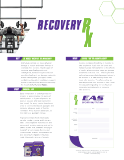
Review
MATBIO-1142; No. of pages: 8; 4C:
Role of gelatinases in pathological and
physiological processes involving the
dystrophin–glycoprotein complex
Manuela Bozzi a , Francesca Sciandra b and Andrea Brancaccio b, 1
a - Istituto di Biochimica e Biochimica Clinica, Università Cattolica del Sacro Cuore, Rome, Italy
b - Istituto di Chimica del Riconoscimento Molecolare (CNR) c/o Istituto di Biochimica e Biochimica Clinica, Università Cattolica
del Sacro Cuore, Rome, Italy
Correspondence to Manuela Bozzi: Istituto di Biochimica e Biochimica Clinica, Università Cattolica del Sacro Cuore,
L.go F. Vito 1, 00168 Rome, Italy. [email protected].
http://dx.doi.org/10.1016/j.matbio.2015.02.005
Edited by W.C. Parks and S. Apte
Abstract
Dystrophin is a cytosolic protein belonging to a membrane-spanning glycoprotein complex, called dystrophin–
glycoprotein complex (DGC) that is expressed in many tissues, especially in skeletal muscle and in the nervous
system. The DGC connects the cytoskeleton to the extracellular matrix and, although none of the proteins of the
DGC displays kinase or phosphatase activity, it is involved in many signal transduction pathways. Mutations in
some components of the DGC are linked to many forms of inherited muscular dystrophies. In particular, a
mutation in the dystrophin gene, leading to a complete loss of the protein, provokes one of the most prominent
muscular dystrophies, the Duchenne muscular dystrophy, which affects 1 out of 3500 newborn males. What is
observed in these circumstances, is a dramatic alteration of the expression levels of a multitude of
metalloproteinases (MMPs), a family of extracellular Zn 2+-dependent endopeptidases, in particular of MMP-2
and MMP-9, also called gelatinases. Indeed, the enzymatic activity of MMP-2 and MMP-9 on dystroglycan, an
important member of the DGC, plays a significant role also in physiological processes taking place in the central
and peripheral nervous system. This mini-review discusses the role of MMP-2 and MMP-9, in physiological as
well as pathological processes involving members of the DGC.
© 2015 Published by Elsevier B.V. This is an open access article under the CC BY-NC-ND license
(http://creativecommons.org/licenses/by-nc-nd/4.0/).
Introduction
Dystrophin is a cytosolic protein associated to a
glycoprotein complex, the dystrophin–glycoprotein
complex (DGC), composed by the intracellular α and
β-syntrophin, α-dystrobrevin and neuronal nitric oxide
synthase (nNOS), the transmembrane β-dystroglycan, α-, β-, γ- and δ-sarcoglycan and sarcospan, and
the extracellular α-dystroglycan [1,2]. The DGC is
expressed in a wide variety of tissues, especially in
skeletal muscle and in the nervous system, and
provides a strong contribution to the sarcolemma
stability. The DGC represents an important link
between the cytoskeleton and the extracellular matrix,
in several respects. It is directly and indirectly involved
in different signal transduction pathways. As an
example, nNOS, an enzyme activated by muscle
contraction, produces nitric oxide from L-arginine,
which in turn triggers the production of cGMP
catalyzed by the guanylyl cyclase; the second
messenger cGMP stimulates vasodilatation favoring
the blood influx into the contracting muscle [3–5].
Furthermore, some DGC members, including dystrobrevin [6], syntrophins [7] and β-dystroglycan [8],
serve as platforms to recruit phosphatases and
kinases involved in signal transduction pathways. In
addition to their indirect involvement in signal transduction pathways, the two dystroglycan subunits, αand β, which represent the DGC inner core, interact in
a non-covalent fashion through the C-terminal domain
of α-dystroglycan and the β-dystroglycan ectodomain
[9], playing a key role in maintaining the connection
between the cytoskeleton and the extracellular matrix
through formation of a multitude of interactions [10].
0022-2836/© 2015 Published by Elsevier B.V. This is an open access article under the CC BY-NC-ND license
(http://creativecommons.org/licenses/by-nc-nd/4.0/).
Matrix Biol. (2015) xx, xxx–xxx
Please cite this article as: Bozzi Manuela, et al, Role of gelatinases in pathological and physiological processes involving the
dystrophin–glycoprotein complex, Matrix Biol. (2015), http://dx.doi.org/10.1016/j.matbio.2015.02.005
2
Gelatinases' role in pathological and physiological processes including DGC
This network of interactions contributes to the
plasma membrane stability in normal conditions, on
the other hand, the connection between the cytosol
and the extracellular matrix needs to be interrupted
when tissue remodeling takes place. In such
circumstances, an overexpression of some matrix–
metalloproteinases (MMPs) is often observed.
These proteins belong to a family of Zn 2+ -dependent
extracellular endopeptidases involved in many
physiological (such as morphogenesis, development, cell migration, proliferation and adhesion) as
well as pathological processes, such as cancer,
neurodegeneration, inflammation and muscular dystrophy [11].
Although dystrophin has recently been proposed
as a new target of MMP-2 during ischemic injury [12]
and a decrease in γ-sarcoglycan levels has been
shown to correlate with an increase of MMP-2
activity, in an animal model of right ventricular failure
[13], dystroglycan remains the only ascertained and
direct DGC target of MMP-2 and MMP-9. In vitro,
the two gelatinases disrupt the recombinant βdystroglycan ectodomain by two distinct molecular
mechanisms. MMP-9 induces a first cleavage
leaving an intact C-terminal region of about 30
amino acids and an N-terminal region that is further
processed [14], whereas MMP-2 produces multiple
early cleavages on the entire protein [15] (see
Fig. 1). MMP-2 has lately also been found to exert
a significant proteolytic activity on native and
recombinant α-dystroglycan in vitro [16].
Dystroglycan degradation is driven by
gelatinases in physiological conditions
The ectodomain of β-dystroglycan represents the
Achille's heel of the DGC. The possibility of a
proteolytic breakdown, likely to take place at the
ectodomain of β-dystroglycan, was first postulated
after observing the electrophoretic behavior of a
30 kDa β-dystroglycan fragment in carcinoma cell
lines [17].
The first direct evidence of an enzymatic activity
driven by an MMP on dystroglycan comes from a
study of Yamada and colleagues who found a
truncated form of β-dystroglycan, devoid of part of
its ectodomain and therefore unable to maintain its
link with α-dystroglycan, in healthy tissues, such as
peripheral nerve, kidney, lung and smooth muscle.
In the same study the authors indicated the
metalloproteinases MMP-2/MMP-14 and MMP-9,
as the main players involved in the production of
this 30 kDa β-dystroglycan fragment [18].
Fig. 1. Scheme of the DGC and β-dystroglycan degradation driven by gelatinases. In vitro, MMP-9 catalyzes a first
cleavage within the β-dystroglycan ectodomain producing an intact C-terminal region of about 30 amino acids and an
N-terminal region that is further processed [14], while MMP-2 induces multiple cleavages on the entire ectodomain [15].
Please cite this article as: Bozzi Manuela, et al, Role of gelatinases in pathological and physiological processes involving the
dystrophin–glycoprotein complex, Matrix Biol. (2015), http://dx.doi.org/10.1016/j.matbio.2015.02.005
Gelatinases' role in pathological and physiological processes including DGC
Further studies highlighted that MMP-2, rather
than MMP-9, is constitutively expressed in injured
neurons and is responsible for the production of the
30 kDa β-dystroglycan fragment. This leads to loss
of pre- and post-synaptic connections preceding the
synaptic remodeling induced by injury [19].
A basal enzymatic activity level of the two
gelatinases producing low amounts of the 30 kDa
β-dystroglycan fragment was also found in healthy
neurons and hippocampus, but, under stimulation
with glutamate or bicuculline, the MMP-9 driven
cleavage of β-dystroglycan was significantly increased in post-synaptic elements and in the
hippocampus, indicating that this process might be
involved in learning and memory processes [20]. A
consistent increase of the MMP-9 levels, induced by
the transcription factor AP-1, with a consequent
increase of the 30 kDa β-dystroglycan fragment, has
also been observed in animals subjected to fear
conditioning [21]. It was hypothesized that the
MMP-9-driven cleavage of β-dystroglycan might
reorganize the network of interactions that dystroglycan, expressed in the post-synaptic elements,
establishes with other proteins, such as neurexin,
expressed in the pre-synapsis. The β-dystroglycan
cleavage might influence also the cytoskeletal
organization; for example, in Schwann cells, it was
found that the 30 kDa β-dystroglycan displays a
higher affinity for a short isoform of utrophin, UP71,
than for the dystrophin isoform Dp116, whose
interaction with full length β-dystroglycan is indeed
stronger than that with its truncated form [22].
Accordingly, it has been revealed that in normal
nerves MMP-2 and MMP-9 (that are differentially
expressed during development) shed the N-terminal
extracellular domain of β-dystroglycan making it
not longer able to bind α-dystroglycan, thus modulating the dystroglycan complex composition and
the size of the Schwann cell compartments [23].
Recently, a gelatinolytic activity targeting β-dystroglycan has been proposed to be involved in the
remodeling of the postsynaptic domain of the
neuromuscular junction after physical exercise [24].
Gelatinases are overexpressed in
DGC-associated muscular dystrophies
Duchenne muscular dystrophy is a lethal X-linked
disease due to a mutation in the dystrophin gene
causing complete loss of protein function [25].
Muscular tissues of patients affected by Duchenne
muscular dystrophy show sarcolemma instability,
loss of Ca 2+ homeostasis and reduced resistance to
mechanical stress with consequent degeneration of
muscular fibers followed by cycles of regeneration
[26]. Complete regeneration of the muscular fibers is
prevented by inflammatory cell invasion that triggers
fibrosis, progressively reducing the amount of func-
3
tional muscular tissue [1]. Many proteases, including
calpains, caspases and lysosomal proteases, increase their enzymatic activity in these conditions;
although controversial, an increased proteasome
activity has also been found [27].
In such circumstances, the integrity of the entire
DGC is severely compromised; besides loss of
dystrophin, the levels of nNOS [28,29] and of one
or both the dystroglycan subunits are dramatically
reduced, when they are not completely lost [30].
In 1999, Kherif and colleagues observed that the
expression and the activity of MMP-9 were increased in mice deficient of the dystrophin gene,
called mdx mice, which represent an animal model of
Duchenne muscular dystrophy [31]. This observation has been corroborated by many other studies
that revealed an upregulation of gelatinases also in
hearts of mdx mice [32–34].
MMP-2 and MMP-9 are overexpressed also in the
brain of mdx mice and they co-localize with the
vascular endothelial growth factor (VEGF), thus
explaining the increased angiogenesis and vascular
permeability observed in the brain of mdx mice [35].
Recently, many studies revealed that MMP-9 also
increases in serum and muscular biopsies of
patients affected by Duchenne muscular dystrophy
and that the MMP-9 levels correlate with the severity
of the disease [36].
Other members of the MMPs family display altered
expression patterns and enzymatic activities in the
dystrophic phenotype. For example, an increased
activity of MMP-1, which limits fibrosis mainly by
degrading collagen I and III, has been reported in
dystrophic muscles, during remodeling of the extracellular matrix [34]. Altered levels and activities of
MMP-1 and MMP-7, favoring a fibrotic phenotype,
have also been found in fibroblasts from Duchenne
muscular dystrophy biopsies [37], whereas overexpression of MMP-10 in dystrophic muscles plays a
role in the muscle regeneration process [38].
Interestingly, alterations of the MMP levels are very
often accompanied by alterations in the levels of
TIMP-1 and TIMP-2, the endogenous inhibitors of
MMPs [39,40]. In general, the increased expression
of MMPs in mdx mice is probably due to macrophages, mast cells and fibroblasts that infiltrate the
muscular tissue in pathological conditions and
secrete different endopeptidases [39]. Indeed, it is
well known that fibroblasts express high amounts of
MMPs [41,42].
What is the exact role of gelatinases in the
pathogenesis of DGC-related muscular dystrophies? This question still awaits a complete and
exhaustive answer. A characteristic shared by many
neuromuscular diseases is the presence of the
aforementioned 30 kDa β-dystroglycan fragment
that therefore represents an interesting biomarker
(see Fig. 1). Indeed, this peptide was revealed in
skeletal muscle of tissues affected by Duchenne
Please cite this article as: Bozzi Manuela, et al, Role of gelatinases in pathological and physiological processes involving the
dystrophin–glycoprotein complex, Matrix Biol. (2015), http://dx.doi.org/10.1016/j.matbio.2015.02.005
4
Gelatinases' role in pathological and physiological processes including DGC
muscular dystrophy [43] and sarcoglycanopathy
[43,44].
The quest for therapeutic targets:
inhibiting the gelatinases activity
by molecular genetics or
pharmacological methods
Important clues regarding the role of gelatinases in
the pathogenesis of DGC-related muscular dystrophies come from studies in which the corresponding
genes have been up- or down-regulated in healthy
and/or mdx mice or have been inhibited by molecular
drugs.
In general, genetic ablation of MMP-9 in mdx mice
improves the dystrophic phenotype, as indicated by a
number of parameters [33,45,46]. For example, disruption of the MMP-9 gene reduces the serum levels of
creatine kinase (a marker of muscular fibers damage),
as well as the amount of macrophages that infiltrate
muscular fibers, promotes muscle regeneration and
ameliorates the fibers' stability increasing the amount of
the two known targets of MMP-9, β-dystroglycan and
collagen IV, this latter being essential for the stability of
the basal membrane. Moreover, genetic depletion of
MMP-9 increases nNOS while reducing the levels of
caveolin-3 [45]. It was also observed that partial
deletion of MMP-9 in 8-week old mdx mice i) improves
the number of satellite cells in dystrophic muscles,
ii) suppresses the pro-inflammatory M1 macrophages
that contribute to muscular necrosis, and iii) increases
the anti-inflammatory M2 macrophages that stimulate
muscle regeneration by reducing the transcript levels
of the inflammatory cytokines INF-γ and IL-6 while
increasing the ones of the anti-inflammatory cytokine
IL-4 [46].
Surprisingly, MMP-9 ablation in mdx mice reduces
fibrosis by reducing the levels of collagen I and III [45],
whereas overexepression of MMP-9 in healthy mice
suppresses fibrosis by reducing the levels of collagen
I and IV but not collagen III [47]. These contradictory
results may be in part explained considering that in
pathological conditions, MMP-9 deletion alters the
levels of other MMPs, whose specificity is often
overlapping, and/or of their endogenous inhibitors
[33]. Deregulation of other cytokines and inflammatory
cells that modulate the entire fibrotic process, makes
this scenario even more complex [47]. Interestingly, in
addition to reducing fibrosis, overexpression of an
active form of MMP-9 in healthy mice stimulates
growth and regeneration of the muscle fibers (also
increasing their cross-sectional area), promotes the
fusion of myoblasts into myotubes, and improves the
contractile force in soleus muscle. Accordingly, an
upregulation of the insulin growth factor and follistatin,
both promoting skeletal muscle regeneration and
growth [48,49], is observed [47].
Disruption of the MMP-2 gene in mdx mice leads to
different results compared to the MMP-9 knockout.
Indeed, ablation of MMP-2 significantly reduces
muscle regeneration, as indicated by decreased
centronuclear regenerating fibers. This is probably
due to decreased levels of vascular endothelial growth
factor-A (VEGF-A) that impair angiogenesis by reducing the mean size of the vessels. Nitric oxide (NO) is a
vasodilator, produced by nNOS from arginine, which
stimulates capillary proliferation [50]. Depletion of
MMP-2 gene in mdx mice further reduces the levels
of nNOS. Surprisingly, degradation of full-length
β-dystroglycan producing the 30 kDa fragment occurs
at 1 month of age but not at 3 months [51].
These studies suggest that gelatinase overexpression compensates for muscular damage. For example, they partially degrade the basal lamina favoring
the migration and fusion of myogenic cells devoted to
regeneration of the damaged fibers [31,52]. A
sustained and prolonged activation of the two
gelatinases, on the other hand, leads to an excessive
protein degradation that exacerbates the dystrophic
phenotype.
In accordance with the knockout studies, suramin,
an antifibrotic drug, restores the diaphragm function,
decreasing the activity of MMP-9 but not that of
MMP-2, and increasing the β-dystroglycan levels [53].
Moreover, spironolactone, an aldosterone antagonist,
and lisinopril, an angiotensin-converting enzyme
(ACE) inhibitor, if co-administered before functional
impairment reduce the damage of cardiac and skeletal
muscle by decreasing the gelatinase activity [54].
Other molecules can be listed that ameliorate the
dystrophic phenotype by suppressing the gelatinase
activity. Among them, drugs that support the signal
transduction pathway mediated by nitric oxide,
which is otherwise attenuated in mdx mice, have
beneficial effects on these animal models. For
example, L-arginine, a target of nNOS enzyme,
reduces the β-dystroglycan cleavage and reinforces
its interaction with utrophin, by suppressing the
MMP-2 and MMP-9 activity. A correct re-localization
of nNOS is also observed in short-term treated mdx
mice [55], although prolonged treatment with this drug
stimulates fibrosis and is no longer effective on the
gelatinase activity [56]. Similar results are obtained
with Sildenafil, an inhibitor of phosphodiesterase 5, an
enzyme that reduces the levels of cGMP, a second
messenger involved in the NO induced signal
transduction pathway, reinforcing NO-driven signaling
[57]. Beneficial effects were obtained also with
Batimastat, a broad-spectrum MMP inhibitor, which
reduces fibrosis and the number of macrophages and
increases the diaphragm force restoring the levels of
nNOS and β-dystroglycan [40]. A mixture of free
radical scavengers, composed of α-lipoic acid and
L-carnitine, also reduces damages of dystrophy,
suppressing the gelatinase activity and increasing
the levels of β-dystroglycan [58].
Please cite this article as: Bozzi Manuela, et al, Role of gelatinases in pathological and physiological processes involving the
dystrophin–glycoprotein complex, Matrix Biol. (2015), http://dx.doi.org/10.1016/j.matbio.2015.02.005
5
Gelatinases' role in pathological and physiological processes including DGC
All these data strongly support the view that
MMPs may contribute to the progression of
DGC-linked muscular dystrophy, mainly by degrading β-dystroglycan.
Downregulation or pharmacological
inhibition of gelatinases suppresses
the MAPK kinase cascades
In various dystrophic models the susceptibility to
contraction-induced injury and the sarcolemma integrity are not always clearly correlated with fiber
degeneration and muscular dystrophy [59-61]. This
suggests that some other factors are likely to contribute
to muscle cell degeneration and its apoptosis.
Detrimental signal transduction pathways have been
indicated as a possible cause of muscular dystrophy.
In mdx mice, an alteration of the mitogen-activated
protein kinase (MAPK) signaling cascades, that
regulate proliferation, differentiation and cell-survival,
has been observed; dystrophic animals in fact display
an increased phosphorylation of the extracellular
signal regulated kinases 1 (ERK1) and 2 (ERK2)
[55,62,63] and of the c-jun N-terminal kinases 1 (JNK1)
and 2 (JNK2) [64–66], and a reduction in the
phosphorylation of p38 [63]. Activation of the ERK1/2
pathway has been found also in limb–girdle muscular
dystrophy, due to a deficiency in γ-sarcoglycan [67].
Moreover, the phosphatidylinositol 3-kinase (PI3K/Akt)
signaling pathway, which regulates cell viability and
protein synthesis [68], resulted altered in DMD and
limb–girdle muscular dystrophy, showing an increased
synthesis and phosphorylation of Akt to compensate
muscular damages [69,70]. Indeed, an increase of the
expression levels of Akt in mdx mice stimulates muscle
regeneration and muscle hypertrophy, by elevating the
levels of utrophin [71].
Interestingly, overexpression of MMP-9 in healthy
mice leads to increased phosphorylation levels of Akt
as well as of mTOR and FOXO1, two of its downstream
phosphorylation targets (the PI3K/Akt/mTOR pathway
promotes protein synthesis and hypertrophy, whereas
FOXO1 stimulates protein degradation, by modulating
the ubiquitin–proteasome pathway) rather than of
ERK1/2 and JNK1/2, whereas p38 MAPK phosphorylation is reduced. Conversely, ablation of MMP-9 in
mdx mice leads to reduced phosphorylation levels of
Akt and p38 MAPK, but does not affect phosphorylation
levels of ERK1/2 and JNK1/2 in the diaphragm of mdx
mice [33]. In the hearts of mdx mice, depletion of
MMP-9 reduces the phosphorylation of ERK1/2 and
Akt, thus stimulating heart hypertrophy, but not the
phosphorylation of JNK1/2 and p38 MAPK [47]. Many
drugs that ameliorate the dystrophic phenotype by
suppressing the gelatinase activity also affect the
MAPK kinase signaling. For example, a mixture of
α-lipoic acid and L-carnitine reduces the phosphory-
lation levels of p38 MAPK, ERK1/2 and JNK1/2
kinases, with a concomitant reduction of the creatine
kinase levels [58]. Moreover, Batimastat reduces
the levels of phosphorylated ERK1/2 and p38
MAPK, but does not affect the levels of phosphorylated JNK1/2 in mdx mice [41], although it should be
noted that this drug may have a more complex effect,
since it inhibits also other MMPs as well as ADAM
proteases [72,73].
However, the reason why and the mechanism by
which downregulation, or pharmacological inhibition
of MMPs suppresses MAPK kinase cascades has
yet to be clarified. There is some evidence that some
transcription factors or cytokines, involved in inflammation and fibrosis, may represent a liaison between
inhibition of MMPs and suppression of MAPK kinase
cascades.
The nuclear factor (NF)-kB, a ubiquitous transcription factor, which activates genes involved in inflammation, immune and stress responses [74,75]
and induces the expression of MMP-9, is overexpressed in mdx mice and in DMD patients [76–78].
MMP-9 expression is also induced by the transcription factor activator protein-1 (AP-1) [79,80]. Genetic ablation of MMP-9 in mdx mice reduces the
activity of NF-kB and AP-1, suggesting that MMP-9
regulates its own expression by a positive feedback
mechanism [45]. On the other hand, activation of
MAPK signaling triggers the expression of many
genes involved in inflammation and fibrosis, including AP-1 and NF-kB.
All these observations suggest that MAPK signaling, as well as the activities of NK-kB, AP-1 and
gelatinases, might be reciprocally regulated. Indeed,
the downregulation of MAPK signaling may produce
beneficial effects both per se and by reducing the
gelatinase levels, with possible important repercussions for the development of effective therapeutic
protocols for muscular dystrophies.
Acknowledgments
The authors wish to thank Maria Giulia Bigotti
(Bristol, UK) and Matt Reed (Gloucester, UK) for
their critical reading of the manuscript.
Received 30 September 2014;
Received in revised form 9 February 2015;
Accepted 10 February 2015
Available online xxxx
Keywords:
Dystrophin;
Dystrophin–glycoprotein complex;
Dystroglycan;
Please cite this article as: Bozzi Manuela, et al, Role of gelatinases in pathological and physiological processes involving the
dystrophin–glycoprotein complex, Matrix Biol. (2015), http://dx.doi.org/10.1016/j.matbio.2015.02.005
6
Gelatinases' role in pathological and physiological processes including DGC
Gelatinase;
Duchenne muscular dystrophy
Present address: School of Biochemistry, Bristol University,
Bristol, UK.
References
[1] Blake DJ, Weir A, Newey SE, Davies KE. Function and
genetics of dystrophin and dystrophin-related proteins in
muscle. Physiol Rev 2002;82:291–329.
[2] Ervasti JM, Sonnemann KJ. Biology of the striated muscle
dystrophin–glycoprotein complex. Int Rev Cytol 2008;265:
191–225.
[3] Thomas GD, Victor RG. Nitric oxide mediates contractioninduced attenuation of sympathetic vasoconstriction in rat
skeletal muscle. J Physiol 1998;506:817–26.
[4] Thomas GD, Shaul PW, Yuhanna IS, Froehner SC, Adams
ME. Vasomodulation by skeletal muscle-derived nitric oxide
requires alpha-syntrophin-mediated sarcolemmal localization of neuronal nitric oxide synthase. Circ Res 2003;92:
554–60.
[5] Sander M, Chavoshan B, Harris SA, Iannaccone ST, Stull JT,
Thomas GD, et al. Functional muscle ischemia in neuronal
nitric oxide synthase-deficient skeletal muscle of children
with Duchenne muscular dystrophy. Proc Natl Acad Sci U S
A 2000;97:13818–23.
[6] Ceccarini M, Grasso M, Veroni C, Gambara G, Artegiani B,
Macchia G, et al. Association of dystrobrevin and regulatory
subunit of protein kinase A: a new role for dystrobrevin as a
scaffold for signaling proteins. J Mol Biol 2007;371:
1174–87.
[7] Oak SA, Russo K, Petrucci TC, Jarrett HW. Mouse alpha1syntrophin binding to Grb2: further evidence of a role for
syntrophin in cell signaling. Biochemistry 2001;40:11270–8.
[8] Bozzi M, Morlacchi S, Bigotti MG, Sciandra F, Brancaccio A.
Functional diversity of dystroglycan. Matrix Biol 2009;28:
179–87.
[9] Di Stasio E, Sciandra F, Maras B, Di Tommaso F, Petrucci
TC, Giardina B, et al. Structural and functional analysis of the
N-terminal extracellular region of beta-dystroglycan. Biochem Biophys Res Commun 1999;266:274–8.
[10] Michele DE, Campbell KP. Dystrophin–glycoprotein complex: post-translational processing and dystroglycan function. J Biol Chem 2003;278:15457–60.
[11] Sbardella D, Fasciglione GF, Gioia M, Ciaccio C, Tundo GR,
Marini S, et al. Human matrix metalloproteinases: an
ubiquitarian class of enzymes involved in several pathological processes. Mol Aspects Med 2012;33:119–208.
[12] Buchholz B, Perez V, Siachoque N, Miksztowicz V, Berg G,
Rodríguez M, et al. Dystrophin proteolysis: a potential target
for MMP-2 and its prevention by ischemic preconditioning.
Am J Physiol Heart Circ Physiol 2014;307:H88–96.
[13] Daicho T, Daisho Y, Kojima S, Takano S, Tejima Y,
Marunouchi T, et al. Alterations in dystrophin-related
glycoproteins in development of right ventricular failure in
rats. J Pharmacol Sci 2009;111:405–15.
[14] Bozzi M, Inzitari R, Sbardella D, Monaco S, Pavoni E, Gioia M,
et al. Enzymatic processing of beta-dystroglycan recombinant
ectodomain by MMP-9: identification of the main cleavage site.
IUBMB Life 2009;61:1143–52.
[15] Sbardella D, Inzitari R, Iavarone F, Gioia M, Marini S,
Sciandra F, et al. Enzymatic processing by MMP-2 and
MMP-9 of wild-type and mutated mouse beta-dystroglycan.
IUBMB Life 2012;64:988–94.
[16] Sbardella D, Sciandra F, Gioia M, Marini S, Gori A,
Giardina B, et al. Alpha-dystroglycan is a potential target
of matrix metalloproteinase MMP-2. Matrix Biol 2015;41:
2–7.
[17] Losasso C, Di Tommaso F, Sgambato A, Ardito R, Cittadini
A, Giardina B, et al. Anomalous dystroglycan in carcinoma
cell lines. FEBS Lett 2000;484:194–8.
[18] Yamada H, Saito F, Fukuta-Ohi H, Zhong D, Hase A, Arai K,
et al. Processing of beta-dystroglycan by matrix metalloproteinase disrupts the link between the extracellular matrix and
cell membrane via the dystroglycan complex. Hum Mol
Genet 2001;10:1563–9.
[19] Paggi P, De Stefano ME, Petrucci TC. Synaptic remodeling
induced by axotomy of superior cervical ganglion neurons:
involvement of metalloproteinase-2. J Physiol Paris 2006;99:
119–24.
[20] Michaluk P, Kolodziej L, Mioduszewska B, Wilczynski GM,
Dzwonek J, Jaworski J, et al. Beta-dystroglycan as a target
for MMP-9, in response to enhanced neuronal activity. J Biol
Chem 2007;282:16036–41.
[21] Ganguly K, Rejmak E, Mikosz M, Nikolaev E, Knapska E,
Kaczmarek L. Matrix metalloproteinase (MMP-9) transcription in mouse brain induced by fear learning. J Biol Chem
2013;288:20978–91.
[22] Hnia K, Hugon G, Masmoudi A, Mercier J, Rivier F, Mornet D.
Effect of beta-dystroglycan processing on utrophin/Dp116
anchorage in normal and mdx mouse Schwann cell
membrane. Neuroscience 2006;141:607–20.
[23] Court FA, Zambroni D, Pavoni E, Colombelli C, Baragli C,
Figlia G, et al. MMP2-9 cleavage of dystroglycan alters the
size and molecular composition of Schwann cell domains. J
Neurosci 2011;31:12208–17.
[24] Yeghiazaryan M, Cabaj AM, Sławińska U, Wilczyński GM.
The expression and function of gelatinolytic activity at the rat
neuromuscular junction upon physical exercise. Histochem
Cell Biol 2015;143:143–52.
[25] Monaco AP, Neve RL, Colletti-Feener C, Bertelson CJ, Kurnit
DM, Kunkel LM. Isolation of candidate cDNAs for portions of
the Duchenne muscular dystrophy gene. Nature 1986;323:
646–50.
[26] Petrof BJ, Shrager JB, Stedman HH, Kelly AM, Sweeney HL.
Dystrophin protects the sarcolemma from stresses developed
during muscle contraction. Proc Natl Acad Sci U S A 1993;90:
3710–4.
[27] Hollinger K, Selsby JT. The physiological response of
protease inhibition in dystrophic muscle. Acta Physiol (Oxf)
2013;208:234–44.
[28] Brenman JE, Chao DS, Xia H, Aldape K, Bredt DS. Nitric
oxide synthase complexed with dystrophin and absent from
skeletal muscle sarcolemma in Duchenne muscular dystrophy. Cell 1995;82:743–52.
[29] Chang WJ, Iannaccone ST, Lau KS, Masters BS, McCabe
TJ, Mc-Millan K, et al. Neuronal nitric oxide synthase and
dystrophin-deficient muscular dystrophy. Proc Natl Acad Sci
U S A 1996;93:9142–7.
[30] Barresi R, Campbell KP. Dystroglycan: from biosynthesis to
pathogenesis of human disease. J Cell Sci 2006;119:199–207.
[31] Kherif S, Lafuma C, Dehaupas M, Lachkar S, Fournier JG,
Verdière-Sahuqué M, et al. Expression of matrix metalloproteinases 2 and 9 in regenerating skeletal muscle: a study in
Please cite this article as: Bozzi Manuela, et al, Role of gelatinases in pathological and physiological processes involving the
dystrophin–glycoprotein complex, Matrix Biol. (2015), http://dx.doi.org/10.1016/j.matbio.2015.02.005
Gelatinases' role in pathological and physiological processes including DGC
[32]
[33]
[34]
[35]
[36]
[37]
[38]
[39]
[40]
[41]
[42]
[43]
[44]
[45]
[46]
[47]
experimentally injured and mdx muscles. Dev Biol 1999;205:
158–70.
Au CG, Butler TL, Sherwood MC, Egan JR, North KN, Winlaw
DS. Increased connective tissue growth factor associated
with cardiac fibrosis in the mdx mouse model of dystrophic
cardiomyopathy. Int J Exp Pathol 2011;92:57–65.
Dahiya S, Bhatnagar S, Hindi SM, Jiang C, Paul PK, Kuang
S, et al. Elevated levels of active matrix metalloproteinase-9
cause hypertrophy in skeletal muscle of normal and
dystrophin-deficient mdx mice. Hum Mol Genet 2011;20:
4345–59.
Zanotti S, Gibertini S, Di Blasi C, Cappelletti C, Bernasconi P,
Mantegazza R, et al. Osteopontin is highly expressed in
severely dystrophic muscle and seems to play a role in muscle
regeneration and fibrosis. Histopathology 2011;59:1215–28.
Nico B, Corsi P, Ria R, Crivellato E, Vacca A, Roccaro AM,
et al. Increased matrix–metalloproteinase-2 and matrix–
metalloproteinase-9 expression in the brain of dystrophic
mdx mouse. Neuroscience 2006;140:835–48.
Nadarajah VD, van Putten M, Chaouch A, Garrood P, Straub
V, Lochmüller H, et al. Serum matrix metalloproteinase-9
(MMP-9) as a biomarker for monitoring disease progression
in Duchenne muscular dystrophy (DMD). Neuromuscul
Disord 2011;21:569–78.
Zanotti S, Gibertini S, Mora M. Altered production of extracellular matrix components by muscle-derived Duchenne
muscular dystrophy fibroblasts before and after TGF-beta1
treatment. Cell Tissue Res 2010;339:397–410.
Bobadilla M, Sáinz N, Rodriguez JA, Abizanda G, Orbe J, de
Martino A, et al. MMP-10 is required for efficient muscle
regeneration in mouse models of injury and muscular
dystrophy. Stem Cells 2014;32:447–61.
Delfín DA, Zang KE, Schill KE, Patel NT, Janssen PM,
Raman SV, et al. Cardiomyopathy in the dystrophin/utrophindeficient mouse model of severe muscular dystrophy is
characterized by dysregulation of matrix metalloproteinases.
Neuromuscul Disord 2012;22:1006–14.
Kumar A, Bhatnagar S, Kumar A. Matrix metalloproteinase
inhibitor batimastat alleviates pathology and improves
skeletal muscle function in dystrophin-deficient mdx mice.
Am J Pathol 2010;177:248–60.
Gallagher GL, Jackson CJ, Hunyor SN. Myocardial extracellular matrix remodelling in ischemic heart failure. Front Biosci
2007;12:1410–9.
Porter KE, Turner NA. Cardiac fibroblasts: at the heart of
myocardial remodelling. Pharmacol Ther 2009;123:255–78.
Matsumura K, Zhong D, Saito F, Arai K, Adachi K, Kawai H,
et al. Proteolysis of beta-dystroglycan in muscular diseases.
Neuromuscul Disord 2005;15:336–41.
Anderson LV, Davison K. Multiplex Western blotting system
for the analysis of muscular dystrophy proteins. Am J Pathol
1999;154:1017–22.
Li H, Mittal A, Makonchuk DY, Bhatnagar S, Kumar A. Matrix
metalloproteinase-9 inhibition ameliorates pathogenesis and
improves skeletal muscle regeneration in muscular dystrophy. Hum Mol Genet 2009;18:2584–98.
Hindi SM, Shin J, Ogura Y, Li H, Kumar A. Matrix
metalloproteinase 9 inhibition improves proliferation and
engraftment of myogenic cells in dystrophic muscle of mdx
mice. PLoS One 2013;8. http://dx.doi.org/10.1371/journal.
pone.0072121.
Dahiya S, Givvimani S, Bhatnagar S, Qipshidze N, Tyagi SC,
Kumar A. Osteopontin-stimulated expression of matrix
metalloproteinase-9 causes cardiomyopathy in the mdx
[48]
[49]
[50]
[51]
[52]
[53]
[54]
[55]
[56]
[57]
[58]
[59]
[60]
[61]
[62]
[63]
7
model of Duchenne muscular dystrophy. J Immunol 2011;
187:2723–31.
Ten Broek RW, Grefte S, Von den Hoff JW. Regulatory
factors and cell populations involved in skeletal muscle
regeneration. J Cell Physiol 2010;224:7–16.
Lee SJ, Lee YS, Zimmers TA, Soleimani A, Matzuk MM,
Tsuchida K, et al. Regulation of muscle mass by follistatin
and activins. Mol Endocrinol 2010;24:1998–2008.
Milkiewicz M, Hudlicka O, Brown MD, Silgram H. Nitric oxide,
VEGF, and VEGFR-2: interactions in activity-induced angiogenesis in rat skeletal muscle. Am J Physiol Heart Circ
Physiol 2005;289:H336–43.
Miyazaki D, Nakamura A, Fukushima K, Yoshida K, Takeda
S, Ikeda S. Matrix metalloproteinase-2 ablation in dystrophindeficient mdx muscles reduces angiogenesis resulting in
impaired growth of regenerated muscle fibers. Hum Mol
Genet 2011;20:1787–99.
Pichavant C, Gargioli C, Tremblay JP. Intramuscular
transplantation of muscle precursor cells over-expressing
MMP-9 improves transplantation success. PLoS Curr 2011.
http://dx.doi.org/10.1371/currents.RRN1275.
Taniguti AP, Matsumura CY, Rodrigues-Simioni L, Santo
Neto H, Marques MJ. Suramin affects metalloproteinase-9
activity and increases beta-dystroglycan levels in the
diaphragm of the dystrophin-deficient mdx mouse. Muscle
Nerve 2012;46:810–3.
Rafael-Fortney JA, Chimanji NS, Schill KE, Martin CD,
Murray JD, Ganguly R, et al. Early treatment with lisinopril
and spironolactone preserves cardiac and skeletal muscle in
Duchenne muscular dystrophy mice. Circulation 2011;124:
582–8.
Hnia K, Gayraud J, Hugon G, Ramonatxo M, De La Porte S,
Matecki S, et al. L-Arginine decreases inflammation and
modulates the nuclear factor-kappaB/matrix metalloproteinase cascade in mdx muscle fibers. Am J Pathol 2008;172:
1509–19.
Wehling-Henricks M, Jordan MC, Gotoh T, Grody WW, Roos
KP, Tidball JG. Arginine metabolism by macrophages promotes cardiac and muscle fibrosis in mdx muscular dystrophy.
PLoS One 2010;5. http://dx.doi.org/10.1371/journal.pone.
0010763.
Percival JM, Whitehead NP, Adams ME, Adamo CM, Beavo
JA, Froehner SC. Sildenafil reduces respiratory muscle
weakness and fibrosis in the mdx mouse model of Duchenne
muscular dystrophy. J Pathol 2012;228:77–87.
Hnia K, Hugon G, Rivier F, Masmoudi A, Mercier J, Mornet
D. Modulation of p38 mitogen-activated protein kinase
cascade and metalloproteinase activity in diaphragm muscle in response to free radical scavenger administration in
dystrophin-deficient Mdx mice. Am J Pathol 2007;170:
633–43.
Gumerson JD, Michele DE. The dystrophin–glycoprotein
complex in the prevention of muscle damage. J Biomed
Biotechnol 2011. http://dx.doi.org/10.1155/2011/210797.
Batchelor CL, Winder SJ. Sparks, signals and shock
absorbers: how dystrophin loss causes muscular dystrophy.
Trends Cell Biol 2006;16:198–205.
Yoshida M, Ozawa E. Glycoprotein complex anchoring
dystrophin to sarcolemma. J Biochem 1990;108:748–52.
Kumar A, Khandelwal N, Malya R, Reid MB, Boriek AM. Loss
of dystrophin causes aberrant mechanotransduction in
skeletal muscle fibers. FASEB J 2004;18:102–13 [2004].
Lang JM, Esser KA, Dupont-Versteegden EE. Altered activity
of signaling pathways in diaphragm and tibialis anterior
Please cite this article as: Bozzi Manuela, et al, Role of gelatinases in pathological and physiological processes involving the
dystrophin–glycoprotein complex, Matrix Biol. (2015), http://dx.doi.org/10.1016/j.matbio.2015.02.005
8
Gelatinases' role in pathological and physiological processes including DGC
[64]
[65]
[66]
[67]
[68]
[69]
[70]
[71]
[72]
muscle of dystrophic mice. Exp Biol Med (Maywood) 2004;
229:503–11.
Kolodziejczyk SM, Walsh GS, Balazsi K, Seale P, Sandoz J,
Hierlihy AM, et al. Activation of JNK1 contributes to dystrophic
muscle pathogenesis. Curr Biol 2001;11:1278–82.
Nakamura A, Yoshida K, Ueda H, Takeda S, Ikeda S. Upregulation of mitogen activated protein kinases in mdx
skeletal muscle following chronic treadmill exercise. Biochim
Biophys Acta 2005;1740:326–31.
St-Pierre SJ, Chakkalakal JV, Kolodziejczyk SM, Knudson
JC, Jasmin BJ, Megeney LA. Glucocorticoid treatment
alleviates dystrophic myofiber pathology by activation of the
calcineurin/NF-AT pathway. FASEB J 2004;18:1937–9.
Griffin MA, Feng H, Tewari M, Acosta P, Kawana M,
Sweeney HL, et al. gamma-Sarcoglycan deficiency increases cell contractility, apoptosis and MAPK pathway
activation but does not affect adhesion. J Cell Sci 2005;118:
1405–16.
Datta SR, Brunet A, Greenberg ME. Cellular survival: a play
in three Akts. Genes Dev 1999;13:2905–27.
Dogra C, Changotra H, Mohan S, Kumar A. Tumor necrosis
factor-like weak inducer of apoptosis inhibits skeletal
myogenesis through sustained activation of nuclear factorkappaB and degradation of MyoD protein. J Biol Chem 2006;
281:10327–36.
Peter AK, Crosbie RH. Hypertrophic response of Duchenne
and limb–girdle muscular dystrophies is associated with
activation of Akt pathway. Exp Cell Res 2006;312:2580–91.
Peter AK, Ko CY, Kim MH, Hsu N, Ouchi N, Rhie S, et al.
Myogenic Akt signalling upregulates the utrophin–glycoprotein complex and promotes sarcolemma stability in muscular
dystrophy. Hum Mol Genet 2009;18:318–27.
Botos I, Scapozza L, Zhang D, Liotta LA, Meyer EF.
Batimastat, potent matrix metalloproteinase inhibitor, exhibits
an unexpected mode of binding. Proc Natl Acad Sci U S A
1996;93:2749–54.
[73] Parvathy S, Karran EH, Turner AJ, Hooper NM. The
secretases that cleave angiotensin converting enzyme and
the amyloid precursor protein are distinct from tumour
necrosis factor-alpha convertase. FEBS Lett 1998;431:63–5.
[74] Karin M, Delhase M. The I kappa B kinase (IKK) and NFkappa B: key elements of proinflammatory signalling. Semin
Immunol 2000;12:85–98.
[75] Vellaichamy E, Khurana ML, Fink J, Pandey KN. Involvement
of the NF-kappa B/matrix metalloproteinase pathway in
cardiac fibrosis of mice lacking guanylyl cyclase/natriuretic
peptide receptor A. J Biol Chem 2005;280:19230–42.
[76] Messina S, Altavilla D, Aguennouz M, Seminara P, Minutoli
L, Monici MC, et al. Lipid peroxidation inhibition blunts
nuclear factor-kappaB activation, reduces skeletal muscle
degeneration, and enhances muscle function in mdx mice.
Am J Pathol 2006;168:918–26.
[77] Kumar A, Lnu S, Malya R, Barron D, Moore J, Corry DB, et al.
Mechanical stretch activates nuclear factor-kappaB, activator
protein-1, and mitogen-activated protein kinases in lung
parenchyma: implications in asthma. FASEB J 2003;17:
1800–11.
[78] Kumar A, Boriek AM. Mechanical stress activates the nuclear
factor-kappaB pathway in skeletal muscle fibers: a possible
role in Duchenne muscular dystrophy. FASEB J 2003;17:
386–96.
[79] Lin CC, Kuo CT, Cheng CY, Wu CY, Lee CW, Hsieh HL, et al.
IL-1 beta promotes A549 cell migration via MAPKs/AP-1- and
NF-kappaB-dependent matrix metalloproteinase-9 expression. Cell Signal 2009;11:1652–62.
[80] Moon SK, Cha BY, Kim CH. ERK1/2 mediates TNF-alphainduced matrix metalloproteinase-9 expression in human
vascular smooth muscle cells via the regulation of NFkappaB and AP-1: involvement of the ras dependent
pathway. J Cell Physiol 2004;198:417–27.
Please cite this article as: Bozzi Manuela, et al, Role of gelatinases in pathological and physiological processes involving the
dystrophin–glycoprotein complex, Matrix Biol. (2015), http://dx.doi.org/10.1016/j.matbio.2015.02.005


© Copyright 2026