
Document 17355
Angiotensin-Converting
Peter
G. Pryde,
P.G
Pryde,
Aileen
CE.
B. Sedman,2
Nugent,
M.
Obstetrics/Gynecology,
cal
Center,
A.B.
M.
M.
Soc,
Jr.,
E. Nugent,
Department
of
Michigan
of
Medical
Center,
of
Pathology,
Center.
Nephrol.
of
Jr. , Department
Barr,
Medical
Clark
Medi-
Ml
Jr. , Department
Barr,
(J. Am.
Arbor,
of Michigan
Michigan
Barr,
University
Ann
Sedman,
University
Enzyme
Ann Arbor,
1993;
Pediatrics,
Ann Arbor,
University
Ml
of
Ml
3:1575-1582)
ABSTRACT
Angiotensin-converting
enzyme
(ACE)
inhibitors
are
widely used for controlling
hypertension.
Their use in
women
who are pregnant
is not without
risk to the
fetus. We describe
three infants exposed
in utero to
ACE inhibitors
who had adverse
outcomes.
These
cases, combined
with other reports in the literature,
suggest
strongly
that these drugs are fetotoxic.
ACE
inhibitor
fetopathy
is characterized
by fetal hypotension,
anuria-oligohydramnios,
monary
hypoplasia,
pocalvaria.
Although
fetal effects
has yet
the debilitating
and
age when it occurs,
ACE inhibitors
not be
in the
second
Key Words:
and
Kidney,
oligohydramnios,
growth
restriction,
pul-
renal tubular dysplasia,
and hythe true frequency
of adverse
to be determined,
because
of
lethal nature
of the fetal damit is our recommendation
that
used in pregnancy,
particularly
third
trimesters.
captopril,
teratogen,
enalapril,
lisinopril,
skull, anuria.
fetotoxin
I
n the decade
since
their introduction,
the angiotensin-converting
enzyme
(ACE)
inhibitors
have
gained
widespread
acceptance
in the management
of
hypertension.
Their
success
(even
as monotherapy)
and relative
paucity
of side effects
have
contributed
to this popularity
(1 -3). Further,
the lack of metabolic
changes
(such
as hyperglycemia
or hyperbipidemla).
as well as a possible
ameliorating
effect
on the progression
of diabetic
nephropathy,
has prompted
the
suggestion
that
ACE inhibitors
be considered
first-
‘Received
August
2Correspondence
0297,
University
25, 1992. Accepted
to Dr. A. B. Sedman,
of Michigan
Medical
November
Pediatric
Center.
30. 1992.
Nephrology,
Ann Arbor,
1046-6673/0309-1575$03.00/0
Journal
of the American
society
of Nephrology
Copyright
© 1993 by the American
Society
of Nephrology
Journal
of the
American
Society
of Nephrology
Room
L2602,
MI 48 109-0297.
Box
Inhibitor
and
Mason
Barr,
Fetopathy’
Jr.
line agents
in the management
of hypertensive
diabetics
(1.3.4).
As early
as 1 980,
there
were
worrisome
reports
of
extraordinary
fetal
loss rates
in a variety
of experimental
animals
exposed
to ACE
inhibitors
during
gestation
(5-7).
In 1 98 1 the first
case
of adverse
fetal outcome
in a human
pregnancy
exposed
to captopril
was reported
(8). Soon,
other
cases
appeared
Implicating
both
captopril
and enalaprib
as possible
fetotoxins
(9-12).
The animal
data,
coupled
with
a
growing
list of adverse
human
pregnancy
outcomes,
prompted
warnings
as early
as 1 985 against
the use
of ACE inhibitors
during
human
gestation
( 1 3). However,
in contacts
with
prescribing
physicians,
our
experience
has been
that many
are not aware
of the
potential
danger
to the fetus
from
ACE inhibitor
exposure.
We report
three
cases
from
our institution
of adverse
fetal
outcome
in hypertensive
pregnancies
exposed
to ACE inhibitors,
and we review
previously
reported
cases
(8-12,14-24).
Further,
we propose
that
sufficient
evidence
exists
to implicate
the ACE
inhibitors
as fetotoxins
and suggest
the designation
“ACE inhibitor
fetopathy.”
,
CASE
Patient
REPORTS
I
The mother
was a 26-yr-old.
gravida
2, para 0. with
an 8-yr history
of systemic
bupus erythematosus.
Her
associated
hypertension
was controlled
by a regimen
of prednisone.
5 to 1 0 mg/day,
atenobol,
1 00 mg/day,
furosemide,
20 mg/day,
and captopril,
1 50 mg/day.
Ultrasonographic
evaluation
at 1 9 wk of gestation
revealed
oligohydramnios
but no fetal structural
abnormalities.
Over the next several
weeks,
there
was
persistence
of the oligohydramnios
and evolution
of
mild growth
restriction.
At 33 wk of gestation.
labor
was induced
because
of a worsening
biophysical
profile. A 1 .440-g
female
was delivered
vaginally
from a
breech
presentation.
Apgar
scores
were
1 and 5 at 1
and 5 mm,
respectively.
Despite
maximal
ventilator
support.
the infant
remained
hypoxic
and
became
progressively
acidotic.
Her hypotenslon
was
unresponsive
to volume
and pressor
therapy,
and during
the 14 h before
death,
there
was no urine
output.
At autopsy,
the hand,
foot, and face deformational
features
of the oligohydramnios
sequence
were identified.
The lungs
were
small
with
histologic
features
of pulmonary
hypoplasia.
A calcified
thrombus
in the
subhepatic
vena
cava
extended
into the renal
veins
bilaterally.
Subdiaphragmatic
venous
return
to the
1575
ACE Inhibitor
Fetopathy
heart
was via a well-developed,
thick-walled
azygous
system
to the superior
vena
cava.
The kidneys
were
enlarged
compared
with
gestationab
age-matched
controls
and had histologic
changes
consistent
with
renal
tubular
dysplasia.
The distal
urinary
tract,
although
slender
and empty.
was structurally
normal
and patent.
The brain
and cranial
base were normal.
However,
the calvarial bones, although
normal
in
shape
and
configuration,
were
remarkably
small,
leaving the top third
of the brain
unprotected
by bone
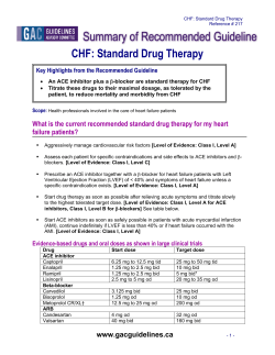
(Figure 1). Microscopically,
these
bones
were
thin
with poor trabeculatlon
and
marrow
development
compared
with
those
of age-matched
controls
(see
reference
38).
Patient
2
The mother
was an 18-yr-old
primigravida
with
renovascular
hypertension
of 10 yr duration.
She had
been managed
on multiagent
therapy with variable
success
until
age 18, at which
time llsinoprib
monotherapy
was initiated
with
the establishment
of excellent
control.
When
she became
pregnant,
lisinopril
was continued.
At 16 wk of gestation,
an ultrasound
examination
revealed
a fetus
of appropriate
size with
a normal
anatomic
survey
and normal
amniotic
fluid
volume.
Subsequently,
there
was
no suspicion
of
problems
until
the onset
of labor
at 32 wk of gestation.
After
a failed
trial of tocolysis,
a 1 .480-g
male
was
delivered
by cesarean
section.
Amniotic
fluid
volume
was not described.
Apgar
scores
were
4 and
7 at 1 and 5 mm,
respectively.
The infant
required
48 h of mechanical
ventilation
for respiratory
distress.
The newborn
period
was complicated
by hypotension and anuria
that
failed
to respond
to fluid chablenge
and
pressor
support.
Ultrasound
evaluation
showed
normal
size and appearance
of the kidneys
and
renal
collecting
system.
Renal
artery
Doppler
studies
demonstrated
bilateral
flow.
At 8 days
of age, the infant
remained
anuric
and
peritoneab dialysis was initiated. By day 12 of life, he
began
to pass
small
amounts
of urine. ACE activity
increased
from a day 12 value
of 7.9 U/mL
(normal,
20 to 50) to 22 U/mL
on day 37. A kidney
biopsy
performed
at 4 wk of age demonstrated
evidence
of
previous
ATN,
patchy
cortical
necrosis,
and lack of
Figure 1. Hypocalvaria
(Patient
1). The skull viewed from above,
with the fibrous tissue of the fontanels
to emphasize
the diminutive
size of the bones. Reprinted
with permission
from Barr and Cohen (38).
1576
Volume
and sutures
3.
Number
removed
9
1993
Pryde
Patient
differentiation
of the proximal
convoluted
tubules
(Figure
2). Although
his urine
volume
increased
modestby over the next
several
weeks,
he remained
dependent
on peritoneal
dialysis
and,
at 22 months
of
age. received
a successful
kidney
transplant.
He mitialby suffered
from intestinal
malabsorption
and Severe
growth
retardation.
After
the transplant.
his
growth
remains
delayed.
but his cognitive
function
is progressing
normally.
At birth,
his fontanels
were
noted
to be barge and
the sutures
excessively
wide.
At 1 month
of age, the
cabvarlal
plates
were
small
but of appropriate
shape
and position.
The width
of the sagittal.
coronal,
and
lambdoidab
sutures
measured
3, 3, and 2 cm, respectively.
The anterior
fontaneb
was 6 cm long by 4 cm
wide,
and the posterior
fontanel
was 4 cm by 4 cm.
Subsequently,
considerable
growth
of the skull bones
was noted
so that
by 3 months
of age. the sutures
had narrowed
to 0.5 cm in width,
with a corresponding reduction
in size of the fontanebs.
At 22 months
of age, his anterior
fontanel
was still patent,
but the
rest of the skull
examination
was unremarkable.
Figure 2. ACE inhibitor-exposed
kidney (Patient 2) showing
of differentiation
between
proximal
and distal convoluted
original magnification,
x20.
Journal
of the
American
Society
of Nephrology
et al
3
The mother
was a 30-yr-old
primigravida
with a 9yr history
of presumed
essential
hypertension
adequately
controlled
by atenobob
until
several
months
before
conception.
At that
time,
she was
changed
electively
to enalapril,
5 mg/day.
Her blood
pressure
was well controlled
until
20 wk of gestation,
when
the daily dose was increased
to 7.5 mg for an elevated
diastolic
pressure.
At 30 wk. fundal
height
was noted
to be subnormal
and ultrasonography
demonstrated
fetal
growth
restriction
and profound
oligohydramnios.
At 32 wk,
a contraction
stress
test
showed
recurrent
decelerations
and the infant
was delivered
by cesarean
section.
The boy infant weighed
only 980 g although
the
physical examination
was appropriate
for 32 wk of
gestation. Apgar scores were 8 and 9 at 1 and 5 mm,
respectively, but mild grunting
progressed
to frank
respiratory
distress
and
by 2 h of age mechanical
ventilation was required. He was
profoundly
hypotensive
(mean
arterial
pressure
of 16 mm Hg) and
remained
so despite administration
of dopammne,
do-
dilated
tubules,
Bowman’s
spaces and tubules,
and increased
mesenchyme.
old tubular necrosis, lack
Periodic
acid-Schiff
stain;
1577
ACE Inhibitor
Fetopathy
butamine,
and
epmnephrmne
and
aggressive
hydration.
Hypotension
and anuria
persisted
until
initiation,
on day 5 of life, of peritoneal
dialysis.
After
14
h of dialysis.
his blood pressure
rose to 40/ 1 7 mm Hg
and
7 mL of urine
was
obtained.
On day
8, the
dialysate
became
cloudy
and
bowel
obstruction
or
perforation
was suspected.
At laparotomy,
a length
of ischemic
jejunum
was
resected.
Postoperatively,
he remained
hypotensive
despite
massive
pressor
support
and died on day 9.
At autopsy,
he had barge fontanebs
(anterior,
5.0 x
3.5 cm: posterior,
2.0 x 1 .5 cm) and widened
sutures
(sagittal.
1 cm; coronals
and
bambdoidabs,
0.3 cm:
metopic,
0.5 cm). His face was wide with a short
nose
and anteverted
nares.
His arms
(150
mm) and begs
(120 mm) were
short
in relation
to his crown-rump
length
(285 mm).
The kidneys
were
barge (combined
weight.
16.1 g; expected,
9.0 ± 1.6 for body weight).
but no other
gross
abnormalities
of the urinary
tract
or renal
vascular
system
were
found.
Microscopic
examinations
of the kidneys
showed
tubular
dysplasia (Figure
3). The lungs,
particularly
the left one,
were small
(right,
13.7 g; left, 6.9 g; combined
weight,
20.6 g; expected,
scopicab
features
25.4 ± 5.2)
of hypoplasia.
and
showed
the
micro-
DISCUSSION
The ACE inhibitors
are competitive
inhibitors
of
kininase
II. Their
complex
pharmacology
has been
reviewed
in detail
by others
( 1 -3). Most importantly.
they
affect
both
the renin-angiotensin-abdosterone
and
the
bradykinin-prostaglandin
systems.
Many
other
antlhypertensive
agents
tend
to increase
peripherab
resistance
and affect
the metabolism
of glucose, lipids,
and electrolytes.
In contrast,
ACE inhibitors
decrease
vascular
resistance
and
have
no
known
adverse
metabolic
consequences
( 1 ,3). In the
absence
of heart
failure,
they
produce
little
change
in heart
rate,
cardiac
output,
or pulmonary
wedge
pressure
(25-27).
An important
effect
of ACE inhibitors
is on the renal
microvasculature,
resulting
in
dilatation
of the gbomerular
efferent
arterioles
and a
decrease
in filtration
pressure.
Excretion
of the ACE inhibitors
is principally
by
the kidney. They are also known
to cross the human
(
,
‘:N.l
..
a
?
-,
,4
_J
a
.
-
-
4
:“
Figure 3. ACE Inhibitor-exposed
kidney
tubules, dilated
tubules, and increased
1578
4
(Patient 3) showing tubular dysplasia:
minimal differentiation
of proximal
mesenchyme.
Periodic acid-Schiff
stain; original magnification,
x20.
Volume
3’
Number
convoluted
9’
1993
Pryde
TABLE
1. Cases
of pregnancy
outcome
Author,
(Ref. No.),
Year
Guignard
1981
Dummy
(9),
Boutroy
Burger
Renovascular
pertension
(10),
1984
Caraman
eta!. (11).
1984
Caraman
eta!.
(11).
1984
Rothberg
and Iorenz
(12), 1984
Fiocchi eta!. (14),
1984
eta!.
(15),
1985
Schubiger
29 wk; cesarean
I 0%
2nd trimester
hy-
eta!.
(16)
Broughton-Pipkmn
a!. (17), 1989
et
Mehta
3-10%
34 wk; cesarean
10-50%
28 wk; cesarean
etiology
section;
No
No
RDS, PDA, survived
section;
No
No
RDS, PDA, survived
section;
Yes
Yes
section
Yes
Yes
Hypocalvaria,
dialysis,
death day 30
Survived
section;
No
No
IUGR, survived
section;
Yes
Yes
section;
Yes
Yes
Decreased
ACE activity, dialysis, survived
Placental
abruption,
severe RDS, death
day 6
IUFD, suspected
renal
tubular dysgenesis
Hypoplastic
lungs, hypocalvaria,
death
day 10
RDS, death day 7
N/A
Yes
Yes
Implied
Yes
Yes
Yes
Systemic
lupus
thematosus
Systemic lupus
thematosus
transplant
(21),
IUFD, intrauterine
patent
ductus
fetal
eryery-
death:
arteriosus;
26 wk; cesarean
section;
3%
34 wk; cesarean
section;
weight not recorded
lUG?, intrauterine
N/A,
not
growth
of the American
Society
of Nephrology
restriction;
RDS. respiratory
Hypoplastic
lungs,
renal tubular dysgenesis
distress syndrome;
PIH, pregnancy-induced
applicable.
placenta.
In the
fetus,
it Is presumed
that
these
agents
are, at beast in part,
also renally
excreted
into
the amniotic
fluid and recycled
by fetal swallowing.
In animal
studies,
the administration
of captopril
in doses
comparable
to those
used In human
therapy
has
been
associated
with
significant
rates
of fetal
loss.
Chronically
cannulated
pregnant
ewes
given
3
mg/kg
of captopril
demonstrated
fetal
loss rates
in
excess
of 80% (7). The administration
of 2.5 to 5.0
mg/kg
of captopril
per day to pregnant
rabbits
yielded
pregnancy
loss rates
ranging
from
37 to 92% (5-7).
Severe
and
prolonged
fetal
hypotension
has
been
demonstrated
in the sheep
model
(7) and
has been
implicated
in the high rate of fetal wastage.
Possible
impairment
of uteroplacental
perfusion
by ACE inhibitors
has also been
suggested
(6,7).
Journal
Yes
Yes
(20),
1989
PDA.
Yes
section:
vaginal
delivery
32 wk; cesarean
section;
3-10%
Scott and Purohit
Abbreviations:
N/A
29 wk IUFD, spontaneous
Renal
hypertension;
N/A
Pulmonary
hypoplasia,
decreased
ACE activity, death day 8
Elective abortion,
hypocalvaria,
transverse limb defect
PDA-surgical
repair,
Renal transplant
(19),
Other
and Anuria
survived
34 wk; cesarean
1989
0
Neonatal
Hypotension
3-10%
PIH
and Modi
or in pairs’
Yes
termination
34 wk; cesarean
310%
34 wk; cesarean
<3%
35 wk; cesarean
10-50%
36 wk; cesarean
1989
Cunniff eta!.
1990
Oligohydramnios
individually
<3%
eta!.
(18),
reported
Yes
<3%
Renovascular
hypertension
Essential hypertension
Essential hypertension
Unclear
section;
34 wk; cesarean
Nephrotic
Syndrome
Renovascular
hypertension
Glomerulonephritis,
1988
Knott
exposure
Birth Weight
hypertension
Ducret
inhibitor
(Percentile)
Preeclampsia
eta!.
ACE
Gestation;
Mode of Delivery;
Maternal
Disease
eta!. (8),
and
1981
after
et al
The true
rates
of adverse
fetal
effects
from
ACE
inhibitor
use in human
pregnancy
cannot
be determined
from available
information.
Certainly,
a number of exposed
pregnancies
have
resulted
in no discernible
adverse
effect.
In the largest
published
series,
there
were
3 1 pregnancies
exposed
either
to
captoprib
(N = 22) or enabapril
(N = 9) (23).
The
reported
adverse
fetal
outcomes
included
nine
cases
of intrauterine
growth
restriction,
three
intrauterine
deaths,
and two infants
with
patent
ductus
arteriosus. The other
cases
in this report
were
presumably
unaffected.
However,
the list of case reports
suggesting adverse
ACE
inhibitor-related
fetal
outcome
continues
to grow
and a consistent
pattern
of manifestations
has
emerged.
We have
compiled
the reported
cases
of ACE inhibitor
use in pregnancy:
Table
1579
ACE Inhibitor
Fetopathy
TABLE 2. Case
series
.-o;-’-.-
,.....
reporting
Author
(Ref. No.),
Year
pregnancy
No. of
Cases
in Series
Plouin and Tchobroutsky (22), 1985
7 cases
not report-
..‘.
outcomes
IUGR
2/7 <3%
after
ACE inhibitor
exposurea
Oh
h
dramnios
Neonatal
Hypotension
and Anuria
-
2/7 Yes
None
noted
ed elsewhere
Other
2/7 Spontaneous
abortion
1/7 IUFD
4/7 survived
2/7 PDA
Kreft-Jais
1988
et a!. (23),
31 cases not
reported
9/31 <3%
Not recorded
Not recorded
elsewhere
Rose et a!. (24),
4 cases not reported elsewhere
1989
#{176}Abbrevlatlons as
in
footnote
I PDA surgery
Weights not 2/4 Yes
recorded
2/4 not recorded
1580
4/4 Yes
2 Full recovery
2 renal insufficiency
4 survivors
to Table 1.
1 summarizes
single
case reports
(8- 1 2. 1 4-2 1 ). and
Table
2 summarizes
case series
(22-24).
Cases
that
were
reported
alone
(in Table
1 ). but that
were
also
included
in a case series,
were
excluded
from
Table
2.
The
most
commonly
reported
adverse
effects
in
ACE
inhibitor-exposed
pregnancies
are middleto
late-trimester
onset
of oligohydramnios
and
intrauterine
growth
restriction,
followed
by delivery
of an
infant
whose
course
is complicated
by prolonged
and
often
severe
hypotenslon
and anuria.
This sequence
was observed
in our 3 cases
as well as in 1 3 prevlously
reported
cases
(Tables
1 and 2). Our first case
and three
previously
reported
cases
(8, 1 9,2 1 ) had the
morphologic
findings
of the oligohydramnios
deformation
sequence,
including
the
neonatally
lethal
component,
pulmonary
hypoplasia.
In each
of our cases,
the immediate
postpartum
period
was complicated
by profound
hypotenslon
and
anuria.
As observed
in 1 3 previous
cases
manifesting
this complication,
the hypotension
was recalcitrant
to both
volume
and pressor
support
therapy.
In Patient
1 death
of the neonate
occurred
before
normotension
could
be established.
In Patients
2 and 3, the
infants
remained
profoundly
hypotensive
and anuric
until
dialysis
was initiated
several
days
after
birth.
However,
the combination
of severe
hypotension
and
anuria
in a premature
and/or
growth-retarded
infant
makes
both peritoneal
dialysis
and hemodialysis
extremeby
difficult,
and the mortality
rate is exceptionally high.
Each
of our patients
had renal
tubular
dysplasia.
Identical
kidney
lesions
have
been
reported
in one
other
case
of neonatal
renal
failure
(2 1 ) and
suspected
in another
(1 8) in which
transplacental
ACE
inhibitors
were
implicated.
Renal
tubular
dysplasia
is characterized
by dilation
of Bowman’s
spaces
and
tubules,
diminished
to absent
differentiation
of prox,
1/7 PDA/surgery
3 Stillborn
2 PDA
imal convoluted
tubules
and
increased
cortical
and
medublary
mesenchyme
(and later fibrosis).
The histologic
changes
in the kidney
are compatible
with
ischemic
injury,
having
the added
component
of deficient
tubular
differentiation.
Renal
tubular
dysplasia is also found
in fetuses
unexposed
to ACE inhibitors but who have
presumably
been
hypotensive
tn
utero
(e.g. . the donor
twin
in the twin-twin
transfusion syndrome)
(28). Although
a histologically
similar
renal
lesion
has been
described
in a rare autosomab
recessive
disorder
(29,30),
the clinical
course
and
histories
of our cases
and those
previously
described
suggest
a related
but etiobogically
distinct
lesion.
The
kidneys
in two of our patients
were
large.
a finding
first noted
by Cunniff
et at. (2 1 ). Nephromegaly
has
also been
noted
as one of the features
of the hereditary form of renal
tubular
dysplasia
(29,30).
Oligohydramnios,
intrauterine
growth
restriction,
and fetal
distress
are not uncommon
complications
of hypertensive
pregnancies.
However,
the finding
of
evolving
oligohydramnios
followed
by the delivery
of
a neonate
with
prolonged
hypotension
and
anuria
has not been
included
in the well-described
list of
complications
of maternal
hypertension
and its tradltional
therapy.
In this setting.
the probability
that
the observed
oligohydramnios
was
in fact
due
to
drug-related
fetal hypotension
and renal
failure
that
persisted
into the neonatal
period
needs
to be serlously considered.
Evidence
in support
of this hypothesis is the reported
occurrence
of profound
hypotension, anuria,
and low ACE activity
in neonates
given
low doses
of ACE
inhibitors
for the treatment
of
hypertension
(3 1 -33).
Similarly
blunted
ACE activity
was observed
in our second
patient.
Additional
evidence
Includes
reported
observations
that
the onset
of oligohydramnios
is temporally
related
to the initiation
of ACE inhibitor
therapy
(8. 1 6). In one case,
the amniotic
fluid
volume
was
observed
to return
Volume
3
‘
Number
9
‘
1993
Pryde
toward
normal
after
the drug was discontinued;
however, that
infant
died of respiratory
insufficiency
on
day 6 of life (17).
It has been emphasized
that the renin-angiotensin
system
becomes
crucially
Important
under
conditions
of low renal
perfusion
pressure
(34,35).
In this
setting,
angiotensin
Il-mediated
efferent
arteriolar
resistance
becomes
essential
to the maintenance
of
gbomerular
filtration
and
the production
of urine.
Under
normal
circumstances,
the fetus
and
early
neonate
have a low renal
perfusion
pressure
(36,37).
Therefore,
it makes
good biologic
sense
to postulate
that transplacental
administration
of ACE inhibitors
would
cause
decreased
angiotension
II levels
in the
fetus,
which
would
have the dual effect
of a further
decrement
in systemic
blood
pressure
as well as the
loss
of compensatory
efferent
arteriolar
tone.
The
result
would
be a marked
decrease
in gbomerular
filtration
pressure
with
the attendant
loss of urine
production,
manifest
as oligohydramnios.
Apart
from the renal
failure
and related
abnormalities
discussed
above,
our patients
and three
previously
reported
patients
(1 2, 1 9) had hypoplasia
of the
membranous
bones
of the
skull,
or hypocalvaria.
This created
symmetrically
enlarged
sutures
and fontanels
and, in severe
cases,
left the brain
essentially
unprotected
by skull
and potentially
liable
to trauma
during
labor
and delivery.
Although
there
are now
only a total
of six cases
In which
this skull
lesion
is
described,
we believe
it may
be more
common
but
unrecognized
by physicians
not alerted
to the phenomenon
and therefore
not looking
for it. The possible pathogenesis
of hypocalvaria
is considered
in
more
detail
by Barr and Cohen
(38).
Occasionally,
there
is a drug whose
record
in pregnancy
is so frequently
associated
with
adverse
outcome
of so specific
a pattern
that
it becomes
clear
that
its use must
be restricted
before
proof
of harm
can be obtained
by epidemiologic
studies.
We believe
this to be the case with the drug class
of ACE inhibitors.
There
are two mammalian
models
suggesting
substantial
fetotoxicity
in a dose-related
fashion.
There
Is a strong
and consistent
pattern
to the reported
human
cases
of ACE Inhibitor-related
adverse
outcomes:
the syndrome
of oligohydramnios-anuria.
neonatal
hypotension,
renal
tubular
dysplasia,
and
hypocalvarla
is too specific
In association
with
the
use of these
drugs
to be ignored.
There
is a plausible
biologic
mechanism
to explain
the relationship:
the
features
of ACE Inhibitor
fetopathy
suggest
that
the
underlying
pathogenetic
mechanism
Is fetal hypotension,
but clearly
more
research
in animal
models
is
warranted
to clarify
the pathophysiology
of the toxic
effect.
Although
all of the features
of the fetopathy
may
not be specific
to ACE inhibitors,
we suggest
that ACE Inhibitors
are particularly
liable
to produce
these
fetal
renal
effects
(with
their
sequels
anuria-
Journal
of the
American
Society
of Nephrology
et al
oligohydramnios.
pulmonary
hypoplasia,
growth
restriction)
and hypocalvaria.
We suggest
that this syndrome
be labeled
“ACE inhibitor
fetopathy”
and recommend
restrictions
on the use of these
drugs
in
pregnancy.
RECOMMENDATIONS
We have
deliberately
used
the word
“fetopathy”
rather
than
“teratogenlcity”
In this discussion
of ACE
Inhibitor-rebated
adverse
outcomes.
To date,
there
is
no convincing
evidence
of a true teratogenic
or firsttrimester
effect
of ACE
inhibitors
on the
human
embryo.
and exposure
during
this
time
is not construed
as an indication
for abortion.
In contrast,
secondand third-trimester
exposure
seems
to carry
an Important.
but as yet unquantified.
risk of fetotoxicity,
resulting
in major
morbidity
and mortality.
Emphasis
on this distinction
is important
because
of
the indisputable
therapeutic
usefulness
of the drugs
of this
class
and
the potentiality
that
women
will
conceive
while
taking
one of them.
Hanssens
et at.
(39) suggest
that
enalapril
may be the more
toxic
of
the clinically
available
ACE inhibitors.
Be that
as it
may,
fetotoxiclty
has been
observed
In association
with captopril
and lisinopril,
as well as enalapril.
and
there
is no reason
to assume
that any ACE Inhibitor
will be really
safe for the fetus.
Women
of reproductive
age who can clearly
benefit
from these
drugs
should
be advised
of the hazards
to
the fetus,
educated
about
the Importance
of periconceptionab
antihypertenslon
regimen
change.
and
counseled
about
the use of safe and reliable
contraception.
With
other
safer
antihypertensive
agents
available,
we feel there
is no justification
for ACE
inhibitor
use once pregnancy
has been documented.
REFERENCES
1 . Williams
GH: Converting
enzyme
inhibitors
in
the treatment
of hypertension.
N Engb J Med
1988:319:1517-1524.
2. Rotmensch
ff1, Viasses
PH, Ferguson
RK: Angiotensin-converting
enzyme
inhibitors.
Med
Clin North
Am 1988:72:399-425.
3. Gavras
H: The place
of angiotensmn
converting
enzyme
inhibitors
In the treatment
of cardlovascular
diseases.
N Engl
J Med
1988:3
19:
1 54 1 -1543.
4. Taguma
Y, Kitamoto
Y, Futaki
G, et at.: Effect
of captopril
on heavy
protelnuria
In azotemic
diabetics. N Engl J Med 1985;313:1617-1620.
5. Broughton-Pipkin
F, Turner
SR. Symonds
EM:
Possible
risk
with
captoprib
during
pregnancy.
Some
animal
data.
Lancet
1 980:2:
[256.
6. Ferris
TF, Weir
EK: Effect
of captoprib
on uterme blood flow and prostaglandmn
E synthesis
in
the
pregnant
rabbit.
J Clin
Invest
1982:71:
809-815.
7. Broughton-Pipkin
F, Symonds
EM, Turner
SR:
The effect
of captopril
(SQ 14,225)
upon
mother
1581
ACE Inhibitor
8.
9.
1 0.
1 1.
1 2.
1 3.
1 4.
1 5.
1 6.
1 7.
1 8.
1 9.
20.
21.
22.
23.
1582
Fetopathy
and fetus
in the chronically
cannubated
ewe and
in the
pregnant
rabbit.
J Physiol
1982:323:
415-422.
Guignard
JP, Burgener
F, Calaine
A: Persistent
anuria
in a neonate:
a side effect
of captopril?
Int J Pediatr
Nephrol
1981:2:133.
Dummy
PC, Burger
P du T: Fetal
abnormality
associated
with the use of captoprib
during
pregnancy. S Afr Med J 1981:60:805.
Boutroy
MJ, Vent
P. Hunault
de Ligny
B, Milton
A: Captopril
administration
in pregnancy
impairs
fetal angiotensmn
converting
enzyme
activity
and neonatal
adaptation.
Lancet
1984:2:
935-936.
Caraman
PL, Milton
A, Hunault
de Ligny
B, et
at. : Grossesses
sous
captopril.
Therapie
1984:
39:59-62.
Rothberg
AD. Lorenz
R: Can
captopril
cause
fetal
and neonatal
renal
failure?
Pediatr
Pharmacol 1 984;4: 189-192.
Lindheimer
MD, Katz
A!: Hypertension
in pre
nancy
(current
concepts).
N Engb J Med
1 98
313:675-680.
Fiocchi
R, Ligney
P. Fagard
R, Staessen
J.
Amery
A: Captopril
during
pregnancy.
Lancet
1984:2:1153.
Ducret
F, Pointet
PH, Laurengeon
B, Jacoulet
C, Gagnaire
J: Grossesse
sous inhibiteur
de l’enzyme de conversion.
Presse
Med 1 985; 14:897.
Schubiger
G, Flury
G, Nussberger
J: Enalapril
for
pregnancy-induced
hypertension
:
Acute
renal
failure
in the neonate.
Ann
Intern
Med
1988:108:215-216.
Broughton-Pipkin
F, Baker
PN, Symonds
EM:
Angiotensmn
converting
enzyme
inhibitors
in
pregnancy.
Lancet
1989:2:96-97.
Knott
PD, Thorpe
SS, Lamont
CAR: Congenital
renal
dysgenesis
possibly
due to captoprib.
Lancet 1989:1:451.
Mehta
N, Modi N: ACE inhibitors
in pregnancy.
Lancet
1989:2:96.
Scott
AA, Purohit
DM: Neonatal
renal
failure:
A
complication
of maternal
antihypertensive
therapy.
Am
J Obstet
Gynecol
1989:160:
12231224.
Cunniff
C, Jones
KL, Phillipson
K, Short
S.
Wujek
J: Oligohydramnios
and renal tubular
malformation
associated
with maternal
enalapril
use.
Am
J Obstet
Gynecol
1990:162:
187-189.
Plouin
PF, Tchobroutsky
C: Inhibition
de b’enzyme
de conversion
de 1 angiotensine
au cours
de la grossesse
humaine.
Qumnze cas. Presse
Med
1985:14:2175-2178.
Kreft-Jais
C, Plouin
C, Tchobroutsky
C, Boutroy MJ: Angiotensmn-converting
enzyme
inhib-
-
24.
25.
26.
27.
28.
29.
30.
3 1.
32.
33.
34.
35.
36.
37.
38.
39.
itors during
pregnancy:
A survey
of 22 patients
given
captopril
and
nine
given
enabaprib.
Br J
Obstet
Gynaecol
1988:95:420-422.
Rosa
FW, Bosco
LA, Fossum-Graham
C, Mustien
JB, Dreis
M, Creamer
J: Neonatal
anuria
with
maternal
angiotensin-converting
enzyme
inhibition.
Obstet
Gynecob
1989:74:371-374.
Vidt DG, Bravo
EL, Fouad
FM: Captopril.
N Engb
J Med 1982:306:214-2
19.
Todd
PA, Heel
RC: Enalapril:
A review
of its
pharmacodynamic
properties.
and
therapeutic
use in hypertension
and congestive
heart
failure.
Drugs l986;3l:198-248.
Gomez
HJ, Cirillo
VJ, Moncloa
F: The clinical
pharmacology
of bisinoprib.
J Cardiovasc
Pharmacol
1987:9(Suppb
3):S27-S34.
Martin
PA, Jones
KL, Mendoza
A, Barr
M, Benirschke
K: The effect
of ACE inhibition
on the
fetal
kidney:
Decreased
renal
blood
flow.
Teratology 1993:46:317-321.
Ailanson
JE, Pantzar
JT, MacLeod
PM: Possible new autosomab
recessive
syndrome
with unusual
renal
histopathobogicab
changes.
Am J Med
Genet
1 983; 16:57-60.
Bernstein
J: Renal
tubular
dysgenesis.
Pediatr
Pathol
1988:8:453-456.
Tack
ED, Perlman
JM:
Renal
failure
in sick
hypertensive
premature
infants
receiving
captopril
therapy.
J Pediatr
1988:1
12:805-8
10.
Penman
JM,
Volpe
JJ:
Neurobogic
complications
of captoprib
treatment
of neonatal
hypertension.
Pediatrics
1989:83:47-52.
Wells
TG, Bunclunan
TE, Kearns
GL: Treatment
of neonatal
hypertension
with enabapril.
J
Pediatr
1990:117:664-667.
Hall JE, Guyton
AC, Jackson
TE, Coleman
TG,
Lohmeier
TE, Tnppodo
NC: Control
of gbomerular filtration
rate by renmn-angiotensin
system.
Am J Physiol 1977;233:F366-372.
Blythe
WB: Captoprib
and renal
autoregubation.
N Enl
J Med 1983:308:390-391.
Rudolph
AM, Heyman
MA, Teramo
KAW,
Barrett CT, Raffia
NCR: Study
on the circulation
of
the previable
human
fetus.
Pediatr
Res 1971:5:
452-465.
Guignard
JP:
Renal
function
in the newborn
infant.
Pediatr
Clin
North
Am
1982:9:
777-790.
Barr M, Cohen
MM: ACE inhibitor
fetopathy
and
hypocalvaria:
The kidney-skull
connection.
Teratology 1991:44:485-495.
Hanssens
M, Keurse
MJNC,
Vankelecom
F, Van
Assch
FA: Fetal
and neonatal
effects of treatment
with
angiotensin-converting
enzyme
inhibitors
in pregnancy.
Obstet
Gynecol
1991:78:
128-135.
Volume
3’
Number
9
1993


© Copyright 2026