
Cln3 associate with the cyclin-dependent kinase (CDK)
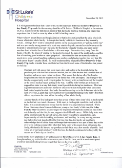
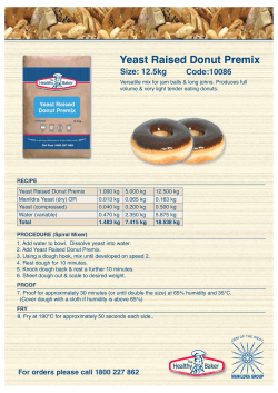
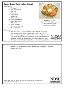
770
Meiosis: how to create a specialized cell cycle
Brian Lee and Angelika Amon*
During the meiotic cell cycle, a single round of DNA replication
precedes two nuclear divisions. Recent work has shown that
the proteins controlling the mitotic cell cycle are either
replaced by homologous proteins only expressed during the
meiotic cell cycle or modulated by meiosis-specific factors to
bring about this specialized cell cycle.
Addresses
Center for Cancer Research, Howard Hughes Medical Institute,
Massachusetts Institute of Technology, E17-233, 40 Ames Street,
Cambridge MA 02139, USA
*Correspondence: [email protected]
Current Opinion in Cell Biology 2001, 13:770–777
0955-0674/01/$ — see front matter
© 2001 Elsevier Science Ltd. All rights reserved.
Abbreviations
APC/C anaphase promoting complex/cyclosome
CDK
cyclin-dependent kinase
ORC
origin recognition complex
SPB
spindle pole body
Introduction
In eukaryotes, a specialized cell cycle, the meiotic cell cycle,
allows for the exchange of genetic material between parental
chromosomes and the formation of haploid gametes. This
generates offspring that are genetically different from their
parents, thus maintaining genetic diversity. During meiosis,
a single round of DNA replication is followed by two
consecutive rounds of nuclear divisions, termed meiosis I
and meiosis II. In the first meiotic division, homologous
chromosomes segregate to opposite poles; during the second
division, which resembles a mitotic division, sister chromatids separate from each other, thereby generating haploid
gametes (Figure 1). This modification of the canonical
G1-S-G2-M mitotic cell cycle requires retooling of the basic
cell cycle machinery to meet meiosis specific requirements.
Although recombination is a key aspect of the meiotic cell
cycle, we will not, due to space limitation, discuss this
topic but refer to recent reviews by Zickler and Kleckner
[1], Smith and Nicolas [2] and van Heemst and Heyting
[3]. Instead, this review focuses on the regulation of the
meiotic cell cycle, in particular on how pre-meiotic DNA
replication and meiotic chromosome segregation differ
from their counterparts in the mitotic cell cycle. How the
meiotic cell cycle is regulated is best understood in the
two yeast model systems, Saccharomyces cerevisiae and
Schizosaccharomyces pombe. Recent progress in both systems
forms the basis of this review and we will refer to other
model organisms when appropriate.
G1: time to get started
Budding yeast enters the mitotic cell cycle when nutrients
are in ample supply. The G1 cyclins Cln1, Cln2 and
Cln3 associate with the cyclin-dependent kinase (CDK)
Cdc28 and trigger bud formation, spindle pole body
(SPB) duplication and DNA replication (Figure 2a).
How Cln–CDKs promote bud formation and SPB
duplication is not yet understood but their role in initiating DNA replication is through phosphorylation of
Sic1, a potent inhibitor of S-phase and mitotic CDKs
(Clb–CDKs), leading to its degradation. The elimination
of Sic1 allows Clb–CDKs to become active to initiate
DNA replication [4].
Since cells can enter either the mitotic or meiotic cell
cycle during G1, a mechanism is necessary to prevent
simultaneous initiation of both cell cycle programs. During
vegetative growth, Cln–CDKs prevent entry into the
meiotic cell cycle by inhibiting the inducer of the meiotic
program, Ime1 [5]. On the other hand, when diploid cells
commit themselves to the meiotic cell cycle, which occurs
upon nitrogen and carbon source starvation, Cln–CDKs are
inactivated by down regulation of CLN transcription [5]
(Figure 2a, 2b). The lack of nutrients and the a/α cell type
signal also lead to activation of the transcription factor
Ime1, which activates transcription of early meiotic genes [6]
(Figure 2b). The three essential functions of Cln–CDKs
during the mitotic cell cycle, bud emergence, SPB
duplication and initiation of S-phase, are either dispensable, as in the case of bud formation, or performed by
the meiosis-specific protein kinase Ime2, as in the case of
S-phase initiation [7] (Figure 2b). Ime2 is thought to initiate
pre-meiotic DNA replication by phosphorylating Sic1.
Indeed, the requirement for IME2 to initiate pre-meiotic
DNA replication can be bypassed by deleting SIC1.
Furthermore, Sic1 is stabilized in cells lacking IME2
(Figure 2b) [7]. The regulation of meiotic SPB duplication
remains largely unknown.
When S. pombe cells are starved for nitrogen and fermentable
carbon sources, a/α diploid cells enter the meiotic cell
cycle through inactivation of the protein kinase Pat1 [8].
As in the case of budding yeast, S. pombe pre-meiotic
DNA replication is initiated in a manner similar to that of
pre-mitotic DNA replication, but one of the key activators
of DNA replication is replaced by a meiosis-specific
factor. Pre-mitotic DNA replication depends upon the
activity of the transcriptional activators Cdc10–Res1 and
Cdc10–Res2–Rep2. These complexes are also required
for pre-meiotic DNA replication, except for Rep2, which
is replaced by a meiosis specific factor, Rep1/Rec16
(Figure 2c,d). The rep1/rec16+ gene is essential for premeiotic DNA replication, whereas rep2+ is required for
pre-mitotic DNA replication, suggesting that fission yeast
also possesses a system to functionally separate pre-mitotic
and pre-meiotic S-phase while using the same basic cell
cycle machinery [8] (Figure 2c,d).
Meiosis: how to create a specialized cell cycle Lee and Amon
771
Figure 1
(a)
S-phase
Mitosis
(b)
Pre-meiotic
S-phase
Recombination
Meiosis I
homolog
segregation
Meiosis II
sister
chromatid
separation
Current Opinion in Cell Biology
The mitotic and meiotic cell cycles. (a) During pre-mitotic DNA
replication, the genetic material is duplicated creating sister
chromatids. At the onset of anaphase, sister chromatids are
separated. Cytokinesis then generates two genetically identical
daughter cells. (b) During pre-meiotic DNA replication, sister
chromatids are generated. During prophase I, homologous
chromosomes (maternal chromosome in red, paternal
chromosome in blue) undergo recombination. At the metaphase I to
anaphase I transition, homologous chromosomes are segregated.
The second meiotic division resembles mitosis; sister chromatids are
segregated. Thus, meiosis leads to the production of four cells that
are genetically not identical.
Pre-meiotic S-phase: a time of courtship
Murakami and Nurse [13••], found that the formation of
DSBs is not affected when DNA replication is inhibited or
delayed. It is possible that coupling of meiotic recombination
to DNA replication is fundamentally different in S. cerevisiae
and S.pombe. It is also possible that differences in methods
of assessing the levels of double strand breaks led to
these contradictory results. Finally Murakami and Nurse
suggest that perhaps in budding yeast DNA replication
per se is not required for the initiation of recombination,
but that S-phase cyclins and origins of replication or
factors assembling onto them play a crucial role in
meiotic recombination [13••].
In all organisms, pre-meiotic S-phase is substantially
longer than pre-mitotic S-phase. An extended S-phase is
thought to be required to establish interhomolog interactions required for meiotic recombination and faithful
segregation of homologous chromosomes. Indeed, when
recombination is abolished in budding yeast, pre-meiotic
DNA replication is shortened, suggesting that preparation
for recombination is one factor responsible for lengthening
pre-meiotic S-phase [9•]. In addition cells deleted for the
S-phase cyclins CLB5 and CLB6 fail to complete premeiotic DNA replication. These cells initiate homologue
synapsis and recombination but display defects in both
processes [10••]. Further support for the idea that premeiotic DNA replication is required for recombination,
came from the observation that removal of origins from the
left arm of chromosome III delays DNA replication. The
formation of recombination-initiating double-stranded
breaks (DSBs) in this region was delayed by the same
amount of time [11••]. This coupling of recombination to
DNA replication is likely to require meiosis-specific factors
such as Mum2. MUM2 genetically interacts with the DNA
replication machinery and is required for normal levels of
meiotic recombination [12•].
In S. pombe, pre-meiotic DNA replication does not appear
to be required for the initiation of meiotic recombination.
Despite the difference in length and added complexity of
meiotic DNA replication, the core replication machinery
appears to be the same between the mitotic and meiotic
cell cycle, at least in S. cerevisiae and S. pombe [11•,13••,14•].
In addition, regulators of DNA replication, such as S-phase
cyclins, are required for both pre-mitotic and pre-meiotic
DNA replication [7,10••,15]. Whether the mitotic and
meiotic cell cycles use the same replication initiation
machinery is, however, controversial. Two reports from
fission yeast reach opposite conclusions. Murakami and
Nurse found that replication initiation proteins such as the
mini chromosome maintenance proteins (MCMs) and
Cdc18 are essential for the initiation of pre-meiotic DNA
replication [13••], whereas a report by Forsburg and
772
Cell multiplication
Figure 2
Control of entry into the mitotic and meiotic
cell cycle in budding and fission yeast.
(a,b) When nutrients are in ample supply,
budding yeast reproduces vegetatively. G1CDKs (Cln–CDKs) promote bud formation,
SPB duplication and DNA replication. The
initiation of DNA replication requires the
activity of another set of CDKs, the
Clb–CDKs. Before Cln–CDKs are active,
Clb–CDKs are inhibited by the CDK inhibitor
Sic1. Cln–CDKs phosphorylate Sic1, thereby
targeting it for ubiquitin-mediated
degradation. This allows Clb–CDKs to initiate
DNA replication. How Cln–CDKs promote
bud formation and SPB duplication is poorly
understood. While promoting entry into the
mitotic cell cycle, Cln–CDKs inhibit cells from
entering the meiotic cell cycle. Cln–CDKs
inhibit the meiosis-specific transcription
factor Ime1 from activating transcription of
early meiotic genes, which include IME2. The
lack of nutrients and the a/α cell type signal
lead to activation of the transcription factor
Ime1, which activates transcription of early
meiotic genes. Nitrogen and carbon source
starvation also lead to inactivation of
Cln–CDKs by down regulation of CLN
transcription. The meiosis-specific protein
kinase Ime2 replaces Cln–CDKs in
promoting DNA replication. (c,d) In
S. pombe, the presence of ample nutrients
promotes vegetative growth. A transcription
factor complex composed of Cdc10, Res2
and Rep2 induces transcription of genes
required for pre-mitotic DNA replication. The
lack of nutrition and mating type signals
induce the meiotic cell cycle by promoting
the accumulation of the meiosis-specific
protein Rep1/Rec16. Rep1/Rec16 replaces
Rep2 in the Cdc10–Res2–Rep2
transcription factor complex, enabling the
complex to promote transcription of genes
required for pre-meiotic DNA replication and
meiotic recombination. Signals indicating the
(a)
(b)
Ample nutrients
Starvation
G1-CDKs
Ime1
Ime2
Bud
formation
Sic1
Mating type
Spindle pole
body
duplication
+ Early meiotic
genes
Sic1
Clb–CDKs
Clb–CDKs
Pre-mitotic
DNA replication
(c)
Ample nutrients
Res2 + Cdc10 + Rep2
Pre-meiotic
DNA replication
(d)
Starvation
Mating type
Rep1/Rec16 + Res2 + Cdc10
Recombination
genes
Pre-mitotic
DNA replication
Pre-meiotic
DNA replication
Current Opinion in Cell Biology
presence of nutrition inhibit the accumulation
of Rep1/Rec16, ensuring that meiosis is not
initiated in the presence of ample nutrients.
Starvation signals, on the other hand, repress
Hodson [14•] suggests that they are not. Both reports
agreed, however, that the origin recognition complex
(ORC) is required.
Rep2, ensuring that Rep1/Rec16 can take its
place in the Cdc10–Res2 transcription factor
complex and promote entry into the meiotic
cell cycle.
fission yeast). Upon degradation of securin, separase is free to
cleave the cohesin subunit Scc1/Mcd1 in budding yeast and
Rad21 in fission yeast, thereby triggering sister-chromatid
separation and the onset of anaphase [16] (Figure 3).
Meiosis I: a time of parting
During mitosis, sister chromatids are segregated to each
daughter cell (Figure 1). This is accomplished by the
pulling force of the mitotic spindle and is resisted by
protein complexes called cohesins. Cohesins hold sister
chromatids together until they are released at the onset of
anaphase (Figure 3). This release is initiated when a
ubiquitin ligase called the anaphase promoting complex or
cyclosome (APC/C), together with its activator, Cdc20,
ubiquitinates the anaphase inhibitor, securin (Pds1 in
budding yeast and Cut2 in fission yeast), thereby targeting
it for degradation by the proteasome. During S-phase, G2
and metaphase, securin binds to and inhibits a protease
known as separase (Esp1 in budding yeast and Cut1 in
During meiosis I, homologous chromosomes and not sister
chromatids are segregated. In fact, sister chromatids
migrate to the same, rather than opposite, poles of the
meiosis I spindle (Figure 1). To accomplish this specialized
division, three events need to occur: a physical linkage
between homologous chromosomes has to be established
to resist the pulling force of the meiosis I spindle; a
linkage between sister chromatids has to be maintained
beyond meiosis I to prevent premature sister chromatid
separation prior to meiosis II; and sister kinetochores have
to attach to microtubules emanating from the same pole
rather than from opposite poles. Over the past two years,
significant progress has been made towards understanding
Meiosis: how to create a specialized cell cycle Lee and Amon
773
Figure 3
Metaphase
Anaphase
Mitosis
Cohesins
Centromere
Sep
c2
Cd
p
Se
PC
p
Se
Sep
A
UU
U
Sec
Sec
U0
Arm Centromeric
cohesins cohesins
cleaved retained
Cohesins
Meiosis I
Control of the metaphase to anaphase
transition during mitosis and meiosis. During
pre-mitotic DNA replication, cohesins
(yellow) are laid down between sister
chromatids. At the onset of anaphase, a
protease called separase cleaves the
cohesin subunit Scc1/Mcd1, thereby
initiating sister-chromatid separation. Prior to
the onset of anaphase, separase is inhibited
by securin. Activation of separase is brought
about by a ubiquitin ligase known as the
anaphase promoting complex or cyclosome
(APC/C), which, together with its specificity
factor Cdc20, ubiquitinates securin, thereby
targeting it for degradation. Cohesins are
also assembled onto chromosomes during
pre-meiotic DNA replication. During
prophase I of meiosis, homologous
chromosomes (blue and red) pair and
recombination occurs, leading to the
formation of chiasmata. At the onset of
anaphase I, separase cleaves the cohesin
subunit Rec8 at chromosome arms (Rec8 is
a homolog of Scc1/Mcd1 that is specifically
expressed during the meiotic cell cycle).
Rec8 is spared from cleavage at
pericentromeric regions, ensuring that sister
chromatids do not separate prematurely.
Furthermore, kinetochores of sister
chromatids attach to microtubules of the
same pole, causing homologous
chromosomes to segregate during anaphase
Centromere
Chiasma
Metaphase I
Sister kinetochore
co-orientation
Anaphase I
Current Opinion in Cell Biology
I. During the second meiotic division, sisterchromatid kinetochores attach to
microtubules from opposite poles, and upon
the molecular mechanisms controlling these events, leading
towards the emergence of the following working model.
First, chiasmata, the physical manifestations of recombination events, provide the physical linkage between
homologous chromosomes. Second, cohesins at centromeres
are protected from cleavage during meiosis I, providing a link
between sister chromatids beyond meiosis I. Lastly, specific
proteins facilitate co-orientation of sister kinetochores during
meiosis I, ensuring that sister chromatids migrate to the same
pole during the first meiotic division (Figure 3b).
Chiasma: the tie that binds
In most organisms, chiasmata and cohesion distal to
chiasmata link homologs together, allowing them to align
on the meiosis I spindle. Indeed, when initiation of recombination is abolished in budding yeast, chromosome
segregates randomly during meiosis I and the first meiotic
division occurs significantly earlier than in cells that
complete recombination successfully [9•,17] The proteins
involved in establishing this linkage between the
homologs are unknown. Additionally, it is important that
each pair of homologous chromosomes experiences at least
one reciprocal exchange. The mechanisms that ensure that
these exchanges occur are also not understood.
Sister chromatid cohesion: one step at a time
It has long been recognized that cohesion along chromosome
arms is lost during meiosis I but that sister chromatids
cleavage of the remaining Rec8 around
centromeres, sister chromatids separate.
Sec, securin; Sep, separase; U, ubiquitin.
remain associated at centromeres until the onset of
anaphase II. It was proposed that this stepwise loss of
cohesion is critical for chromosome segregation during
both meiotic divisions [18]. Loss of arm cohesion is
required for the resolution of chiasmata and thus meiosis I
chromosome segregation, whereas maintenance of cohesion at centromeres is important for proper meiosis II
segregation [18]. The recent isolation and characterization
of a meiosis specific cohesin subunit, Rec8, provides
evidence in support of this model. rec8+ was isolated in
S. pombe as a gene affecting meiotic recombination, particularly at the centromeric region [19]. Further studies
revealed that Rec8 is a conserved protein with homology
to the mitotic cohesin subunit Rad21 and is required for
meiotic cohesion [20,21,22]. Studies in S. cerevisiae
revealed that Rec8 and another cohesin subunit, Smc3,
are required for meiotic cohesion since rec8∆ and smc3-73
cells show precocious sister chromatid separation [23].
Rec8 in higher eukaryotes is likely to perform a similar
function. Inactivation of REC8 by RNAi in C. elegans
leads to precocious separation of sister chromatids [24•].
Mammalian REC8 homologs have been isolated but
await further characterization [22]. Additional meiosisspecific cohesion subunits are likely to exist in mammalian
cells. Stag3/Sa3, a protein with homology to the meiotic
cohesion subunit Sa1/Sa2 is only expressed during meiosis
and was recently shown to be associated with meiotic
chromosomes [25,26].
774
Cell multiplication
Immunolocalization of Rec8 in S. cerevisiae and S. pombe
showed that it is lost from chromosomes in a stepwise
manner. Rec8 is removed from chromosome arms at the
onset of anaphase I but is retained at centromeric regions
until sister chromatids separate at the onset of anaphase II
[20,23]. In budding yeast, Rec8, like its mitotic counterpart
Scc1/Mcd1, is cleaved by separase (Esp1), and this cleavage
is essential for progression into anaphase I [27••].
Mutations that render Rec8 uncleavable by Esp1 arrest at
metaphase I. When recombination is abolished, cells
expressing an uncleavable version of REC8 progress
through anaphase I and arrest at metaphase II [27••]. Thus,
in the absence of chiasmata, Rec8 cleavage is dispensable
for meiosis I, although it is essential for sister-chromatid
separation at the onset of anaphase II. Together, these
data strongly support the idea that loss of arm cohesion
allows for homolog segregation, and subsequent loss of
centromeric cohesion at anaphase II allows for sisterchromatid separation (Figure 3).
The finding that ESP1 is required for progression into
anaphase I suggests that the metaphase I to anaphase I
transition may be regulated in a manner similar to that in
mitosis (Figure 3). Accordingly, budding yeast securin is
present in the nucleus in metaphase I and metaphase II
cells, but is absent from anaphase I and anaphase II nuclei
[28•]. This pattern of Pds1 localization is consistent with
the model that Esp1 is inhibited prior to anaphase I and
anaphase II but becomes active during both meiotic
divisions when Pds1 is destroyed. In addition, low level
expression of a form of Pds1 resistant to ubiquitination by
APC/CCdc20 during meiosis causes a delay in metaphase I
[29••]. This suggests that APC/CCdc20 is also likely to
regulate the metaphase I to anaphase I transition in
budding yeast. In support of this model, Cdc20 accumulates in the nucleus just prior to Pds1 destruction during
both meiotic divisions [28•]. Evidence that APC/C regulates the onset of anaphase I has also been found in
C. elegans. Mutations in several APC/C subunits lead to a
metaphase I arrest [30•,31•].
In vertebrates, however, APC/CCdc20 does not appear to
regulate the metaphase I to anaphase I transition.
Immunodepletion of the Xenopus Cdc20 homolog Fizzy, or
injection of antisense oligonucleotide against FIZZY mRNA,
cause a metaphase II arrest [32•,33•]. Immunodepletion
of the APC/C core subunit, Cdc27 or microinjection of a
non-degradable version of securin also fails to arrest cells at
metaphase I, suggesting that APC/C and separase activity
are not required for entry into anaphase I in Xenopus
oocytes [32•]. This difference could be explained by the
existence of two mechanisms to remove cohesins in
vertebrates. An APC/C–separase-independent mechanism
preferentially removes cohesins from chromosome arms
[34,35] and an APC/C–separase-dependent mechanism
removes cohesins from centromeric regions [36]. Perhaps
during meiosis I, arm cohesion is removed from chromosomes by the APC/C–separase-independent mechanism
and during meiosis II, centromeric cohesin is removed by
the APC/C–separase-dependent mechanism.
Meiotic checkpoint
As during the mitotic cell cycle, surveillance mechanisms
exist that function to prevent cell cycle progression when
meiotic recombination or kinetochore attachment are
incomplete or defective. The ‘pachytene checkpoint’
inhibits cell cycle progression when meiotic recombination
is ongoing or when defects in recombination or chromosome synapsis occur [37]. The cell cycle is halted by the
phosphorylation of the tyrosine 19 (Y19) residue in Cdc28.
Phosphorylation of Y19 inhibits Cdc28 activity leading to
cell cycle arrest in prophase I.
Defects in mitotic spindle formation or attachment of
chromosomes to the mitotic spindle activate a surveillance
mechanism, the spindle checkpoint, that halts cell cycle
progression at the metaphase to anaphase transition.
Checkpoint proteins such as Mad2 bind to and inhibit
APC/CCdc20, thereby preventing the degradation of
securin [38]. Recent work has shown that the spindle
checkpoint also functions during meiosis I and is, in fact,
essential for meiosis. In S. cerevisiae, cells deleted for the
spindle checkpoint component MAD2 show a 10-fold
increase in non-disjunction during meiosis I, suggesting
that the spindle checkpoint is required to delay homolog
segregation until all chromosomes have attached to the
meiosis I spindle [29••]. Similar results were obtained in
S. pombe, where mad2∆ and bub1∆ cells show moderate
non-disjuction during meiosis I [39•].
Centromeric cohesin: linking sisters beyond meiosis I
A key aspect of the regulation of sister-chromatid cohesion
during meiosis is that cohesins at pericentromeric regions
are protected from cleavage during anaphase I. This is a
special property of Rec8-mediated cohesion since expression of SCC1/MCD1 instead of REC8 during meiosis results
in sister-chromatid cohesion being lost along the entire
chromosome during the first meiotic division [40••]. This
suggests that either Rec8 itself is refractory to cleavage
when bound to pericentromeric regions or, more likely,
that factors that specifically bind to Rec8 around centromeres protect Rec8 from being cleaved. Such factors are
likely to be or are regulated by Drosphila MEI-S332 and
S. cerevisiae Spo13 and Slk19. MEI-S332 has been shown to
localize to centromeres and mutations in MEI-S332 lead to
premature separation of sister chromatid during meiosis II
suggesting that cohesion is lost between the meiotic
divisions [41,42]. Slk19 in budding yeast also localizes to
meiotic kinetochores prior to meiosis I. Furthermore, in
cells lacking SLK19, Rec8 staining is greatly reduced
around centromeres during anaphase I suggesting that
Slk19 plays a role in protecting Rec8 from cleavage at
centromeres [43••]. Budding yeast SPO13 has also been
implicated in the regulation of Rec8 cleavage since spo13∆
cells loose Rec8 along the entire length of the chromosome
during anaphase I [23].
Meiosis: how to create a specialized cell cycle Lee and Amon
Even though it remains unclear how centromeric Rec8 is
protected from cleavage during anaphase I, experiments in
grasshopper spermatocytes show that whether or not
cohesion is lost at centromeres is a chromosome intrinsic
property suggesting that the protector of centromeric
cohesin is likely to be chromosome associated. When a
meiosis I chromosome is transferred onto a meiosis II
spindle, this chromosome segregates in a reductional
(meiosis I-like) manner even though the other chromosomes
on the same spindle segregate in an equational (meiosis IIlike) manner. The reciprocal result is obtained when a
meiosis II chromosome is transferred onto a meiosis I
spindle; while the meiosis II chromosome segregates
equationally, the other chromosomes on the meiosis I
spindle segregate reductionally [44••]. Further characterization and isolation of factors like Spo13, Slk19 and
MEI-S332 will be critical for determining the exact
mechanisms by which linkage between sister chromatids is
retained at centromeres until anaphase II and for understanding the meiotic pattern of chromosome segregation.
Co-orientation of kinetochores: sister chromatids unite
To ensure that sister chromatids segregate as a single unit
during the first meiotic division, the kinetochores of sister
chromatids have to attach to microtubules emanating from
the same centrosome/SPB. Recently, a mutant, mam1, has
been isolated in budding yeast that is defective in coorientation of sister kinetochores. In mam1∆ cells, sister
chromatids attach to the meiosis I spindle as if it were a
mitotic spindle, with kinetochores attaching to microtubules emanating from opposite rather than the same pole
[40••]. The stepwise loss of Rec8, however, is not affected
in these cells, as Rec8 disappears from chromosome arms
but not from centromeres during this division. This result
suggests that co-orientation of kinetochores is genetically
separable from the protection of Rec8 at centromeres. The
regulation of co-orientation of kinetochores and protection
of Rec8 at centromeres are likely, however, to be intimately
linked as mutations in SPO13 or SLK19 affect both
processes. Cells lacking SPO13 or SLK19 undergo a single
meiotic division in which chromosomes are segregated
largely in an equational rather than a reductional manner.
This indicates that Rec8 is not protected at centromeres
and that a significant percentage of sister kinetochores
attaches to microtubules emanating from opposite poles
in these mutants [43••,45•,46].
Evidence that protection of Rec8 at the centromeres and
co-orientation of kinetochores are intimately linked processes
is also found in S. pombe. In fission yeast, rec8∆ mutants
undergo an equational instead of a reductional division at
meiosis I, indicating that in the absence of Rec8, sister
kinetochores are oriented to opposite poles [20]. In addition,
cells lacking the spindle checkpoint component Bub1, which
localizes to meiotic kinetochores, are defective in sister kinetochore co-orientation and protection of Rec8 at centromeres
in S. pombe [39•]. In bub1∆ cells, a high percentage of
chromosomes undergoes an equational division at meiosis I.
775
In summary, these findings suggest that proteins at the
kinetochore, such as Bub1, Slk19, and Mam1, are required
to force sister kinetochores to face the same pole in
meiosis I. They also suggest that sister kinetochore coorientation and protection of Rec8 are likely to be
coordinately regulated since many mutants affect both
processes. How one influences the other, however, is at
present unclear and awaits characterization of protein
complexes bound to meiosis I kinetochores.
Conclusion
In the past two years, it has become clear that the cell cycle
machinery controlling progression through the mitotic cell
cycle is also employed to regulate progression through the
meiotic cell cycle with meiosis-specific modifications. The
exact mechanism by which cells modulate the mitotic
cell cycle machinery to bring about a meiotic cell cycle
and the factors involved in this process are still largely
uncharacterized. It is clear, however, that the field is
progressing at a rapid pace. New tools, such as genomewide expression analysis and functional genomic approaches
[47,48] has and will lead to the identification of meiotic cell
cycle regulators [49] making it likely that we will soon
know ‘how meiosis can work’ [50].
Acknowledgements
We are grateful to members of the Amon lab for their critical reading of
this manuscript.
References and recommended reading
Papers of particular interest, published within the annual period of review,
have been highlighted as:
• of special interest
•• of outstanding interest
1.
Zickler D, Kleckner N: Meiotic chromosomes: integrating structure
and function. Annu Rev Genet 1999, 33:603-754.
2.
Smith KN, Nicolas A: Recombination at work for meiosis. Curr Opin
Genet Dev 1998, 8:200-211.
3.
van Heemst D, Heyting C: Sister chromatid cohesion and
recombination in meiosis. Chromosoma 2000, 109:10-26.
4.
Koepp DM, Harper JW, Elledge SJ: How the cyclin became a
cyclin: regulated proteolysis in the cell cycle. Cell 1999,
97:431-434.
5.
Colomina N, Gari E, Gallego C, Herrero E, Aldea M: G1 cyclins block
the Ime1 pathway to make mitosis and meiosis incompatible in
budding yeast. EMBO J 1999, 18:320-329.
6.
Vershon AK, Pierce M: Transcriptional regulation of meiosis in
yeast. Curr Opin Cell Biol 2000, 12:334-339.
7.
Dirick L, Goetsch L, Ammerer G, Byers B: Regulation of
meiotic S phase by Ime2 and a Clb5, 6-associated
kinase in Saccharomyces cerevisiae. Science 1998,
281:1854-1857.
8.
Murakami H, Nurse P: DNA replication and damage checkpoints
and meiotic cell cycle controls in the fission and budding yeasts.
Biochem J 2000, 349:1-12.
9.
•
Cha RS, Weiner BM, Keeney S, Dekker J, Kleckner N: Progression of
meiotic DNA replication is modulated by interchromosomal
interaction proteins, negatively by Spo11p and positively by
Rec8p. Genes Dev 2000, 14:493-503.
Meiotic DNA replication in budding yeast is ~3.5 times longer than mitotic
S-phase. Additionally, proteins involved in the initiation of recombination and
meiotic cohesion affect the length of pre-meiotic S-phase. Deletion of
SPO11 shortens it by ~25% whereas rec8∆ lengthens it by ~10%, suggesting
that preparation for recombination is one factor responsible for the longer
meiotic S-phase.
776
Cell multiplication
10. Smith KN, Penkner A, Ohta K, Klein F, Nicolas A: B-type cyclins
•• CLB5 and CLB6 control the initiation of recombination and
synaptonemal complex formation in yeast meiosis. Curr Biol
2001,11:88-97.
S-phase cyclins, Clb5 and Clb6 are essential not only for pre-meiotic DNA
replication but also for homolog synapsis and recombination as clb5∆ cells
show defects in both processes. This could be due to either incomplete
DNA replication or an additional role of S-phase CDKs in recombination
and synapsis.
11. Borde V, Goldman AS, Lichten M: Direct coupling between meiotic
•• DNA replication and recombination initiation. Science 2000,
290:806-809.
The timing of the appearance of recombination initiating DNA DSBs and
DNA replication are directly coupled. When replication is delayed, either by
the deletion of origins or transplantation of telomeric sequences to a nontelomeric region, appearance of DSBs in those regions are also delayed by
the same amount of time, indicating that replication and DSB formation are
locally coupled.
12. Davis L, Barbera M, McDonnell A, McIntyre K, Sternglanz R, Jin Q,
•
Loidl J, Engebrecht J: The Saccharomyces cerevisiae MUM2 gene
interacts with the DNA replication machinery and is required for
meiotic levels of double strand breaks. Genetics 2001,
157:1179-1189.
Mum2 is dispensable for pre-mitotic DNA replication but is essential for
pre-meiotic replication. Cells deleted for MUM2 can partially bypass orc2-2
and lower the restrictive temperature of pol1-17 suggesting that it interacts
with the DNA replication machinery. Recombination frequency and homolog
synapsis is reduced in the mum2∆ mutant suggesting that it either
influences meiotic recombination directly or through its effect on replication.
13. Murakami H, Nurse P: Regulation of pre-meiotic S phase and
•• recombination-related double-strand DNA breaks during meiosis
in fission yeast. Nat Genet 2001, 28:290-293.
This paper reports that MCMs and Cdc18, which are required for mitotic
DNA replication, are also required for pre-meiotic DNA replication. Meiotic
DNA replication is incomplete at the restrictive temperature in temperature
sensitive alleles of these genes. Also in contrast to budding yeast, appearance
of double strand breaks is not associated with meiotic DNA replication as
these mutants produce DSBs despite incomplete DNA synthesis.
14. Forsburg SL, Hodson JA: Mitotic replication initiation proteins are
•
not required for pre-meiotic S phase. Nat Genet 2000,
25:263-268.
Mitotic DNA replication initiation proteins such as MCMs, Cdc18 and Cdt1
are apparently not required for pre-meiotic S-phase in S. pombe as
temperature sensitive alleles in these genes can complete meiotic DNA
replication at their restrictive temperature.
15. Stuart D, Wittenberg C: CLB5 and CLB6 are required for premeiotic
DNA replication and activation of the meiotic S/M checkpoint.
Genes Dev 1998, 12:2698-2710.
16. Nasmyth K, Peters JM, Uhlmann F: Splitting the chromosome:
cutting the ties that bind sister chromatids. Science 2000,
288:1379-1385.
17.
Galbraith AM, Bullard SA, Jiao K, Nau JJ, Malone RE: Recombination
and the progression of meiosis in Saccharomyces cerevisiae.
Genetics 1997, 146:481-489.
18. Miyazaki WY, Orr-Weaver TL: Sister-chromatid cohesion in mitosis
and meiosis. Annu Rev Genet 1994, 28:167-187.
19. DeVeaux LC, Smith GR: Region-specific activators of meiotic
recombination in Schizosaccharomyces pombe. Genes Dev 1994,
8:203-210.
20. Watanabe Y, Nurse P: Cohesin Rec8 is required for
reductional chromosome segregation at meiosis. Nature 1999,
400:461-464.
21. Parisi S, McKay MJ, Molnar M, Thompson MA, van der Spek PJ,
van Drunen-Schoenmaker E, Kanaar R, Lehmann E, Hoeijmakers JH,
Kohli J: Rec8p, a meiotic recombination and sister chromatid
cohesion phosphoprotein of the Rad21p family conserved from
fission yeast to humans. Mol Cell Biol 1999, 19:3515-3528.
22. Molnar M, Bahler J, Sipiczki M, Kohli J: The rec8 gene of
Schizosaccharomyces pombe is involved in linear element
formation, chromosome pairing and sister-chromatid cohesion
during meiosis. Genetics 1995, 141:61-73.
23. Klein F, Mahr P, Galova M, Buonomo SB, Michaelis C, Nairz K,
Nasmyth K: A central role for cohesins in sister chromatid
cohesion, formation of axial elements, and recombination during
yeast meiosis. Cell 1999, 98:91-103.
24. Pasierbek P, Jantsch M, Melcher M, Schleiffer A, Schweizer D, Loidl J:
•
A Caenorhabditis elegans cohesion protein with functions in
meiotic chromosome pairing and disjunction. Genes Dev 2001,
15:1349-1360.
A Rec8 homolog was isolated from C. elegans by sequence homology and
was shown to be expressed only in meiotic tissues. Inactivation of Rec8 by
RNAi showed that Rec8 is required for recombination and cohesion.
25. Eijpe M, Heyting C, Gross B, Jessberger R: Association of
mammalian SMC1 and SMC3 proteins with meiotic chromosomes
and synaptonemal complexes. J Cell Sci 2000, 113:673-682.
26. Prieto I, Suja JA, Pezzi N, Kremer L, Martinez-A C, Rufas JS,
Barbero JL: Mammalian STAG3 is a cohesin specific to sister
chromatid arms in meiosis I. Nat Cell Biol 2001, 3:761-766
27.
••
Buonomo SB, Clyne RK, Fuchs J, Loidl J, Uhlmann F, Nasmyth K:
Disjunction of homologous chromosomes in meiosis I depends
on proteolytic cleavage of the meiotic cohesin Rec8 by separin.
Cell 2000, 103:387-398.
Rec8 is cleaved by the separase Esp1. Cleavage by Esp1 was shown to be
required for the metaphase I to anaphase I transition as expression of an
uncleavable Rec8 mutant lead to cell cycle arrest at metaphase I. In spo11∆
cells that fail to initiate recombination bypass the metaphase I arrest of
uncleavable Rec8 and arrest at metaphase II, suggesting that cleavage of
Rec8 is only required at MI to resolve chiasmata by removing arm cohesion.
28. Salah SM, Nasmyth K: Destruction of the securin Pds1p occurs at
•
the onset of anaphase during both meiotic divisions in yeast.
Chromosoma 2000, 109:27-34.
Budding yeast Securin, Pds1, was shown to localize to metaphase I and
metaphase II nuclei but is absent in anaphase I and anaphase II cells.
Cdc20, the APC/C specificity factor responsible for targeting Pds1 for
degradation, is localized to meiotic nuclei prior to Pds1 destruction in both of
the meiotic divisions, suggesting that APC/CCdc20 is activated twice to
degrade Pds1, which allows for the activation of Esp1 in both meiotic divisions.
29. Shonn MA, McCarroll R, Murray AW: Requirement of the spindle
•• checkpoint for proper chromosome segregation in budding yeast
meiosis. Science 2000, 289:300-303.
The spindle checkpoint component Mad2 was shown to be essential for
preventing progression through MI in response to meiotic spindle damage.
mad2∆ cells have a modest increase in MI non-disjuction in unchallenged
cells. Treatment of cells with the microtubule depolymerizing drug benomyl
caused a MI arrest that is dependent upon Mad2. Spo11∆ mutants, which
do not initiate recombination, bypass this checkpoint induced arrest and
undergo a ‘pseudo anaphase I’ without Pds1 degradation, which does not
occur in wild-type cells.
30. Golden A, Sadler PL, Wallenfang MR, Schumacher JM, Hamill DR,
•
Bates G, Bowerman B, Seydoux G, Shakes DC: Metaphase to
anaphase (mat) transition-defective mutants in Caenorhabditis
elegans. J Cell Biol 2000, 151:1469-1482.
Temperature sensitive maternal effect embryonic lethal mutants that arrest at
the one cell stage in C. elegans were isolated. This screen revealed several
orthologs of APC subunits. These mutants arrest in metaphase I in both
oogenesis and spermatogenesis, indicating that the APC is required for
meiosis I in C. elegans.
31. Furuta T, Tuck S, Kirchner J, Koch B, Auty R, Kitagawa R, Rose AM,
•
Greenstein D: EMB-30: an APC4 homologue required for
metaphase-to-anaphase transitions during meiosis and mitosis in
Caenorhabditis elegans. Mol Biol Cell 2000, 11:1401-1419.
Analysis of the emb30 mutant showed that it arrests at metaphase I in
meiosis and metaphase in mitosis. Neither arrest depends on the spindle
checkpoint. Subsequent cloning of emb30 showed that it is the C. elegans
ortholog of the APC/C subunit APC4/LID1, indicating that the APC/C is
essential for metaphase I to anaphase I transition in C. elegans.
32. Peter M, Castro A, Lorca T, Le Peuch C, Magnaghi-Jaulin L, Doree M,
•
Labbe JC: The APC is dispensable for first meiotic anaphase in
Xenopus oocytes. Nat Cell Biol 2001, 3:83-87.
Immunodepletion of the APC subunit Cdc27 and the APC/C specificity
factor Fizzy/Cdc20 showed that the APC/C is dispensable for MI in
Xenopus oocytes but is required for metaphase II to anaphase II transition.
Microinjection of non-degradable Securin or a destruction box peptide
failed to arrest oocytes at MI, indicating that APC/C activity and Securin
degradation are dispensable for entry into anaphase I in Xenopus oocytes.
33. Taieb FE, Gross SD, Lewellyn AL, Maller JL: Activation of the
•
anaphase-promoting complex and degradation of cyclin B is not
required for progression from Meiosis I to II in Xenopus oocytes.
Curr Biol 2001, 11:508-513.
Injection of antisense oligonucleotide directed against the APC/C specificity
factor Fizzy/Cdc20 did not cause oocytes to arrest at metaphase I, indicating
that Fizzy/Cdc20 is not required for metaphase I to anaphase I transition in
Xenopus despite high levels of Cdc20 protein during metaphase I.
Meiosis: how to create a specialized cell cycle Lee and Amon
34. Sumara I, Vorlaufer E, Gieffers C, Peters BH, Peters JM:
Characterization of vertebrate cohesin complexes and
their regulation in prophase. J Cell Biol 2000,
151:749-762.
35. Losada A, Yokochi T, Kobayashi R, Hirano T: Identification
and characterization of SA/Scc3p subunits in the Xenopus
and human cohesin complexes. J Cell Biol 2000,
150:405-416.
36. Waizenegger IC, Hauf S, Meinke A, Peters JM: Two distinct
pathways remove mammalian cohesin from chromosome arms in
prophase and from centromeres in anaphase. Cell 2000,
103:399-410
37.
Roeder GS, Bailis JM: The pachytene checkpoint. Trends Genet
2000, 16:395-403
38. Amon A: The spindle checkpoint. Curr Opin Genet Dev 1999,
9:69-75.
39. Bernard P, Maure JF, Javerzat JP: Fission yeast Bub1 is essential in
•
setting up the meiotic pattern of chromosome segregation. Nat
Cell Biol 2001, 3:522-526.
The spindle checkpoint components Mad2 and Bub1 were shown to be
functional in MI in S. pombe as non-disjuction at MI slightly increased in
mad2∆ and bub1∆ cells. Bub1 was also shown to localize to meiotic kinetochores and to be required for co-orientation of sister kinetochores at MI.
40. Toth A, Rabitsch KP, Galova M, Schleiffer A, Buonomo SB,
•• Nasmyth K: Functional genomics identifies monopolin: a
kinetochore protein required for segregation of homologs during
meiosis I. Cell 2000, 103:1155-1168.
Mam1 was identified in a systematic screen in which genes induced during
meiosis were deleted. mam1∆ cells are specifically defective in the coorientation of sister kinetochores at MI without affecting the stepwise loss of
Rec8 from chromosomes. In addition, the stepwise loss of cohesion was shown
to be a property of Rec8 mediated cohesion since cells that express Scc1
from the REC8 promoter loose cohesion along the entire chromosome at MI.
41. Moore DP, Page AW, Tang TT, Kerrebrock AW, Orr-Weaver TL: The
cohesion protein MEI-S332 localizes to condensed meiotic and
mitotic centromeres until sister chromatids separate. J Cell Biol
1998, 140:1003-1012.
42. Tang TT, Bickel SE, Young LM, Orr-Weaver TL: Maintenance
of sister-chromatid cohesion at the centromere by the
Drosophila MEI-S332 protein. Genes Dev 1998,
12:3843-3856
777
43. Kamieniecki RJ, Shanks RM, Dawson DS: Slk19p is necessary to
•• prevent separation of sister chromatids in meiosis I. Curr Biol
2000 10:1182-1190.
In S. cerevisiae, Slk19 was shown to localize to meiotic kinetochores prior to
anaphase I and to spindle pole bodies afterward. slk19∆ cells exhibit normal
levels of recombination suggesting that they complete prophase I., however,
they undergo a single mixed meiotic division generating diploid dyads. Thus,
indicating a defect in kinetochore orientation. In addition, Rec8 staining was
significantly reduced in anaphase I of slk19∆ cells suggesting that they are
also defective in the retention of centromeric cohesion after MI as well.
44. Paliulis LV, Nicklas RB: The reduction of chromosome number in
•• meiosis is determined by properties built into the chromosomes.
J Cell Biol 2000, 150:1223-1232.
Meiotic chromosome behavior was shown to be a chromosome intrinsic
property in grasshopper spermatocytes. When a MI chromosome is
micro-manipulated to a MII spindle, it segregates reductionally with the loss
of arm cohesion but retention of centromeric cohesion. When a meiosis II
chromosome is moved to a MI spindle, it segregates equationally, whereas
the other chromosomes on this spindle segregate reductionally, indicating
that stepwise loss of cohesion and co-orientation of sister kinetochores are
intrinsic properties of meiotic chromosomes.
45. Zeng X, Saunders WS: The Saccharomyces cerevisiae centromere
•
protein Slk19p is required for two successive divisions during
meiosis. Genetics 2000, 155:577-587.
Budding yeast Slk19 was shown to be required for meiosis as slk19∆
cells produced a large amount of diploid dyads that have undergone an
equational division. This indicates that slk19∆ cells are defective in sister
kinetochore orientation.
46. Klapholz S, Esposito RE: Recombination and chromosome
segregation during the single division meiosis in SPO12-1 and
SPO13-1 diploids. Genetics 1980, 96:589-611.
47.
Chu S, DeRisi J, Eisen M, Mulholland J, Botstein D, Brown PO,
Herskowitz I: The transcriptional program of sporulation in
budding yeast. Science 1998, 282:699-705.
48. Primig M, Williams RM, Winzeler EA, Tevzadze GG, Conway AR,
Hwang SY, Davis RW, Esposito RE: The core meiotic transcriptome
in budding yeasts. Nat Genet 2000, 26:415-423.
49. Rabitsch KP, Toth A, Galova M, Schleiffer A, Schaffner G, Aigner E,
Rupp C, Penkner AM, Moreno-Borchart AC, Primig M et al.: A screen
for genes required for meiosis and spore formation based on
whole-genome expression. Curr Biol 2001, 11:1001-1009
50. Kleckner N: Meiosis: how could it work? Proc Natl Acad Sci USA
1996, 93:8167-8174.


© Copyright 2026