
RELATIONSHIP OF MATERNAL SERUM FATTY ACIDS AND BODY MASS INDEX
RELATIONSHIP OF MATERNAL SERUM FATTY ACIDS AND BODY MASS INDEX
A Thesis
Submitted to the Graduate Faculty of the
Louisiana State University and
Agricultural and Mechanical College
in partial fulfillment of the
requirements for the degree of
Master of Science
In
The School of Human Ecology
By
Emily Fontenot Gilbert
B.S., Louisiana State University and Agricultural & Mechanical College, 2007
August 2009
ACKNOWLEDGMENTS
I would first like to thank my major professor, Dr. Carol Lammi-Keefe, for giving me the
chance to continue my education and for supporting me through this process. In addition to Dr.
Lammi-Keefe, I would like to thank my committee members for their continued assistance in the
research process. Also, I would like to thank my fellow graduate students for their help and
advice during my studies.
My entire education would not be possible without my parents, especially my mother. I
would like to thank her for her continuous support and encouragement in achieving my goals.
My husband deserves a special thanks for the inspiration he provides for me everyday. Lastly, I
would like to thank my friends for understanding that my time was better spent in the lab or at
my desk.
ii
TABLE OF CONTENTS
ACKNOWLEDGMENTS…………………………………………………………………. ii
ABSTRACT……………………………………………………………………………….. v
CHAPTER
1 INTRODUCTION……………………………………………………………………….. 1
Justification………………………………………………………………………… 1
Objectives…………………………………………………………………………. 2
Research Statement………………………………………………………………… 2
Limitations…………………………………………………………………………. 2
Definitions…………………………………………………………………………..2
Assumptions……………………………………………………………………….. 3
2 REVIEW OF LITERATURE……………………………………………………………. 4
Lipids and Fatty Acids……………………………………………………………... 4
Synthesis of Long-Chain Polyunsaturated Fatty Acids (LCPUFA)………………..5
Importance of ω3 and ω6 Fatty Acids……………………………………………...7
Importance of LCPUFAs during Pregnancy………………………………………. 8
Placental Transfer of ω3 and ω6 Fatty Acids……………………………………… 9
Obesity, BMI, and Insulin Resistance………………………………………………12
Obesity and Pregnancy…………………………………………………………….. 12
Influence of BMI and Insulin Resistance on LCPUFA Transfer…………………... 13
3 METHODS………………………………………………………………………………. 15
Participants and Sampling…………………………………………………………..15
Procedures………………………………………………………………………….. 15
Statistical Analysis…………………………………………………………………. 17
4 RELATIONSHIP OF MATERNAL SERUM FATTY ACIDS AND BODY MASS
INDEX……………………………………………………………………………………...18
Introduction………………………………………………………………………… 18
Subjects and Methods……………………………………………………………… 19
Results……………………………………………………………………………… 20
Discussion………………………………………………………………………….. 22
5 SUMMARY……………………………………………………………………………… 24
iii
REFERENCES…………………………………………………………………………….. 27
VITA………………………………………………………………………………………..33
iv
ABSTRACT
Maternal supply of nutrients is critical for the developing fetus during all stages of
gestation. The altered lipid metabolism that is often seen in pregnancies complicated by obesity
and insulin resistance may negatively impact maternal nutrient supply to the fetus. More women
are entering pregnancy overweight or obese. Recently body mass index (BMI) has been found to
be a positive predictor for decreased maternal plasma phospholipid concentrations of the longchain polyunsaturated fatty acids (LCPUFA) docosahexanoic acid (DHA) and arachidonic acid
(ARA). These nutrients play important roles in early development and their availability is
critical to fetal growth and development. The purpose of the present study was to assess if
maternal serum fatty acids in the second trimester of pregnancy are associated with BMI.
Serum samples are frequently stored after blood draws during pregnancy. The
availability of these samples provided the opportunity to examine if serum samples collected
from non-fasting, pregnant women could provide information similar to what has previously
been reported for BMI and fatty acid status in pregnancy.
Sera from 265 women from the Foundation for Blood Research (FBR, Maine) were
analyzed for fatty acid content using gas chromatography (GC). The BMIs of each participant at
approximately 13-18 weeks of gestation were provided by FBR. Participants were grouped by
BMI category and fatty acid concentrations were compared across BMI categories.
There were significant differences for weight percent of the LCPUFA eicosapentanoic
acid (EPA) as well as for the total ω3 LCPUFA. In this data set BMI was a negative predictor of
maternal serum concentrations of DHA and EPA. These results support previous findings for
fatty acids in plasma phospholipids of pregnant women. We conclude that increased BMI may
negatively impact maternal LCPUFA concentrations and stored sera may be used to further
assess this relationship.
v
CHAPTER 1
INTRODUCTION
Lipids have long been recognized as important dietary substances for growth and
development. Recently, there has been considerable interest in the role of dietary lipids,
specifically essential fatty acids, in the earliest phases of life. The long chain polyunsaturated
fatty acids (LCPUFA) docosahexaenoic acid (DHA, 22:6ω3) and arachidonic acid (ARA,
20:4ω6) are important structural components of membrane phospholipids. The highest
concentrations of these LCPUFA, especially DHA, are found in the brain. Exponential fetal
accumulation begins during the last trimester of pregnancy and continues until the age of two.
During the prenatal period the fetus depends on the maternal supply of preformed DHA and
ARA to meet its needs. These fatty acids are transferred across the placenta by placental fatty
acid binding proteins (p-FABP).
Recent findings suggest that insulin resistance may result in altered lipid metabolism and
thus impact fetal LCPUFA status. Insulin resistance in pregnancy is often characterized by
increased maternal body mass index (BMI). Due to the increase in the number of women
entering pregnancy as overweight or obese, concern has been raised regarding insulin resistance,
which can lead to gestational diabetes mellitus (GDM). These conditions could prove
detrimental to the developing fetus in part because of their impact on nutrient supply to the fetus.
The purpose of the present study is to assess if maternal serum fatty acids are associated with
maternal BMI in the second trimester of pregnancy.
Justification
It is well established that the LCPUFAs DHA and ARA are critical nutrients during early
development. They play important roles throughout the lifespan, especially in early infant
growth, brain, and retinal development. With more women entering pregnancy as overweight or
1
obese, it is imperative that there be research directed at determining the impact of these
conditions on the developing fetus. Because overweight and obesity are often accompanied by
insulin resistance and impaired lipid metabolism there is the possibility that maternal supply of
DHA and ARA may be impacted. Inadequate supply of these nutrients may be detrimental to
fetal growth. Results from this study may lend support to the growing body of evidence for
decreased ω3 LCPUFA status in pregnancies complicated by overweight or obesity.
Objectives
1. To measure fatty acids in sera collected in the second trimester of pregnancy from
a large cohort of pregnant women living in Maine.
2. To investigate whether serum fatty acid concentrations are associated with
maternal BMI.
Research Statement
It is hypothesized that women with higher BMIs will have lower concentrations of serum
DHA and ARA than women with lower BMIs.
Limitations
1. This is a relational study so we cannot infer causality.
2. The results of the present study will only be applicable to women of similar age, race,
parity and health status.
Definitions
1. Gestation: the period of fetal development in the uterus from conception to delivery.
2. Body Mass Index (BMI): weight in kilograms divided by height in meters squared
(kg/m2). The most widely used weight-height measure of adiposity.
3. Essential fatty acids (EFA): those fatty acids that cannot be synthesized by humans and
must be obtained from the diet to prevent development of disease. In humans the parent
2
EFAs from which all other fatty acids are derived are linoleic acid (LA, C18:2ω6) and
alpha-linolenic acid (ALA, C18:3ω3).
4. Docosahexaenoic acid (DHA, C22:6ω3): A 22-carbon polyunsaturated fatty acid that has
its first double bond at the third carbon from the omega or methyl end.
5. Arachidonic Acid (ARA, C20:4ω6): A 20-carbon polyunsaturated fatty acid that has its
first double bond at the sixth carbon from the omega or methyl end.
6. Diabetes mellitus: A metabolic disorder characterized by inadequate insulin secretion by
the pancreas or the inability of certain cells to use insulin resulting in abnormally high
serum glucose levels. Diabetes mellitus can be classified as type 1 diabetes, type 2
diabetes, or GDM.
Assumptions
1. The sample size is adequate to reflect the relationship between the variables under
investigation.
2. The Hewlett Packard 5890 gas chromatograph is a reliable instrument for measuring
the concentration of fatty acids in serum samples.
3. The BMI of each participant was calculated and recorded correctly.
3
CHAPTER 2
REVIEW OF LITERATURE
Lipids and Fatty Acids
Lipids have long been recognized as important and essential dietary substances for
growth and development. Although these compounds are chemically diverse, they all share a
common feature, insolubility in water. These compounds are necessary for a variety of functions
in organisms including providing a storage form of energy and providing structural support for
biological membranes. The most commonly used fats and oils for stored forms of energy are
derivatives of fatty acids.
Fatty acids are the simplest class of lipid whose basic structure is that of a chain of
carbon atoms linked together and flanked by hydrogen atoms. One end of the fatty acid chain is
designated the acid end and contains a carboxyl group, while the other end is designated the
omega (ω) end and contains a methyl group. A large proportion of the fatty acids found in
nature contain an even number of carbon atoms in an unbranched chain.
Fatty acids are subdivided based on their chain length, either short (less than six carbons),
medium (six to ten carbons) or long (12 or more carbons). Saturated fatty acids are those that
have single bonds between carbons and have the maximum amount of possible hydrogen atoms
linked to all the carbon atoms. Therefore, monounsaturated fatty acids contain one carboncarbon double bond (site of unsaturation) and are missing two hydrogen atoms from the carbon
chain. Carbon chains with two or more double bonds are thus termed polyunsaturated fatty acids
(PUFA) and contain two fewer hydrogen atoms per carbon-carbon double bond or site of
unsaturation. The physical properties of fatty acids and also of the compounds that contain them
are determined by the length and degree of unsaturation of the carbon chain.
4
Fatty acids are further classified based on the location of the first carbon-carbon double
bond. An omega-3 (ω3 or n-3) fatty acid is one which has its first double bond at the third
carbon from the methyl group (omega) end while an omega-6 (ω6 or n-6) fatty acid has its first
double bond at the sixth carbon from the methyl group end. These compounds are most often
identified by their chain length, number of double bonds, and ω3 or ω6 family.
Human cells lack the enzymes necessary to synthesize some long-chain polyunsaturated
fatty acids (LCPUFA) and, therefore, depend on the diet to obtain the precursor ω3 and ω6 fatty
acids. These fatty acids are thus termed essential fatty acids (EFA). Alpha-linolenic acid
(C18:3ω3, ALA) is the ω3 EFA and linoleic acid (C18:2ω6, LA) is the ω6 EFA. Humans can
desaturate and elongate the parent EFAs to form several important LCPUFAs.
Synthesis of Long-Chain Polyunsaturated Fatty Acids (LCPUFA)
The conversion pathway of LA and ALA to LCPUFA as described by Voss (1991)
involves a series of elongation and desaturation enzymes and includes several intermediate
compounds (1). These chain elongation and desaturation systems occur mainly in the cytosol of
cells, but can also be found in the mitochondria of brain cells (2-6). One of the products of LA
conversion is the 20 carbon LCPUFA arachidonic acid (C20:4ω6, ARA) which can be seen in
Figure 1. It is formed by adding two carbon atoms of acetate to the carboxyl group of LA and
removing four hydrogen atoms (7). ALA is converted to the LCPUFAs eicosapentaenoic acid
(C20:5ω3, EPA) and docosahexaenoic acid (C22:6ω3, DHA) (Figure 2).
Figure 1. A schematic of the structure of arachidonic acid. Upper numbers refer
to the numbering system from the methyl carbon and lower numbers refer to the number
system from the acid carbon end, which is known as the delta system.
5
Figure 2. A schematic of the structure of docosahexaenoic acid.
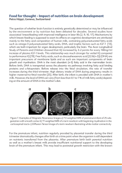
A schematic of the conversion pathway of fatty acids can be seen in Figure 3. It is
important to note that the synthesis pathways of ARA and DHA share enzymes which compete
for substrates. Also of importance is the fact that synthesis of ω3 and ω6 fatty acids is not
interconvertible, meaning an LCPUFA of the ω3 series cannot be synthesized from an LCPUFA
of the ω6 series and vice versa. Humans obtain ARA and DHA either from synthesis from their
parent EFAs or from the consumption of animal tissues, with fatty fish being the richest source
of DHA. Because humans have a low capacity for de novo lipogenesis and are unable to
synthesize the EFA, the dietary fatty acid supply is critical for the incorporation of fatty acids
into membrane bilayers.
Figure 3. A schematic of the conversion pathway of LA and ALA to the LCPUFA
C20:4ω6, C20:5ωn and C22:6ω3.
6
Importance of ω3 and ω6 Fatty Acids
The ω3 and ω6 class of fatty acids are important components of storage lipids, cell
membrane phospholipids, intracellular cholesterol esters and plasma lipids (8). Animal studies
have provided evidence that EFAs and their longer chain derivatives have a structural role in cell
membranes, a role in the transport of other lipids and both direct and indirect roles in the activity
of enzyme systems (7).
The long-chain fatty acids have also been found to play a role in regulation of gene
expression. These LCPUFA enhance the DNA binding affinity of peroxisome proliferatoractivated receptors (PPARs), which regulate the genes involved in fatty acid and glucose
oxidation, fatty acid uptake, fatty acid activation, triacylglycerol biosynthesis, and lipoprotein
metabolism. There is also some evidence that EPA enhances the transcriptional activity of this
family of transcription factors; however, these findings need to be substantiated by live animal
studies. Synthesis and activation of the sterol regulatory element binding protein (SREBP)-1 is
also influenced by unsaturated fatty acids. The SREBP transcription factors regulate
transcription of several of the genes involved in lipid, cholesterol, bile, and lipoprotein
biosynthesis. There is evidence that dietary ω3 fatty acids suppress hepatic lipogenesis by
inhibition of SREBP-1 gene transcription and by increasing SREBP-1 mRNA decay (9). The
activity and abundance of hepatic nuclear factor 4α (HNF4α), a steroid receptor that enhances
expression of genes involved in hepatic lipogenesis and carbohydrate metabolism, is modulated
by the ω3 class of fatty acids. Taken together, the genomic effects of ω3 fatty acids on hepatic
metabolism involve a shift from triacylglycerol synthesis, storage, and apolipoprotein secretion
towards hepatic oxidation of lipids. This shift may influence the risk of developing several
chronic diseases including coronary heart disease and obesity.
7
ARA and DHA make up the majority of the lipid content of the nervous system (10, 11).
Fetal brain accumulation of LCPUFA rapidly increases during the third trimester of gestation
and continues into the first two years after birth. These LCPUFA are incorporated into highly
specialized membrane phospholipids found in the retina and synapses. ARA is found in
relatively large amounts in most tissues, while DHA is distributed preferentially to specific
tissues. Compared to ARA, DHA is found in higher amounts in the cerebral cortex, retina and
testis (12). Within each of these tissues, DHA is especially abundant in rod photoreceptors and
synaptic membranes. These findings suggest an important role for the LCPUFA in visual and
cognitive development.
There is ample evidence that these LCPUFA are important for normal brain development,
visual acuity and cognitive function. Studies show that deficiency of the LCPUFAs is related to
degenerative diseases of the retina (13-15), poor performance on a test of retinal function and
tests of learning capabilities (10), memory loss, and diminished cognitive function (16). Infant
supplementation studies point to relationships between LCPUFAs and better visual acuity (1725), and mental development (21, 25-29).
Importance of LCPUFAs during Pregnancy
Substrate transfer from the mother to the fetus is critical for successful conception and
proper fetal growth. The accretion of LCPUFA is especially important during the prenatal
period for both the mother and the growing fetus. There is a growing body of evidence that these
nutrients are necessary for a variety of processes during gestation. For the mother, these
nutrients have been shown to play a role in the proper development of the placenta, in the
initiation of labor and delivery, and possibly development of the mammary gland (30). Studies
of increased dietary LCPUFA and supplementation have shown positive effects in the prevention
of pregnancy-induced hypertension and prolonging gestation in those women at risk of
8
delivering preterm (31). Recent work found that low maternal plasma ω3 fatty acids and high
ARA during early pregnancy were associated with a 40-50% increase in risk of small for
gestational age (SFA) infants (32). For the fetus these nutrients are important for proper growth
(33, 34), are significantly related to anthropometric measurements at birth (35) and also
influence neurological conditions at birth (36). There is also a large pool of evidence that
suggests the LCPUFA are especially important for postnatal growth and development of preterm
infants (35, 37).
Placental Transfer of ω3 and ω6 Fatty Acids
The elongation and desaturation enzymes necessary for EFA conversion to DHA and
ARA are present in the fetal liver during the early stages of gestation; however, their activity is
very low before birth (38). For this reason, the ω3 and ω6 LCPUFA that the fetus accumulates
come from placental transfer from the mother. To meet the additional needs of the growing
fetus, the maternal diet should contain sufficient EFAs and their long chain derivatives. If intake
is too low to meet the physiological needs, maternal body stores may be mobilized to supply the
required fatty acids (39). These stores are usually made available during the third trimester of
pregnancy when fetal growth reaches its maximum rate and fetal requirements for fatty acids,
especially DHA and ARA, are greatly increased (40). Evidence suggests that the accumulation
of brain DHA and ARA is far more efficient from preformed, dietary DHA and ARA rather than
their EFA precursors, LA and ALA. Additionally, recent evidence points to an inefficiency in
the conversion of DHA from ALA. This raises the question of whether DHA may be considered
conditionally essential during pregnancy when the requirement is increased.
Extensive research has investigated the amount of LCPUFA that is transferred to the
fetus in relation to the amount available in maternal circulation. Results of these studies show
that the amounts in cord blood (plasma phospholipids) are strongly correlated with maternal
9
plasma phospholipids (41, 42) and are influenced by the maternal diet (43). This evidence
supports previous findings of Otto et al. who showed that in an international population, the ω3
LCPUFA concentrations in umbilical plasma, vein, and artery phsospholipids were the highest in
those countries that had the highest maternal plasma ω3 LCPUFA concentrations (44). In the
same international population, ω6 LCPUFA concentrations in umbilical material seemed less
dependent on the maternal ω6 LCPUFA concentrations in blood phospholipids, which
researchers suggested could be due to some type of fetal autonomy with respect to establishing
its ARA supply (44).
In the very early stages of pregnancy, maternal plasma and erythrocyte DHA
concentrations begin to increase without any concomitant change in dietary intake. The increase
is most probably due to the release of fatty acids from the maternal stores to provide the
requirements for highly proliferating and differentiating tissues during this stage (45).
Compounding the issue of insufficient maternal dietary DHA are the findings by Al et al
(41). In a prospective, longitudinal study the total concentration (mg/L) of fatty acids in
maternal plasma phospholipids increased by ~51% throughout the course of pregnancy. The
absolute amount of ARA increased by 23% while DHA increased by 52%. It was thought that
the significant increase in DHA was due to an increase in fish intake; however, the dietary
information for this population showed no increase in fat intake during pregnancy.
Taken together, the body of evidence regarding DHA points to reduced maternal status
during pregnancy with suboptimal supply for the fetus (46). For this reason, the LCPUFA are
often thought of as conditionally essential during pregnancy. Further, the reduction in maternal
EFA status over the course of pregnancy appears to be independent of cultural differences in
dietary intake and ethnic origin (44).
10
The concentrations of DHA and ARA in fetal plasma phospholipids are much higher
compared to the concentration in maternal phospholipids; however, the LA and ALA precursors
are lower. Research supports the notion that higher LCPUFA concentrations in fetal plasma
reflect selective placental transfer, synthesis in placental or fetal tissues, or selective fetal
retention. Most evidence points to the selective transfer of ARA and DHA by membrane
associated proteins with higher affinity and binding capacity for these LCPUFA than for other
fatty acids. Findings by Ghebremeskel et al. (2000) suggest the possible role of a physiological
mechanism that works to maintain an appropriate balance between ARA and DHA (47).
Early research suggested the existence of a mechanism within the placenta that allowed
for fetal uptake of ARA by selective incorporation of ARA into phosphoglycerides and export to
the fetal circulation (48). These findings were followed by the purification and characterization
of a fatty acid-binding protein (FABP) from human placentas (49). This protein was isolated
from aborted and term placentas and was found to have at least three distinct fractions: one that
is nearly lipid-free, one that binds LCPUFA nonspecifically and one that binds mainly ARA.
Further studies suggested a saturability of fatty acid binding and the occurrence of binding sites
specific to different fatty acids (50). Later research revealed that the FABP is located
exclusively in the maternal facing microvillous membranes which may result in the
unidirectional flow of maternal free fatty acids to the fetus (51).
It was recently established that placental FABP preferentially binds both ARA and DHA
(52). These findings are further supported by the work of Haggerty (1997) and Dutta-Roy
(2000) (53, 54). Most recently, the involvement of two fatty acid transport proteins has been
suggested to play a role in the placental transfer of LCPUFA. The supply of DHA during
pregnancy affects the expression of fatty acid transport proteins in the placenta and therefore
plays a role in placental transfer (55, 56). Taken together, these findings clearly indicate that the
11
fetus is dependent upon the maternal supply of substrates and also on adequate placental
transport.
Obesity, BMI, and Insulin Resistance
Obesity is a condition marked by excess body fat. Measures of height and body weight
are widely used to identify the overweight and obese because it is difficult to obtain accurate
measures of body fatness in populations. Body Mass Index (BMI) is a measure used to assess
body weight relative to height. The number, obtained by dividing weight in kilograms (kg) by
height in meters squared (m2), has a relatively high correlation with estimates of body fatness.
As BMI increases above 25 kg/m2, there is a concomitant, gradual increase in risk of morbidity
from conditions such as type 2 diabetes mellitus, hypertension, coronary heart disease, stroke,
gallbladder disease, osteoarthritis, sleep apnea and some forms of cancer (prostate, colon,
endometrial and breast). See Table 1 for classification of BMI.
Table 1. Classification of Overweight
and Obesity by Body Mass Index.
BMI (kg/m2)
Underweight
< 18.5
Normal
18.5-24.9
Overweight
25.0-29.9
Obese
≥ 30.0
Adapted from: Nutritional Assessment,
3rd Edition.
Obesity and Pregnancy
Current estimates, based on data from the Pregnancy Risk Assessment Monitoring
System, show a 69% increase in pre-pregnancy obesity between 1993 and 2003 (57). These data
are supported by the finding that nearly 30% of women of childbearing age (20-39 years) were
obese between 1999 and 2002. The highest prevalence of obesity occurred in non-Hispanic
black women (49%) followed by Mexican-Americans (38.9%) and non-Hispanic white women
12
(31.3%) (58). Pre-pregnancy BMI is an important measure that is used by the Institute of
Medicine to make weight gain recommendations for pregnancy (59). Excess weight during
pregnancy poses a risk for both the mother and the developing fetus. Several adverse pregnancy
outcomes including miscarriage, neural tube defects, preeclampsia, GDM, macrosomia, shoulder
dystocia, and fetal death have been linked to high maternal BMI.
Influence of BMI and Insulin Resistance on LCPUFA Transfer
Little is known about the maternal factors that may influence supply and transfer of
substrates to the fetus during gestation. There is evidence of altered lipid metabolism in
pregnancies complicated by type 2 diabetes mellitus (NIDDM) and GDM (60). In the animal
model, increasing maternal glycemia has been associated with a decrease in the unidirectional
transfer of both EFA and non-essential fatty acids. Holman et al (1983) found that maternal
diabetes was associated with an overall decrease in PUFA status in the whole animal (61).
Decreased insulin sensitivity, seen in conditions of diabetes and obesity, has been associated
with decreased concentration of PUFA in skeletal muscle and plasma phospholipids (62), (63).
While little is known about the underlying mechanisms of these phenomena, it is suggested that
there is a possible impairment in the enzymes that regulate EFA metabolism (64). In a study of
Pima Indians, a negative correlation was reported between high percentage of body fat and
impaired activity of an enzyme involved in the EFA to LCPUFA conversion pathway. Both
pregnancy and obesity are often accompanied by decreased insulin sensitivity. Any impairment
in the enzyme system that results in decreased ARA or DHA would mean a smaller pool of these
essential nutrients available for transfer to the fetus and, thus, could have severe, deleterious
effects on fetal growth. Increased insulin resistance is a phenomenon associated with normal
pregnancies and to a greater extent in those complicated by diabetes (65).
13
Abnormalities in placental transfer may also be a consequence of pregnancies
complicated by insulin resistance and obesity. Bitsanis et al (2006) found that women diagnosed
with GDM had higher placental concentrations of both ARA and DHA when compared to
control women (66). These results could not be explained by differences in ethnicity or
enhanced placental synthesis of the LCPUFAs. Because neonates born to women with GDM
tend to have low blood levels of AA and DHA, the authors hypothesize that GDM enhances
uptake of the two fatty acids from maternal circulation and causes them to be retained instead of
transferred to the fetus. These results are supported by the findings of increased placental
incorporation of ARA in pregnancies complicated by type 1 diabetes mellitus (IDDM) (67) and
enhanced expression of placental FABP in pregnancies complicated by GDM (68).
In a cohort of pregnant women, BMI appears to be a significant predictor of maternal
concentrations of plasma phospholipid ARA and DHA (69), as well as fetal erythrocyte
phospholipid DHA (70). More recently, both IDDM and NIDDM have been shown to
compromise maternal red blood cell (RBC) DHA, as well as cord plasma and RBC ARA and
DHA (71). These effects seem to be independent of ethnicity or diet and related more to the
disease itself (72).
14
CHAPTER 3
METHODS
Participants and Sampling
Sera from pregnant women living in Maine were collected in the decade between 1996
and 2006. The fatty acids of sera from 265 women who delivered term infants who were
appropriate for gestational age were analyzed. The sample population was selected from a larger
population of 400 women who participated in a similar study. Samples were collected during the
second trimester of pregnancy from non-fasting women and stored in blood banks at FBR in
Maine. The 265 samples were shipped from FBR and upon arrival at the Louisiana State
University nutrition lab were immediately placed in storage at -80oC. Data for participants’ ages
and BMIs, which were calculated at 13-18 weeks gestation, were also provided by FBR.
Assessment of samples that had been in storage for five to ten years provided assurance that fatty
acids were detectable and quantifiable.
Procedures
Sample Collection
Samples were shipped from FBR to the Knapp Hall laboratory overnight on dry ice.
When samples arrived they were immediately transferred to a freezer and stored at -80oC until
analysis for fatty acids began.
Sample Methylation
Sera fatty acids were methylated according to Lepage (73) using an internal standard. In
brief, 100 µl of the stored sera were transferred to 9 ml glass tubes with Teflon lined caps. Two
ml of methanol:benzene (4:1, v/v) containing 40 µg/ml of internal standard (heptadecanoic acid,
C17:0) was added to the sample. Working under a ventilated hood, 200 µl of acetyl chloride was
added slowly over a period of one minute. After the addition of acetyl chloride, samples were
15
capped tightly and mixed thoroughly. Samples were heated for one hour at 100 oC in an analog
heat block (VWR). After one hour the samples were removed from the heat block and allowed
to cool. When samples were cool to the touch, 5 ml of 6% (w/v) potassium carbonate (K2CO3)
was added to neutralize the mixture and stop the reaction. Samples were then centrifuged in a
Beckman refrigerated centrifuge for 10 minutes at ~3500 rpm. After centrifuging, the methyl
esters in the solvent were pipetted off as the top layer and placed in a glass vial labeled with the
sample number. Fatty acids were separated by gas chromatography, identified based on
retention times and quantified against the internal standard. Fatty acids were expressed as
relative weight percent (wt%).
Sample Analysis
Analysis was performed using a Hewlett Packard 5890 series gas chromatograph (GC)
equipped with flame ionization detection (FID). Samples were injected into a 30 m fused silica
column with an internal diameter of 0.32 mm (Omegawax 320). The initial oven temperature
was programmed at 190oC and increased to 210oC in increments of 2o/minute with a final hold of
20 minutes. Helium was used as the carrier gas with a flow set at 1.0 ml/minute and hydrogen
was used as the make-up gas. The split ratio was set at 1:50, which means one part in fifty
entered the column. The injection port and detector temperatures were set to 250oC and 280oC,
respectively. Each sample was injected into the chromatograph three times. Peak identification
of serum fatty acids was based on comparison with the relative retention times of single fatty
acid methyl esters (FAME). Relative weight percents were calculated using the following
formula:
(area % of single FAME * 100)/(total area % of fatty acids – area % of C17:0)
16
Statistical Analysis
The data set was analyzed using JMP® 8 from Statistical Analysis Software (SAS).
Differences in relative weight percents of individual fatty acids and the ratios of fatty acids were
determined using one-way analysis of variance (ANOVA). Tukey’s Post-Hoc analyses were
performed to determine where significant differences occurred between BMI groups.
17
CHAPTER 4
RELATIONSHIP OF MATERNAL SERUM FATTY ACIDS AND BODY MASS INDEX
Introduction
The polyunsaturated fatty acids docosahexaenoic acid (DHA, C22:6ω3) and arachidonic
acid (ARA, C20:4ω6) are important components of the nervous system and have critical roles in
fetal growth and in brain and retinal development and function. Exponential fetal accretion of
these fatty acids begins during the third trimester of gestation and continues into the first two
years after birth. Fetal conversion of the ω3 and ω6 fatty acids is low; therefore, fetal
accumulation depends on the maternal supply. The placenta plays an important role in
transferring these fatty acids from maternal diet or body stores to the growing fetus. Because the
fetus depends on the maternal supply, the importance of identifying factors that influence fatty
acid transfer is underscored.
During pregnancy there is a natural decrease in maternal LCPUFA status, in particular,
DHA. A suboptimal supply of EFAs as well as their long chain derivates could be detrimental to
the development of the fetus. In addition to this, some maternal conditions may impact placental
transfer of nutrients to the fetus. One such condition is insulin resistance that is seen in diabetes
mellitus. Several studies support an overall decrease in EFA and LCPUFA status in these
conditions (60-65, 69, 70, 74).
During pregnancy it is normal to experience some insulin resistance and this resistance is
exacerbated by excess weight. Data document that the number of women entering pregnancy as
overweight or obese is increasing. Together, these conditions, insulin resistance and excess
weight, may negatively influence the efficiency of nutrient transfer. Recently, maternal BMI has
been shown to be negatively related to plasma phospholipid concentrations of ARA and DHA
(69) as well as fetal erythrocyte phospholipid DHA (70). The objective of the current study was
18
to determine if there is a similar relationship between maternal serum fatty acids and BMI. If
these results mimic the results of studies of phospholipid fatty acids, more studies using total
lipids in stored sera may be feasible as a more convenient and relatively rapid way to investigate
the maternal factors that influence substrate transfer to the fetus as well as fetal outcomes.
Subjects and Methods
Subjects
Sera from pregnant women living in Maine were collected in the decade between 1996
and 2006. Samples were collected during the second trimester of pregnancy from non-fasting
women. These samples were shipped from the Foundation for Blood Research in Maine and
upon arrival at the Louisiana State University nutrition lab were immediately placed in storage at
-80oC. Data for women’s ages and BMIs, which were determined at approximately 13-18 weeks
gestation, were provided by FBR. Prior assessment of samples that had been in storage for five
to ten years provided assurance that length of storage would not be a significant factor. The fatty
acids in sera from 265 women who delivered term infants who were appropriate for gestational
age were analyzed for this study.
Sample Analysis
Samples were directly methylated following the LePage method (73). In brief, 100 µl of
the stored sera were transferred to 9 ml glass tubes with Teflon lined caps. Two ml of
methanol:benzene (4:1, v/v) containing 40 µg/ml of internal standard (heptadecanoic acid,
C17:0) was added to each tube. Working under a ventilated hood, 200 µl of acetyl chloride was
added slowly over a period of one minute. After the addition of acetyl chloride, samples were
capped tightly and mixed thoroughly. Samples were heated for one hour at 100 oC in an analog
heat block (VWR). After one hour the samples were removed from the heat block and allowed
to cool. Once cooled, 5 ml of 6% (w/v) potassium carbonate (K2CO3) was added to neutralize
19
the mixture and stop the reaction. Samples were centrifuged in a Beckman refrigerated
centrifuge for 10 minutes at ~3500 rpm. After centrifuging, the methyl esters in the solvent were
pipetted off as the top layer and placed in a glass vial labeled with the sample number.
Fatty acid analysis was performed using a Hewlett Packard 5890 series gas
chromatograph equipped with flame ionization detection. Samples were injected into a 30 m
fused silica column with an internal diameter of 0.32 mm (Omegawax 320). The initial oven
temperature was programmed at 190oC and increased to 210oC in increments of 2o/minute with a
final hold of 20 minutes. Helium was used as the carrier gas with a flow set at 1.0 ml/minute and
hydrogen was used as the make-up gas. The split ratio was set at 1:50. The injection port and
detector temperatures were set to 250oC and 280oC, respectively. Each sample was injected into
the chromatograph three times. Peak identification of serum fatty acids was based on
comparison with the relative retention times of single fatty acid methyl esters (FAME). Relative
weight percents were calculated using the following formula:
(area % of single FAME * 100)/(total area % of fatty acids – area % of C17:0)
Statistical Analysis
The data were analyzed using JMP® 8 from Statistical Analysis Software (SAS).
Differences between BMI categories in relative weight percents of individual fatty acids and the
ratios of fatty acids were determined using one-way analysis of variance (ANOVA). Tukey’s
Post-Hoc analyses were performed to determine where significant differences occurred between
BMI groups.
Results
Sample Characteristics
The 265 participants had an average age of 27.7 years and an average BMI of 27.5 kg/m2.
Participants were grouped according to BMI category (Table 2; < 18.5, underweight; 18.5-24.9,
20
normal; 25.0-29.9, overweight; ≥ 30.0, obese). For statistical analysis, participants under age 18
were excluded as well as six participants in the obese class III category with outlying BMIs and
two participants with outlying weight percents for several fatty acids.
Table 2. Characteristics of sample population
BMI Category
Samples (n)
% of Population
Normal weight
Overweight
Obese
110
74
65
44.2
29.7
26.1
Serum Fatty Acid Analysis
Saturated, ω3, and ω6 fatty acids are presented in Tables 3, 4, and 5, respectively. There
were significant differences for the saturated fatty acid lignoceric acid (C24:0) in normal versus
obese participants (Table 3, p = 0.03). Weight percent of the ω3 fatty acid eicosapentanoic acid
(EPA, C20:5n3) was significantly different between the overweight and obese participants
(Table 4, p = 0.02) while the total relative weight percent of DHA trended toward significant
difference (p=.08) (Table 4). In the overall model there was a significant difference for the total
ω3 LCPUFA (p =.04); however, Tukey’s Post-Hoc analysis failed to determine significant
differences between BMI categories. In general, participants with higher BMIs tended to have
lower total ω3 LCPUFA (∑ C20:5ω3 and C22:6ω3).
Table 3. Average relative weight percent (wt %) of saturated fatty acids by BMI category1
Normal
Overweight
Obese
C16:0
27.37 ± 0.32 [110]
28.13 ± 0.39 [74]
28.33 ± 0.42 [65]
C18:0
5.52 ± 0.13 [110]
5.42 ± 0.17 [72]
5.42 ± 0.17 [65]
C20:0
2.06 ± 0.14 [109]
1.98 ± 0.17 [73]
2.01 ± 0.18 [63]
C22:0
1.27 ± 0.05 [106]
1.18 ± 0.06 [71]
1.19 ± 0.07 [62]
a
a
C24:0
0.95 ± 0.06 [110]
1.09 ± 0.07 [74]
0.81 ± 0.08 [65]b
1
Mean ± standard error. Means within a row with different superscript letters indicate
significant differences among BMI categories, p ≤ 0.05. Sample number in brackets.
21
Table 4. Average relative weight percent (wt %) for the ω3 fatty acids by BMI category1
Normal
Overweight
Obese
C18:3ω3
1.70 ± 0.16 [100]
1.75 ± 0.20 [67]
1.99 ± 0.21 [62]
C20:5ω3
1.38 ± 0.04 [110]a
1.27 ± 0.05 [74]a
1.17 ± 0.06 [65]b
C22:6ω3
1.59 ± 0.04 [110]
1.73 ±0.05 [74]
1.57 ± 0.06 [65]
1
Mean ± standard error. Means within a row with different superscript letters indicate
significant differences among BMI categories, p ≤ 0.05. Sample number in brackets.
Table 5. Average relative weight percent (wt %) for the ω6 fatty acids by BMI category1
Normal
Overweight
Obese
C18:2ω6
24.07 ± 0.37 [110]
23.71 ± 0.45 [74]
23.42 ± 0.48 [65]
C18:3ω6
1.59 ± 0.19 [26]
1.25 ± 0.22 [19]
1.37 ± 0.28 [12]
C20:2ω6
1.14 ± 0.14 [100]
0.84 ± 0.16 [72]
0.89 ± 0.17 [65]
C20:3ω6
1.32 ± 0.06 [110]
1.37 ± 0.07 [74]
1.29 ± 0.08 [64]
C20:4ω6
2.88 ± 0.09 [110]
2.99 ± 0.11 [74]
2.96 ± 0.12 [65]
C22:2ω6
0.53 ± 0.09 [10]
0.59 ± 0.11 [6]
0.38 ± 0.20 [2]
1
Mean ± standard error. Sample number in brackets.
Table 6. Sums and ratios of serum fatty acid content (wt %)1
Normal
Overweight
Obese
Total ω3 PUFA
4.48 ± 0.16 [110]
4.53 ± 0.19 [74]
4.58 ± 0.20 [65]
Total ω6 PUFA
29.73 ± 0.37 [110] 29.26 ± 0.45 [74]
28.81 ± 0.48 [65]
SFA:ω3 PUFA
9.22 ± 0.30 [110]
9.18 ± 0.36 [74]
9.30 ± 0.38 [65]
SFA:ω6 PUFA
1.27 ± 0.02 [110]
1.32 ± 0.03 [74]
1.33 ± 0.03 [65]
ω3:ω6
0.15 ± 0.01 [110]
0.16 ± 0.01 [74]
0.16 ± 0.01 [65]
Total ω3 LCPUFA
2.97 ± 0.06 [110]
3.00 ± 0.08 [74]
2.74 ± 0.08 [65]
Total ω6 LCPUFA
5.28 ± 0.22 [110]
5.23 ± 0.27 [74]
5.14 ± 0.29 [65]
ω3 LCPUFA:ω6 LCPUFA 0.65 ± 0.02 [110]
0.64 ± 0.03 [74]
0.58 ± 0.03 [65]
DHA:ARA
0.60 ± 0.02 [110]
0.63 ± 0.03 [74]
0.56 ± 0.03 [65]
Total SFA
37.14 ± 0.33 [110] 37.59 ± 0.41 [74]
37.64 ± 0.43 [65]
Total PUFA
34.19 ± 0.42 [110] 33.76 ± 0.51 [74]
33.38 ± 0.54 [65]
SFA:PUFA
1.11 ± 0.02 [110]
1.15 ± 0.03 [74]
1.15 ± 0.03 [65]
1
Mean ± standard error. PUFA, polyunsaturated fatty acid; SFA, saturated fatty acid;
LCPUFA, long-chain polyunsaturated fatty acid; DHA, docosahexaenoic acid; ARA,
arachidonic acid. Sample number in brackets.
Discussion
In this study we have established that sera collected from pregnant women and stored for
a decade or so may be used to study relationships of fatty acid status, maternal risk factors, and
pregnancy outcomes. In previous research, positive relationships were found between higher
22
BMI and lower ω3 fatty acid concentrations in plasma phospholipids (69, 70). Similar results for
total serum lipids were found in this study with participants in the highest BMI category tending
to have lower total ω3 LCPUFA, and statistically, significantly lower EPA.
The findings of this study support the theory that BMI is a predictor of maternal
LCPUFA status and suggest that stored serum may be a feasible medium to further explore the
relationships between maternal factors that influence pregnancy and fetal outcomes. These
results are limited by the lack of dietary intake information in this group as well as the absence of
pre-pregnancy BMI. The nature of the sample is also a limitation because of the influence of
recent dietary intake. The serum contains fatty acids bound to triglycerides, which would reflect
recent intake, as well as fatty acids contained in phospholipid membranes.
In this study, phospholipids were not extracted from the sera. The samples were analyzed
for total fatty acid content as a convenient and relatively quick way of investigating the
relationship between maternal BMI and fatty acid content of sera. It is interesting to note that a
similar relationship was found using the srea as was found in previous studies using plasma
phospholipids. This suggests the plausibility of analyzing serum in this population to investigate
maternal factors influencing LCPUFA status. Sera is often stored after routine blood collections
and archived over many years and demographic areas. Using stored sera to explore fatty acid
status during pregnancy provides a unique opportunity to find relationships in a large number of
samples that include many different populations. In addition to the availability of a large number
of samples, the methodology for total serum fatty acid analysis is less laborious than the
methodology for plasma phospholipids. To further investigate these relationships, future studies
should include a more diverse sample population and control for dietary intake, supplementation,
and diagnosis of diabetes.
23
CHAPTER 5
SUMMARY
The purpose of the present study was to investigate the relationship between maternal
serum fatty acids and BMI. The fatty acid content of serum was analyzed in 265 women in their
second trimester of pregnancy. Serum samples were provided by the Foundation for Blood
Research in Maine along with maternal age and BMI. Sera were collected between 1996 and
2006 and maternal BMIs were calculated at 13-18 weeks gestation. These samples provided a
unique opportunity to examine a relationship that has previously been reported in studies of
plasma phospholipids. Participants were grouped by BMI category and after analysis using gas
chromatography, fatty acid concentrations were compared across BMI categories.
In the sample population, women with the highest BMIs tended to have lower total
weight percent of the ω3 LCPUFA DHA and statistically, significantly lower EPA. These
findings are in line with recent reports that maternal BMI negatively influences plasma
phospholipid concentrations of DHA and ARA in the third trimester of pregnancy (69).
DHA is especially important for fetal growth and development. During the last trimester
of pregnancy, brain accumulation rapidly increases and continues until the age of two. Maternal
supply of preformed LCPUFA is critical for the growing fetus. Activity of the elongation and
desaturation enzymes needed for production of LCPUFAs from EFAs is low in the fetus;
therefore, the ω3 and ω6 LCPUFAs that the fetus accumulates must be transferred from the
mother across the placenta. Maternal dietary intake and body stores provide sources of the
LCPUFAs. Because tissue accumulation of the fatty acids is more efficient from preformed
DHA and ARA than from their 18-carbon parent fatty acids, it is important that the mother
provide sufficient amounts of these LCPUFAs. There is strong evidence that the amount of
24
LCPUFAs transferred to the fetus is strongly related to maternal plasma phospholipid
concentrations and dietary intake (41-44).
Because the fetus relies on maternal supply of LCPUFA, there are many maternal factors
that may negatively impact the amount that is transferred. Animal studies investigating the
influence of maternal insulin resistance on substrate transfer found an overall decrease in PUFA
status in the presence of diabetes (61). In human models, decreased insulin sensitivity has been
associated with decreased PUFA concentrations in skeletal muscle (62) and plasma
phospholipids (63). Insulin resistance is often seen in conditions of diabetes and obesity and it is
normal to see decreased insulin sensitivity during pregnancy. There is great concern considering
the 69% increase in prepregnancy obesity reported between 1993 and 2003 (57). If maternal
LCPUFA supply is inadequate, the consequences could be deleterious since these nutrients are
important for proper fetal growth (34, 75) and are significantly related to anthropometric
measures (35) and neurological conditions (36) at birth.
This study lends support to findings that BMI is a predictor of maternal ω3 LCPUFA
concentrations. However, these data are limited by several factors. Serum is not the preferred
sample for assessing fatty acid status as it is a reflection of both dietary intake and fatty acids of
cell membranes rather than fatty acids of the membrane phospholipids that are known to reflect
fatty acids of tissues cells, i.e. fatty acid status (76). In addition, ideally, blood should have been
drawn after a fast. Also, the maternal BMIs provided were calculated well into pregnancy at
approximately 13-18 weeks gestation. This BMI is likely to represent baseline BMI plus the
amount of weight the mother gained up to that point in pregnancy. Perhaps a more accurate BMI
would have been calculated before pregnancy, which would represent baseline BMI. Finally,
these results are only applicable to women of similar age, race, parity, and health status. Based
on these findings we conclude that women with increased BMI may be at risk for inadequate
25
PUFA supply to the fetus. More studies are needed to determine the impact of maternal BMI
and insulin resistance on placental transfer of LCPUFAs as well as the physiologic mechanisms
causing these results.
26
REFERENCES
1.
Voss A, Reinhart M, Sankarappa S, Sprecher H. The metabolism of 7,10,13,16,19docosapentaenoic acid to 4,7,10,13,16,19-docosahexaenoic acid in rat liver is
independent of a 4- desaturase. J. Biol. Chem. 1991;266:19995-20000.
2.
F. Yatsu SM. Elongation of Mitochondrial Fatty Acids During Brain Development.
Journal of Neurochemistry 1971;18:1895-1901.
3.
F. M. Yatsu SM. Chain elongation of linoleic and linolenic acids by brain mitochondria.
Journal of Neurochemistry 1972;19:1813-1815.
4.
F. Yatsu SM, E. Connolly, L. Nelson,. Elongation of fatty acids in human brain tissue.
Journal of Neurochemistry 1973;20:621-624.
5.
Cook HW, Spence MW. Biosynthesis of fatty acids in vitro by homogenate of developing
rat brain: desaturation and chain-elongation. Biochimica Et Biophysica Acta
1974;369:129-141.
6.
H. W. Cook. In Vitro Formation of Polyunsaturated Fatty Acids by Desaturation in Rat
Brain: Some Properties of the Enzymes in Developing Brain and Comparisons with
Liver. Journal of Neurochemistry 1978;30:1327-1334.
7.
Aaes-Jorgensen E. Essential fatty acids. Physiological Reviews 1961;41:1-51.
8.
Heird WC, Lapillonne A. The role of essential fatty acids in development. Annual
Review Of Nutrition 2005;25:549-571.
9.
Pawar A, Botolin D, Mangelsdorf DJ, Jump DB. The role of liver X receptor-alpha in the
fatty acid regulation of hepatic gene expression. The Journal Of Biological Chemistry
2003;278:40736-40743.
10.
Bourre J-M, Francois M, Youyou A, et al. The Effects of Dietary {alpha}-Linolenic Acid
on the Composition of Nerve Membranes, Enzymatic Activity, Amplitude of
Electrophysiological Parameters, Resistance to Poisons and Performance of Learning
Tasks in Rats. J. Nutr. 1989;119:1880-1892.
11.
Sastry PS. Lipids of nervous tissue: composition and metabolism. Progress In Lipid
Research 1985;24:69-176.
12.
Tinoco J. Dietary Requirements and Functions of alpha-linolenic acid in Animals.
Progress In Lipid Research 1982;21:1-45.
13.
Anderson RE, Maude MB, Lewis RA, Newsome DA, Fishman GA. Abnormal plasma
levels of polyunsaturated fatty acid in autosomal dominant retinitis pigmentosa.
Experimental Eye Research 1987;44:155-159.
14.
Bazan NG, Scott BL, Reddy TS, Pelias MZ. Decreased content of docosahexaenoate and
arachidonate in plasma phospholipids in Usher's syndrome. Biochemical and Biophysical
Research Communications 1986;141:600-604.
27
15.
Converse CA, Hammer HM, Packard CJ, Shepherd J. Plasma lipid abnormalities in
retinitis pigmentosa and related conditions. Transactions Of The Ophthalmological
Societies Of The United Kingdom 1983;103 ( Pt 5):508-512.
16.
Chamberlain JG. The possible role of long-chain, omega-3 fatty acids in human brain
phylogeny. Perspectives In Biology And Medicine 1996;39:436-445.
17.
Birch EE, Garfield S, Hoffman DR, Uauy R, Birch DG. A randomized controlled trial of
early dietary supply of long-chain polyunsaturated fatty acids and mental development in
term infants. Developmental Medicine And Child Neurology 2000;42:174-181.
18.
SanGiovanni JP, Berkey CS, Dwyer JT, Colditz GA. Dietary essential fatty acids, longchain polyunsaturated fatty acids, and visual resolution acuity in healthy fullterm infants:
a systematic review. Early Human Development 2000;57:165-188.
19.
SanGiovanni JP, Parra-Cabrera S, Colditz GA, Berkey CS, Dwyer JT. Meta-analysis of
dietary essential fatty acids and long-chain polyunsaturated fatty acids as they relate to
visual resolution acuity in healthy preterm infants. Pediatrics 2000;105:1292-1298.
20.
Birch EE, Hoffman DR, Uauy R, Birch DG, Prestidge C. Visual acuity and the
essentiality of docosahexaenoic acid and arachidonic acid in the diet of term infants.
Pediatric Research 1998;44:201-209.
21.
Uauy R, Hoffman D, Peirano P, Birch D, Birch E. Essential fatty acids in visual and brain
development. Lipids 2001;36:885-895.
22.
Mitchell DC, Niu S-L, Litman BJ. DHA-Rich phospholipids optimize G-Protein-coupled
signaling. The Journal of Pediatrics 2003;143:80-86.
23.
Makrides M, Neumann MA, Gibson RA. Is dietary docosahexaenoic acid essential for
term infants? Lipids 1996;31:115-119.
24.
Hoffman DR, Birch EE, Castañeda YS, et al. Visual function in breast-fed term infants
weaned to formula with or without long-chain polyunsaturates at 4 to 6 months: A
randomized clinical trial. The Journal of Pediatrics 2003;142:669-677.
25.
Judge MP, Harel O, Lammi-Keefe CJ. A docosahexaenoic acid-functional food during
pregnancy benefits infant visual acuity at four but not six months of age. Lipids
2007;42:117-122.
26.
J.S. Forsyth PW. Do LC-PUFA Influence Infant Cognitive Behavior. In: J.G. Bindles
ACG, H.K. Visser, ed. Recent Developments in Infant Nutrition. London: Kluwer
Academic Publishers, 1996:225-234.
27.
Willatts P, Forsyth JS, DiModugno MK, Varma S, Colvin M. Effect of long-chain
polyunsaturated fatty acids in infant formula on problem solving at 10 months of age.
Lancet 1998;352:688-691.
28.
Uauy R, Hoffman DR, Peirano P, Birch DG, Birch EE. Essential fatty acids in visual and
brain development. Lipids 2001;36:885-895.
28
29.
Judge MP, Harel O, Lammi-Keefe CJ. Maternal consumption of a docosahexaenoic acidcontaining functional food during pregnancy: benefit for infant performance on problemsolving but not on recognition memory tasks at age 9 mo. The American Journal Of
Clinical Nutrition 2007;85:1572-1577.
30.
Knazek RA, Liu SC, Bodwin JS, Vonderhaar BK. Requirement of essential fatty acids in
the diet for development of the mouse mammary gland. Journal Of The National Cancer
Institute 1980;64:377-382.
31.
Reece MS, McGregor JA, Allen KG, Harris MA. Maternal and perinatal long-chain fatty
acids: possible roles in preterm birth. American Journal of Obstetrics and Gynecology
1997;176:907-914.
32.
van Eijsden M, Hornstra G, van der Wal MF, Vrijkotte TG, Bonsel GJ. Maternal n-3, n-6,
and trans fatty acid profile early in pregnancy and term birth weight: a prospective cohort
study. The American Journal Of Clinical Nutrition 2008;87:887-895.
33.
Agostoni C, Marangoni F, Stival G, et al. Whole blood fatty acid composition differs in
term versus mildly preterm infants: small versus matched appropriate for gestational age.
Pediatric Research 2008;64:298-302.
34.
Dirix CE, Kester AD, Hornstra G. Associations between neonatal birth dimensions and
maternal essential and trans fatty acid contents during pregnancy and at delivery. The
British Journal Of Nutrition 2009;101:399-407.
35.
Foreman-van Drongelen MMHP, van Houwelingen AC, Kester ADM, Hasaart THM,
Blanco CE, Hornstra G. Long-chain polyunsaturated fatty acids in preterm infants: Status
at birth and its influence on postnatal levels. The Journal of Pediatrics 1995;126:611-618.
36.
Dijck-Brouwer DAJ, Hadders-Algra M, Bouwstra H, et al. Lower fetal status of
docosahexaenoic acid, arachidonic acid and essential fatty acids is associated with less
favorable neonatal neurological condition. Prostaglandins, Leukotrienes and Essential
Fatty Acids 2005;72:21-28.
37.
Crawford MA, Costeloe K, Ghebremeskel K, Phylactos A, Skirvin L, Stacey F. Are
deficits of arachidonic and docosahexaenoic acids responsible for the neural and vascular
complications of preterm babies? Am J Clin Nutr 1997;66:1032S-1041.
38.
Uauy R, Mena P, Wegher B, Nieto S, Salem N, Jr. Long chain polyunsaturated fatty acid
formation in neonates: effect of gestational age and intrauterine growth. Pediatric
Research 2000;47:127-135.
39.
Hornstra G. Essential fatty acids in mothers and their neonates. The American Journal Of
Clinical Nutrition 2000;71:1262S-1269.
40.
Herrera E, Amusquivar E, López-Soldado I, Ortega H. Maternal lipid metabolism and
placental lipid transfer. Hormone Research 2006;65 Suppl 3:59-64.
29
41.
Al MD, van Houwelingen AC, Kester AD, Hasaart TH, de Jong AE, Hornstra G.
Maternal essential fatty acid patterns during normal pregnancy and their relationship to
the neonatal essential fatty acid status. The British Journal Of Nutrition 1995;74:55-68.
42.
Al MD, Badart-Smook A, von Houwelingen AC, Hasaart TH, Hornstra G. Fat intake of
women during normal pregnancy: relationship with maternal and neonatal essential fatty
acid status. Journal Of The American College Of Nutrition 1996;15:49-55.
43.
Krauss-Etschmann S, Shadid R, Campoy C, et al. Effects of fish-oil and folate
supplementation of pregnant women on maternal and fetal plasma concentrations of
docosahexaenoic acid and eicosapentaenoic acid: a European randomized multicenter
trial. The American Journal Of Clinical Nutrition 2007;85:1392-1400.
44.
Otto SJ, Houwelingen AC, Antal M, et al. Maternal and neonatal essential fatty acid
status in phospholipids: an international comparative study. European Journal Of Clinical
Nutrition 1997;51:232-242.
45.
Otto SJ, van Houwelingen AC, Badart-Smook A, Hornstra G. Changes in the maternal
essential fatty acid profile during early pregnancy and the relation of the profile to diet.
Am J Clin Nutr 2001;73:302-307.
46.
Hornstra G, van Houwelingen AC, Simonis M, Gerrard JM. Fatty acid composition of
umbilical arteries and veins: possible implications for the fetal EFA-status. Lipids
1989;24:511-517.
47.
Ghebremeskel K, Crawford MA, Lowy C, et al. Arachidonic and docosahexaenoic acids
are strongly associated in maternal and neonatal blood. European Journal Of Clinical
Nutrition 2000;54:50-56.
48.
Kuhn DC, Crawford M. Placental essential fatty acid transport and prostaglandin
synthesis. Progress In Lipid Research 1986;25:345-353.
49.
Das T, Sa G, Mukherjea M. Purification and characterization of fatty acid-binding protein
from human placenta. Lipids 1988;23:528-533.
50.
Campbell FM, Taffesse S, Gordon MJ, Duttaroy AK. Plasma Membrane Fatty-AcidBinding Protein in Human Placenta: Identification and Characterization. Biochemical and
Biophysical Research Communications 1995;209:1011-1017.
51.
Campbell FM, Dutta-Roy AK. Plasma membrane fatty acid-binding protein (FABPpm) is
exclusively located in the maternal facing membranes of the human placenta. FEBS
Letters 1995;375:227-230.
52.
Campbell FM, Gordon MJ, Dutta-Roy AK. Placental membrane fatty acid-binding
protein preferentially binds arachidonic and docosahexaenoic acids. Life Sciences
1998;63:235-240.
53.
Haggarty P, Page K, Abramovich DR, Ashton J, Brown D. Long-chain polyunsaturated
fatty acid transport across the perfused human placenta. Placenta 1997;18:635-642.
30
54.
Dutta-Roy AK. Transport mechanisms for long-chain polyunsaturated fatty acids in the
human placenta1,2,3. Am J Clin Nutr 2000;71:315S-322.
55.
Larqué E, Demmelmair H, Klingler M, De Jonge S, Bondy B, Koletzko B. Expression
pattern of fatty acid transport protein-1 (FATP-1), FATP-4 and heart-fatty acid binding
protein (H-FABP) genes in human term placenta. Early Human Development
2006;82:697-701.
56.
Larque E, Krauss-Etschmann S, Campoy C, et al. Docosahexaenoic acid supply in
pregnancy affects placental expression of fatty acid transport proteins. Am J Clin Nutr
2006;84:853-861.
57.
Kim SY, Dietz PM, England L, Morrow B, Callaghan WM. Trends in Pre-pregnancy
Obesity in Nine States, 1993-2003[ast]. Obesity 2007;15:986-993.
58.
Hedley AA, Ogden CL, Johnson CL, Carroll MD, Curtin LR, Flegal KM. Prevalence of
overweight and obesity among US children, adolescents, and adults, 1999-2002. JAMA:
The Journal Of The American Medical Association 2004;291:2847-2850.
59.
Parker JD, Abrams B. Prenatal weight gain advice: an examination of the recent prenatal
weight gain recommendations of the Institute of Medicine. Obstetrics And Gynecology
1992;79:664-669.
60.
Knopp RH, Chapman M, Bergelin R, Wahl PW, Warth MR, Irvine S. Relationships of
lipoprotein lipids to mild fasting hyperglycemia and diabetes in pregnancy. Diabetes Care
1980;3:416-420.
61.
Holman RT, Johnson SB, Gerrard JM, Mauer SM, Kupcho-Sandberg S, Brown DM.
Arachidonic Acid Deficiency in Streptozotocin-Induced Diabetes. Proceedings Of The
National Academy Of Sciences Of The United States Of America 1983;80:2375-2379.
62.
Borkman M, Storlien LH, Pan DA, Jenkins AB, Chisholm DJ, Campbell LV. The
Relation between Insulin Sensitivity and the Fatty-Acid Composition of Skeletal-Muscle
Phospholipids. N Engl J Med 1993;328:238-244.
63.
Bassi A, Avogaro A, Crepaldi C, et al. Short-term diabetic ketosis alters n-6
polyunsaturated fatty acid content in plasma phospholipids. J Clin Endocrinol Metab
1996;81:1650-1653.
64.
Pan DA, Lillioja S, Milner MR, et al. Skeletal muscle membrane lipid composition is
related to adiposity and insulin action. The Journal Of Clinical Investigation
1995;96:2802-2808.
65.
Boden G. Fuel metabolism in pregnancy and in gestational diabetes mellitus. Obstetrics
And Gynecology Clinics Of North America 1996;23:1-10.
66.
Bitsanis D, Ghebremeskel K, Moodley T, Crawford MA, Djahanbakhch O. Gestational
diabetes mellitus enhances arachidonic and docosahexaenoic acids in placental
phospholipids. Lipids 2006;41:341-346.
31
67.
Kuhn DC, Crawford MA, Stuart MJ, Botti JJ, Demers LM. Alterations in transfer and
lipid distribution of arachidonic acid in placentas of diabetic pregnancies. Diabetes
1990;39:914-918.
68.
Magnusson AL, Waterman IJ, Wennergren M, Jansson T, Powell TL. Triglyceride
Hydrolase Activities and Expression of Fatty Acid Binding Proteins in the Human
Placenta in Pregnancies Complicated by Intrauterine Growth Restriction and Diabetes. J
Clin Endocrinol Metab 2004;89:4607-4614.
69.
Wijendran V, Bendel RB, Couch SC, et al. Maternal plasma phospholipid
polyunsaturated fatty acids in pregnancy with and without gestational diabetes mellitus:
relations with maternal factors. The American Journal Of Clinical Nutrition 1999;70:5361.
70.
Wijendran V, Bendel RB, Couch SC, Philipson EH, Cheruku S, Lammi-Keefe CJ. Fetal
erythrocyte phospholipid polyunsaturated fatty acids are altered in pregnancy
complicated with gestational diabetes mellitus. Lipids 2000;35:927-931.
71.
Min Y, Lowy C, Ghebremeskel K, Thomas B, Offley-Shore B, Crawford M. Unfavorable
effect of type 1 and type 2 diabetes on maternal and fetal essential fatty acid status: a
potential marker of fetal insulin resistance. Am J Clin Nutr 2005;82:1162-1168.
72.
Min Y, Nam JH, Ghebremeskel K, Kim A, Crawford M. A distinctive fatty acid profile in
circulating lipids of Korean gestational diabetics: a pilot study. Diabetes Research and
Clinical Practice 2006;73:178-183.
73.
Lepage G, Levy E, Ronco N, Smith L, Galeano N, Roy C. Direct transesterification of
plasma fatty acids for the diagnosis of essential fatty acid deficiency in cystic fibrosis. J.
Lipid Res. 1989;30:1483-1490.
74.
Phinney S, Davis P, Johnson S, Holman R. Obesity and weight loss alter serum
polyunsaturated lipids in humans. Am J Clin Nutr 1991;53:831-838.
75.
Agostoni C. Role of long-chain polyunsaturated fatty acids in the first year of life.
Journal Of Pediatric Gastroenterology And Nutrition 2008;47 Suppl 2:S41-4.
76.
Holman RT, Johnson SB, Ogburn PL. Deficiency of essential fatty acids and membrane
fluidity during pregnancy and lactation. Proceedings Of The National Academy Of
Sciences Of The United States Of America 1991;88:4835-4839.
32
VITA
Emily Fontenot Gilbert was born in Baton Rouge, Louisiana, in 1985. In May 2007, she
obtained her Bachelor of Science Degree in dietetics from Louisiana State University in Baton
Rouge, Louisiana. In the fall of 2007 she began pursuing a master’s degree in the School of
Human Ecology with a concentration in human nutrition and food at Louisiana State University.
She will continue her pursuit of becoming a registered dietitian with a dietetic internship in the
fall of 2009. She is a member of the American Dietetic Association, the Louisiana Dietetic
Association and the American Oil Chemists’ Society.
33


© Copyright 2026