
The Westie Health EBook Common health problems, how to recognize them and what to do about them John Robertson, VMD PhD and Elizabeth McStay, BS
The Westie Health EBook Common health problems, how to recognize them and what to do about them John Robertson, VMD PhD and Elizabeth McStay, BS VirginiaMaryland Regional College of Veterinary Medicine (VMRCVM) Virginia Tech, Blacksburg, VA 240610442 About this “EBook” (electronic book) Welcome to the first edition of the Westie EBook, sponsored by generous support from the Westie Foundation of America. This electronic book is meant to serve as a source of information for Westie owners, breeders, veterinarians and for anyone who loves Westies. The EBook (really an electronic website) is organized by topic. A list of topics follows and then the link of that portion of the site will take you there with a click of a button. The material is organized by topics of general interest, and then by specific diseases that Westie seem more prone to than other dogs. Under each disease heading, is information intended for all readers. There are then areas of more detailed content (for veterinarians and scientists), but all are welcome to read through this material. References used to prepare the EBook are listed, as are short summaries of some key articles. This is a work in progress! Comments (wag, wag, wag!) and criticisms (grrrr!) are welcome! Health and disease in dogs – a brief overview Dogs are amazingly hearty and healthy. The owner of any new puppy is amazed at the number of “delectable” items puppies eat – and how they grow and prosper. There are several things that determine the health of all dogs. First, and foremost, dogs need a nutritionally adequate and complete diet, plenty of clean, fresh water, and a safe environment (See Diet and Environment, below). Second, dogs need human attention to their health problems. Owners of dogs are the front line in providing regular visits to the veterinarian for vaccinations and physical examinations (See You and Your Veterinarian, below). Third, the breed of dogs can have a very strong influence on the development of some health problems. We know that some types of health problems, such as cancer or skin disease, are more common in some purebred dog breeds than others. One example of this is an increased incidence of bladder cancer in Scottish terrier breeds, such as the Scottie and Westie (See How Breed Influences Health in Dogs, below) (reference here). Diet and Environment Revision by Korrin Saker, DVM PhD DACVN Dogs prosper on many different diets. How can you be assured the diet you choose will provide adequate nutrition for your dog? AAFCO, the American Association of Feed Control Officials, has developed policies for regulating the manufacture, labeling, distribution and sale of animal feeds. The Nutrition Claim or Nutrition Statement on all pet food labels will state if and to what extent the manufacturer has followed AAFCO guidelines in the formulation and testing of that diet. You will be able to ascertain, to some extent, if the diet is appropriate for your dog at a particular lifestage. Most commercially available diets (dry, semimoist, or canned) are formulated to provide “complete” nutrition for a specific life stage. A separate category of commercial diets, termed “therapeutic or prescription” are formulated to address specific health concerns. Diets that are formulated to provide “complete and/or balanced nutrition” do not require supplementation with vitamins, minerals or other nutrients for maintaining adequate health. Some owners prefer to prepare the diet for their dog from ingredients from the market. Homemade recipes have their place in pet nutrition, but can easily be incomplete or “unbalanced”; therefore, it is important that you discuss your dog’s diet with your veterinarian to be sure it provides all the nutrients the dog will need to maintain good condition. The National Academy of Sciences maintains a website for pet owners on the subject of dog and cat nutrition. It contains information on nutrient requirements and nutrient problems. The website (called “Petdoor”) is worth a visit if you have questions about nutrition and diets for your Westie. It is found at The National Academy of Sciences, Board of Agriculture and Natural Resources site (http://www.dels.nas.edu/banr/petdoor.html). In general, most dogs thrive on the same diet – day after day. In fact, some dogs don’t do very well if their diet is changed from one food to another, and will sometimes get stomach upsets and diarrhea if their diet changes. Most owners realize that there are some dietary nono’s – too much food (exceeding body needs and leading to weight gain), doggie junk food and snacks, and fatty scraps. Although they love them, many dogs will get upset stomachs and diarrhea if they eat bones. Soft bones, such as those from poultry, may actually be a danger to dogs. These soft bones can be broken into sharp pieces during chewing and injure the digestive tract and mouth of dogs. All of these should be avoided. Numerous pet food companies maintain very good information on dog nutrition on their websites. They also provide information for dog owners on their products. Pet food websites and product information can be obtained by accessing the American Academy of Veterinary Nutrition (AAVN) website ( www.aavn.org). Dogs need clean, fresh water. Dogs should have their water changed several times daily, and regularly checked by their owner for debris, cloudiness or discoloration that might indicate the water is unpalatable or potentially contaminated. Water bowls made of stainless steel are generally easier to keep clean and to resist chewing by enthusiastic dogs. One of the most important things owners can do to insure the health of their animal is to keep it in a safe environment. Dogs that are allowed to run free may encounter other dogs with potentially infectious diseases. Although Westies have the courage of African lions and will stand their ground against much larger dogs, they may get severely injured in dog or cat fights. Dogs that run free also run the risk of being struck by automobiles, ingesting dangerous substances (like antifreeze, for example), or being injured from falls or from malicious acts. So, keep your Westie safe! You and Your Veterinarian You and your veterinarian are a team dedicated to maintaining the health of your Westie. Veterinarianstobe take four years of classes and mentored practice experiences before they take licensing examinations and begin to practice veterinary medicine. Most new veterinarians will work with more seasoned practitioners to hone their skills. Some veterinarians will take additional years of training (as interns and residents) to learn a veterinary specialty like dermatology, oncology (the study of cancer), or orthopedic surgery. If you are interested in veterinary training or in the scope of the veterinary profession, several very good websites are maintained by The American Veterinary Medical Association (www.avma.org) and the American Animal Hospital Association (www. healthypet.com). These websites also contain a wealth of information on the health of companion animals (such as dogs, cats, and horses) and you may want to visit them. Regular visits to your veterinarian are critical in maintaining the health of your Westie. These visits allow the veterinarian to get to know your dog and to know you. The visits allow you to communicate to the veterinarian what a special dog you have and to allow the dog to understand the environment and examination procedures. It is well known that dogs that know their veterinarian and his practice environment are more at ease with visits. This lowers the stress levels your dog might have when going to a place where there are other dogs and cats, strange people and strange smells. Regular visits also help the veterinarian do a good job in assessing the health of your dog, because they can develop a baseline of health and potential medical problems, detect diseases at early stages, and, most importantly, gain the trust of you and your Westie. How often you and your Westie visit your veterinarian depends on the age and the health of your dog. Most veterinarians would like to see your dog frequently (every few months) as a puppy, for vaccination against infectious diseases, to provide information on diet, to detect early signs of health problems, and to assess whether or not your puppy is affected by parasites. When dogs become mature (between 12 years) visits to the veterinarian may only be needed every 612 months. Of course, you and your dog should always see the veterinarian if there are any health problems, so they can be accurately diagnosed and treated. Most dogs in the United States are now regularly maintained on medication to prevent the development of canine heartworm disease (“dirofilariasis”), a disease spread from one dog to another by mosquito bites. Veterinarians may also recommend the use of medications applied regularly to minimize the effects of fleas and ticks on dogs that go outdoors. Breeding, spaying and neutering Breeding, spaying and neutering are critical topics for discussion between you and your veterinarian. If you are an experienced dog breeder, you have a wealth of knowledge regarding breeding – perhaps more than your veterinarian. Most veterinarians will readily acknowledge this and will be happy to learn from your experiences. They may also have questions and observations that will foster dialogue, including optimum timing for breeding, frequency of breeding, suggestion on nutrition for dam, sire and pups, vaccination schedules and protocols (to optimize puppy immunity), and a number of other topics. The outcome of breeder and veterinarian dialogue is happy, health Westie pups, Westie moms, and their human families! If you do not intend to breed your dog, then spaying and neutering may have important health consequences for your dog and for the dog population in general. We are sure most people realize there is a serious problem of pet overpopulation in the United States. Although this is rarely a problem for Westies (because they have very devoted owners!), there are many dogs that do not have homes and which may stray. Pet owners who spay and neuter their pets play a major role in preventing pet overpopulation. Spaying your female dog (“ovariohysterectomy”) removes the ovaries and the uterus of the dog, so that she will not have puppies. Neutering male dogs removes the testis, and these dogs are sterile. These operations are done by your veterinarian in his hospital. Veterinarians first examine your dog to be sure that the dog is healthy enough for surgery, and then schedule the operation. Dogs that are spayed/neutered are given a general anesthesia and prepared for sterile (aseptic surgery). After the operation, dogs will have a portion of their fur shaved, a sutured/stapled surgical site, and will require observation and aftercare. This will all be discussed with you by your veterinarian. As noted above, there are important health consequences of spaying and neutering. Several studies have noted that the incidence of uterine infections (“pyometra”) and mammary gland tumors is markedly reduced in female dogs that have been spayed. The beneficial effect on the development of canine mammary gland tumors is seen in dogs that are spayed in the first year of life and somewhat in dogs spayed between 12 years of age. Female dogs of any age have a reduced risk of developing uterine infections and inflammation (“pyometra”), since the spaying operation removes the uterus. The benefits (aside from preventing pet overpopulation) of neutering male dogs may be a reduction in the incidence and growth of some types of skin tumors (“perianal gland adenoma”), decreased incidence of perianal fistula and on problems associated with benign enlargement of the canine prostate, including prostatic cysts and abscesses. Recent research has not shown that neutering of male dogs decreases prostatic cancer in dogs; in fact, some data shows that neutered male dogs may be at a slightly increased risk for developing this very uncommon tumor. The effect of spaying and neutering on the development of other diseases and on pet behavior (such as aggression) is less clear cut. Once again, a discussion of these topics with your veterinarian will help you make important decisions on pet breeding and pet neutering. How Breed Influences Health in Dogs One of the many things that are very important in determining the health of every dog is their genetic makeup. Each cell in every dog (and person and everything else) contains a “blueprint” for the cell and for the dog. These “blueprints” are made up of DNA, formed into specific genes contained in chromosomes. The entire set of genes that contain the “blueprint” for each individual dog is known as its genome. The genome specifies how cells are made, how the cells form tissues, and how the tissues (like the heart or skin) function. Selective breeding of dogs, following domestication from wild dogs and wolves, has resulted in the evolution of specific dog breeds, like the West Highland White Terrier. The genome of one Westie is likely to be very similar to other Westies, because selective breeding over several hundreds of years has focused the genome on certain desirable characteristics that make them Westies. For example, the pale and white coat color of Westies, the shape of their body, and even things like their lifespan are encoded in their genome (Westies tend to live longer than Great Danes!). It is very likely that the differences in the genome of Westies from other dog breeds are small and caused by the variable expression of certain key genes. These variations in gene expression are termed “mutations” or “polymorphisms” by genomic scientists. Many such variations in the genome are good, conferring selective advantages in appearance, performance and health. On the other hand, some variations are not advantageous for dogs. It is well known that cancer, for example, is the result of mutation in certain specific genes that control cell growth, cell division, and cell lifespan. Unfortunately, some breeds of dogs, as a result of selective breeding, develop diseases that are associated with their breed. Two examples are shown below to illustrate this. The information is a little technical, but it helps show strong associations between breed and disease. How this all relates to Westies follows these examples. Example: Breed predispositions as a significant factor in the development of ML Golden Retrievers, in comparison to many other dog breeds, are disproportionately affected by neoplasms as causes of morbidity and mortality. In 1998, the Golden Retriever Club of America (GRCA) developed and distributed a questionnairebased health survey to Club members (Glickman, 1998). Health, husbandry, morbidity, and mortality data was received from 746 dog owners, documenting information on 1444 dogs. Pet owners confirmed information on morbidity and mortality of dogs with veterinary practitioners. The results of this survey indicated that approximately 61.4% of all deaths reported in Golden Retrievers were caused by neoplasms. In dogs that died, the lifetime risk of developing a neoplasm was 1 in 2, with the risk of development of hemangiosarcoma (the most common tumor) being 1 in 5, and of developing ML 1 in 8. According to survey results, only about 12% of dogs which developed ML were cured by therapy. ML ranked as the most significant Golden Retriever disease in terms of years of potential life lost in the study population, and caused roughly 4 times more potential years lost than any other single cause of mortality. Other data also indicates Golden Retrievers may be more likely to develop ML than other breeds. The five most popular breeds of dogs (20034, in descending order) were Labrador Retrievers, Golden Retrievers, German Shepherd Dogs, Beagles, and Yorkshire Terriers (Source: American Kennel Club registration data, 2005). These breeds have been among the most popular for more than a decade. They accounted for 15.6%, 5.6%, 4.8%, 4.8%, and 4.4%, respectively, of the total of 1,873,711 purebred dogs registered in those years. If all breeds had a similar risk for the development of ML, one would expect that the proportion of cases of ML recorded in databases would be roughly proportional to breed popularity. For example, one would expect that Labrador Retrievers would contribute about 3x more cases of ML to the database than Golden Retrievers. However, an examination of data in the VCR database indicates a substantially increased risk for ML in Golden Retrievers. Currently, there are 1,141 cases of ML in the database (18% of the total cases). Labrador Retrievers account for 8.2% of the ML cases, while Golden Retrievers account for 10.2%. Based solely on breed registrations (representing numbers of popular purebred dogs) it appears that Golden Retrievers may be 3 times more likely than Labrador Retrievers to develop ML (Table, below). Unfortunately, actuarial data, including estimates of the total ‘at risk’ canine population, and reports of causes of death do not exist, making calculation of incidence, prevalence, or frequency, difficult and imprecise. Labrador Retriever Golden Retriever German Shepherd Beagle Yorkshire Terrier AKC reg. (%) 15.6 5.6 4.8 4.8 4.4 ML in VCR (%) Obs/Exp 8.2 10.2 3.7 2.2 0.6 1.2 4.0 1.7 1.0 0.3 Table. Representation of different dog breeds among total AKC registrations, and cases of ML in the Veterinary Cancer Registry. The ratio of observed ML frequency to expected ML frequency (based on AKC registration) is normalized relative to Beagle. The overrepresentation of ML in the Golden Retriever breed was confirmed in a retrospective study of ML at our institution. Three hundred ninetyfive (395) dogs were diagnosed with ML in a 15year period of study (19862001). This comprised 1.2% (n=32,823) of all canine admissions. More than 80 breeds of dog were diagnosed with and treated for ML. Breeds diagnosed with ML included: Mixed Breed (n=79), Golden Retriever (n=38), Labrador Retriever (n=31), Cocker Spaniel (n=23), Rottweiler (n=16), German Shepherd Dog (n=12), Doberman Pinscher (n=11), Boxer (n=11), Shih Tzu (n=11), and other breeds (n=157). Of the total number of cases in our study population, Golden Retrievers accounted for 9.6% of total cases seen, a figure close to that represented in the VCR database. It is entirely possible that phenotypic selection of desirable characteristics in some breeds may have also selected for genomic abnormalities associated with the development of ML. As previously noted, the fundamental basis for all neoplasms is mutation of critical genes that control growth and differentiation Conventional pedigreebased linkage studies have been used successfully to identify genes that are associated with a variety of canine disorders, including a rare cancer (reviewed by Sutter and Ostrander, 2004). References Glickman, L, Glickman, N, Thorpe, R, “19981999 Golden Retriever Club of America National Health Survey”, Golden Retriever Club of America Health and Genetics Committee, 1998 Sutter NB, Eberle MA, Parker HG, Pullar BJ, Kirkness EF, Kruglyak L, Ostrander EA, “Extensive and breedspecific linkage disequilibrium in Canis familiaris, “ Genome Res 14: 238896, 2004 Example: Breed predispositions in the development of brain tumors in dogs Spontaneous primary brain tumors are common in dogs, accounting for 13% of all deaths in aged dogs where necropsy is performed (Koestner, et al, 2002). In one study, an incidence rate of 14.5 cases/100,000 dogs at risk was reported, although this may be somewhat imprecise as actuarial data is not collected on dogs at the time of death (Vandevelde, 1984). Over 70% of primary tumors occur in dogs aged 6 years or more, a period in lifespan comparable to middle age in humans. Astrocytomas, oligodendrogliomas and meningiomas are most common, with astrocytomas accounting for approximately 10% of primary brain tumors in some series (Luginbuhl, H et al, 1968). Astrocytomas classified as grades IIIV in the revised WHO Classification system have been recognized and reported in dogs, and are histologically identical to similarly graded tumors in humans (Kleihues, P et al, 1993). Most canine astrocytomas occur as cortical lesions, with many forming in the temporal and frontal regions. An increased incidence of gliomas has been noted in brachycephalic breeds (Boston Terriers, Boxers, Bulldogs) (Summers, BA, et al, 1995) as well as in the Golden Retriever, Doberman Pinscher, Scottish Terrier and Old English Sheepdog breeds (Summers, BA, et al, 1991). Both the clinical presentation and progression of these astrocytomas is similar to humans (LeCouteur, RA, 2001). Unfortunately, patterns of survival are also similar, likely a combination of limitations in providing therapy to veterinary patients and inherent tumor behavior. Despite the known predisposition of certain breeds to specific tumors, there have been no definitive investigations of gene expression in normal canine brain tissue. Extrapolating from the human experience, it is possible that some dog breeds have inherited defects in tumor suppressor gene function or may be predisposed to overexpress other critical growth regulating genes, eventually leading to tumor development. References Koestner, A, Higgins, RJ, “Tumors of the nervous system,” in Tumors of Domestic Animals, 4 th ed., Meuten, DJ (ed.), Iowa State University Press, Ames, IA, 2002, p. 697 Vandevelde, M, “Brain tumors in domestic animals: An overview,” Proceedings of the Conference on Brain Tumors in Man and Animals, Research Triangle Park, NC, September, 1984 Luginbuhl, H, Fankhauser, R, McGrath, JT, “Spontaneous neoplasms of the nervous system of animals,” Prog Neurology Surgery 2: 85164, 1968 Kleihues, P, Burger, PC, Scheithauer, BW, “The new WHO classification of brain tumours”, Brain Pathology 3: 255268, 1993 Summers, BA, Cummings, JF, De Lahunta, A, “Tumors of the central nervous system,” in Veterinary Neuropathology, MosbyYear Book Publishers, Inc., St. Louis, MO, 1995, p. 364 Summers, BA, Kornegay, JN, et.al, “Analysis of survival in a retrospective study of 86 dogs with brain tumors,” J Veterinary Internal Medicine 5: 219 226, 1991 LeCouteur, RA, “Tumors of the nervous system,” in Small Animal Clinical Oncology, 3 rd ed., Withrow, SJ, MacEwen, EG, (eds), WB Saunders Co, Philadelphia, PA, 2001 How does this relate to Westies? There are some diseases that occur in Westies that are more common than in other breeds. The reason for this is undoubtedly tied up in the genome of the breed. Selective breeding over hundreds, if not thousands of years, has developed the Westie with certain characteristics such as size, stature, coloration and even personality. At the same time that these desirable characteristics were selected by careful breeding, other less desirable characteristics also developed. Some of these less desirable mutations were linked (literally, in the DNA and chromosomes) to more desirable breed characteristics – sort of ‘hitching a ride’ in the Westie genomic pattern. Because of these linked mutations, Westies are predisposed to the development of some diseases, just like Golden Retrievers get more malignant lymphoma and Bulldogs get more brain tumors. We know that while dogs may be predisposed genetically to developing some diseases, there are also many identified and unidentified environmental influences on disease development, expression and severity. This complex interplay between genome and environment is an area of intense scientific study. A very good first step in making progress in understanding which diseases are common and for beginning the study of genomeenvironmental relationships are health surveys, conducted by the Westie Foundation of America and also the West Highland White Terrier Club of America. The results of recent studies are a kicking off point for discussion of diseases that follow. It is now our job to find the specific genes in the Westie genome that are related to common diseases. Once this is done, more effective treatments for these diseases can be found, and concerned breeders, owners, veterinarians and scientists can work together to eliminate these problem genes, while maintaining happy, healthy populations of Westies for centuries to come. With all this background information down, we are now ready to discuss some Common diseases of Westies. The information is organized by disease topic. Before each topic, there is a brief summary of information which may help readers understand some of the terminology and concepts used in the discussions. Common diseases of Westies The Westie Foundation of America (WFA) and the West Highland White Terrier Club of America (WHWTCA) have each conducted several surveys of Westie owners and breeders to identify common diseases. The fact that owners and breeders recognize diseases that occur more commonly in Westies means that there are likely to be complex genomic factors and environmental factors that interact to produce disease. In 1999 (and again in 2004) the WFA conducted a health survey of Westies. The results of the 1999 Survey were both interesting and important in terms of focusing research on Westie health problems. Owners and their veterinarians were asked to report the disease both by importance to them and prevalence. A response rate on this survey (27.1%) is considered low for the purposes of statistical analysis. According to the 1999 WFA Westie Health Survey Diseases ranked by importance as reported by Westie owners: 1. Atopic Dermatitis 2. Pulmonary fibrosis 3. Copper toxicosis and LeggCalvePerthes (tied) 4. Addison’s disease (hypoadrenocorticism) 5. Aggression 6. Diabetes mellitus 7. Whiteshaker syndrome 8. Dry eye (keratoconjunctivitis sicca) 9. Allergies 10. Luxated patella 11. Globoid Cell Leukodystrophy 12. Cancer Targeted diseases were ranked by prevalence (mainly diagnosed by veterinarians) as follows: 1. Atopic Dermatitis 2. Aggression 3. Luxated patella 4. Dry eye (keratoconjunctivitis sicca) 5. LeggCalvePerthes 6. Cranial mandibular osteopathy 7. Pulmonary fibrosis 8. Whiteshaker syndrome, diabetes, Addison’s disease (hypoadrenocorticism) (tied) 9. Copper toxicosis 10. Juvenile cataracts 11. Globoid Cell Leukodystrophy 12. Deafness According to WHWTCA, the following diseases have been identified as of particular concern to Westie owners (these diseases are listed alphabetically, not in order of occurrence or importance) (bolded items are discussed in this first draft of the Westie Ebook): Alphabetical listing of diseases of concern to Westie owners Aggression Addison's disease (Hypoadrenocorticism) Atopic Dermatitis Bladder Cancer (TCC) Cleft Palate Copper Toxicosis (CT) Craniomandibular Osteopathy (CMO) Deafness Diabetes Mellitus Ear Infections Epidermal Dysplasia Globoid Cell Leukodystrophy (GCL) Heart Disease Hip Dysplasia Immune System Inflammatory Bowel Disease (IBD) Hernia (inguinal and umbilical) Juvenile Cataracts Keratoconjunctivitis Sicca (KCS, Dry Eye) Kidney Disease LeggCalvePerthes Luxated Patella Portosystemic Shunt Pulmonary Fibrosis (Westie Lung Disease) Pyruvate Kinase (PK) Deficiency Seborrhea, Primary and Secondary Skin and Allergy Problems Teeth and Gums White Shakers Syndrome Common diseases of Westies, categorized by Body System Alimentary (Digestive) System Cleft Palate Copper Toxicosis Craniomandibular Osteopathy Diabetes Mellitus Inflammatory Bowel Disease (IBD) Pyruvate Kinase (PK) Deficiency Teeth and Gums Cardiovascular System Heart Disease Portosystemic Shunt Pulmonary Fibrosis (Westie Lung Disease) Endocrine System Addison's disease (Hypoadrenocorticism) Immune System Locomotor System Hip Dysplasia LeggCalvePerthes Luxated Patella Nervous System Aggression Globoid Cell Leukodystrophy (GCL) White Shakers Syndrome Special Senses Deafness Ear Infections Juvenile Cataracts Keratoconjunctivitis Sicca (KCS, Dry Eye) Skin and Soft Tissue Atopic Dermatitis Ear Infections Epidermal Dysplasia Hernia (inguinal and umbilical) Keratoconjunctivitis Sicca (KCS, Dry Eye) Seborrhea, Primary and Secondary Skin and Allergy Problems Urogenital System Bladder Cancer (TCC) Kidney Disease Common diseases of Westies, categorized by underlying pathologic process Congenital/Developmental Atopic Dermatitis Cleft Palate Craniomandibular Osteopathy (CMO) Deafness Globoid Cell Leukodystrophy (GCL) Heart Disease Hip Dysplasia Hernia (umbilical) Juvenile Cataracts LeggCalvePerthes Luxated Patella Portosystemic Shunt Seborrhea, Primary and Secondary Hemodynamic Infectious Ear Infections Inflammatory Epidermal Dysplasia Inflammatory Bowel Disease (IBD) Keratoconjunctivitis Sicca (KCS, Dry Eye) Pulmonary Fibrosis (Westie Lung Disease) Skin and Allergy Problems (Atopic dermatitis) White Shakers Syndrome Metabolic/Endocrine Addison's disease (Hypoadrenocorticism) Neoplastic Bladder Cancer (TCC) Nutritional/Toxic Copper Toxicosis (CT) Diabetes Mellitus Globoid Cell Leukodystrophy (GCL) Pyruvate Kinase (PK) Deficiency Other/Unknown Aggression Hernia (inguinal) LeggCalvePerthes Luxated Patella Pulmonary Fibrosis (Westie Lung Disease) Teeth and Gums White Shakers Syndrome ******************************************************** Specific diseases ******************************************************** Topic #1:The Itchy Flaky Dog Dermatitis Basics and Atopic Dermatitis in Westies Dermatitis (inflammation of the skin) is one of the most common medical problems in dogs. It has many causes, can take many forms, and is often complicated to diagnose and treat. Many owners experience the frustration of searching for what is causing their Westie to itch and scratch and for effective means to control and cure the problem. This brief overview describes the basics of dermatitis, causes of dermatitis, and how veterinarians diagnose and treat dermatitis. A detailed discussion of atopic (allergic) dermatitis in Westies is emphasized. Skin is a complex organ, consisting of several types of cells with various jobs and the inflammatory response may involve all or just some of these cells. Inflammation is one of the body’s most important protective responses to negative stimuli in its environment. In fact, without the protection offered by inflammation, people and dogs could not survive the constant assault of cuts, bruises and other daily traumas, as well as exposure to infectious organisms like bacteria and fungi. Common signs of acute inflammation are familiar to everyone and include redness, swelling, heat and pain at the site of injury. There are an almost infinite number of things in the environment that the body can perceive as negative stimuli. This paper will discuss the types of dermatitis (inflammation of the skin) associated with reactions to food, inhaled substances, parasites, hormones and bacteria. One type of dermatitis often seen in humans, but uncommon in dogs is “urticaria”, also known as hives. Dogs with urticaria have patches of skin (called wheals) which are itchy and elevated and may or may not be reddened. These wheals are dry and may group together to form larger flattopped patches called plaques. In a related condition, “angioedema”, these patches will be moist and appear more swollen. People with severe allergies (like Dr. Robertson!) to some substances like bee venom, will develop urticaria (hives) and angioedema when stung. Both urticaria and angioedema result from environmental irritants, such as food, medication, insects, and plants, time in the sun or extreme high or low temperatures. The body mounts a defense against the irritant by releasing histamine from special cells in the skin called mast cells. The presence of histamine outside the mast cells triggers a series of events in the body that produce the signs of inflammation. The treatment of urticaria and angioedema ideally involves avoiding the offending environmental stimulus and medicating with epinephrine or glucocorticoids and possibly antihistamines. Canine atopic dermatitis is a more common disorder, affecting at least 10% of dogs, with some breeds more susceptible that others, West Highland White Terriers among them. Canine atopic dermatitis is the dog version of “allergies.” Dogs are more likely to experience allergies to things in the environment, such as pollens and dust. When exposed to these allergens, dogs are more likely to develop itchy red patches on their skin (like hives) than to experience sneezing and stuffy head, like humans. A Westie with severe atopic dermatitis is shown here, in a photograph courtesy of Dr. William Miller, New York State Veterinary College at Cornell University. Dogs with atopic dermatitis will usually scratch and lick themselves enough to create other skin problems, including hair loss, hair staining from saliva, elevated or pussfilled lesions, dark or particularly rough or thickened patches of skin (lichenification). In the photograph at the left (courtesy of Dr. William Miller, New York State Veterinary College at Cornell University) areas of pigmentation and lichenification are seen in the groin and hind legs of this Westie with severe longstanding atopic dermatitis. These problems usually show up on the face, paws, lower legs, elbows and belly. Some dogs may also experience ear infections and have a bad smell to their skin. Canine atopic dermatitis is considered a Type I hypersensitivity, meaning that the onset of symptoms after exposure to the allergen (negative stimulus) is immediate. The symptoms of atopic dermatitis appear as a result of the inflammatory process. That process is triggered by expression of hypersensitivity (allergies) to particular allergens. Some dogs (like Westies) are genetically programmed to be more sensitive to some things in their environments. It can be very difficult to understand what is causing atopic dermatitis or even if a dog has it. In the photograph on the left (courtesy of Dr. Tom Manning), a dog with non specific but atopic/allergic dermatitis is shown, with reddened and itchy lesion under the front legs. The exact mechanism (underlying processes that produce the signs of disease) of atopic dermatitis is not yet known. The current understanding is that when a sensitive dog first inhales, ingests or absorbs the allergen it’s sensitive to, its body makes proteins to fight it, called allergenspecific immunoglobulin E (IgE). On subsequent exposures that IgE, mast cells (a specific type of inflammatory cell containing histamine and other pro inflammatory molecules), along with high numbers of certain white cells called T lymphocytes (T cells), cause the inflammatory response to be produced. Current research is looking into other factors that may play a role in producing the inflammatory response, including other genetically programmed immune problems and skin barrier issues. We now know that up to 90% of cases of canine atopic dermatitis can be controlled with appropriate and longterm treatment. Treatment includes avoiding contact with the allergen, bathing with medicated shampoo, and giving some combination of fatty acids in the diet, as well as administration of anti histamines, glucocorticoids and possibly more powerful immunosuppressive drugs like cyclosporin. Allergic contact dermatitis, or contact hypersensitivity, differs from atopic dermatitis in that the allergen comes from something that has touched the dog’s skin, such as a plant, medication or fabric. Allergic contact dermatitis is rare in dogs but when it is present, the skin will be reddened and have small flat lesions that are colored differently from normal skin or similar larger lesions or, rarely, large fluidfilled lesions. Over time, this type of contact dermatitis can produce hair loss, greater discoloration and raw or thickened skin. The areas affected are common contact areas: the bottoms of paws, the belly and the outsides of the ears. The chemical and plastics contained in old style flea collars used to be a common cause of this type of dermatitis, with lesions appearing around the neck, but newer types are less like to produce contact dermatitis around the neck. Patients that develop contact dermatitis may or may not be itchy, and this is very specific both to the contact allergen and to the individual dog. Allergic contact dermatitis is considered a Type IV hypersensitivity, which means it is a delayed reaction to an allergen and may take a long time to develop (and to eliminate). The contact allergen is thought to be attacked by special skin cells called Langerhan’s cells, which then present molecules of the allergen to T cells, which then launch an immune and inflammatory reaction. More research needs to be done to determine the exact mechanism of the disorder. Contact allergic dermatitis is treated by avoiding the allergen, if it can be identified, and medicating with glucocorticoids. Dogs can also develop itchy, flaky dermatitis from food allergies; this is known as canine food hypersensitivity. This form of dermatitis primarily means itchy skin for dogs, although some may also have many different kinds of lesions or thickening of the skin, changes in coloring, scales, crusts or redness. The ears, rump, lower legs and groin are the most commonly affected areas. Canine food hypersensitivity is considered a Type I hypersensitivity, producing immediate reactions to ingested allergens. Common foods that produce this type of dermatitis are beef, dairy, chicken, eggs, wheat, corn and soy all normal ingredients of commercial dog foods, unfortunately. Food additives have not been shown to be a contributor to food allergies. When the offending food ingredient is ingested, abnormalities in the immune system similar to those involved in other hypersensitivities, or in the gastrointestinal tract’s barrier allow it to pass into the blood stream rather than be attacked and eliminated. This exposure can lead to mast cells releasing their histamine and causing an inflammatory reaction in the skin. Future research may determine why the skin in particular is so affected. Like all allergies, determining the allergen and avoiding it is an important aspect of treatment for canine food hypersensitivity. Medications to control itching may also be employed. The other part of treatment is a food change, either to a commercial dog food made with proteins the dog’s immune system doesn’t have a sensitivity to or to a custom hypoallergenic food, one that doesn’t contain the offending allergen, once it is determined. Medication with glucocorticoids has only been completely successful for controlling food hypersensitivity dermatitis in about half of dogs and cats with food allergies. Parasites, such as fleas, ticks and other insects can also cause dermatitis in sensitive dogs from their bites. This is known as parasitic hypersensitivity or more commonly “flea allergy”. Dogs with flea saliva sensitivity are itchy and have larger elevated domeshaped or flattopped lesions on their backs by their tails, the inner rear thighs and on the belly. Tick bites can produce dead skin around the bite and ulceration and possibly itching as well. Dermatitis can also occur in response to intestinal parasites, although this is rare. There does not appear to be any breed predilection for parasitic hypersensitivity. While the complete mechanism of this parasitic hypersensitivity dermatitis is unknown, it is thought to occur in similar fashion to other allergies, with the body producing allergenspecific IgE and mounting an inflammatory response. Treatment means parasite control. Topical flea and tick preventatives work well. Medication with glucocorticoids may also be administered. Another rare hypersensitivity is related to levels of sex hormones and is referred to as hormonal hypersensitivity. Ninety percent (90%) of cases of this rare type of dermatitis are seen in intact females. Patients are itchy and have small elevated lesions on the rump, inner back of the thighs and in the genital and anal areas. Enlargement of the vulva and nipples is common. How the skin becomes inflamed is unknown but treatment by neutering is very successful. Administration of testosterone to female patients and estrogen to male patients is another option. Finally, bacterial hypersensitivity is the last type of dermatitis discussed here. Dogs with bacterial hypersensitivity are sensitive to a group of bacteria known as Staphylococcus. Their skin is itchy and has discrete pusfilled lesions. Your veterinarian may sometimes refer to this as “pyoderma” – literally translating from medical jargon into “pusfilled skin”. Again, the mechanism is not well understood, although it is thought to be Type III hypersensitivity, meaning it is immune system related. These patients can be treated with antibiotics. Each of the types of dermatitis discussed here involve skin inflammation, often in the form of visible skin disturbances and, especially, itching. Dermatitis is the result of the body’s abnormal or overly sensitive defense against irritants or allergens in the environment. Some of these sensitivities are genetically passed on and can be avoided by careful breeding while all can be addressed by avoiding specific allergens. Treatment Atopic dermatitis is a difficult condition to treat. The first focus of treatment is usually on alleviating the patient’s itching, and then treating secondary infections and inflammation. If your dog is diagnosed with atopic dermatitis, your veterinarian will develop a treatment plan with you that may involve several types of therapy including avoidance of allergens, bathing with medicated shampoo, immunotherapy (allergy shots) and giving some combination of fatty acids, antihistamines, and glucocorticoids. Veterinarians can use skin tests to determine which allergens a dog is allergic to. A typical skin test for a dog is shown in the photograph on the left (courtesy of Dr. Tom Manning). The bumps seen here represent positive reactions to small amounts of injected allergens. Fleas, dust mites and pollens are common allergens. If what’s bothering a dog can be determined, avoiding that substance is the best way to treat any allergy. There are many ways to control your dog’s exposure to these negative stimuli and they are similar to measures taken for people with allergies, like washing all bedding regularly in hot water, vacuuming all carpets and upholstered furniture, and keeping grass cut short. Unfortunately, avoiding allergens is not very successful as a sole treatment for dogs with atopic Dermatitis. The allergens can not always be definitively determined and these dogs’ systems are so sensitive that other measures are usually needed for relief. However, putting in an effort to avoid known or common allergens does lower the load for the dog and can help in combination with other treatments. Topical treatments are the next line of defense for the atopic dog. Regular bathing with a shampoo with antiitch or moisturizing properties may go a long way towards easing your dog’s discomfort. Bathing not only removes debris and allergens from contact with the skin, but water itself can be cooling and soothing to inflamed skin. Other topical treatments include fatty acids and glucocorticoids (discussed below). These substances may also be administered orally. Fatty acids, specifically γlinolenic acid (found in evening primrose) and eicosapentaenoic acid (fish oil), added to the diet, have been shown to improve the skin condition of atopic canine patients in several studies. It is not known exactly how these fats work and there is debate over the most beneficial dose but it is thought that they are metabolized in the body into substances which have antiinflammatory properties. Fatty acids are available as diet supplements and can also be included in commercial diets. Glucocorticoids are drugs which have antiinflammatory properties. They also act to suppress the immune system (suppress cell activity and antibody production) and in this way go to the source of the problem for atopic patients, rather than just treat the symptoms of inflammation. Prednisone, prednisolone and methylprednisolone are commonly used glucocorticoids. Cyclosporin, another immunosuppressant drug, has also recently been employed for treatment of atopic dogs. While these drugs can be very effective for treating several disorders, they also carry risks, including pancreatitis, gastrointestinal ulceration and muscle and skin problems. Even dogs on shortterm glucocorticoid therapy can experience some sideeffects. They will usually drink and urinate and defecate more frequently and may also pant and exhibit behavioral changes. Because these drugs suppress the immune system, dogs being treated with them are more prone to getting bacterial and other infections. Due to these potential sideeffects, glucocorticoids are used when other treatments are not working and then only in the lowest dose necessary. Another route for treatment of atopic dermatitis is immunotherapy, which uses vaccines made from the offending allergens to desensitize the dog over a period of time. Vaccines are given repeatedly at increasing time intervals, gradually teaching the dog’s immune system to tolerate the allergens. Immunotherapy, also known as hyposensitization, has been accepted as a safe and effective treatment for allergies, and has been used for many years on highly allergic people to lower their response to allergens. Hyposensitization therapy (“allergy shots”) usually need to be administered regularly (weekly to monthly) for the life of the dog, as the beneficial effects may diminish if injections are stopped. Finally, it is common to include antihistamines in a treatment plan for atopic dermatitis. Antihistamines serve as antiitch medications. Traditional anti histamines block skin cells’ receptors for histamine, the chemical released by mast cells that can initiate the inflammatory response. Unfortunately, anti histamines aren’t the most effective treatment and may only be effective for 10% of dogs with allergies. Newer versions also block histamine release from mast cells in humans but have not been shown to be particularly effective in dogs and tend to cost more than older antihistamines. All types of anti histamines carry the sideeffect of mild sedation for the patient. While properly diagnosing and treating atopic dermatitis is a challenge for veterinarians, the hardest part of treatment is the burden of care it places on the owner. Owner of dogs suffering from allergies need to understand that caring for an atopic dog is a lifelong endeavor which will involve trials of several drugs and doses, bathing, environmental changes and cost. What works for your dog in the winter may not be enough in the spring, when allergens abound. Staying the course on therapies you and your veterinarian agree on will be the biggest determinant in your dog’s prognosis for healthy skin. Current Research on Atopic Dermatitis (Further reading/Scientific background literature and summaries) Medical research on canine topic dermatitis over the last few years has focused mainly on a new understanding of the mechanism of the disorder and new treatment options. The theory that atopic dogs have elevated levels of allergen specific IgE has been challenged continuously and other theories, especially those involving skin cell dynamics, are moving to the forefront. While we still don’t have a perfect picture of the mechanism of the disorder, it appears that many more factors are in play than was originally thought. Treatment with fatty acids has garnered a lot of attention as well. Several studies have also looked into the safety and efficacy of cyclosporine and other drugs. While many studies have found that hypersensitive dogs have high levels of allergenspecific IgE, other studies have not found a correlation between dogs’ general blood level of IgE and skin health (Ledin, et al, 2006). Some researchers started looking into IgG, another immunerelated protein. Two recent studies concluded that IgG levels were not a reliable indicator of atopic dermatitis (Hou, et al, 2006; Hou, et al, 2005). While it’s still commonly agreed that IgE is playing a role in the skin disorder, it’s becoming clear that there may be other factors also involved. As in human AD, it seems that atopic dogs tend to have an imbalance of immune cells and chemicals. Special cell receptors (CCR4), TARC (and immune system chemical), increased leukotriene B4 production, lysosomal enzymes and C3b have each been found in the skin of atopic dogs (Maeda, et, 2004; Maeda, et al, 2005; Breathnach, et, al, 2006; Marsella, et al, 2006). Further research is this area of cell and chemical dynamics continues to be done. A complete knowledge of the mechanism of atopic dermatitis will lead to the best and most targeted treatments. Current treatments continue to be evaluated for their efficacy and safety while new ones are being developed. Many studies have shown fatty acid supplementation to improve the clinical presentation of the skin of atopic patients (Mueller, et al, 2004; Saevik, et al, 2004; Abba, et al, 2005, Mueller, et al, 2005). Research has focused on trying to understand the mechanism behind the clinical improvement (Gueck, et al, 2004) and determine the best doses and balance of omega3 and omega6 fatty acids. One recent study concluded that neither total amount nor ratio was important. For now, fatty acids remain a valuable, but mysterious, aid in the treatment of AD. Cyclosporine, a systemic immunosuppressive agent, has recently been compared with methylprednisolone (Steffan, et al, 2004). Researchers found that dogs treated with cyclosporine were less likely to relapse or did so after a greater amount of time after stopping treatment. Several other studies have shown cyclosporine to be effective and relatively safe for dogs (Steffan, et al, 2005; Guaguere, et al, 2004; Radowicz, et al, 2005; Burton, et al, 2004, Steffan, et al, 2006). It is thought to have a wider safety margin in dogs than in humans, where regular blood tests are necessary to monitor a patient’s health (Steffan, et al, 2004). One study showed 83.9% improvement in dogs’ symptoms after about 40 days of treatment (Burton, et al, 2004). The sideeffects found have tended to be gastrointestinal in nature. However, all current research recommends the use of cyclosporine for shortterm therapy only, up to about six months. Scientific summaries of a few important references on atopic dermatitis for veterinarians Pathogenesis: IgE Ledin, A., Generation of therapeutic antibody responses against IgE in dogs, an animal species with exceptionally high plasma IgE levels. · · High IgE levels (total serum) did not correlate with health status (atopic, healthy, parasites) Immunized 9 high IgEbeagles with new vaccine to reduce level which decreased it by 65% average. May be helpful treatment to look into further. Marsella, Rosanna, Pilot investigation of a model for canine atopic dermatitis: environmental house dust mite challenge of highIgEproducing beagles, mite hypersensitive dogs with atopic dermatitis and normal dogs, 2006 This study evaluated the reaction of highIgE beagles, diagnosed atopic derma dogs and normal dogs to dust mites (sprayed on kennel floor). The dogs were subjected to four exposures of dust mites of increasing concentration and time. Normal and AD dogs were used as controls. The dogs were evaluated for erythema, macules, papules, excoriations, alopecia, lichenification, scaling and pruritis. Biopsies were also taken to evaluate lesions. All high IgE dogs and all AD dogs developed lesions and pruritis after exposure while no normal dogs did. The study only included 6 IgE dogs, 3 AD dogs and 3 normal dogs. This study was unique in its ability to produce positive APT (atopy patch tests) and dermatitis in all IgE dogs. The experimenters think their results are different from past experiments because of the way the dogs were initially sensitized to dust mites (epicutaneously). Other important points: · Humidity level plays a role in the amount of mites in an environment (higher humidity = more mites) · There may be mutations other than high IgE that facilitate the development of skin lesions · References Willemse’s guidelines for diagnosing atopic dermatitis. Cell Dynamics Maeda S., Increase of CC chemokine receptor 4positive cells in the peripheral CD4 cells in dogs with atopic dermatitis or experimentally sensitized to Japanese cedar pollen. · “CONCLUSION: The proportion of CCR4+ cells in peripheral blood CD4+ cells was measured in dogs with allergic conditions. The present findings indicate that CCR4+ cells may be involved in the pathogenesis of allergy in dogs as in humans.” Maeda S., Production of a monoclonal antibody to canine thymus and activation regulated chemokine (TARC) and detection of TARC in lesional skin from dogs with atopic dermatitis. · “Immunohistochemical analysis using the monoclonal antibody CTA1 demonstrated that keratinocytes were major TARC producing cells in lesional skin of dogs with AD.” Breathnach, R., Increased leukotriene B4 production, complement C3 conversion and acid hydrolase enzyme concentrations in different leucocyte subpopulations of dogs with atopic dermatitis This study measured several markers of the inflammatory response in 31 atopic dogs, including 6 Westies. Dogs with AD had all three enzymes measured in increased amounts vs. normal values. C3 to C3b conversion (unlike in humans) and LTB4 concentration were significantly increased in atopic dogs. · · Results indicate a potentially significant role for LTB4 (in early phases), lysosomal enzymes and C3b in the pathogenesis of AD Warrents studying using leukotriene antagonists for treatment Marsella, Rosanna, Cellular and cytokine kinetics after epicutaneous allergen challenge (atopy patch testing) with house dust mites in highIgE beagles. This study looked at the cellular and cytokine dynamics of reactions from atopy patch testing with house dust mites in 6 highIgE beagles. “In human AD, it is currently accepted that imbalances in lymphocyte populations and cytokine production play in important role in the pathogenesis of the disease.” Eosinophils should dominate an allergic reaction, not neutrophils. · · Results indicate that there is “an immunological response in which allergens appear to be epicutaneously captured by epidermal Langerhan’s cells triggering a progressive infiltration of granulocytes and lymphocytes.” (116) Hyperplasia of LC occurred (produce IL12—may be involved in progression to later phases) Cytokines present depend on age of lesions: early lesions had proinflammatory cytokines (IL6) and Th2 cytokines (IL13), later lesions · had an increase in Th1 cytokines (IL18) and increased mRNA expression of TARC The authors concluded they had a good model for human and canine AD Atopic dermatitis references for owners and their veterinarians – For further reading Abba C, Mussa PP, Vercelli A, Raviri G, “Essential fatty acids supplementation in differentstage atopic dogs fed on a controlled diet” Journal of Animal Physiology and Animal Nutrition (Berl) 89(36):2037, 2005 Bensignor E, Olivry T, “Treatment of localized lesions of canine atopic dermatitis with tacrolimus ointment: a blinded randomized controlled trial” Veterinary Dermatology 16(1):5260, 2005 Breathnach R, Donahy C, Jones BR, Bloomfield FJ, “Increased leukotriene B4 production, complement C3 conversion and acid hydrolase enzyme concentrations in different leucocyte subpopulations of dogs with atopic dermatitis” Veterinary Journal 171(1):10613, 2006 Burton G, Burrows A, Walker R, Robson D, Bassett R, Bryden S, Hill A, “Efficacy of cyclosporine in the treatment of atopic dermatitis in dogs—combined results from two veterinary dermatology referral centres” Australian Veterinary Journal 82(11):6815, 2004 Cook CP, Scott DW, Miller WH Jr, Kirker JE, Cobb SM, “Treatment of canine atopic dermatitis with cetirizine, a second generation antihistamine: a single blinded, placebocontrolled study” The Canadian Veterinary Journal 45(5):4147, 2004 DeBoer DJ., “Canine atopic dermatitis: new targets, new therapies” The Journal of Nutrition 134(8 Suppl):2056S2061S, 2004 Farver K, Morris DO, Shofer F, Esch B, “Humoral measurement of type1 hypersensitivity reactions to a commercial Malassezia allergen” Veterinary Dermatology 16(4):2618, 2005 Fraser MA, McNeil PE, Gettinby G, “Examination of serum total IgG1 concentration in atopic and nonatopic dogs” The Journal of Small Animal Practice 45(4):18690, 2004 Guaguere E, Steffan J, Olivry T, “Cyclosporin A: a new drug in the field of canine dermatology” Veterinary Dermatology 15(2):6174, 2004 Gueck T, Seidel A, Baumann D, Meister A, Fuhrmann H, “Alterations of mast cell mediator production and release by gammalinolenic and docosahexaenoic acid” Veterinary Dermatology (5):30914, 2004 Hou CC, Day MJ, Nuttall TJ, Hill PB, “Evaluation of IgG subclass responses against Dermatophagoides farinae allergens in healthy and atopic dogs” Veterinary Dermatology 17(2):10310, 2006 Hou CC, Pemberton A, Nuttall T, Hill PB, “IgG responses to antigens from Dermatophagoides farinae in healthy and atopic dogs” Vet Immunology and Immunopathology 106(12):1218, 2005 Ledin A, Bergvall K, Hillbertz N., Hansson H, Andersson G, Hedhammar A, Hellman, L, “Generation of therapeutic antibody responses against IgE in dogs, an animal species with exceptionally high plasma IgE levels” Vaccine 24(1):66 74, 2006 Maeda S, Ohmori K, Yasuda N, Kurata K, Sakaguchi M, Masuda K, Ohno K, Tsujimoto H, “Increase of CC chemokine receptor 4positive cells in the peripheral CD4 cells in dogs with atopic dermatitis or experimentally sensitized to Japanese cedar pollen” Clinical and Experimental Allergy: Journal of the British Society for Allergy and Clinical Immunology 34(9):146773, 2004 Maeda S, Tsukui T, Saze K, Masuda K, Ohno K, Tsujimoto H, Iwabuchi S, “Production of a monoclonal antibody to canine thymus and activationregulated chemokine (TARC) and detection of TARC in lesional skin from dogs with atopic dermatitis” Veterinary Immunology and Immunopathology 103(12): 8392, 2005 Marsella R, Nicklin C, Lopez J, “Atopy patch test reactions in highIgE beagles to different sources and concentrations of house dust mites” Veterinary Dermatology 16(5):30814, 2005 Marsella R, Nicklin CF, Saglio S, Lopez J, “Investigation on the clinical efficacy and safety of 0.1% tacrolimus ointment (Protopic) in canine atopic dermatitis: a randomized, doubleblinded, placebocontrolled, crossover study” Veterinary Dermatology 15(5):294303, 2004 Marsella R, Olivry T, Maeda S, “Cellular and cytokine kinetics after epicutaneous allergen challenge (atopy patch testing) with house dust mites in highIgE beagles” Veterinary Dermatology 17(2):11120, 2006 Marsella R, Olivry T, Nicklin C, Lopez J, “Pilot investigation of a model for canine atopic dermatitis: environmental house dust mite challenge of highIgE producing beagles, mite hypersensitive dogs with atopic dermatitis and normal dogs” Veterinary Dermatology 17(1):2435, 2006 McEwan NA, Kalna G, Mellor D, “A comparison of adherence by four strains of Staphylococcus intermedius and Staphylococcus hominis to canine corneocytes collected from normal dogs and dogs suffering from atopic dermatitis” Research in Veterinary Science 78(3):1938, 2005 McEwan NA, Mellor D, Kalna G, “Adherence by Staphlococcus intermedius to canine corneocytes: a preliminary study comparing noninflamed and inflamed atopic canine skin” Veterinary Dermatology 17(2):1514, 2006 Mueller RS, Fettman MJ, Richardson K, Hansen RA, Miller A, Magowitz J, Ogilvie GK, “Plasma and skin concentrations of polyunsaturated fatty acids before and after supplementation with n3 fatty acids in dogs with atopic dermatitis” American Journal of Veterinary Research 66(5):86873, 2005 Mueller RS, Fieseler KV, Fettman MJ, Zabel S, Rosychuk RA, Ogilvie GK, Greenwalt TL, “Effect of omega3 fatty acids on canine atopic dermatitis” The Journal of Small Animal Practice 45(6):2937, 2004 Mueller RS, Veir J, Fieseler KV, Dow SW, “Use of immunostimulatory liposome nucleic acid complexes in allergenspecific immunotherapy of dogs with refractory atopic dermatitis a pilot study” Veterinary Dermatology 16(1):618, 2005 Olivry T, Deangelo KB, Dunston SM, Clarke KB, McCall CA, “Patch testing of experimentally sensitized beagle dogs: development of a model for skin lesions of atopic dermatitis” Veterinary Dermatology 17(2):95102, 2006 Olivry T, Jackson HA, Murphy KM, Tater KC, Roberts M, “Evaluation of a pointof care immunodot assay for predicting results of allergenspecific intradermal and immunoglobulin E serological tests” Veterinary Dermatology 16(2):11720, 2005 PucheuHaston CM, Shuster D, Olivry T, Brianceau P, Lockwood P, McClanahan T, de Waal Malefyt R, Mattson JD, Hammerberg B, “A canine model of cutaneous latephase reactions: prednisolone inhibition of cellular and cytokine responses” Immunology 117(2):17787, 2006 Patterson AP, Schaeffer DJ, Campbell KL, “Reproducibility of a commercial in vitro allergenspecific assay in immunoglobulin E in dogs.” The Veterinary Record 157(3):815, 2005 Radowicz SN, Power HT, “Longterm use of cyclosporine in the treatment of canine atopic dermatitis” Veterinary Dermatology 16(2):816, 2005 Saevik BK, Bergvall K, Holm BR, SaijonmaaKoulumies LE, Hedhammar A, Larsen S, Kristensen F, “A randomized, controlled study to evaluate the steroid sparing effect of essential fatty acid supplementation in the treatment of canine atopic dermatitis” Veterinary Dermatology (3):13745, 2004 Schnabl B, Bettenay SV, Dow K, Mueller RS, “Results of allergenspecific immunotherapy in 117 dogs with atopic dermatitis” Veterinary Record 21;158(3):815, 2006 Seidel A, Gueck T, Fuhrmann H, “The influence of longchain polyunsaturated fatty acids on total lipid fatty acid composition on a canine mastocytoma cell line” Journal of Veterinary Medicine. A Physiology, Pathology, Clinical Medicine 52(5):21924, 2005 Shida M, Kadoya M, Park SJ, Nishifuji K, Momoi Y, Iwasaki T, “Allergenspecific immunotherapy induces Th1 shift in dogs with atopic dermatitis” Veterinary Immunology and Immunopathology 102(12):1931, 2004 Simou C, Thoday KL, Forsythe PJ, Hill PB, “Adherence of Staphylococcus intermedius to corneocytes of healthy and atopic dogs: effect of pyoderma, pruritis score, treatment and gender” Veterinary Dermatology 16(6):38591, 2005 Steffan J, Favrot C, Mueller R, “A systematic review and metaanalysis of the efficacy and safety of cyclosporine for the treatment of atopic dermatitis in dogs” Veterinary Dermatology 17(1):316, 2006 Steffan J, Horn J, Gruet P, Strehlau G, Fondati A, Ferrer L, Noli C, “Remission of the clinical signs of atopic dermatitis in dogs after cessation of treatment with cyclosporin A or methylprednisolone” The Veterinary Record 154(22):6814, 2004 Steffan J, Parks C, Seewald W; North American Veterinary Dermatology Cyclosporine Study Group, “Clinical trial evaluating the efficacy and safety of cyclosporine in dogs with atopic dermatitis.” Journal of the American Veterinary Medical Association 226(11):185563, 2005 Steffan J, Strehlau G, Maurer M, Rohlfs A, “Cyclosporin A pharmacokinetics and efficacy in the treatment of atopic dermatitis in dogs” Journal of Veterinary Pharmacology and Therapeutics 27(4):2318, 2004 Swinnen C, Vroom M, “The clinical effect of environmental control of house dust mites in 60 house dust mitesensitive dogs” Veterinary Dermatology 15(1):316, 2004 Taieb A, Hanifin J, Cooper K, Bos JD, Imokawa G, David TJ, Ring J, Gelmetti C, Kapp A, Furue M, de Prost Y, Darsow U, Werfel T, Atherton D, Oranje AP, “Proceedings of the 4 th Georg Rajka International Symposium on Atopic Dermatitis, Arcachon, France, September 1517, 2005” The Journal of Allergy and Clinical Immunology 117(2):37890, 2006 ******************************************************** Topic #2: Understanding cancer in dogs We all know what cancer is. It is among the most frightening diseases we encounter in our dogs and in ourselves. The term “cancer” refers to the progressive, relentless and uncontrolled growth of cells. A more accurate term that is now used by scientists to characterize uncontrolled cell growth is “neoplasia”. This term literally translates from Latin as “new cells”. Neoplasms (tumors) can be either benign or malignant. All neoplasms are caused by mutations in the genome – the gene blueprint of cells, tissues and individuals. These mutations can be inherited or acquired by exposure to viruses (warts are caused by papilloma viruses), to chemical carcinogens in the environment, and by exposure to excessive amounts of radiation. Examples of how some forms of cancers seem to be inherited is discussed in Examples #1 and #2, under Dermatitis. Benign tumors, such as warts (officially known as “cutaneous papillomas”), are characterized by excessive cell growth in a local area. Many (but not all) benign neoplasms form discrete lumps and bumps and these are frequently treated by surgical removal, local chemotherapy, radiation, cryosurgery (freezing), or procedures using a combination of these treatments. Benign tumors usually respond very well to treatment, being wellcontrolled for long periods of time or cured completely. Shown to the left is a benign tumor on the upper eyelid of an 8 year old Westie. This tumor grew slowly over a period of a couple of months, from a small pink nodule to this rather ugly, raised and ulcerated mass. The surface of the mass is ulcerated – not covered by normal skin cells – and is mostly hairless. This tumor is also inflamed, probably because it is both infected with bacteria (which did not cause it) and because this dog has been scratching it. Although it looks pretty nasty, it was removed by veterinary surgeons and is very unlikely to recur. This surgical site should heal in about 4 weeks, with gradual return of hair to the site that was shaved for surgery. The pathologist (drbob!) who read the slide interpreted this as a benign cutaneous histiocytoma, a common and easily treatable benign tumor. (Photo courtesy of Dr. Christine Sandberg). Malignant neoplasms are a different story. Malignant neoplasms are those types of neoplasms that are officially “cancers”. Malignant neoplasms first start as local uncontrolled cell clusters, but may spread (infiltrate) into the tissue around them. Malignant neoplasms may also spread to distant sites, a process called “metastasis”, by way of the blood stream and lymphatic channels. Sometimes, veterinarians will use the terms “carcinoma” or “sarcoma” when discussing malignant neoplasms. These additional terms help define the type of tissue that the cancer originates from and relates to terminology that pathologists use when describing biopsies. Malignant neoplasms are much more difficult to control and to cure in dogs and in people. One reason for this is that malignant cells tend to infiltrate normal tissues around the site of tumor growth early in the life span of the tumor. In fact, the word “cancer” is derived from the Latin word for crab, since physicians and veterinarians, hundreds of years ago, recognized the infiltrative growth of malignant cells resembled the legs of a crab projecting from the body of the crab. In any event, because malignant neoplasms infiltrate tissue, they are much more difficult to remove with surgery or radiation therapy. Malignant neoplasms frequently require extensive surgical resection, and then additional radiation and chemotherapy to control tumor growth. Unfortunately, many malignant tumors are found after they have grown for a while, and they may be large, highly infiltrative, or have already sent clusters of tumors cells to distant sites, like the lung, liver, brain, or bones (tumor metastases). When these malignant tumors are spread, they are more difficult, if not impossible, to get under control and to cure. Westies, like other dogs, get all types of neoplasms. A summary of tumor types was obtained in June, 2006 from the Veterinary Cancer Registry database (www.vetcancerregistry.info). This electronic database contains records of tumors from over 6000 dogs of all breeds. Here is what was in the neoplasm database for Westies: Westies—62 total cases (less than 1% of the total number of dogs in the database) Sex of Westies with neoplasms · · · · Male (M)—5 dogs Female (F)—7 dogs Male, castrated (MC)—20 dogs Female, neutered (FN)—30 dogs Age of the dog at the time neoplasm was noticed and owner sought veterinary consultation (age data not available for 2 dogs) · ≤12 mo.—2 · 1260 mo.—4 · 6172 mo.—5 · 73108 mo.—15* · 109132 mo.—18 * · 133144 mo.—4 · 145156 mo.—6 · 157170 mo.—5 · 171194 mo.—1 * 33/60 (55%) of neoplasms occurred in dogs between the ages of 6 – 11 years Neoplasms by site of occurrence Digestive System (7) · Malignant lymphoma · Papillary adenocarcinoma · Perianal carcinoma · Sarcoma · Squamous cell carcinoma (3) Endocrine System (1) · Neuroendocrine carcinoma Epithelial and Melanocytic Tumors of the Skin (9) · Adenocarcinoma · Basal cell carcinoma · Carcinoma/adenocarcinoma · Mast cell sarcoma (3) · Pilomatricoma · Squamous cell carcinoma (2) Mesenchymal Tumors of Skin and Connective Tissue (3) · Fibrosarcoma · Hemangiopericytoma · Osteosarcoma Mammary Glands (5) · Adenocarcinoma (2) · Complex adenoma & ductal papilloma · Complex mammary adenoma · Invasive mammary ductular adenocarcinoma Hematopoietic/lymphoreticular system including malignant lymphomas (14) · Malignant lymphoma (12) · Hemangiosarcoma (1) · Histiocytic sarcoma (1) Male Genital System (2) · Prostatic Adenocarcinoma Nervous System (4) · Granulomatous meningoencephalitis · Meningioma · Neurofibroma · Schwannoma Respiratory System (6) · Bronchial Adenocarcinoma · Carcinoma (2) · Nasal adenocarcinoma (2) · Sarcoma Urinary System (11) · Transitional Cell Carcinoma (11) Summary of neoplasms in Westies by site of occurrence (62 animals total) (number in parentheses is the number of neoplasms) · Digestive System (7) · Endocrine System (1) · Epithelial and Melanocytic Tumors of the Skin (9) · Mesenchymal Tumors of Skin and Connective Tissue (3) · Mammary Glands (5) · Hematopoietic/lymphoreticular system including malignant lymphoma (14) · Male Genital System (2) · · · Nervous System including eye (4) Respiratory System (6) Urinary System (11) The most common types of neoplasms, based on percentages were: · Hematopoietic/lymphoreticular system neoplasms including malignant lymphoma (14/62 = 22.6%) · Urinary system neoplasms (11/62 = 17.7%) A similar type of analysis was done for two other shortlegged Scots terrier breeds, Scottish Terriers and Cairn Terriers. Summary of neoplasms in Scottish Terriers by site of occurrence (97 animals total; some with multiple tumors) (number in parentheses is the number of neoplasms) · Digestive System (17) · Endocrine System (1) · Epithelial and Melanocytic Tumors of the Skin (8) · Mesenchymal Tumors of Skin and Connective Tissue (18) · Mammary Glands (5) · Hematopoietic/lymphoreticular system including malignant lymphomas (22) · Female Genital System (1) · Nervous System including eye (6) · Respiratory System (7) · Urinary System (19) (Photo of Duncan, a happy and healthy Scottie, a courtesy of The Scottish Terrier Club of America) The most common types of neoplasms, based on percentages were: · Hematopoietic/lymphoreticular system neoplasms including malignant lymphoma (22/97 = 22.6%) · Urinary system neoplasms (19/97 = 19.6%) · Digestive system neoplasms (17/97 = 17.5%) Summary of neoplasms in Cairn Terriers by site of occurrence (33 animals total; some with multiple tumors) (number in parentheses is the number of neoplasms) · Digestive System (5) · Endocrine System (1) · · · · · · · · Epithelial and Melanocytic Tumors of the Skin (2) Mesenchymal Tumors of Skin and Connective Tissue (8) Mammary Glands (1) Hematopoietic/lymphoreticular system including malignant lymphomas (8) Male Genital System (2) Nervous System including eye (4) Respiratory System (4) Urinary System (1) The most common types of neoplasms, based on percentages were: · Hematopoietic/lymphoreticular system neoplasms including malignant lymphoma (8/33 = 24.2%) · Mesenchymal Tumors of Skin and Connective Tissue neoplasms (8/33 = 24.2%) · Digestive system neoplasms (5/33 = 15%) While the information in the Veterinary Cancer Registry database is very useful for identifying overall trends in the incidence of neoplasms in dogs, it has limitations. First, only a small number of total cases are submitted for entry into the database, and it is very likely that there are many more dogs with tumors whose records are not submitted for inclusion. Second, only cases in which there has been a biopsy confirmation of the tumor type are included; many dogs with suspect masses may not be biopsied and their information may not end up in the database. Third, it is very hard to tell if the numbers presented in the Veterinary Cancer Registry database represent all of the dogs at risk. There is no way to know how many Westies (or Scotties, or Cairns, or dogs of mixed heritage) are actually in the United States and we can only make rough estimates of ‘dogs at risk’ for developing neoplasms. The work of breed clubs like the WFA in conducting surveys of health problems in specific breeds is a great help in making more accurate data available. Bladder cancer in Westies and Scotties One type of cancer that is of very serious concern to owners of Westies and Scotties is bladder cancer. The medical designation of this type of malignant neoplasm is “transitional cell carcinoma” (TCC) of the urinary bladder. TCC can occur in any dog breed, but is more common in Shetland Sheepdogs, Scottish Terriers and Westies. The median age of occurrence for dogs is around 8 years old. There are several excellent websites which discuss TCC in dogs, how this tumor is diagnosed and how it is treated. Two such sites are: · www.petplace.com/dogs/urinarybladdercancerindogs/ , a site that is authored by Dr. Jeffrey Philibert · www.veterinarypartner.com, a site that is authored by Dr. Wendy Brooks. On this site you will need to search for diseases of dogs and then look for urinary bladder cancer A brief summary of important aspects of this disease will help to alert Westie owners that their dog may have a problem. Transitional cell carcinoma of the urinary bladder develops from cells that line the urinary bladder and the kidney. There appear to be a number of factors that influence whether or not this neoplasm will develop. In dogs, the genome appears to play a major role, since some breeds (the short legged Scots breeds like Westies and Scotties) seem to have a higher incidence per capita than other breeds of dogs. This increased breed incidence, once again, suggests that during the development of the breed, certain mutations in the genome were acquired and linked to desirable breed characteristics. It is very likely that there are several mutations that may be present and research scientists are actively looking for them, in order to see what is causing cancer to develop. Remember, not every dog will inherit mutations that can lead to the development of cancer, and it may take the complex interactions of several mutations to lead to the initiation and development of neoplasms. One other important factor in the development of bladder neoplasms in Scotties is exposure to certain environmental chemicals. Glickman and his colleagues at the Purdue University School of Veterinary Medicine have shown that repeated exposure to one type of common lawn chemical – phenoxy herbicides – may lead to an increased risk for developing bladder cancer. (Photo of Rosie, a happy and healthy Scottie, courtesy of the Scottish Terrier Club of America) Here is a summary of important findings from work conducted at Purdue University’s School of Veterinary Medicine (From: http://www.vet.purdue.edu/news.html). “Herbicide Exposure and Urinary Bladder Cancer” Investigators at the Purdue University School of Veterinary Medicine have recently conducted a case control study in Scottish Terriers to determine risk factors for the development of urinary bladder cancer (transitional cell carcinoma, TCC). Results of this case control study were recently published in the Journal of the American Veterinary Medical Association (April 15, 2004; volume 224; pages12901297). Scottish Terriers have previously been found to be at higher risk of TCC than other breeds of dogs. The study was performed to determine if exposure to lawn chemicals further increases the risk of TCC in this breed of dogs. Environmental exposure histories were compared between 83 Scottish Terrier dogs with TCC (cases) and 83 Scottish Terrier dogs of approximately the same age with other healthrelated conditions (controls). A significantly increased risk of TCC was found for dogs exposed to lawns or gardens treated with herbicides and insecticides or herbicides alone, but not with insecticides alone, compared with dogs exposed to untreated lawns or gardens. These findings suggest that Scottish Terrier dogs, as well as other dogs of high risk breeds for TCC, be restricted from lawns treated with herbicides until additional risk studies are conducted. With the publication of our findings, the large number of requests for information related to this study has exceeded our capacity to respond on an individual basis. Further information can be obtained from: http://www.vet.purdue.edu/epi/herbicide_tcc_scotties.doc; or from http://clubs.akc.org/stca/TCCStudy_Page_1.htm. (Complete scientific citation: “Herbicide exposure and the risk of transitional cell carcinoma of the urinary bladder in Scottish Terriers,” Glickman, LT, Raghavan, M, Knapp, DW, Bonney, PL, Dawson, MH Journal of the American Veterinary Medical Association 224: 12901297, 2004) Westie owners need to understand this has only been wellstudied in Scotties, but is the subject of very intense investigation by several groups of research scientists. In humans, occupational exposure to several types of chemicals such as dyes and plasticizers has been associated with an increased risk of developing TCC of the urinary bladder. Diagnosing urinary bladder cancer (TCC) in dogs The first signs that there may a problem with the health and function of the urinary bladder may be one or more of the following clinical signs: · difficulty urinating · frequent attempts to urinate (a change in the pattern of urination) · dribbling urine · loss of housebreaking in adult dogs · the presence of blood (“hematuria) in the urine (seen as pink or red spots on floors and carpets) · abdominal tenderness Of course, these signs only indicate there is potentially a problem with the health and function of the bladder and are not specific for any disease. For example, these signs might indicate bladder infection, the presence of stones, a neurologic problem leading to altered bladder function, or the presence of a neoplasm, among other diseases. The important thing for Westie owners to note is that if their dog is developing or having any of these signs, they should take their dog to their veterinarian for further evaluation. The veterinarian will perform a physical examination of your dog, and also suggest some additional tests to narrow down what is causing your dog to have signs of bladder disease. During the physical examination, it is very likely the veterinarian will gently palpate the dog’s abdomen, paying attention for signs of tenderness, especially around the area of the urinary bladder. The veterinarian may suggest collecting a urine sample, either by catching urine in a pan or a cup during spontaneous urination (a “freecatch” specimen), by passing a catheter into the bladder, or by taking a small sample with a syringe and needle, through the abdominal wall (this is known as “cystocentesis”). Urine samples collected by the use of a catheter or by cystocentesis can be used for bacterial culture – to see if there is an infection present. Urine samples can also be analyzed for the presence of blood and to see what types of cells and other suspended materials are present. At times, veterinarians and clinical pathologists can see clumps of cells that may indicate the presence of tumors. It is very likely that your veterinarian will also suggest additional tests. Recently, a test called the bladder tumor antigen test (“VBTA”) was developed to help detect the presence of some unique proteins associated with TCC in dogs. This may be especially helpful in screening for the presence of a neoplasm. It is quite common now for veterinarians to do radiographs (the old term was “x rays”) and to do ultrasound studies to look for masses in the bladder. Shown below is a radiographic of the urinary bladder of a Sheltie dog which was seen by a veterinarian for blood in the urine. In this radiograph, the urinary bladder has been filled with a contrast dye. The arrow in this image points to a dark mass (called a “filling defect”) is a transitional cell carcinoma projecting into the center of the bladder. These imaging studies are very helpful in differentiating between stones and tumors. (Radiograph courtesy of Dr. Chris Ober) Definitive diagnosis and options for therapy If there is a high likelihood that a tumor is present, your veterinarian may want to perform a surgical biopsy. This will involve general anesthesia, an exploratory surgical procedure of the abdomen, and opening of the urinary bladder. Some veterinarians will remove as much tumor as possible during this procedure. Others may chose to take a small biopsy to be sent to a pathologist, and then to treat the bladder with one or more chemotherapeutic agents. Chemotherapeutic drugs used to treat cancer of the urinary bladder in dogs are identical to drugs used to treat this neoplasm in people. All cancer chemotherapy drugs are given to kill tumor cells. They do this in a variety of ways, including interrupting tumor cell division, blocking tumor cell metabolism, breaking down tumor cell DNA and genes, or poisoning other tumor cell activities. Cancer chemotherapy drugs are usually given by mouth or injection, or a combination of these methods. Treatment may continue for months, depending on the extent of the tumor, response of the tumor to therapy, and tolerance for the side effects of the drugs. Typical unpleasant side effects seen in some dogs may include vomiting and diarrhea, loss of energy, changes in patterns of urination, and potentially increased susceptibility to infections. It is very important to know that veterinarians are very experienced in treating cancer, that they understand the effects and side effects of drug therapy, and that they are trying to help you and your pet overcome a serious disease problem. Most side effects of drug therapy are transient and temporary, and can be managed with supportive care. You need to discuss this with your veterinarian when deciding and if to treat your dog. Most veterinarians will also discuss the use of medications to control discomfort and will be candid about the probability of the treatments being effective. The outlook (“prognosis”) for dogs with bladder cancer is guarded and depends a great deal on: · initial size and location of the tumor · the amount of invasion of the bladder wall and surrounding tissues in the abdomen · metastasis of tumor cells to lymph nodes and other locations · age and overall health of the dog · histologic type of tumor, including degree of differentiation and cellular patterning · response of tumor cells to chemotherapy · toxic side effects of chemotherapy According to Dr. Deborah Knapp (“Tumors of the Urinary System”, Chapter 25 in Small Animal Clinical Oncology, 3 rd ed., Withrow, SJ and MacEwen, EG, WB Saunders Co, Philadelphia, PA, 2001) the median survival of dogs (of all breeds) with the early stages of TCC is 218 days. For dogs with more advanced disease, the survival is about half of that interval. Of course, the outcome for any individual dog is hard to predict, but TCC of the urinary bladder is one of the most serious health problems affecting Westies and other short legged Scots breed terriers. (Shortie The Scottie, courtesy of Lisa and Michael Cerone, Minglewood Kennels) We collectively (owners, breeders, veterinarians, and research scientists) need to put forth our best efforts to identify the causes and to find effective treatments for neoplasms in our dogs. We owe them that. ******************************************************** Topic #3: Idiopathic Pulmonary Fibrosis – “Westie Lung Disease” Lung Disease Breathing problems can arise from a number of factors including developmental problems, injury, obstruction of airways, circulation problems, viral, bacterial and fungal infections, and interstitial disease. Interstitial disease refers to abnormalities within the interstitium, the “stuff” between the air sacks (the alveoli) which function in breathing. This interstitium is made up of elastin, collagen, smooth muscle cells, mast cells and a few other types of less common cells and gives the lungs structure and strength. Pulmonary fibrosis, also known as “Westie Lung Disease” or acute interstitial pneumonia, is a disease of the lung interstitium in which there is injury to those cells and fibers. As a result of this injury, scaring occurs. The scar tissue decreases the ability of the lungs to function normally, causing difficulty breathing and, eventually, death. Fibrosis We do not know what injury originally causes the scaring, or fibrosis, to take place, so the disease is often referred to as “idiopathic” pulmonary fibrosis. The word idiopathic means that one does not know what causes the disease. (Other forms of pulmonary fibrosis occur in humans, but the injury can be traced to exposure to things like cigarette smoke or mining dusts.) It was once thought that idiopathic pulmonary fibrosis was a sort of inflammatory reaction to a one time injury. It is now understood that fibrosis is most likely to be the result of repeated injury by some unknown agent or agents (perhaps a combination of allergens in the air, pollution, and infectious organisms). The body’s natural response to injury is to replace damaged cells and fibers with new fibrous connective tissue (scar tissue). While the scar tissue fills in the space where damage occurred, it usually leaves the area less flexible and less functional. Similar to a scar on the skin, scar tissue formation heals the wound, but doesn’t leave the area quite the same. Overactive and repeated scarring, as in pulmonary fibrosis, leaves the lungs in a debilitated state, unable to expand fully or to contract properly, and with impaired ability to bring oxygen into the body or to expel waste gases. There appears to be a genetic component to pulmonary fibrosis and Westies appear, in particular, to be at risk. Clinical Symptoms and Diagnosis The major symptom of dogs with pulmonary fibrosis is difficulty in breathing – a clinical sign known as “dyspnea”. Some canine patients will also cough frequently and may have a fever. These dogs tire quickly and get out of breath from things they once could do with ease (like running around or going up and down stairs). Lung sounds heard through a stethoscope are abnormal. As the disease progresses, patients will develop enlarged hearts. The right ventricle, in particular, enlarges, as this is the part of the heart that pumps blood into the increasingly resistant lungs. Patients may also have congestion in their veins around their organs, the result of lowgrade circulatory and cardiac dysfunction. Hypersensitivity pneumonitis, a disorder in which the patient has an allergic reaction to inhaled organic dusts, has similar symptoms to pulmonary fibrosis and can lead to chronic and debilitating scaring as well. A veterinarian will use the clinical signs and history to diagnose pulmonary fibrosis. In the case of hypersensitivity, pulmonary fibrosis may be prevented by eliminating exposure to the allergen. In some cases, skin testing may be used to determine allergic sensitivity (see the section of this EBook that discusses Atopic Skin Disease) Lung function studies and xrays (radiographs) are also used to make the diagnosis. A veterinarian may look at the patient’s level of oxygen in the blood, the respiratory rate, how well oxygen is being taken in, and how well carbon dioxide is eliminated from the body. Chest radiographs from those suffering with pulmonary fibrosis show a particular type of shadowing in both lungs instead of clear air space. In some cases, veterinarians may consider doing a lung biopsy, to collect tissue for histologic interpretation by a veterinary pathologist. A biopsy of the lung tissue is the gold standard used to make the diagnosis of idiopathic pulmonary fibrosis in humans. While this procedure is invasive (it requires sedation/anesthesia and surgical preparation, at a minimum), it is the only way to look at the actual lung cells for abnormal changes indicating disease. Veterinarians may also use a technique bronchiolar lavage to look at other types of cells involved in the disease process. This diagnostic procedure involves sedating the dog, washing dilute physiologic sterile saline solution into the patient’s lungs, and then suctioning it out with a syringe. When the saline sample is then retrieved, it contains cells and debris from the lungs and airways. Experienced veterinary clinical pathologists can then look at the fluid and the contents of the fluid and make an interpretation about the types of disease that may be present, based on the contents of the lavage fluid. Treatment and Prevention Idiopathic pulmonary fibrosis is a very serious and potentially progressive disease in Westies. Prevention of this devastating disease may start with careful breeding. Current research is looking into exactly how to determine which animals’ offspring are at risk. While most canine pulmonary fibrosis is idiopathic, smoking is a common cause of the disease in humans (hot gases from cigarette smoke damage lung tissue and this leads to scarring and lung remodeling – emphysema). Living with a human who smokes will dramatically increase any dog’s risk of developing pulmonary fibrosis and other lung diseases. There is no cure for pulmonary fibrosis and treatment is difficult. Currently, patients can be helped by the use of corticosteroids, to suppress the immune system and help interrupt the cycle that leads to fibrosis. Systemic anti inflammatory therapy (such as the use of drugs like aspirin or ibuprofen) has not been shown to be particularly effective in treating the signs of disease or arresting the progressive scarring associated with the disease. Without a good understanding of the cause of this disease, therapies are not specific to the disease and do not completely alleviate symptoms. Bronchodilators and cough suppressants may help make some patients more comfortable. Each patient will have a different response to therapy. Cases diagnosed in earlier stages of the disease can often be managed more successfully than cases which have progressed significantly. Dogs with longstanding disease may be at risk for heart problems and these, too, will need to be detected and managed. Current Research in Canine Idiopathic Pulmonary Fibrosis – For further reading Idiopathic pulmonary fibrosis has only been discovered to exist in dogs in the past several years. Most literature on the subject is case studies, profiling dogs, often Westies, with the symptoms and progression that indicate pulmonary fibrosis. Veterinarians are still sharing this type of information in the hopes of gathering more data to explain the possible causes and genetic link. In 2005, a study was run which compared the findings in lung tissue of six Westies diagnosed with pulmonary interstitial fibrosis to those normally associated with the human version of the disease, usual interstitial pneumonia (Norris, et al, 2005). i The authors concluded that “concentric deposition of ECM [extracellular matrix] around alveolar capillaries” (39) was the main feature of Westie chronic pulmonary disease. This means that the tissues showed excess buildup of connective tissue between air sacks and blood vessels. The extra material thickens the bloodair barrier, making gas exchange more difficult. The authors saw no evidence of damage to basal lamina, which is one of the hypothesized sites of repeated injury which can lead to fibrosis. Only mild inflammation was observed, challenging the hypothesis that fibrosis stems from an overactive fight against infection. The authors suggested more research be done on abnormal collagen regulation, as increased synthesis of collagen or decreased degradation of it must be playing a role. Two other recent studies examined new ways to diagnose pulmonary fibrosis. In 2002, researchers used a keyhole biopsy method on 13 animals with suspected interstitial lung disease (ILD) (Norris, et al, 2002). They promoted this procedure as necessary in diagnosis of ILD while other diagnostics proved useful in ruling out infection or cancer. Complications from the surgery included insufficient oxygenation of the blood (hypoxemia), poor wound healing in those patients taking immunosuppressants, air in the pleural cavity (pneumothorax) and pulmonary hemorrhage. Two dogs in the study were euthanized following surgery because their already severe cases worsened postoperatively. The most promising new diagnostic may be thoracic highresolution computed tomography (HRCT). HRCT uses a “tightly collimated xray beam with high spatial frequency reconstruction algorithms” (382) to take images of the area of the lungs. A 2005 study examined eight Westies and two Cairn Terriers with symptoms indicating canine idiopathic pulmonary fibrosis (IPF) (Johnson, et al, 2005). By comparing HRCT images of cases classified as mild, moderate or severe from other diagnostics, the researchers concluded that THCT was a superior imaging tool to radiographs. With more research, it could become the definitive diagnostic for IPF and replace biopsy. References Norris, AJ, naydan, DK, Wilson, DW, “Interstitial lung disease in West Highland White Terriers,” Veterinary Pathology 42: 3541, 2005 Norris, CR, Griffey, SM, Walsh, P, “Use of keyhole biopsy for diagnosis of interstitial lung diseases in dogs and cats: 13 cases (19982001),” Journal of the American Veterinary Medical Association 221: 14531459, 2002 Johnson, VS, Corcoran, BM, Wotton, PR, Schwarz, T, Sullivan, M, “Thoracic high resolution computed tomographic findings in dogs with canine idiopathic pulmonary fibrosis,” Journal of Small Animal Practice 46: 381388, 2005 ******************************************************** Topic #4: LeggCalvePerthes Disease (LCP) (Aseptic femoral head necrosis) Young Westies, and other breeds of purebred dogs, can develop a debilitating condition in which the bones that form the top of the hind leg (the femurs) deteriorate. This condition also occurs in children and was first diagnosed in 1910 by the physician/scientists who the disease is named after. Bone growth and skeletal maturation All dogs are born with an immature skeletal system. Healthy developing long bones, like those in the legs, grow by way of a process known as “endochondral ossification.” When animals are first developing their skeleton (this occurs during gestation [pregnancy]), all of the “bone” is made entirely of cartilage. As growth and maturation occur, the cartilage that forms the modeling structures becomes calcified from the center of the bone outward. As this occurs, a thin layer of bone is laid around the outside of the shaft of the bone (also known as the diaphysis). Bone marrow, containing stem cells and maturing blood cells) develops inside the forming bone, as are blood vessels. Eventually, immature bone is formed in as a lacey, woven matrix for further development. The bone ends, or epiphyses, are still made of cartilage and continue to grow. The area where the cartilage epiphysis meets the boney diaphysis meet is known as the growth plate. The bone grows by way of replacing cartilage with bone at the growth plate and within the epiphysis, which has its own center of bone development, or ossification. Eventually, immature woven bone is remodeled into mature regular bone. When the dog is mature, the growth plates slow down or cease activity, and bones stop growing. Depending on the breed, this maturation of the skeleton and bones generally occurs between 12 years of age. It is important to realize that all bone, in animals of any age, is “alive” – made up of cells, using nutrients and oxygen from the circulation, responding to demands for support and strength, constantly renewing itself, and capable of repairing injuries, like fractures. Bone growth is a very complex and wellregulated activity. The genome of each dog contains instructions for bone and cartilage cells, for the organization of these cells into bones, and for the orderly growth of the skeleton to support the dog’s activities. The proper formation of bone is heavily dependent on an adequate supply of nutrients such as protein, calcium and several other minerals, as well as the activity of one or more vitamins and hormones. Bone formation is also controlled by the growth of blood vessels in and around bone and in the covering of bone, the periosteum. The pathogenesis of LCP disease As noted above, bone is a living tissue and, like other body tissues, is vulnerable to all kinds of diseases, ranging from developmental and congenital problems to cancer, infection, metabolic, or biochemical problems. LeggCalvéPerthes is classified as a developmental orthopedic (bonerelated) disease. Each stage of bone development requires healthy blood vessels to bring nutrients, including oxygen, to the working cells and carry waste from those cells. In LCP disease, blood flow to developing bone is disrupted. Young cartilage and bone cells don’t get the nutrients they need, and consequently they die. As a result of this cell death (also known as “necrosis”), the head(s) of the femurs do not properly develop and are weak. As a result of this boney weakness, the heads of the femurs in dogs with LCP may fracture, even with normal activity. The blood flow problem that seem to be the inciting cause of LCP appears to be temporary, because the bone will eventually heal itself and remodel. However, this remodeling leaves the femoral head deformed and leads to degenerative joint disease and pain for the patient. It is not known exactly what causes the disruption in blood flow to the femoral head region, despite the fact this disease has been observed in people and in animals for nearly a century. Theories include destruction of the growth plate, repeated blockage of blood flow, compression of the veins, effects of sex hormones (in people, boys are more affected than girls), infection of the hip joint or increased pressure in the joint. A genetic component is suspected because several breeds are overrepresented in presentation, including Westies. Clinical signs LeggCalvéPerthes disease typically affects smallbreed dogs. Onset is usually between four and eleven months and males and females are at equal risk. Only 1216% of dogs have both legs affected. Patients experiencing these bone changes will be irritable, may limp and may be in pain. As the disease progresses, the gluteal and quadriceps muscles may atrophy, making them appear smaller. In 68 weeks, the patient may not put weight on the affected limb at all. Diagnosis The principal diagnostic tool for LeggCalvéPerthes disease is an xray or radiograph. Veterinarians look for increased joint space in the hip of the problem leg, decreased density of the bone (giving a grey appearance instead of a strong white), subtle or overt fracture of the head of the femur, and possible dislocation of the hip joint (subluxation). In early stages, patients will experience pain when a veterinarian manipulates the limb outward away from the body (abducts the limb), restricted limb motion, and there may be a crinkly or grating feeling or sound in the hip joint (crepitus). In the radiographic image of a dog, shown to the left, there is a very good example of aseptic necrosis of the femoral head. This radiograph is from a 10month old dog (not a Westie) with the typical radiographic signs. At the arrow, there is degeneration of the top of the femur and detachment (subluxation) from the hip socket. (This radiograph is courtesy of Dr. Chris Ober). Treatment/Prognosis/Prevention The accepted treatment for LeggCalvéPerthes is surgical removal of the femoral head and neck, the section of bone right under the head. Once the surgery is completed, aftercare involves exercising the dog’s legs with a gentle program of exercise that may include walking, and then running and swimming. The exercise encourages the growth of a fibrous false joint, also known as a “pseudoarthrosis”. Scar tissue actually takes the place of the head of the femur and fits into the hip bone. While this procedure may seem pretty dramatic, patients can have full use of the leg in as little as four weeks. This is dependent, however, on the degree of bony changes in the femur and hip before surgery, and may vary by individuals. Surgery relieves the pain and lameness in dogs suffering from LeggCalvéPerthes in 84100% of cases, regardless of the progression of the condition or age of the patient. Proper surgical technique is critical; many veterinarians are very experienced with the procedure of femoral head resection. Without surgery, dogs with LCP disease patients can be kept comfortable with rest, the judicious use of antiinflammatory drugs and medication to control pain, and attention proper nutrition (to promote natural healing). Lameness resolves with this course of treatment in less than 25% of patients. Recent Research on LeggCalvéPerthes Disease Unfortunately, there has not been much research on canine LeggCalvéPerthes disease in recent years. It was discovered to exist in dogs in the 1960s, but only a handful of studies have been done to explore the causes and treatments for the condition in the last decade. As recently as 2002, researchers looked at the prevalence of developmental orthopedic disorders in dogs for the first time. By exhaustively reviewing veterinary teaching hospital medical records from 1986 to 1995, veterinary researchers were able to rank susceptibility of breeds for being diagnosed with twelve developmental orthopedic disorders. West Highland White Terriers are 33.2 times more likely to have LeggCalvéPerthes than mixed breed dogs. Of the breeds that had been diagnosed with the condition, Westies were the seventh most represented breed. While breed susceptibility suggests a genetic component to any disease, the researchers pointed out that “the ultimate method to characterize genetic etiology for a disease is the determination of its heritability and mode of inheritance” (Lefond, et al, 2002). This means that mapping the genome of Westies would give us the best opportunity to see what mutations are associated with the development of the disease and how it is inherited. LeggCalvéPerthes has been linked to overactive blood clotting in humans. Clotted blood can create thrombosis, a traveling clot that blocks blood vessels and causes the tissues around the block area to go without nutrients and die. A 1999 study examined the clotting factors in 18 dogs with LeggCalvéPerthes but found clotting to be entirely normal (Bertram, et al, 1999). Based on this work, it appears that factors other than clotting are contributing to the condition in dogs. A promising new diagnostic tool for early detection of LCP disease has come out of human medicine (Isola, et al, 2005). Dualenergy xray absorptiometry is a technique for imaging precise bone mineral density. It can be used for humans and dogs. Quantifying bone mineral density allows veterinarians to track the progress of boneweakening diseases and potentially diagnose such problems earlier. A 2005 study found no significant difference in mineral density between dogs’ good and bad legs, but attributed this to bone remodeling in the bad leg. Bone remodeling may not be easily seen with conventional radiographic methods used in routine veterinary practice. A better understanding of why patients with LeggCalvéPerthes have blood flow disruptions could potentially greatly contribute to our ability to treat the disorder in a less invasive way than surgery. More research in the area of genetics and genomics would potentially help us wipe out the disorder and keep Westies from experiencing the pain associated with it. Reference articles on LCP disease, with selected short summaries, – For further reading Bertram, B, Leeb, T, Jansen, S, Kopp, T, “Analysis of blood clotting factor activities in canine LeggCalvePerthes’ Disease” Journal of Veterinary Internal Medicine 13:570573, 1999 Based on the theory that ischemia results from vascular compression or occlusion and the link to hypercoagulability in humans, researchers looked at mediators of thrombosis. All dogs (18) studied had normal plasma clotting factors and clotting activity. The results of this study appear to indicate that hypercoagulability/thrombosis is not a primary factor in the development of LCP disease in dogs. Demko, J, McLaughlin, R, “Developmental orthopedic disease” The Veterinary Clinics of North America Small Animal Practice 35: 1111135, 2005 Overview of several diseases, well laidout for differential diagnoses. This work defines LCP (avascular necrosis of the femoral head) as a disease of young dogs, 411 months old at the time of onset, and as a bilateral (both hips) disease in 1216% of dogs studied. This work also summarizes current theories and possible causes of LCP disease: · Compromise of vessels entering epiphysis, · Synovitis · Sustained abnormal limb position leading to increase in intraarticular press collapsing veins · Transient vascular problem because of cyclical reparative fibrosis and osteoblastic activity after initial phase. These authors also outlined current effects and outcomes of treatment. Conservative treatment: rest, pain killers, good food. Lameness associated with disease resolves in less than 25% of dogs managed conservatively. Surgery relieves pain and lameness in 84100% regardless of progression and age. Proper surgery technique is critical. A good reference radiograph of an affected animal is shown on pg. 1121 of this reference. Isola, M, Zotti, A, Carnier, E, Baroni, E, Busetto, R, “Dualenergy xray absorptiometry in canine LeggCalvePerthes Disease” Journal of Veterinary Medicine 52:40710, 2005 Used dualenergy xray absorptiometry (recent advancement in imaging for precise bone mineral density measurement for humans and dogs) to “quantify bone changes produced by osteonecrosis in the proximal femur on the affected and unaffected side.” Describes radiographic signs/stages used for diagnosis. Found no differences between good and bad legs and concluded that even when radiographic changes are seen, the overall mineral density is not affected. This could be due to bone remodeling that can’t be detected in an xray. LeFond, E, Breur, G, Austin, C, “Breed susceptibility of developmental orthopedic diseases in dogs” Journal of the American Animal Hospital Association 38: 467 477, 2002 Epidemiological study on breed risk for 12 developmental orthopedic diseases in dogs. Examined medical records from veterinary teaching hospitals for the period of 19861995. Differential analysis of breed susceptibility suggests genetic predisposition. Westies have odds ratio of 1,313/1 for craniomandibular osteopathy, the only breed to have a higher incidence of that disease. They have an odds ratio of 33.2/1 for LeggCalvePerthes, the 7 th highest ratio for that disease. They have a 1.7/1 ratio for panosteitis (acquired selflimiting inflammatory condition of undetermined cause that affects the diaphyseal and metaphyseal regions of long bones in young dogs) and 1.8/1 for patella luxation. “The ultimate method to characterize genetic etiology for a disease is the determination of its heritability and mode of inheritance.” Liu, S, H, T, “The role of venous hypertension in the pathogenesis of Legg Perthes Disease” The Journal of Bone and Joint Surgery 73A (2): 194200, 1991 This study examined 32 human patients with LeggPerthes and looked at the venography, intraosseous and intraarticular pressures, arthrography and bone imaging. They determined that the difference in arterial blood flow between good and bad femoral heads was not statistically significant, but that there was a marked disturbance of the venous drainage, and, higher intraosseous pressure and intraarticular pressure in the bad (affected) hip compared to nonaffected hips. Other general reference articles: Fujikawa K, “Comparative vascular anatomy of the hip of the miniature dog and of the normalsize mongrel” 38(3):15965, 1991 Naito M, Schoenecker PL, Owen JH, Sugioka Y, “Acute effect of traction, compression, and hip joint tamponade on blood flow of the femoral head: an experimental model” Journal of Orthopedic Research 10(6):8006, 1992 Topic #5: Craniomandibular osteopathy (CMO) in Westies and other Scottish terrier breeds The ability to eat and to drink is essential to staying alive! Eating and drinking are complex activities, dependent on coordination of the processes of hunger and thirst by the brain and also on the function of nose, mouth, esophagus and gastrointestinal tract. The bones of the skull (the temporal bone, temporomandibular joint, tympanic bullae, and mandible), muscles of chewing (temporal muscles, primarily), attached ligaments, and tendons, allow dogs to hold and chew their food effectively. Malfunction in any part of the “eating/drinking” system is serious. When disease develops, it can affect the dog’s ability to eat, gain weight, grow, and in extreme cases, to live. West Highland White Terriers are very highly affected with craniomandibular osteopathy (CMO), a nonneoplastic (not a tumor) disease altering form and function of the bones of the skull and mandible (jaw bone). This disease is known by several synonyms, such as ‘mandibular periostitis’, ‘Westie jaw’, ‘Scottie jaw’ and ‘lion’s jaw’ (Alexander, 1983). Other Scottish terrier breeds (Scottish Terriers and Cairn Terriers) are affected more commonly than other breeds of dogs, such as Irish Setters (TronwaldWigh, G, et al, 2000), Labrador Retrievers (Alexander, et al, 1975), Doberman Pinschers (Watson, ADJ, et al, 1975), English Bulldogs (Hathcock, 1982), Pyrenean Mountain Dogs (Franch, et al, 1998), and Shetland Sheepdogs (Taylor, et al, 1995). CMO was first recognized in England in 1958 ((Littlewort, MC, 1958) and the pathology of the disease was studied extensively by Wayne Riser and his colleagues at the University of Pennsylvania. Drs. Riser and Newton have written a very extensive review of CMO. You can read this review online at http://www.ivis.org/special_books/ortho/chapter_54/ivis.pdf (Riser and Newton, 1985). In addition to this Web citation, there have been excellent reviews of this subject, and these are listed in References for this Topic (Alexander, JW, wt al, 1983; Watson, ADJ, et al, 1995; LaFond, et al, 2002, Schwarz, T, et al, 2002;). The incidence of CMO: Not rare in Westies! In a review of literature on CMO, Watson and colleagues (Watson, et al, 1995) found reports of 81 affected dogs: 44 of 81 dogs were West Highland White Terriers, 22/81 were Scottish Terriers, 2/81 were Cairn Terriers. Taken together, these three Scottish terrier breeds comprised about 84% of cases described in literature reviewed. In a very large study of information on developmental orthopedic diseases in dogs contained in medical records, and collected over a tenyear period of time, LaFond and colleagues (LaFond, et al, 2002), found that Westies were the only breed with CMO. They identified 35 cases of CMO among 24,373 records of dogs of all breeds with developmental orthopedic diseases. It is clear that the incidence of CMO among dogs is low, but a significant problem for owners of Westies (see more below). Onset and clinical signs CMO is a developmental disease, primarily affecting the form and function of bones of the skull of young dogs. When we say the disease is “developmental” we mean that there is a genetic predisposition to the disease in some dogs, and it usually becomes a problem at a particular stage of life. In this case, CMO usually begins to “develop” beginning around 6 months of age and may be fully developed in affected dogs by 18 months of age (see the Table 1, found below, from Watson, et al, 1995, modified slightly for this Topic). Data from this Table show that slightly more than 90% of dogs with CMO develop the disease by 1 year of age. Table 1. Age at diagnosis of 153 dogs with craniomandibular osteopathy included in the Veterinary Medical Data Base (VMDB) at Purdue University Percentage of total dogs in database (modified from original text figure) Breed Age 26 Age 612 Age 12 Older than category months months years 2 years Terriers 37.9% 33.3% 3.3% 3.9% Other breeds 9.2% 10.5% 1.3% 0.6% Total 47.1% 43.8% 4.6% 4.5% (Reference: Watson, ADJ, Adams, WM, Thomas, CB, “Craniomandibular osteopathy in dogs,” Compendium Vet Med Small Animal (July): 911922, 1995) In considering the data contained in Table 1, it is important to understand that while young West Highland White Terriers and other Scottish terriers are predisposed to developing CMO, the disease cannot be considered common in dogs as a species. The data summarized in this Table included 153 affected dogs, but this disease represented a very small fraction (0.014%) of the 1,080,396 canine entries in the VMDB at the time the table was compiled. Similarly, LaFond and colleagues only found CMO in 35 Westies in a study of 300,122 VMDB records (19861995) (an incidence of 0.01%). The fact that this particular disease is very uncommon has implications for research, discussed later (See Research and Additional Information, below). Individual dogs may vary widely in the severity of disease, the time of onset, and the rate of progression (more on this below). In one review, there was no sex predisposition noted, with males and females being represented in approximately equal numbers (Watson, et al, 1995). This lack of gender predisposition has been confirmed by others (LaFond, et al, 2002). The disease can affect several individual dogs in the same litter (TronwaldWigh, et al, 2000). A study of the genetics of this disease, using retrospective pedigree analysis, conducted by Padgett and coworkers (Padgett, et al, 1986) has shown that the disease is inherited as a simple autosomal recessive characteristic. As breeders know, this means that the breeding of dogs that may possess the genetic mutation causing CMO, but not showing signs of disease (carriers of a defective gene), are the source of disease. Diseases caused by the expression of recessive genes occur when dogs acquire mutated genes from both parents. The diagnosis of CMO The first signs of CMO noted by owners of affected dogs are difficulty eating (grasping, holding and chewing food), drooling, and swelling of the face around the jaw. Some affected dogs may display signs of pain when the mouth is opened, and there may be restriction in the amount the jaws can open. Padgett and Mostosky (Padgett, GA, et al, 1986) report that palpation of the jaw and temporomandibular joint, with pain on palpation, is a dependable method that can be used clinically to diagnose dogs with CMO. Fever may or may not be present. Affected dogs may show swelling of the face over the jaw, at the joint of the jaw and the skull (the temporomandibular joint), and at the base of the skull. This swelling is due to the proliferation of disorganized bone (see below). Laboratory tests for serum and hematologic (blood) abnormalities are not useful in diagnosing this disease. Analysis of indicators of bone remodeling or proliferation, such as serum inorganic phosphorus or alkaline phosphatase gives inconsistent results among affected dogs. Definitive diagnosis is usually made by characteristic radiographic changes, described below (Schwarz, et al, 2002). Radiographs (xray images) of the temporal bones and mandible of dogs with CMO show disorderly proliferation on bone surfaces, compromising the function of the joint linking these bones (the temporomandibular joint) (Figures 13, courtesy of Drs. Greg Daniel and Martha Moon Larson, VirginiaMaryland Regional College of Veterinary Medicine [VMRCVM]). This proliferation of bone is not a tumor (neoplasm). Other bones in the body, including long bones in legs (radius and ulna) may also show similar disorderly proliferations, although this is not common (Padgett, et al, 1986). In some dogs, bony changes may be present on radiographs, but dogs do not display typical clinical signs; this is uncommon. Figure 1. Drawings of the skull of a dog with CMO, lateral and ventral views. Arrows and darkened lines show sites of body proliferation in mandible, temporal bones, tympanic bullae and temporomandibular joints. (Figure courtesy of Dr. Greg Daniels, VMRCVM; all rights reserved) Figure 2. Lateral radiographic view of the disorderly bony proliferation (white arrow) on the mandible (lower jaw) of a dog with CMO. The top of the skull is at the top of the figure, and the teeth/tooth roots clearly visible for reference. (Figure courtesy of Dr. Greg Daniel, VMRCVM; all rights reserved) Figure 3. Radiograph of anesthetized dog with CMO, with mouth open and view taken from the nose toward the back of the mouth. An endotracheal tube can be seen in the mouth (center, lower). Bilateral (both sides) disorderly bone growth on the mandibles and temporomandibular joints (white arrows) can be seen. The growth of this bone limits opening and closing of the mouth, ability to eat, is associated with pain and can be irreversible. (Figure courtesy of Dr. Martha Moon Larson, VMRCVM; all rights reserved) Proliferating bone may decrease movement of the temporomandibular joint, limiting the ability of the affected dog to open its mouth, grasp and hold food and to chew. In very severe cases, dogs with CMO can only open their mouth to a limited degree and experience very significant pain when trying to grasp food and chew. Dogs experiencing pain and limited mobility of the jaw may develop secondary atrophy (shrinkage) of the muscles that allow chewing, due to disuse. This loss of muscle in the head and jaw may further compromise the affected dog’s ability to eat. Riser and colleagues described histologic bony changes as bony proliferation, bone remodeling, increased connective tissue within and surrounding bone, and variable degrees of inflammation in and around bone (Riser, et al, 1967). The inflammation seen may play a role in the development of disease, perhaps supplying growth factors stimulating bone growth. It is pretty clear that treating dogs with antiinflammatory agents may help control pain, fever, and perhaps the progression of disease (See Treatment of CMO). Bony lesions progress at differing rates for different dogs. In some dogs, lesions are minimal and cause only minimal loss of function. In more severely affected dogs, bony changes may fuse the bones of the jaw to those of the skull. These severely affected dogs rarely show improvement clinically. Your veterinarian needs to consider a variety of possible diseases when presented with a Westie with difficulty eating and with mouth pain. A good saying to remember is “Common things happen commonly” and that although Westies, as a breed are most likely to develop CMO, it is a relatively rare condition. Some possible causes of difficulty eating and/or mouth and jaw pain are (in no particular order): · Bad teeth and bad gums · Strains, sprains and fractures due to trauma · Mouth, nose and throat infections · Tonsillitis · Tumors of soft tissues and bones of the skull and jaw · Inflammation of the muscles of the head and neck · Sprains, strains and fractures of the neck · Palatability of food items offered · Oral ulcers or ulcers elsewhere in the digestive tract · Exposure to toxins in the diet or environment · And many others…. It’s very important to see your veterinarian if you notice poor weight gain, trouble eating, pain, or drooling. Treatment of CMO There is some controversy regarding the progression of CMO. Riser and colleagues write that the disease is selflimiting (slows and stops with advancing age), often regresses and at times completely resolves (Riser, et al, 1967). Alexander noted a lot of variation in the progression of disease. He felt that some dogs might be minimally affected and the disease only seen with radiographs. Some dogs, unfortunately, had variable amounts of bony fusion of the temporomandibular joint and severely affected dogs were euthanized. Alexander, too, concluded that in most cases, CMO was selflimiting and that as dogs matured, the disease stopped progressing (Alexander, 1983). Padgett stated “With corticosteroid treatment (or sometimes spontaneously) remodeling occurs, the new bone is removed and the jaw line returns to a normal or near normal appearance” (Padgett, et al, 1986). In their review, Watson and colleagues note that bony proliferations may show smoothing and remodeling in some bones of some dogs (Watson, et al, 1995). Most authors agree that the use of antiinflammatory agents can be helpful (see Treatment of CMO). Many of the authors cited in this Topic advocate the use of antiinflammatory drugs to help control pain, fever and swelling in dogs with CMO. The judicious use of these drugs (both corticosteroids and nonsteroidal drugs such as aspirin) allows affected dogs to eat and drink. Several articles have suggested the use of these drugs may interfere with progression of disease, but according to Riser and Newton “Most animals can be made comfortable using aspirin or corticosteroids; however, treatment does not result in cure” (Riser, et al, 1985). There have been scattered reports of attempts to surgically free up bony fusions in the temporomandibular joint of severely affected dogs. This does not appear to help. It is very clear that the best treatment for CMO in Westies is prevention by selective breeding. Current research and additional Information This disease attracted considerable attention between 1958, when it was first described, and 2002. Some of the features of CMO in Westies resembled skeletal diseases seen in other dog breeds and in people. A disease of Bull Mastiffs, known as “idiopathic hyperostosis” or “calvarial hyperostotic syndrome (CHS)”, causes asymmetrical thickening of some bones of the skulls in young, male dogs (Pastor, et al, 2000). McConnell and co workers (McConnell, JF, et al, 2006) described magnetic resonance findings in two dogs, one of which had bony proliferation in the femur (sometimes seen in several skeletal diseases, including CMO). Huchkowsky (2002) successfully treated CMOlike syndrome in a 6month old Bull Mastiff with antiinflammatory drugs. While some of the proliferative bony lesions seen in CHS of Bull Mastiffs are similar histologically to CMO in Westies, there are important differences including the sex of affected dogs (only male Bull Mastiffs get CHS, while both sexes of Westies get CMO), and the bones affected (CHS in Bull Mastiff affects frontal, temporal and occipital bones asymmetrically; CMO in Westies affects the mandible and temporal bones, and usually symmetrically). A comparison of the underlying genomic mutations controlling each of these diseases (CHS, CMO) should be done to assess potential links and possible molecular therapies. CMO was described as part of a complex pattern of disease in 12 dogs from 6 litters of Irish Setters (TronwaldWigh, et al, 2000). All of these dogs suffered from a primary disease condition known as “canine leukocyte adhesion deficiency (CLAD)”. All of the dogs were less than 15 weeks old when they developed a variety of infections, including bone (‘osteomyelitis’) and mouth (‘gingivitis’) infections. The susceptibility of these dogs to infection was due to the fact that their white blood cells (‘leukocytes’) did not function properly to protect against infectious agents like bacteria in the environment. Eleven of 12 dogs studied developed proliferative bony growth on mandibles, with the characteristic cobblestone appearance of CMO in 5/12 dogs. Histologic sections of bone showed increased activity of bone remodeling cells, bone death, and inflammation. The results of studying these dogs is of some interest, since inflammation is a relatively common feature in CMO and one cannot exclude the possibility that one initial trigger for CMO might be exposure to infectious agents. This has relevance to observations of people with Padget’s disease (see next paragraph). Some aspects of the development of CMO in Westies have interested researchers studying human bone diseases. For example, Padget’s disease is a relatively common nonneoplastic proliferative bone disease, generally seen in older people (Inzucchi, 2006). The radiographic and histologic appearance of newlyformed and disorderly bone in people with Padget’s disease resembles the appearance of newlyformed bone in CMO. In people, the bony growth occurs in the skull, pelvis and long bones of the legs and arms – somewhat similar in location to the location of CMO in Westies. Some research done on people with Padget’s disease has suggested, but not proven, that a viral infection might trigger the beginning of the disease. A role for viral infection in CMO has not been studied. Another human disease, infantile cortical hyperostosis, resembles some aspects of the development of CMO in Westies. It is a genetic disease – an autosomal dominant disease, which results in nonneoplastic bony proliferation (Padgett, et al, 1986). Food for thought It’s pretty clear that CMO is a Westie problem. We understand the genetics of it and how to diagnose it. We know that selective breeding can reduce the incidence of the disease. What we don’t know is what if the key problem with the genome (one or more mutations) which trigger it and allow development. Understanding this could be important not only for diagnosis and treatment, but also to see if some of these same genomic mutations might be associated with other Westie and dog diseases. Unfortunately, the rarity of this disease among dogs and the absence of an identical disease in people mean that CMO is not likely to get studied much. Veterinarians and researchers may think they have the complete story. We think the book on CMO and other genetic disorders may only be open be on the first page! Acknowledgements The authors thank Drs. Martha Larson and Greg Daniel of the Department of Small Animal Clinical Sciences at the VirginiaMaryland Regional College of Veterinary Medicine, Virginia Tech, Blacksburg, Virginia, for images provided to illustrate this Topic. References Alexander, JW, Kallfelz, F, “A case of craniomandibular osteopathy in a Labrador Retriever,” Vet Med Small Animal Clinician 70: 560563, 1975 Alexander, JW, “Selected skeletal dysplasia: Craniomandibular osteopathy, multiple cartilaginous exostosis, and hypertrophic osteodystrophy,” Vet Clin North America: Small Animal 13:5570, 1983 Franch, J, Cesari, JR, Font, J, “Craniomandibular osteopathy in two Pyrenean mountain dogs,” Vet Rec 142: 455459, 1998 Hathcock, JT, “Craniomandibular osteopathy in an English Bulldog,” Journ Amer Vet Med Assoc 181: 389, 1982 Huchkowsky, SL, “Craniomandibular osteopathy in a bullmastiff,” Can Vet Journ 43:883885, 2002 Inzucchi, SE, “Diseases of calcium metabolism and metabolic bone disease,” American College of Physicians ACP Medicine 2006, Dale, DC, Federman, DD (eds.), New York, 2006, p. 681 LaFond, E, Breur, GJ, Austin, CC, “Breed susceptibility for developmental orthopedic diseases in dogs,” Jour American Animal Hosp Assoc 38:467477, 2002 Littlewort, MC, “Tumourlike exostosis on the bones of the head in puppies,” Vet Rec 70:977978, 1958 McConnell, JF, Hayes, A, Platt, SR, Smith, “Calvarial hyperostosis syndrome in two Bullmastiffs,” Vet Radiol and Ultrasound 47: 7277, 2006 Padgett, FA, Mostosky, UV, “Animal model: The mode of inheritance of craniomandibular osteopathy in West Highland White Terriers,” Amer J Med Genetics 25: 913, 1986 Pastor, KF, Boulay, JP, Schelling, SH, Carpenter, JL, “Idiopathic hyperostosis of the calvaria in five young Bull Mastiffs,” Journ Amer Anim Hosp Assn 36:439 445, 2000 Riser, WH, Parkes, JL, Shirer, BS, “Canine craniomandibular osteopathy,” J Am Vet Radiol Soc 8: 2331, 1967 Riser, W, Newton, CD, “Craniomandibular osteopathy,” in Newton, CD, Nunamaker, DM (eds), Textbook of Small Animal Orthopedics, Lippincott Co, Philadelphia, PA, 1985 pp. 621626 Schwarz, T, Weller, R, Dickie, AM, Konar, M, Sullivan, M, “Imaging of the canine and feline temporomandibular joint: A review,” Vet Radiol Ultrasound 43: 8597, 2002 Taylor, SM, Remedios, A, Myers, S, “Craniomandibular osteopathy in a Shetland Sheepdog,” Can Vet Journ 36: 437439, 1995 TronwaldWigh, G, Ekman, S, Hansson, K, Hedhammar, A, Hard, C, Segerstad, AF, “Clinical, radiographical, and pathological features in 12 Irish Setters with canine leukocyte adhesion deficiency,” J Small Animal Practice 41: 211217, 2000 Watson, ADJ, Huxtable, CR, Farrow, BR, “Craniomandibular osteopathy in Doberman Pinschers,” J Small Anim Pract 16:1119, 1975 Watson, ADJ, Adams, WM, Thomas, CB, “Craniomandibular osteopathy in dogs,” Compendium Vet Med Small Animal (July): 911922, 1995 Topic #6: Luxating Patella The basics – patellar luxation (‘dislocated kneecap’) A luxating patella, or dislocating knee cap, is a common cause of lameness in small breed dogs, including Westies. Most patellar luxations are medial, meaning that the knee cap has been dislocated towards the inside of the joint (and leg) versus lateral, when the knee cap has moved towards the outside of the joint. While a traumatic event, like a fall or being hit by a car, can dislocate the knee cap, patellar luxation is usually caused by muscle and skeletal problems that dogs may be born with. Needless to say, the condition can be painful and be a cause of lameness. Fortunately, it can be diagnosed and treated effectively in most dogs. How does patellar luxation happen? The bones of the knee joint include the thigh bone (the femur), the shin bones (the tibia and fibula), and the knee cap (the patella), which sits in a groove in the femur called the trochlear groove. Ligaments connect all these bones and serve to stabilize the joint. The quadriceps muscles are a set of four muscles that lie on the front of the femur and all come together around the patella to form the patellar ligament, which can easily be felt running from the patella to a crest on the tibia (the tibial tuberosity) over the front of the knee. The quadriceps muscles serve to extend, or straighten, the knee, while the patella acts as a lever arm for the quadriceps and also creates even tension from the muscles on the patellar ligament. Healthy knee joints work because all of these components are lined up properly. In congenital cases of patellar luxation, dogs may be born with or develop malformation or dysfunction of one or more of the components (bones, ligaments, tendons, muscles) of the knee joint. Of course, many dogs may develop this condition later in life, due to things like trauma or a combination of inherited knee joint malformation and subsequent wear. Dogs with luxating patella may have one or several of the following abnormalities: poor angle of the head of the femur, medial displacement of the quadriceps, lateral twisting of the lower femur, lateral bowing of the lower femur, a shallow trochlear groove, or a medial displacement of the tibial tuberosity (Figure 1, right, courtesy of Dr. Daniel Degener, Auburn Hills Hospital, Auburn Hills, Michigan, diagrammatically shows normal components in the diagram on the left, with the patella (small maroon oval) under the quadriceps femoris muscle and attached by the patellar ligament to the tibia. On the right side of the figure, the patella and quadriceps are displaced – a classic case of patellar luxation). Veterinarians have multiple theories as to which type of deformity initiates the problem and cause progression. The most widely accepted theory is that abnormal angulation between the head of the femur and the shaft of the bone causes the quadriceps muscle to move medially and pull the patella with it. Other theories suggest that hormonal influences on bone formation and growth lead to the development of a shallow trochlear groove, or that hip problems cause dogs to walk abnormally, making the knee compensate inappropriately, or that the existence of muscle attachments in the wrong place on bones pull the patella medially. No one theory explains all cases. No matter what initiates patellar luxation, there are several consequences if it is left untreated. With the patella out of place, the cartilage on the surface of the patella and other bones gets worn down, causing pain and restricting movement, eventually leading to arthritis. Other problems that may develop include rupture of the cranial cruciate ligament, a ligament that attaches the femur to the tibia. This ligament can be stretched too far and break if the patella is displaced. There are special consequences of patellar luxation for dogs which are still growing. Any preexisting bone deformity that may be causing the luxation will only worsen without correction as the bones continue to grow. Also, without normal wear from the patella, the trochlear groove will not appropriately deepen, eventually leaving it too shallow to hold the patella, even if it is popped back into place. It is very important to get a good diagnosis and follow up with treatment in dogs of any age, but especially in young dogs. Signs and appearance of patellar luxation – the severity grading system Dogs with luxating patellae will have various signs, depending on the severity of the problem. Veterinarians have designated four grades of signs, from the mild cases of Grade I to the severe cases of Grade IV luxations. · · · Grade I luxations are usually only discovered in patients during routine physical examinations. The patella can be forced out of place manually but rarely moves out of place during regular daily activity. All the components of the knee discussed earlier are normal so patients with Grade I luxations will appear totally normal and healthy with no lameness. Grade II luxations, however, are characterized by some sort of mild femur bone deformity. Again the patella may be luxated manually, but also may move on its own occasionally. Some patients will be able to replace the patella themselves through normal movement of the joint and some will need the patella manually replaced for them. These patients will occasionally “skip” when they’re walking or running, whenever the patella has moved out of place and until they can pop it back in. Grade III luxations are more severe and usually remain luxated most of the time, although the patella can often be manually replaced. The · quadriceps muscles of these patients are also displaced medially and there are bone deformities as well. Patients with Grade III luxations may skip like those with Grade II luxations, although more frequently. If the patella is luxated more often than it is not, they will develop a weight bearing lameness, meaning that they will put some weight on the bad leg but walk with a limp. Grade IV luxations are the most severe. The patella is always luxated and cannot be replaced manually. There are significant deformities of the bones and other components involved in the joint. These patients will walk with the hind legs crouched in a permanently flexed position. They are unable to extend the rear legs due to the permanent luxation of the patella. Diagnosis The diagnosis of a luxating patella alone is simple, as the patella can be felt or moved out of place during a physical examination. Radiographs will reveal joint deformities and help determine the grade of the luxation. Your veterinarian will look into other problems that often occur simultaneously with luxating patellae, such as hip dysplasia, cranial cruciate ligament rupture and LeggPerthes disease (see this Topic in the EBook). Treatment Recommended treatment depends on the grade of the luxation and the patient’s age. Dogs with no overt signs of lameness can simply be monitored for any changes that would indicate an increase in the severity of the luxation. Surgery is not recommended for these patients. Older patients with Grade I luxations do fine without any intervention as well. Surgery is recommended for any dog which is lame and especially for those whose bones are still growing. The goal of any surgery for a luxating patella is to put the patella back in its proper place and realign the other knee joint components in order to keep it there. A veterinary surgeon will use a combination of the following various techniques and procedures: recentering the part of the tibia (shin) bone that the patellar ligament attaches to, cutting or reinforcing the ligaments on the sides of the joint, deepening the trochlear groove on the tibia in which the patella is supposed to sit, and shortening and realigning the tibia and or femur bones. (Figure 2, above, courtesy of Dr. Daniel Degener, Auburn Hills Hospital, Auburn Hills, Michigan, diagrammatically shows the goals and results of patellar luxation corrective surgery). After surgery, patients will have to be leashwalked only for up to six weeks, in order for the joint to recover. After that, patients should be brought back up to their regular speed slowly, so as not to reinjure the area. Surgery is usually very successful in restoring patients with Grades IIII luxations back to normal activity. As small dogs, Westies have an advantage in healing and tend to have a higher success rate than large breed dogs. Grade IV luxations may not do as well, even after surgery. As many as 50% of all patients may have Grade I, or incidental, luxations after the surgery. Arthritis can not be avoided completely by surgery, but it may not be as severe as if the patella had remained luxated. Veterinarians may prescribe antiinflammatory drugs to help control pain following surgery. Current research on luxating patella There has not been a significant amount of research on patellar luxation, especially in recent years. While the exact causes of this condition remain a subject of discussion and debate, treatment in many cases has been successful and there is not a strong incentive to prevent patellar luxation. Most of the research that has been published on the matter focuses on treatment. It is largely accepted that a combination of surgical techniques produces the best outcome for patients, and current research has confirmed this by examining and quantifying the benefits of the available techniques. Overlapping with the surgical focus, there has been interest in understanding how surgery and different surgical procedures in particular, may or may not increase the amount of degenerative joint disease a patient will experience posttreatment. Other topics include frequency and distribution of the disease in different breeds and re examining some assumptions about the pathogenesis. Surgery and pathogenesis. Publications on new surgical techniques and on quantifying the value of existing techniques were the focus on recent research on surgical treatment of patellar luxation. A 2004 study presented an alternative to existing softtissue correction. (Bevan, et al, 2004) The authors advocated arthroscopic release of the medial femoropatellar ligament instead of the medial retinaculum or joint capsule. The following advantages were cited by these authors: less tissue trauma, quicker healing time, rapid return to function with significant use of limb in 45 days, and normal use in 10 days (although the authors recommend 6 weeks of restricted exercise). The outcome was similar to that of traditional softtissue techniques and the main complication was fluid extravasation into medial subcutaneous tissue of the stifle in all 5 cases reported. Another “new” surgical technique was examined in recent studies. Trochlear block recession was advocated over other wedge recession techniques in an article published in 2000 (Talcott, et al, 2000). The authors state “Rectangular recession trochleoplasty is simple to perform, achieves adequate trochlear depth and width, results in maximal preservation of hyaline articular cartilage and utilizes and osteochrondral autograft that is securely “press fit” into recipient subchondral bone.” A 2001 study, used cadaver models to compare trochlear block recession (TBR) and trochlear wedge recession (TWR).(Johnson, et al, 2001) The authors used computed tomography and biomechanical evaluation of 12 large breed cadaver hind limbs to determine that “TBR increases proximal patellar depth, increases patellar articular contact with the recessed proximal trochlea, recesses a larger percentage of trochlear surface area, and results in a greater resistance to patellar luxation in an extended position as compared with TWR.” Two recent studies evaluated several surgical techniques. A retrospective medical record study detailed the complications experienced by 109 dogs with luxated patellas (Arthurs, et al, 2006). The authors found the frequency of patellar relaxation to be only 8%, much less than the accepted 50% reported by other studies. Complications were more likely for larger dogs (over 20 kg) than smaller dogs and for dogs with higher grades of luxation. (Another recent study similarly correlated grade with outcome of surgery (Alam, et al, 2007)). This paper reported that while trochlear sulcoplasty and tibial tuberosity transposition resulted in lower frequency of patellar relaxation, retinacular/capsular release actually resulted in higher frequency of major complications. The authors attributed this result to “reduced soft tissue support.” A 2005 prospective study used radiographic and computed tomographic images to take measurements on eight luxated patellas, before and immediately after surgery (Towle, et al, 2005). The authors of this study stated “Radiographic measurements included angle of inclination, Norberg angle, quadriceps angle (QA), anteversion angle, ratio of the length of the patellar tendon (PT) to the length of the patella, and change in patella tendon angle. CT measurements included angle of inclination, Norberg angle, QA, anteversion angle, depth of the femoral trochlear groove, ratio of the middle femoral trochlear groove depth to the patella thickness, and tibial crest alignment.” The authors concluded that surgery “restored the alignment of the quadriceps and adequately deepened the femoral trochlear groove. Tibial crest transposition resulted in caudalization of the patella tendon and lateralization of the tibial tuberosity,” but that the conformation of the hip joint was not affected by surgery. The results of the Towle study (Towle, et al, 2005) generally support and amplify those of a 2001 study in which used magnetic resonance images were used to take measurements of luxated stifles. The 2001 study concluded “that the AT angle [anteversion angle] of the femoral neck does not influence patellar instability.”(Kaiser, et al, 2001) Degenerative joint disease (DJD) – a consequence of patellar luxation Knowing that a luxated patella can cause significant DJD, it is assumed that surgically reducing the patella would help avoid some or all of the associated pain and damage. Sparing patients DJD has been one of the motivating factors in promoting hyaline cartilagesaving surgical techniques over traditional trochlear recession techniques. A study conducted in 1999 examined the cartilage of nine dogs that had undergone corrective surgery for luxating patellas in the past and compared it to two healthy dogs’ cartilage (Wander, et al, 1999). These authors found there was no correlation between grade of luxation and health of cartilage but the surgically treated dogs’ cartilage did show some cell loss in superficial layers of their cartilage, an early sign on osteoarthritis. This finding leads the authors to recommend hyaline cartilagesaving surgical techniques over traditional techniques. The study mentioned above, which used cadavers to compare trochlear block and wedge recessions, recommended the block recession to limit the development of DJD for surgically treated dogs.(Johnson, et al, 2001) This work showed block recession to give a greater proximal depth to the trochlea, making luxation less likely in the extended limb position and therefore making DJD less likely. In 1992, researchers published “A retrospective evaluation of stifle osteoarthritis in dogs with bilateral medial patellar luxation and unilateral surgical repair.”(Roy, et al, 1992) They found that both the surgically corrected and other stifles had developed comparable DJD. The grade of luxation didn’t seem to matter either in how much DJD had developed. Age at surgery was the only positive correlation with progression of osteoarthritis that the authors found. This lead to their conclusion that “In this study, surgery significantly improved limb use in dogs with lameness caused by medial patellar luxation… However, surgical correction did not prevent progression of osteoarthritis in stifles of dogs with medial patellar luxation.” Frequency and distribution of patellar luxation in dogs Recent studies have verified accepted ideas about the frequency and breed distribution of patellar luxation. A 1994 paper examined the medical records of 124 dogs with patellar luxation. (Hayes, et al, 1994) The authors found that 98% of patellar luxations in small breed dogs were medial. Lateral luxation, although less common that medial was more found more often in larger breed dogs. Another study published in 2007 confirmed these findings after examining 134 cases(Alam, et al, 2007). Researchers in Indiana reported on the breed risk for several developmental orthopedic diseases in dogs in 2002 (LaFond, et al, 2002). They used odds ratios to describe the frequency of a disease in a particular breed to that among mixedbreed dogs. Westies were reported to have a 1.8:1 odds ratio for patella luxation. While this is significant, it is a much smaller odds ratio that other breeds, such as Great Pyrenees (64:1) and Pomeranian (18.6:1). References used and further reading Alam MR, Lee JI, Kang HS, Kim IS, Park SY, Lee KC, Kim NS, “Frequency and distribution of patellar luxation in dogs – 134 cases (2000 to 2005)” Veterinary and Comparative Orthopaedics and Traumatology 20(1): 5964, 2007 Arthurs, GI, LangleyHobbs SJ, “Complications associated with corrective surgery for patellar luxation in 109 dogs” Veterinary Surgery 35(6):559–66, 2006 Bevan JM, Taylor, RA “Arthroscopic release of the medial femoropatellar ligament for canine medial patellar luxation” Journal of the American Animal Hospital Association 40(4):32130, 2004 Harasen G, “Patellar luxation: pathogenesis and surgical correction” The Canadian Veterinary Journal 47(10):10379, 2006 Hayes AG, Boudrieau RJ, Hungerford LL, “Frequency and distribution of medial and lateral patellar luxation in dogs: 124 cases (19821992)” Journal of the American Veterinary Medical Association 205(5):71620, 1994 Hulse DA, “The Stifle Joint” in: Olmstead ML ed. Small Animal Orthopedics St. Louis: Mosby, 1995: 395404 Johnson AL, Probst CW, Decamp CE, Rosenstein DS, Hauptman JG, Weaver BT, Kern TL, “Comparison of trochlear block recession and trochlear wedge recession for canine patellar luxation using a cadaver model” Veterinary Surgery 30(2):14050, 2001 Kaiser S, Cornely D, Golder W, Garner MT, Wolf KJ, Waibl H, Brunnberg L, “The correlation of canine patellar luxation and the anteversion angle as measured using magnetic resonance images” Veterinary Radiology and Ultrasound 42(2):1138, 2001 LaFond E, Breur GJ, Austin CC, “Breed Susceptibility for Developmental Orthopedic Diseases in Dogs” Journal of the American Animal Hospital Association 38: 467477, 2002 Roush JK, “Canine patellar luxation” The Veterinary Clinics of North America. Small Anim Practice 23:855868, 1993 Roy RG, Wallace LJ, Johnston GR, Wickstrom SL, “A retrospective evaluation of stifle osteoarthritis in dogs with bilateral medial patellar luxation and unilateral surgical repair” Veterinary Surgery 21(6):4759, 1992 Talcott KW, Goring RL, de Haan JJ, “Rectangular recession trochleoplasty for treatment of patellar luxation in dogs and cats” Veterinary and Comparative Orthopaedics and Traumatology 13:3943, 2000 Towle A, Griffon DJ, Thomas MW, Siegel AM, Dunning D, Johnson A, “Pre and postoperative radiographic and computed tomographic evaluation of dogs with medial patellar luxation” Veterinary Surgery 34(3)265–72, 2005 Wander KW, Powers BE, Schwartz PD, “Cartilage changes in dogs with surgically treated medial patellar luxations” Veterinary and Comparative Orthopaedics and Traumatology 12:1837, 1999 de Bruin T, de Rooster H, van Bree H, Cox E, “Interleukin8 mRNA expression in synovial fluid of canine stifle joints with osteoarthritis” Veterinary Immunology and Immunopathology 108(34):38797, 2005 Topic #7: Addison’s disease (Adrenal gland insufficiency) The basics of Addison’s – a problem for Westies and people Addison’s disease, also known as hypoadrenocorticism, is an uncommon condition in which the patient’s adrenal glands no longer supply the body with very important hormones, called glucocorticoids and mineralocorticoids. These hormones help regulate metabolism and electrolyte balance in the body. According to the Merck Veterinary Manual (Merck, 2005) this disease develops with nonspecific signs of gastroenteritis (vomiting and diarrhea), loss of body condition, lethargy and weakness, and inability to respond to stress in many affected dogs. Addison’s was first reported in a dog in 1953 but is the same condition as human Addison’s disease. It is a difficult condition to diagnose, as the symptoms can indicate many possible diseases. However, when it is properly diagnosed, effective treatment is available and dogs with Addison’s can lead healthy normal lives. How does this disease develop? The adrenal glands are complicated, multifunctional organs. There are two adrenal glands, one on top of each kidney (‘ad renal’ – near the kidney). The outer layer of the gland (the cortex) produces three types of hormones: glucocorticoids, mineralocorticoids and small amounts of sex hormones. The problem in Addison’s disease is a lack of production of the glucocorticoids and mineralocorticoids. Basic physiology of hormone release from the adrenals In the healthy animal, glucocorticoid production is regulated by the brain. The hypothalamus in the brain produces a hormone called corticotrophin releasing hormone (CRH) which stimulates another part of the brain, the pituitary gland, to release a hormone called adrenocorticotrophic hormone (ACTH). ACTH travels to the adrenal glands in the blood and stimulates them to release glucocorticoids in the form of cortisol. When there is a healthy amount of cortisol circulating in the blood, the brain stops sending the CRH and ACTH to stimulate the production of cortisol. This is called a negative feedback system. (The healthy level of cortisol in the blood is a negative influence on the production of more CRH and ACTH.) When there’s not enough cortisol in the blood, the hypothalamus and pituitary glad are stimulated to make more CRH and ACTH, respectively, and that production stimulates the adrenal glands to produce more cortisol until the blood levels reach a healthy value. In contrast, mineralocorticoids are not regulated by the brain, but are instead controlled by a system that starts with special cells in the kidneys, called juxtaglomerular cells. These special cells can sense when blood pressure is too low, primarily by detecting the amount of sodium and oxygenated blood near them. When these cells detect inadequate flow and changes in blood composition, they will produce a chemical called renin, an enzyme which then reacts with another chemical in the blood, called angiotensinogen, and converts it to angiotensin I. With some help from the lungs, angiotensin I is converted to angiotensin II and it is angiotensin II which stimulates the adrenal glands to produce mineralocorticoids in the form of aldosterone. (Activated forms of angiotensin are very important in regulating blood pressure. They not only stimulate mineralocorticoids from the adrenal glands, but also can cause arteries and arterioles to constrict, raising blood pressure. It is pretty obvious that maintaining the right blood pressure helps dogs and people stay alive!) Addison’s disease can be either a primary or secondary adrenal insufficiency. In secondary adrenal insufficiency cases, the main problem starts in the brain, with insufficient production of either stimulating hormone, CRH or ACTH. Without enough CRH or ACTH to encourage the adrenal glands to produce cortisol, the glands think it’s not needed and they will begin to atrophy (shrink in size and reduce function). However, most cases of Addison’s are primary adrenal insufficiency, meaning that the adrenal glands have been damaged somehow and are no longer able to make cortisol and aldosterone, even when stimulated by CRH/ACTH and angiotensin II, respectively. Most veterinarians and physicians think that an autoimmune process is responsible for destroying the adrenal glands in canine and human patients with Addison’s. That means that the patient has developed antibodies (like those that are normally used to fight diseases like the flu, etc.) which attack and destroy their own adrenal cells. The triggers for the development of this autoimmune attack on the adrenals are not known, but are the subject of active research (see below). While autoimmune disease is uncommon, it is more likely in females; females are twice as likely to develop Addison’s as males. Unfortunately, Westies seem to be at a higher risk than other breeds for developing Addison’s, so there is likely to be some genetic component to the disease, as has been shown in other breeds. Very rarely, other events, such as granulomatous diseases (a special type of chronic inflammation), hemorrhagic infarctions (blood clots forming and lodging in the adrenals and other tissues), cancer of the adrenals or nearly tissues, and trauma can induce enough damage to the adrenal glands to cause Addison’s. What are common clinical signs of Addison’s disease in Westies and other dogs? The typical canine Addison’s patient is young to middleaged (average age is 45 years old) and female. The most common sign that dogs show is lethargy. Affected dogs may seem listless, reluctant to exercise or even do normal activities, and may seem more tired and sleepy than they previously did. Many of the signs of Addison’s are not specific to the disease and all relate to the deficiencies of glucocorticoids (cortisol) and mineralocorticoids (aldosterone). Because cortisol is important to the body’s metabolism, cortisol deficiency often manifests in a loss of appetite, vomiting, stomach pain, weight loss and lethargy. Aldosterone (the primary mineralocorticoid) enables the kidneys to retain sodium and excrete potassium, balancing the body’s electrolytes and maintaining proper blood pressure. With too little aldosterone, due to Addison’s disease, there is too little sodium in the blood and blood pressure drops due to insufficient circulating blood volume. Low blood pressure means all the patient’s organs won’t receive enough nutrients and will suffer. This effect is especially a problem for the kidneys. Patients with low blood sodium may lose weight, feel weak, have smaller than normal hearts and produce dilutelooking urine even though they may be dehydrated. High levels of potassium in the blood can cause heart rhythm problems (called ‘arrhythmias’), which can be lifethreatening. Addison’s disease is usually a slowly worsening problem that may appear to wax and wane. Early cases may only become evident when the patient is stressed, such as from undergoing surgery, being sick or even boarding or showing. Other cases may manifest suddenly as an “Addisonian crisis” due to severe dehydration (signs of kidney problems) or heart problems. How is Addison’s disease diagnosed? Addison’s disease is a difficult disease to diagnose because of the myriad of signs dogs can have, and these signs are nonspecific and can occur in many other diseases besides Addison’s disease. Blood tests can easily indicate whether a dog has low sodium and high potassium, which would raise the suspicion of Addison’s. When a veterinarian suspects Addison’s disease, he or she can run other fairly simple blood tests to confirm it. First, a base line cortisol level is measured in the blood. Then, the veterinarian will give the dog the hormone ACTH to mimic the pituitary gland’s secretion of ACTH. Healthy adrenal glands should respond to the ACTH like they would to naturally occurring ACTH from the pituitary and produce cortisol, raising the level of cortisol in the blood. The cortisol level in the blood is measured again one hour after administration of the ACTH. If the adrenal glands have not responded to the ACTH by raising the blood cortisol level, a diagnosis of Addison’s is made. While the ACTH stimulation test only tests cortisol levels, it is considered to indirectly assess aldosterone levels, since damage to the adrenal cortex should effect production of both hormones simultaneously. Unfortunately, the ACTH stimulation test does not distinguish between primary and secondary adrenal insufficiency. Patients with secondary adrenal insufficiency may eventually respond to enough ACTH given by the veterinarian, while those with primary adrenal insufficiency will not. Veterinarians may also use radiographs and electrocardiograms (measurements of the heart’s electrical output) to help make a definitive diagnosis of Addison’s disease. Treatment – current thinking and successes Fortunately, Addison’s disease in can be effectively treated simply by giving patients the hormones they can’t produce themselves. Glucocorticoids can be replaced with prednisone (a manufactured form of the hormone) tablets given orally every other day. Mineralocorticoids can be replaced either with an injectable drug called desoxycorticosterone pivalate (DOCP) or fludrocortisone pills given twice daily. DOCP requires a trip to the vet’s office about once a month, while fludrocortisone may be given at home, like prednisone. While fludrocortisone is more convenient for owners, it does carry some side effects, such as increased urination, drinking, and possible incontinence. It is very important that owners of Westies with this disease provide an adequate supply of drinking water to meet the needs of the dog. This may require frequent refilling of water bowls or providing additional water bowls. Likewise, you and your veterinarian will have a discussion of the right diet, and especially the right amount of minerals like salt, that your dog will need. Working out the right treatment for each dog and owner is a process that may involve a few months of tweaking doses and making sure the dog’s blood work looks healthy. Fludrocortisone, while mainly a mineralocorticoid also has some glucocorticoid effects and some patients may not need additional glucocorticoid replacement while on fludrocortisone. Medications will need to be increased during times of stress for patients, such as boarding and travel. Treating Addison’s disease in your dog is a lifelong commitment. Patients with this disease are dependant on their owners and veterinarians to provide the hormones their bodies need to sustain their metabolism and electrolyte balance, which are essential to life. The good news is that once patients with Addison’s are stabilized and properly diagnosed, they can lead healthy and normal lives. The average life span after diagnosis with Addison’s disease is approximately seven years (there is some individual variation) – about a typical life expectancy. Current research on Addison’s disease Recent research on Addison’s disease has focused on four areas: neurologic complications of treatment of Addisonian crisis, the genetic contribution to the disease, new tools for diagnosis and maintenance, and reviewing treatment options. Neurologic complications of Addison’s disease Treating hyponatremia (low serum sodium) is one of the major goals of addressing a hypoadrenocorticism emergency, or an Addisonian crisis. When dogs experience a ‘crisis’, they have very abnormal blood electrolyte levels and this can be lifethreatening, especially in terms of cardiac and renal failure. Several recent case studies have illustrated the dangers of overaggressive treatment of dogs in crisis and rapid blood sodium level correction. One recent paper described a case of a 3yearold mixedbreed dog in Addisonian crisis which was treated to restore cardiovascular stability and serum electrolyte balance. During treatment, this dog developed severe neurologic signs from rapid increases in serum sodium (Brady, et al, 1999). The dog eventually recovered fully, but brain lesions were demonstrated even at 23 weeks after the initial presentation. Another case study followed the treatment of an 18month old Westie which was diagnosed with myelinolysis (breakdown of the material that covers nerves in the brain and spinal cord) caused by rapid administration of IV fluids (MacMillan, 2003). This study noted that existing literature varied on recommendations for electrolyte restoration and provides calculations for determining a safer restoration rate. A review article published in 2007 recommends correcting serum sodium at a rate no faster than12 mEq/L (milliequivalents/liter) per day (Meeking, 2007). The author recommended withholding mineralocorticoid administration until serum electrolyte balance has been restored, so as not to increase the rate of correction. New research into the genetics of canine Addison’s disease One of the main areas of interest in canine Addison’s disease is the suspected genetic inheritability of the disease. Recent research has indicated that the disease is “highly heritable” in Bearded Collies (Oberbauer, et al, 2002) and in Standard Poodles (Famula, et al, 2003). Another study proposed a “genetic comparison of Addison’s disease in the Portuguese Water Dog to the analogous disease in humans” (Chase, et al, 2006). These authors describe the genetic aspect as complex and similar in humans and dogs. When we speak about “inheritability” we need to mention that among the potential factors that might be inherited (passed in the genome) are: · One or more mutations of genes controlling basic physiologic function of the brain (hypothalamus and pituitary) and adrenal glands · One or more mutations controlling hormone secretion from these endocrine glands · One or more mutations controlling tissue sensitivity to hormone effects · One or more mutations controlling the structure of glands and tissues they effect · One or more mutations that predispose individual dogs to developing automimmune disease · And other effects not listed here! Genetic research for canine disease is a rapidly emerging field, now that the entire canine genome has been mapped. At the time of publication of this Topic, The Westie Foundation of America is seeking genetic samples from Westies to contribute to research on the genetic heritability of Addison’s (and other diseases) in the breed. See the Foundation’s website for more information on this and other studies. Understanding the role of the gene(s) that may contribute to the development and progression of Addison’s disease in Westies and other dog breeds will help breeders and veterinarians prevent the disease. Advances in diagnosing Addison’s disease – responding to the challenge As we have mentioned, making the diagnosis of Addison’s disease can be a challenge for veterinarians. The signs are not pathognemonic (absolutely indicative of a particular disease) and vary from individual to individual. New tools for diagnosis have been a major interest in current research on Addison’s and will hopefully contribute to easier recognition of the disease. A recent paper reported on the ultrasonographic evaluation of adrenal glands in healthy and Addison’s patients (Hoerauf, et al, 1999). The authors found that Addison’s patients had shorter and thinner adrenal glands than healthy dogs, when examined with ultrasonography, and that these differences were statistically significant in the left adrenal glands. In the same year, a separate study examined imaging of other organs in Addison’s patients (Melian, et al, 1999). Melian and coauthors quantified the presence of radiographic abnormalities in dogs with Addison’s disease, using a variety of measurement and computational methods. According to there report, 81.8% of untreated dogs with primary hypoadrenocorticism had one or more radiographic abnormalities. These included small heart, small cranial lobar pulmonary artery, small vena cava and small liver. Renal blood flow may also be another tool for both diagnosis of Addison’s and management of the disease, since the kidneys are ultimately responsible for balancing serum electrolytes like sodium and potassium and also insuring the state of body hydration. Koch and coauthors measured intrarenal blood flow in a Tibetan terrier with Addison’s using duplex Doppler (Koch, et al, 1997), an imaging method that allows visualization and measurement of blood flow. They report that, using a “resistive index” system, blood flow can be used to evaluate proper control of Addison’s disease patients with pharmaceuticals. Once the disease was properly controlled with treatment, the patient’s resistive index fell within a normal range. Traditionally, the definitive diagnosis of Addison’s is made by measuring blood cortisol levels before and after administration of ACTH. Aldosterone is not typically measured but is assumed to be indirectly accounted for as most patients with hypoadrenocorticism have uniform damage to the adrenal cortex which affects the production of both hormones simultaneously. However, Thompson and coworkers, in a study on 46 dogs with hypoadrenocorticism, determined that 35 (76%) were deficient in both glucocorticoids and mineralocorticoids and that 11 (24%) were only glucocorticoid deficient. These results indicate a higher than expected presence of patients with only glucocorticoid deficiency. Two Westies were among the 11 patients with primary glucocorticoid deficiency (Thompson, et al, 2007). Similarly, a paper by Javadi, et al, offered a new set of parameters for assessing Addison’s in dogs (Javadi, et al, 2006) This study measured the traditional plasma concentration of cortisol and of ACTH but also measured the concentration of aldosterone and renin. The authors found that while these values “overlapped” with those of healthy dogs, the cortisoltoACTH ratio and the aldosteronetorenin ratio did not. These ratios definitively separated the two groups of dogs, allowing “the specific diagnoses of primary hypocortisolism and primary hypoaldosteronism.” Making progress in the treatment of Addison’s disease in dogs Research to develop new treatments for affected dogs has been very active for over a decade. Kintzer and Peterson published data on the longterm treatment of 205 dogs with Addison’s (Kintzer, et al, 1994). According to these authors, 190 dogs were initially treated with fludrocortisone acetate and treatment lasted a median of 2.6 years. Twenty seven (27) of these dogs were switched to DOCP due to “adverse effects, poor response or financial considerations.” Median treatment with DOCP was 3.5 years. 200 dogs were treated with prednisone but treatment was stopped in 22 dogs due to adverse effects. The good news is that 80% of cases were found to have a good to excellent response to therapy and the median survival time was 4.7 years, with choice of treatment making no difference. Unfortunately, 31% of cases had one or more signs of iatrogenic (caused by medical therapy) hyperadrenocorticism but these were generally resolved by decreasing or stopping prednisone or by switching to DOCP. Researchers in New Zealand may be offering a new treatment for Addison’s: licorice. Licorice contains glycyrrhizinic acid and its metabolite, glycyrrhetinic acid, can increase mineralocorticoid activity, as has been shown in humans. At the time of publication, the researchers had had success with a 4yearold male with Addison’s but were seeking participants for a larger study (Jarrett, et al, 2005) In summary… Addison’s disease (hypoadrenocorticism) may be difficult to diagnose in early stages of disease, due to relatively nonspecific clinical signs. However, once detected, it can be effectively managed through a combination of hormone replacement therapy, diet, good husbandry, and regular veterinary care. We need to find out why Westies, as a breed, seem to be more prone to develop this disease than many other dog breeds, but we are now using powerful diagnostic and genomic tools to do this. References and Resources Brady CA, Vite CH, Drobatz KJ, “Severe neurologic sequelae in a dog after treatment of hypoadrenal crisis” J Am Vet Med Assoc. 215(2):2225, 210, 1999 Chase K, Sargan D, Miller K, Ostrander EA, Lark KG, “Understanding the genetics of autoimmune disease: two loci that regulate late onset Addison's disease in Portuguese Water Dogs” International Journal of Immunogenetics 33(3):17984, 2006 Famula TR, Belanger JM, Oberbauer AM, “Heritability and complex segregation analysis of hypoadrenocorticism in the standard poodle” Journal of Small Animal Practice 44:8, 2003 Feldman EC, Nelson RW, “Hypoadrenocorticism (Addison’s disease)” in Canine and Feline endocrinology and reproduction. 3 rd ed. Philadelphia. WB Saunders Co., 2004; 394439 Greco DS, “Hypoadrenocorticism in small animals” Clinical Techniques in Small Animal Practice 22(1):325, 2007 Hoerauf A, Reusch C, “Ultrasonographic evaluation of the adrenal glands in six dogs with hypoadrenocorticism” Journal of the American Animal Hospital Association 35(3):2148, 1999 Javadi S, Galac S, Boer P, Robben JH, Teske E, Kooistra HS, “Aldosteroneto renin and cortisoltoadrenocorticotropic hormone ratios in healthy dogs and dogs with primary hypoadrenocorticism” Journal of Veterinary Internal Medicine 20(3):55661, 2006 Jarrett RH, Norman EJ, Squires RA, “Licorice and canine Addison's disease” New Zealand Veterinary Journal 53(3):214, 2005 Kintzer PP, Peterson ME, “Diagnosis and management of primary spontaneous hypoadrenocorticism (Addison's disease) in dogs” Seminars in Veterinary Medicine and Surgery (Small Animal) 9(3):14852, 1994 Kintzer PP, Peterson ME, “Treatment and longterm followup of 205 dogs with hypoadrenocorticism” Journal of Veterinary Internal Medicine 11(2):439, 1997 Koch J, Jensen AL, Wenck A, Iversen L, Lykkegaard K, “Duplex Doppler measurements of renal blood flow in a dog with Addison's disease” The Journal of Small Animal Practice 38(3):1246, 1997 MacMillan KL, “Neurologic complications following treatment of canine hypoadrenocorticism” The Canadian Veterinary Journal 44(6):4902, 2003 Meeking S, “Treatment of acute adrenal insufficiency” Clinical Techniques in Small Animal Practice 22(1):369, 2007 Melian C, Stefanacci J, Peterson ME, Kintzer PP, “Radiographic findings in dogs with naturallyoccurring primary hypoadrenocorticism” Journal of the American Animal Hospital Association 35(3):20812, 1999 Merck Veterinary Manual, 9 th edition, Kahn, CM, Line, S, (eds.), Merck & CO, Whitehouse Station, NJ, 2005, p.436437 Oberbauer AM, Benemann KS, Belanger JM, Wagner DR, Ward JH, Famula TR, “Inheritance of hypoadrenocorticism in bearded collies” American Journal of Veterinary Research 63(5):6437, 2002 Riesen SC, Lombard CW, “ECG of the Month. Atrial fibrillation secondary to hypoadrenocorticism” Journal of the American Veterinary Medical Association 229(12):18902, 2006 Thompson AL, ScottMoncrieff JC, Anderson JD, “Comparison of classic hypoadrenocorticism with glucocorticoiddeficient hypoadrenocorticism in dogs: 46 cases (19852005)” Journal of the American Veterinary Medical Association 230(8):11904, 2007 gut, an infectious agent, or a dog with the equivalent of ‘hyperactivity disorder’. (We are sure this last theory applied strictly to Jack Russell terriers, not Westies). (We apologize to owners of Jack Russell terriers for this joke at their expense. JRTs are wonderful dogs!). Currently, it is understood that IBD is an immune related disorder. In a healthy dog, the small and large intestines, which includes the colon, have their own local part of the immune system. This gastrointestinal portion of the immune system is meant to protect the body from viruses, bacteria or other antigens (unwelcome outsider proteins and complex molecules) that may be consumed in food and water. The healthy intestinal tract is, fortunately, inhabited by friendly bacteria at all times. This friendly bacteria, or normal flora, help keep antigens out simply by occupying all the good living space in the intestines. Under normal circumstances, the intestinal immune system does not react to the friendly bacteria, allowing the many species of these beneficial bacteria an environment do to their job. It is thought that in animals with IBD, a problem has developed in one of three areas: the local intestinal immune system or its regulation (the body may be attacking itself or friendly bacteria), the integrity of the intestines themselves (through some injury), or the disruption of the balance of normal flora in the intestines. Any one of these problems can trigger an unwanted immune response that gets out of control and becomes selfperpetuating. Much of the current research in the pathogenesis (what causes IBD and how it develops) is devoted to understanding the role of the immune system (See Current research in the pathogenesis and treatment of IBD, below) What are the signs that IBD may be developing? The major clinical sign in IBD is small bowel diarrhea. (Yes, veterinarians can tell from diarrhea if there’s a problem with the small or large intestines.) If it’s the earliest part of the small intestines that’s affected, vomiting may be the most prominent sign and there may be no diarrhea at all. Dogs with LPC will suffer from large bowel diarrhea, developing soft stools which may have blood or mucus in them. Often the signs come and go seemingly randomly. In early stages of IBD, dogs may appear perfectly healthy except for a change in stool consistency and frequency. As time goes on and if the disease is undiagnosed or left untreated, some dogs may loose weight because of the loss of nutrients in the stool. Eventually, any patient can develop vitamin and mineral deficiencies which manifest as malnutrition. Another longterm problem that can occur is lymphangiectasia (the dilation of lymphatic vessels) which can eventually result in the development of one or more masses in the affected area. How is IBD diagnosed? IBD is a diagnosis made by ruling out other diagnoses. Dogs can exhibit similar signs (vomiting and diarrhea) from problems with intestinal parasites, food allergies, dietary changes, stresses associated with moving/traveling/boarding, and even changes in household occupants (like the arrival of new babies). In many affected dogs, a definitive diagnosis of IBD is made by examining a section or sections of the intestines taken from a biopsy. These procedures are time consuming, require sedation/anesthesia, are invasive (biopsies are ‘snipped’ from the surface of the gastrointestinal tract), and can be expensive (Allenspach, et al, 2004). Veterinary pathologists examine the sections for an overpopulation of immune cells. Unfortunately, the determination of too many immune cells is a subjective one and can be confused with lymphocytic lymphoma, a form of intestinal cancer. When a veterinarian suspects IBD, he or she may perform a number of diagnostic tests to rule out other diseases. A fecal test will help rule out parasites. Blood work will reveal a high count of immunerelated cells, indicating inflammation. In some cases, xrays may be taken after the patient has received a barium enema, which will highlight the colon on the xray. Ultrasound and regular xrays are not usually as helpful in the diagnosis of IBD, but may reveal other problems. The most important diagnostic test is a thorough examination of the intestines with an endoscope. An endoscope is a camera that can be inserted into the intestines of a patient in order to allow a veterinarian to view the health of the intestinal tissue and to take a biopsy, if necessary. Shown in Figure 1, Dr. Michael Leib, an expert in the diagnosis and therapy of small animal gastrointestinal diseases, examine the inside of a dog’s colon with the endoscope in his left hand and looking at the monitor on the right of Figure 1.. Figure 2, on the left, shows an endoscopic view of inflamed intestine (red spots and rough surface). The endoscopist may biopsy these areas through the endoscope and put these small biopsy pieces in fixative for preparation of microscope slides. A veterinary pathologist will examine the biopsy sample under a microscope and determine what type of disease process is going on. An unexplained inflammatory process is likely to be IBD. A major differential diagnosis in dogs with some of these signs is intestinal canine malignant lymphoma, and it requires a skilled pathologist, working with the clinical veterinarian, to be sure the correct diagnosis is made. The appearance of intestinal canine malignant lymphoma in endoscopic biopsies is an increased number of abnormal lymphocytes present. A major differentiating feature of IBD is the presence of mixed populations of normal lymphocytes, plasma cells, and sometimes cells like neutrophils and eosinophils (Craven, et al, 2004). If there is any doubt about the diagnosis, it is an excellent idea to request a second opinion on the biopsy diagnosis. A veterinarian may also employ the canine IBD activity index (CIBDAI) to “score” a patient’s clinical signs and determine the severity of the disease (Jergens; Jergens et al, 2002, 2003, 2004). The veterinarian assigns a number from one to three to each of six clinical signs: attitude/activity, appetite, vomiting, stool consistency, stool frequency and weight loss. The scores are added up and the total determines if the disease is considered clinically insignificant, mild, moderate or severe. This index is based on others designed to quantify Crohn's disease in humans and can be used to asses the progress a patient is making. Simple blood tests generally aren’t very helpful for diagnosing IBD specifically. However, a very small number of affected dogs (2.5% in one study) had decreased numbers of circulating platelets (Ridgway, et al, 2001). Treatment of these dogs for IBD resolved this problem (thrombocytopenia). Another case report study found two dogs with anemia, presumably due to blood loss through the gastrointestinal tract (Ristic, et al, 2002). Foster and coworkers (Foster, et al, 2003) studied serum antibodies (IgE and IgG) in three populations of dogs – normal dogs, dogs with allergies and dogs with one of four types of gastrointestinal disease. The four types of gastrointestinal disease were small bowel bacterial overgrowth, IBD, food responsive disease, and infectious diarrhea. They found no difference in antibody levels between dogs with any of the types of gastrointestinal disease but there was a statistically significant increase in IgG levels of dogs with gastrointestinal disease, compared to either normal dogs or those with allergies. Treatment of IBD – sometimes frustrating for owners Unfortunately, there is little data on the effectiveness of particular treatments for IBD so treatment is based on empirical evidence and the clinical experience of the veterinarian. Treatment of IBD is usually multifaceted and will likely include a combination of diet change, antibiotics and immunosuppressive drugs, including the use of corticosteroids such as prednisone, all described in more detail below. Management with drugs alone is not recommended and usually has limited value in stopping IBD. Diet. Initial treatment of an acute bout of IBD may require complete resting of the intestines by withholding food and providing IV nutrients. For longterm care, feeding a special diet is one of the most important things that can be done to manage IBD. An ideal diet should include limited sources of highly digestible protein and carbohydrates. Often, it is necessary to provide a novel protein source, one the animal hasn’t had before and so will not have developed an allergy to. A veterinarian can explain how to create an appropriate homemade diet and some commercial prepared diets may work as well. (Figure 3. at left – home cooking of diets may be a useful part of managing IBD). Commercial diets with hydrolyzed proteins (proteins with a molecular weight smaller than is thought to be recognized by the body as an antigen) are now available but little data exists as to their effectiveness. Other diet changes such as increasing fiber content (to decrease diarrhea), changing levels of omega3 and omega6 fatty acids (to reduce inflammation), and feeding probiotics (such as Lactobacillus) may also be tried. A very comprehensive review of nutritional therapy in gastrointestinal disease in dogs was recently published (Zoran, 2003). Antibiotics. Antibiotics are thought to suppress the numbers both friendly and unfriendly bacteria in the intestines. A lowered number of bacteria and the antigens associated with them are thought to allow the patient to reduce its immune response to bacterial antigens, reduce inflammation and reduce signs associated with IBD. For many years, the most commonly used antibiotic in cases of IBD has been sulphasalazine (Azulfidine) because it is thought to have both antibiotic and antiinflammatory properties. The most common side effect from the use of sulphasalazine is dry eyes. Other common drugs used included the antibiotics metronidazole and tylosin (Craven, et al, 2004). Immunosuppressive drugs. Corticosteroids are given to suppress the immune system, but carry dangerous potential side effects when given over long periods of time, such as hyperadrenocorticism, gastrointestinal ulceration and pancreatitis. Azathioprine, an immunosuppressive drug sometimes used to treat autoimmune diseases and cancer, is often given in conjunction with corticosteroids in order to reduce the doses of corticosteroids needed and avoid sideeffects as much as possible. Bone marrow suppression is the most common sideeffect of azathioprine. Managing canine IBD requires a lifelong commitment from owners. The prognosis for a dog with IBD depends on the severity of the disease and the progression at the time of diagnosis. A change in diet and close monitoring of the patient may be all that’s needed to manage many cases of IBD. For others, only minimal medication must be added. However, if an animal is in very poor body condition, it is harder for it to recover. Dogs with LPC do better than those with LPE and dogs with granulomatous enteritis/gastritis are thought to not fare well, although not much data is available. The good or moderately good news is that with a combination of therapies, many dogs with IBD will improve. Data published in a retrospective case study review of 80 dogs with IBD, by Craven and coworkers (Craven, et al, 2004) showed the following: · 21/80 dogs went into disease remission with therapy (median observation time 14 months) · 40/80 dogs had intermittent signs of disease (median observation time 17 months) Unfortunately, these authors also found in their study that 3/80 dogs had disease that could not be controlled by therapy and 10/80 dogs were euthanized due to failure to improve. Good followup was not found on the remainder of dogs in this study. Current research in the pathogenesis and treatment of IBD Much of the current research on IBD is focused in two main areas: · understanding characteristics and functions of immune cells found in biopsies of dogs with IBD · novel diagnostic methods to detect IBD or response to therapy of IBD · testing of new therapies for treatment of IBD Characteristics and functions of mucosal immune cells In a series of papers, Allenspach and colleagues, (Allenspach, et al, 2004, 2006a, 2006b, 2006c; Luckschander, et al, 2006) looked for markers of immune cell activity or responsiveness in dogs with IBD. They found that roughly half of dogs studied had circulating perinuclear antineutrophilic cytoplasmic antibodies (PANCAs) in their serum, indicating reactivity to inflammatory cells. These antibodies had been detected in people with IBD, ulcerative colitis, and also in those with systemic lupus erythematosus. They looked for antibodies against a common yeast (Saccharomyces cerevisiae) (ASCAs), which are known to be seen in people with Crohn’s disease, and found them in 17/39 dogs studied. They did not consider the levels of ASCAs significant in IBD dogs. They found that dogs with IBD that had relatively low levels of a cellular protein, p glycoprotein (pgp), were more likely to respond to drug therapy than dogs with high levels of pgp. German and coworkers (German, et al, 2000a, 2000b, 2001, 2003) studied biopsies from the duodenum from dogs with IBD. They found increased numbers of IgG+ plasma cells, CD3+ tlymphocytes, CD4+ lymphocytes, macrophages and neutrophils, using a variety of special cell stains. Of interest, they noted a decreased number of mucosal mast cells, this differing from findings of Locher and coworkers (Locher, et al, 2001). German and his colleagues found that in Boxer dogs with the histiocytic variant of ulcerative colitis, there were also increased numbers of IgG+ plasma cells, CD3+ tlymphocytes, and periodic acid Schiff’s positive macrophages. Not only were increased numbers of immune effector cells present, but inflammatory cytokines (IL2, IL5, IL12p40, TNF alpha, and TGF beta) produced by these cells were seen over expressed in German Shepherd dogs with IBD. However, McCann and his coworkers (McCann, et al, 2007) did not feel that serum TNF alpha levels were good indicators of disease severity in IBD. Greger and his colleagues (Greger, et al, 2006) also noted increased expression of peroxisome proliferator associated receptor alpha (PPAR alpha) in mucosal samples of dogs with IBD. They interpreted this as an indication of increased nuclear receptor activity mediating mucosal inflammation. Likewise, Spichiger and coworkers (Spichiger, et al, 2005) found increased expression of growth hormone receptors, IGF1, and IGF2 in samples of inflamed gastrointestinal tract. They, too, felt that the presence of these mediators was indicative of cycles of inflammation and healing in dogs with IBD. Novel diagnostic methods Gaschen and coworkers (Gaschen, et al, 2005) used Doppler blood flow measurements of two of the main arteries (the celiac and cranial mesenteric arteries) that supply blood to the gastrointestinal tract. Of significant interest, they found attenuated blood flow during several fasting and feeding intervals in dogs with IBD not seen in normal dogs. This may indicate that changes in blood flow, coupled with changes in motility of gut segments, may contribute to the development and progression of IBD and signs associated with it. New therapies for IBD Some dogs do not respond well to therapies for IBD. Dogs treated with corticosteroids like prednisone are prone to development of gastric ulcers and to Topic #8: Inflammatory Bowel Disease (IBD) The basics – the dog with vomiting, diarrhea, weight loss, and unhappy owners Inflammatory bowel disease is an immunerelated disorder in which the intestines are chronically or intermittently inflamed. A synonym, with the same abbreviation (IBD), is ‘irritable bowel disease’. Canine patients may experience vomiting, diarrhea, weight loss or a combination of any (or all) of these signs. There is a great deal of individual variation in the severity, duration, response to therapy, and longterm effects of having IBD. IBD can come in different forms, but the most common is called lymphocytic plasmocytic enteritis (LPE) and is named for the main immune cell types that are found in biopsies of the affected sections of the small intestine. Also very common is a form in which there are mixed inflammatory cells (Craven, et al, 2004). Lymphocytes are cells that typically function to detect and kill viruses, fungi, and even tumor cells. When they are exposed to infectious agents, including bacteria and some complex molecules like foreign proteins and complex carbohydrates, they can transform into antibodyproducing plasma cells. Lymphocytes also interact with other immune and inflammatory cells to create a very active defense system of the body, protecting people, dogs, and other animals against disease. Other types of IBD include lymphocyticplasmocytic colitis (LPC), which primarily affects the colon (a portion of the large intestine) and granulomatous enteritis/gastritis, where there can be lesions in the small intestines and/or stomach. Granulomatous enteritis/gastritis is very rare. Canine IBD, especially granulomatous enteritis/gastritis, is similar in some respects to the human disorder, known as Crohn’s disease. Humans experience many of the same symptoms as dogs with IBD and are often cared for with the same treatments. Much of our veterinary knowledge of IBD comes from research on Crohn’s disease using dogs and other animals with spontaneous and experimental disease as translational animal models. Crohn’s disease is thought to have a genetic component and researchers are looking into this possibility in dogs. How does your dog develop IBD? It is not fully known why some dogs get inflammatory bowel disease and so it is another “idiopathic” condition. (A running joke among medical and veterinary medical professionals is that ‘idiopathic’ really means ‘the idiots treating me don’t have a clue about why I’m sick!). Different theories on the cause of this disease have been popular over time, including preexisting problems with blood vessels causing disruption in the intestines, overproduction of mucus, a simply overactive have excessive thirst (polydipsia), excessive and sometimes uncontrolled urination (polyuria) and excessive hunger (polyphagia). Longterm use of corticosteroids is associated with increased chance of infection and may increase the risk for developing cancer (this has been shown in people). Veterinarians have looked for new drugs to help dogs that may not tolerate corticosteroids. Allenspach and coworkers (Allenspach, et al, 2006) administered the immunosuppressive drug, cyclosporine A, to dogs not responding well to corticosteroid therapy. Improvement in clinical signs was seen in 12/14 dogs treated and biopsies of the intestinal tract showed decreased mucosal t lymphocytes in responding dogs. Tumulty and colleagues (Tumulty, et al, 2004) studied the effect of the centrally acting hormone mediator, budesonide, on clinical signs in dogs with IBD. No beneficial effect of this drug was seen. In summary, research into the causes and treatments of IBD is very active in the past few years. An increasing understanding of the critical role that immune reactivity plays in the development of IBD is possible with new molecular techniques. This knowledge will help in the design of molecular therapies in the near term and understanding why Westies, as a breed, may be predisposed to develop IBD. References used and resources for further study Allenspach K, Bergman PJ, Sauter S, Gröne A, Doherr MG, Gaschen F, “P glycoprotein expression in lamina propria lymphocytes of duodenal biopsy samples in dogs with chronic idiopathic enteropathies” Journal of Comparative Pathology: 134(1):17, 2006a. Allenspach K, Luckschander N, Styner M, Seibold F, Doherr M, Aeschbach D, Gaschen F, “Evaluation of assays for perinuclear antineutrophilic cytoplasmic antibodies and antibodies to Saccharomyces cerevisiae in dogs with inflammatory bowel disease” American Journal of Veterinary Research: 65(9):127983, 2004. Allenspach K, Rüfenacht S, Sauter S, Gröne A, Steffan J, Strehlau G, Gaschen F, “Pharmacokinetics and clinical efficacy of cyclosporine treatment of dogs with steroidrefractory inflammatory bowel disease” Journal of Veterinary Internal Medicine: 20(2):23944, 2006b. Allenspach K, Steiner JM, Shah BN, Berghoff N, Ruaux C, Williams DA, Blum JW, Gaschen F, “Evaluation of gastrointestinal permeability and mucosal absorptive capacity in dogs with chronic enteropathy” American Journal of Veterinary Research: 67(3):47983, 2006c. Cave NJ, “Chronic inflammatory disorders of the gastrointestinal tract of companion animals” New Zealand Veterinary Journal: 51(6):26274, 2003 Cave NJ, “Hydrolyzed protein diets for dogs and cats” Veterinary Clinics of North America. Small Animal Practice: 36(6):125168, 2006. Craven M, Simpson JW, Ridyard AE, Chandler ML, “Canine inflammatory bowel disease: retrospective analysis of diagnosis and outcome in 80 cases (1995 2002)” Journal of Small Animal Practice: 45(7):33642, 2004. Foster AP, Knowles TG, Moore AH, Cousins PD, Day MJ, Hall EJ, “Serum IgE and IgG responses to food antigens in normal and atopic dogs, and dogs with gastrointestinal disease” Veterinary Immunology and Immunopathology: 92(3 4):11324, 2003. Gaschen L, Kircher P, Lang J, Gaschen F, Allenspach K, Gröne A, “Pattern recognition and feature extraction of canine celiac and cranial mesenteric arterial waveforms: normal versus chronic enteropathya pilot study” Veterinary Journal: 169(2):24250, 2005. German AJ, Hall EJ, Day MJ, “Chronic intestinal inflammation and intestinal disease in dogs” Journal of Veterinary Internal Medicine: 17(1):820, 2003. German AJ, Hall EJ, Day MJ, “Immune cell populations within the duodenal mucosa of dogs with enteropathies” Journal of Veterinary Internal Medicine: 15(1):1425, 2001. German AJ, Hall EJ, Kelly DF, Watson AD, Day MJ, “An immunohistochemical study of histiocytic ulcerative colitis in boxer dogs” Journal of Comparative Pathology: 122(23):16375, 2000a. German AJ, Helps CR, Hall EJ, Day MJ, “Cytokine mRNA expression in mucosal biopsies from German shepherd dogs with small intestinal enteropathies” Digestive Diseases and Sciences: 45(1):717, 2000b. Greger DL, Gropp F, Morel C, Sauter S, Blum JW, “Nuclear receptor and target gene mRNA abundance in duodenum and colon of dogs with chronic enteropathies” Domestic Animal Endocrinology:31(4):32739, 2006. Jergens AE, “Clinical assessment of disease activity for canine inflammatory bowel disease” Journal of the American Animal Hospital Association: 40(6):437 45, 2004 Jergens AE, “Underestimating gastrointestinal inflammationimplications for therapy” Journal of Feline Medicine and Surgery: 4(3):17982, 2002. Jergens AE, Schreiner CA, Frank DE, Niyo Y, Ahrens FE, Eckersall PD, Benson TJ, Evans R, “A scoring index for disease activity in canine inflammatory bowel disease” Journal of Veterinary Internal Medicine:17(3):2917, 2003 Locher C, Tipold A, Welle M, Busato A, Zurbriggen A, GriotWenk ME, “Quantitative assessment of mast cells and expression of IgE protein and mRNA for IgE and interleukin 4 in the gastrointestinal tract of healthy dogs and dogs with inflammatory bowel disease” American Journal of Veterinary Research: 62(2):2116, 2001. Luckschander N, Allenspach K, Hall J, Seibold F, Gröne A, Doherr MG, Gaschen F, “Perinuclear antineutrophilic cytoplasmic antibody and response to treatment in diarrheic dogs with food responsive disease or inflammatory bowel disease” Journal of Veterinary Internal Medicine: 20(2):2217, 2006. McCann TM, Ridyard AE, Else RW, Simpson JW, “Evaluation of disease activity markers in dogs with idiopathic inflammatory bowel disease” Journal of Small Animal Practice: Jun 30 2007 [Epub ahead of print]. Peters IR, Helps CR, Calvert EL, Hall EJ, Day MJ, “Cytokine mRNA quantification in duodenal mucosa from dogs with chronic enteropathies by real time reverse transcriptase polymerase chain reaction” Journal of Veterinary Internal Medicine: 19(5):64453, 2005. Münster M, Hörauf A, Bilzer T, “[Assessment of disease severity and outcome of dietary, antibiotic, and immunosuppressive interventions by use of the canine IBD activity index in 21 dogs with chronic inflammatory bowel disease]” Berliner und Münchener tierärztliche Wochenschrift: 119(1112):493505, 2006. Ridgway J, Jergens AE, Niyo Y, “Possible causal association of idiopathic inflammatory bowel disease with thrombocytopenia in the dog” Journal of the American Animal Hospital Association: 37(1):6574, 2001. Ristic JM, Stidworthy MF, “Two cases of severe irondeficiency anaemia due to inflammatory bowel disease in the dog” The Journal of Small Animal Practice: 43(2):803, 2002. Rudorf H, van Schaik G, O'Brien RT, Brown PJ, Barr FJ, Hall EJ, “Ultrasonographic evaluation of the thickness of the small intestinal wall in dogs with inflammatory bowel disease” The Journal of Small Animal Practice: 46(7):3226, 2005. Spichiger AC, Allenspach K, Ontsouka E, Gaschen F, Morel C, Blum JW, Sauter SN, “Abundance of mRNA of growth hormone receptor and insulinlike growth factors1 and 2 in duodenal and colonic biopsies of dogs with chronic enteropathies” Journal of Veterinary Medicine A, Physiology, Pathology, Clinical Medicine: 52(10):4917, 2005. Strombeck DR, “Chronic inflammatory bowel disease” In: Strombeck DR, (ed.) Small Animal Gastroenterology, Davis, CA: Stonegate Publishing, 1979:240 261. Tumulty JW, Broussard JD, Steiner JM, Peterson ME, Williams DA, “Clinical effects of shortterm oral budesonide on the hypothalamicpituitaryadrenal axis in dogs with inflammatory bowel disease” Journal of the American Animal Hospital Association: 40(2):1203, 2004. Zoran D, “Nutritional management of gastrointestinal disease” Clinical Techniques in Small Animal Practice: (4):2117, 2003. Topic 9: Juvenile Cataracts in West Highland White Terriers Lindsey Buracker and John Robertson Introduction to this Topic To be able to prosper in their environment, dogs need healthy eyes. Evolution has provided dogs with a very keen sense of vision and the ability to see in extreme conditions of light and darkness and to be highly perceptive of movement. Although we generally don’t think of them as primal predators, every Westie is born with this in their blood, and predators need sharp vision. At times, injuries to the eye and the surrounding lids and gland, or disease, will disrupt vision. Unfortunately, highly complex structures such as the eye, may also suffer from defects in ocular development, such as the formation of juvenile cataracts. Simply defined, a cataract is an irregularity and opacity in the lens. It may be helpful to readers to give a few short definitions that will help when reading material included in this Topic. Most cataracts are seen in older dogs. If the cataract forms as a result of disease (like infections or diabetes mellitus), exposure to some chemicals and intense irradiation (cataracts are an unwanted side effect of radiation therapy of the head and neck), or injury, the cataract is considered to be “acquired” as a result of the injurious process. A juvenile cataract is one that has formed before birth (congenital juvenile cataract) or which develops shortly after birth, as the eyes of the dog mature (a developmental juvenile cataract). Juvenile cataracts may be caused by the expression of defective genes and/or problems affecting the dam during gestation. In some breeds of dogs, the incidence of cataracts increases with age and is considered hereditary (inherited as a genetic disease from one or both parents). The outcome of all is the same – a decrease in visual acuity for the dog. The anatomy of the eye and the phenomenon of vision The parts that make up the eye are shown in the figure appearing below (Source: The School of Biomedical Engineering at Georgia Tech). The eye is essentially made up of two connected parts. The smaller part, called the anterior chamber, is bounded by the transparent cornea at the front of the eye and the lens. The larger part of the eye, the posterior chamber is formed externally by the tough white part called the sclera, and contains the lens, the gelatinous vitreous humor, and the neural receptor membrane of vision, the retina. The eye is composed of several layers of cells, blood vessels (in the uveal tract), and connective tissue. The lens is a living tissue. It is a tight clustering of specialized cells, enclosed in a capsule, located behind the iris and in front of the vitreous body, and is held in place by fibers, the anterior vitreous face, and the iris (Magrane, 1972). The lens is normally quite flexible. Tension and relaxation on the edges of the lens, controlled by the small muscles and fibers, alters lens shape. By changing its shape, the lens can alter its refractive power. As a living tissue, the lens grows with advancing age, needs nutrition (acquired by diffusion through the aqueous and vitreous humors, can be damaged by injury and disease, and can show limited healing. Lens shape varies throughout life. Let’s discuss a few important points about the lens, since cataracts form within the lens. The capsule, surface layer or epithelium, and fibers are the three main parts components of the lens. The capsule is a thickened smooth membrane made of collagen and produced by the lens epithelium and fibers. It completely surrounds the lens and possesses elastic properties, so when not under tension, the lens assumes a rounded shape. The epithelium is comprised of cells that elongate over time and are eventually transformed into lens fibers, which contain high concentrations of the protein crystallin (which help the lens to refract and transmit light). These fibers are tightly packed and extend the full length of the lens. Continual growth of the lens adds more elongated cells and fibers and produces an arrangement similar to the layers of an onion (Magrane, 1972). Damage to any of the components of the lens can result in formation of a cataract. One final point – the formation of the lens helps orchestrate the overall development of the eye. The lens forms relatively early during the development of the eye and helps induce the formation of both chambers and many of the components of the eye. This pivotal role of the lens in controlling development of the eye is important for several reasons. First, if the lens does not properly form, this can affect the development of other components of the eye. Second, disease or defective gene expression that occurs during pregnancy can significantly affect lens formation and, by extension, the development of healthy eyes. Third, the presence of cataracts at the time of birth clearly indicates problems with lens development, but also may signal the potential for problems elsewhere in the eye. Finally, it is important to remember that puppies are born with incompletely matured eyes and some of the process of development takes place after birth. A good rule of thumb is that formation of the eye is complete by about 12 weeks of age, in most breeds of dog. Dogs should have good visual acuity by this age. Normal vision – summarized! Light energy from the surroundings produces electrochemical changes in specialized nerve cells (the rods and cones) in the retina. These changes result in the generation of signals (called ‘nerve action potentials’) that are relayed to the brain, where they are processed and consciously appreciated as a vision (Magrane, 1972). The lens is a key part of the system that focuses and transmits light to the retina so that signals that eventually produce vision are received on the retina. Examination of eyes – you and your veterinarian Owners and breeders are often the first to detect a problem with the eyes and vision. Some common signs that something is not right include: • The eyes, lids, and membranes of the eye just don’t look right; there may be discoloration, irregularities in shape and size, or perhaps the eyes are inappropriately proportioned to the size of the head. Shown at the right is the eye of a dog in which there is a cataract, causing the lens to appear milky white/opaque, and in which the pupil is dilated. • Puppies may bump into things in their path (this has to be differentiated from just clumsiness or poor coordination), • Puppies appear reluctant to move about or are overly shy (most Westie puppies are pretty affable and playful), • Puppies are reluctant to go in to darkened areas during exploration, • Puppies appear to cue interactions based on hearing rather than on both hearing and seeing (that is, they may search for sounds instead of searching visually). If a problem with vision is suspected, you should take your Westie to your veterinarian. All thorough physical examinations of dogs include an evaluation of the eyes. Most evaluations by veterinarians have common elements, and some of the routine evaluation is done with simple tests: • Evaluation of the gross appearance of both eyes, comparison of one eye with the other and with the head in terms of size, shape, coloration, tonicity (see below), and integration with facial shape, • Detection of visual signal processing: the veterinarian will shine a light in one eye and then the other to see if there is constriction of the pupils in response to light. This is actually a simple test of a complex process, since it tests whether light is focused on the retina, which then creates a visual ‘signal’ that is then transmitted on nerve fibers to the brain. There the signal is interpreted as vision, at the same time that automatic mechanisms in the central nervous system regulate the size of the pupils, (Figure, right, [not a Westie!] • The ability to track movement in a lighted area is assessed by the response of the dog to hand movements near the eyes. In many examinations, the veterinarian will see if the dog will blink in response to movement near the eyes – assessing both visual perception and the automatic blink response, • Tonicity (firmness) of the eyes can be first evaluated by gently applied pressure to the eyes through the lids. Most dogs do not mind this part of • the examination and it helps determine if the eyes are firm, but not too firm, and if there are irregularities or pain, Ophthalmic evaluation of the anterior chamber, posterior chamber, and intraocular structures (lens, iris, pupil, retina). Many eye problems can be detected with these simple tests. In some cases, owners and veterinarians may wish to seek out a specialist (a veterinary ophthalmologist) for further testing. Specialists commonly see patients by referral and have specialized equipment and facilities for examination and treatment of eye diseases. Many specialists are certified by examination boards after years of advanced training on treating diseases of the eye. Shown at the right, Dr. Phillip Pickett, a board-certified veterinary ophthalmologist, and his veterinary ophthalmology technician, Ms. Betsy Midkiff, perform an examination of a Golden Retriever service dog, using an external lens and light source, which allows the retina to be more closely examined. In the next figure, shown below, left, a hand-held ophthalmoscopic evaluation is conducted. The device being used is a more sophisticated version of the ophthalmoscope used by most veterinarians and has increased magnification to allow detection of smaller defects. The value to Westie owners in seeking regular evaluations of their dogs should be evident. Health problems can be detected, diagnosed and many (most) are treated effectively. When more complex treatments, such as surgery, are needed to treat problems and to correct defects, it makes sense to seek the services of a specialist. These specialists may have facilities and tools for treatment and, since they concentrate exclusively on treating diseases of the eye, they are generally more practiced (have seen more cases and more difficult cases), and may have access to the latest research findings and tools. Cataracts - An Overview A cataract is an opacity occurring in the lens, usually appearing as a cloudy white discoloration, and is a site of injury and cell death within the lens. Unfortunately, cataracts are one of a number of diseases that Westie owners have to consider when breeding and when raising Westies. Cataracts can affect only one or both eyes. Cataracts are among the most common intraocular lesions and are a leading cause of vision loss in dogs. They can be classified according to several criteria: age at presentation, localization, etiology (cause), appearance, and stage (Adkins, et al., 2005). In the diagram at the lower right (Source: http://www.animaleyecare.net/diseases/cataract.htm) several different types of cataracts are diagrammed. In the eye shown, a small, focal, incipient (developing) cataract is shown. Unfortunately, the position of this cataract, in the center of the lens, will interfere with the direct path of light energy to the retina at the back of the eye, and will interfere with visual acuity. Also in this figure are simplified diagrams of immature and mature cataracts. These terms, ‘immature’ and ‘mature’, refer to the developmental stage of the cataract. Immature cataracts are newly formed and may occupy only a portion of the lens; mature cataracts have been present longer and may involve the entire lens. In some mature cataracts, the cells in the lens have degenerated and liquefied. This debris is held within the lens capsule, a membrane that surrounds the lens. Cataracts are more common in adult dogs than young dogs. Common causes of cataracts in adult dogs include direct penetrating trauma to the eye that damages the lens capsule and lens cells, ocular diseases that interfere with nutrition of the lens (such as glaucoma) or inflame the eye (including panophthalmitis and uveitis, see following), and some systemic diseases such as diabetes mellitus. Cataracts diagnosed in younger dogs (usually less than 6 months old), called juvenile or congenital cataracts, can be hereditary or occur as a result of problems during gestation. Cataracts may also be acquired in association with viral infections during gestation or in newborns In many species (including people and dogs), cataracts can be associated with other ocular congenital abnormalities. These include coloboma, which present as holes or craters in the posterior lining of the eye; retinal dysplasia, or failure of the retina to mature; and aniridia, when the iris fails to develop normally (Magrane, 1972). Exposure to chemical substances (both local and systemic exposures), radiation, and electricity have been known to cause cataracts (Magrane, 1972). Uveitis, an inflammation of the vascular (‘uveal’) layer of the eye, can be caused directly by degeneration of the lens, in some cases. With the formation and disruption of mature and hypermature cataracts, lens protein can leak from inside the lens capsule into the anterior chamber, spontaneously or as a result of trauma, and induce severe inflammation. The relationship between glaucoma and cataract formation is complex. Glaucoma is a disease condition characterized by elevated intraocular pressure. In some cases of glaucoma, interference with the production and drainage of fluids within the eye is the primary disease process that increases intraocular pressure. The increased pressure within the eye may interfere with lens health, resulting in the formation of cataracts. In other cases, cataracts and other lens diseases may be a cause of glaucoma. For example, lenses with cataracts may become dislodged from their normal fibrous connections and migrate into the pupil, occluding fluid flow from the posterior chamber to the anterior chamber. This essentially plugs up the drainage of fluid and pressure increases within the eye, resulting in glaucoma. Juvenile Cataracts As previously stated, some juvenile cataracts are hereditary, passed on as one or more mutated genes. If the genomic abnormality is a recessive gene, this means that both parents could carry a single defective copy of one of a number of genes that regulate lens development, but show no signs of juvenile cataract formation. However, breeding this pair can produce puppies that have juvenile cataracts, when copies of the defective recessive gene are combined from the normal parents with the single recessive mutation (see figure below) (Gelatt, et.al., 1983). Father = Carrier J + Mother = Carrier j J J No cataract gene J j J Carrier J j Carrier j j j AFFECTED J = dominant gene, no cataracts j = recessive gene, can express cataracts JJ = no recessive cataract gene present J j = carries the cataract gene but is not expressed j j = CATARACTS This means that if both parents are carries of the disease 1 in 4 of their puppies will have juvenile cataracts. Causes of juvenile cataracts Recent studies in several breeds of dogs, and in people, have identified multiple defective genes that may participate in the generation of cataracts, suggesting that in some cases the development of cataracts has a complex mode of inheritance (that is, it is a polygenic trait). Gelatt and coworkers (2003), and others (Gelatt, et. al., 1985; Barnett, et.al., 1985, Leppanen, et. al., 2001; Mellersh, et. al., 2006; Starr, et.al. 2007; Muller, et.al, 2008) noted that in the 300 breeds of dogs, 20 are known to have hereditary cataracts and the presence of defective genes leading to cataract formation is suspected to affect another 125 breeds, making hereditary/congenital cataracts one of the most common genetic diseases in dogs. Commonly affected breeds are shown in Table 1, found near the end of this chapter. Hejtmancik (2008) recently reviewed the molecular genetics of congenital cataracts in people. In people, congenital or infantile cataracts are those that occur within the first year of life, and juvenile cataracts are defined as those occurring up to age 10. Approximately 8-25% of congenital cataracts were considered to be inherited – the result of transmission and expression of faulty genes. Mutations in 26 specific genes have been identified as causes of human congenital cataracts, with about half of the mutations affecting the development of lens crystallin proteins, and the remainder affecting expression of connexins, heat shock proteins and other structural proteins. Hunter and coworkers (2006) took a first step to identify linkages between breed and cataract formation in dogs, using a highly complex genomic analysis, known as radiation hybrid mapping. Their preliminary work allowed them to locate 21 canine homolog regions to human cataract gene defects and to begin analysis of these homolog regions for specific defects. This work is ongoing. Several groups of researchers have conducted genetic and genomic analyses on dogs to see if similar mutations cause juvenile cataracts. In an early study, Zhang and coworkers (1991) were unable to find mutations in key genes encoding lens crystallins in Miniature Schnauzers. More recently, Sidjanin and coworkers (2005) were unable to identify mutations in a human gene linked to congenital cataracts (galactokinase-1) in a pedigree of Labrador Retrievers affected with juvenile cataracts. Mellersh and coworkers (2006) were able to identify mutations in genes regulating heat shock proteins (HSF4) in Staffordshire, Bull, and Boston Terriers affected with hereditary cataracts. However, Muller and coworkers (2008) were not able to identify mutations in HSF4 in Dachshunds or Enttlebucher Mountain Dogs with hereditary cataracts. Clearly, the search for the complex mix of genes responsible for causing hereditary cataracts in dogs must continue. This is not going to be a trivial (or inexpensive) task. Havanese dogs are a toy breed variant of the Bichon breed that have multiple congenital abnormalities, including cataracts, liver shunts, heart murmurs, and a high incidence of osteochondrodysplasia. Gelatt and coworkers (2005) noted an incidence of hereditary cataracts in over 11% of Havanese dogs, a figure substantiated by the breed club. Starr and coworkers (2007), using a canine specific microarray, found 113 differentially regulated genes in this breed and now are attempting to determine the single, multiple and linked expressions that may be responsible for the multiple congenital anomalies. Given a cost of about $1000 per analysis using the Affymetrix Canine GeneChip 2.0, it may take time to sort this out. Prevention of Cataracts Since we know that some types of cataracts have a hereditary basis, it’s essential for dog breeders to keep thorough records of litters and diseases affecting each pup. Breeders should keep in regular contact with the owners of pups from their litters throughout the life of the dogs. The presence of cataracts in young dogs (less than 6 months old) and in multiple dogs from the same breeding is very suggestive of an underlying genetic problem. One caveat – if, during gestation, there is evidence of ill-health in the dam, cataracts may be the result of damage to the developing puppies. Most experienced breeders are very aware of the need to keep pregnant dams well-nourished and free from exposure to potentially damaging viruses and chemicals in the environment. In the event that a litter is delivered and one or more pups develop cataracts, the breeder has a responsibility to 1) seek veterinary diagnosis and discuss treatment options for affected pups, 2) examine the breeding and pedigree of both dam and sire for similar problems (or the presence of other congenital defects from this paired breeding), and 3) refrain from breeding either sire or dam until the relationship of breeding to cataract development can be clearly determined. As noted above, hereditary cataracts were identified as a significant problem in the Miniature Schnauzer breed in the 1970s and 1980s. Following the leadership provided by breed associations, veterinarians and research scientists, this autosomal recessive trait was identified and bred against, resulting in a substantial decrease in the incidence of the disease in Miniature Schnauzers today. By identifying those dogs that are carriers of the disease and not breeding them, juvenile cataracts can be controlled and eventually eliminated. The importance of keeping accurate breeding records and long-term follow-up on litters cannot be understated. Potential owners need to do an extensive “background check” before purchasing a Westie from a breeder. These potential Westie owners need to be sure there are accurate records for each damn and sire, a solid bloodline, and no overt problems or diseases noted in each litter from the time of whelping. If Westie owners and breeders work together, this disease will eventually be eliminated from breeding stock. Treatment of Cataracts Although there is no cure for cataracts, treatment can be highly successful and is recommended for two reasons. First, the presence of cataractous lenses can lead to the development of serious eye diseases such as glaucoma. The breakdown of cataracts can cause inflammation in the eye (panophthalmitis), which leads to squinting, tearing or watering of the eye, and increased redness of the conjunctiva, the “white part” of the eye. Both glaucoma and panophthalmitis can be serious and progressive problems, potentially leading to blindness. Second, the presence of even small cataracts can lead to losses of visual acuity for affected dogs. In many cases, dogs will learn to compensate for small visual defects by head turning and memorizing their environment. Well-compensated dogs may not need to have treatment for their small cataracts. However, if there is evidence that dogs with cataracts cannot find their way in the environment (bumping into objects, reluctance to walk or run, difficulty in finding sources of food and water), consultation should be sought with your veterinarian. Surgery to remove lenses with cataracts and replacement with artificial lenses is a common and effective method of treatment for dogs with cataracts. Surgery can be performed to remove the cataract, as long as the rest of the eye is healthy and the dog is healthy enough to undergo general anesthesia (although cataract removal in humans does not require general anesthesia, canines must be completely anesthetized for lens removal/replacement). A good outcome of surgery is dependent upon the selection of the patient, surgical technique, and perioperative (referring to the time period surrounding surgery, before, during, and after) screening and therapy. Patients are selected based on a number of different criteria including age, health, and progression of the disease. As with any procedure, the risk of failure for the operation increases with age. Increased age makes surgery slightly more risky because it will be harder for the animal to tolerate anesthesia and for the body to recover and heal itself post operatively. In order to prepare for surgery, your veterinarian may want you to administer a few medications to your pet. These could include a topical non-steroidal antiinflammatory agent or corticosteroids, placed on the eye, and you could be asked to give aspirin orally to reduce the pain (Na-Young, et al., 2006; Kecova, et al., 2004. Your veterinarian will also be likely to perform a complete ophthalmic examination, collect blood for a complete blood count and serum chemistry profile (to help evaluate anesthetic risk), and urinalysis, in order to determine that your animal is healthy enough to undergo surgery (Adkins, et al., 2005). An electroretinogram (ERG) may be obtained by the veterinary ophthalmologist before cataract surgery is performed. This measurement involves placing one or more sensors on the head of the dog and then stimulating the visual pathway (generally with variably pulsed light). The dog is generally sedated while this procedure is done, to minimize movement, allow placement of sensors, and to maximize transmission of light to the retina. The reason that the ophthalmologist may recommend an ERG is to be sure that the dog has an intact vision system and that the only significant ocular problem is the presence of the cataract. Dogs that have other eye problems (such as glaucoma with retinal degeneration leading to “retinal blindness”) will not have significant improvement in vision after cataract surgery. However, if the lens with cataract is an element in causing glaucoma, lens extraction may be helpful in preserving the eye, even with limited or no vision. Several different surgical treatments are used for cataracts. Typically, dogs are anesthetized and prepared for surgery. The pupil is dilated with drugs and topical anesthetics and anti-infective are placed on the surface of the eye. Then, the surgeon will make a small incision that will allow access to the inside of the eye. For some dogs, the surgeon will then extract the lens (and cataract) by cutting any ligament connections to the lens, holding the lens capsule, and gently removing the lens, taking care not to disturb fluids in the posterior portion of the eye. Depending on the dog and surgeon, a prosthetic lens (see figure on the right) may then be implanted and the surgical site closed with very fine suture. (Source of this figure: http: //www.animaleyecare.net/ diseases/ cataract.htm Phacoemulsification refers to a type of cataract surgery in which the lens is emulsified (essentially liquefied) with an ultrasonic instrument, and then the lenticular debris is removed from the eye. After the removal of all the residual debris from the emulsified lens, an intraocular prosthetic lens is implanted, in order to reduce the possibility of the patient suffering from extreme farsightedness. The surgical site is sutured after lens replacement. In some cases, owners and surgeons may elect not to have a prosthetic lens implanted. Dogs without a functional lens are unable to focus light on the retina, and will only be able to perceive vague patterns due to variations in light and darkness. While this may seem to be very undesirable to us (people are very visual), many dogs can get along well with limited vision. In fact, one of the coauthors of this Topic (JR) has examined dogs in his clinical practice and found them to be blind – without owners knowing this! These dogs, if blindness has had gradual onset, compensate with memory and other senses. With every surgery there is a risk, and cataract surgery is no exception. The prognosis for a return of some degree of visual acuity after surgery is normally very good (approximately 90%) depending on the stage of the cataract and other concurrent findings. Complications of surgery may include (Williams, et al., 1996): • • • Post-operative pain and infection (diligent aftercare of the affected eye is needed for several weeks) (see below), Glaucoma, uveitis or panophthalmitis, or keratitis, Retinal detachment Post-operative care is very important in the healing process. The eye will be bandaged in order to immobilize the eye, provide added warmth (conducive to healing) and prevent infection. In some cases, the veterinarian may temporarily suture the eyelid closed to provide additional protection to the eye during healing. Your veterinarian may ask that you apply eyedrops, ocular lubricant, or other topical treatment to the eye following surgery. Oral corticosteroids and antibiotics may also be given for a short time after surgery. A week following surgery, the eyelid suture (if used) will be removed. Owners must be cautious in handling their dogs and may wish to use a protective collar to prevent the dog from scratching at the eye. They should also restrict movement and lessen excitement during the first week prior to surgery (Na-Young et al., 2006). Cataract surgery has significantly improved over the past few decades. At first, surgeons simply removed the intracapsular lens and used irrigation-aspiration techniques to extract any remaining lens cortical material. Recently, phacoemulsification and aspiration have become common methods for lens extraction. New surgical instruments have also been developed that allow surgeons to choose from a one-handed or two-handed system for fragmentation and aspiration. An operating microscope allows the surgeon to perform delicate work on the eye and lens fragments during surgery (see below). (Source: http:// www.spokesmanreview.com /stories/2005/dec/15/cit_cataracts.IMG_12 -15-2005_8F69DK0.jpg Research is currently underway to develop ocular surgery lasers which optimize cutting, welding, and coagulation during surgery. Not only would this make cataract surgery more precise, but it would decrease the chances of certain post-operative complications. Research is also being done to compare the different types of intraocular lenses available. These include acryl foldable, silicone, and polymethylmethacrylate (PMMA) (Na-Young, et al., 2006). Developing the best surgical technique, as well as material for the intraocular lenses, will be significant steps in ensuring cataract surgery will be even more successful and cause fewer problems in the future. New techniques for cataract surgery are currently being tested to minimize the size of the incision and create a more accurate suture placement. Wilkie and colleagues (2008) have modified the current approach for suture fixation of an intraocular lens (IOL) implant in canines. This new technique uses smaller suture needles in order to suture the IOL into the eye and position it appropriately. Surgeons, using an intraoperative microscope, could visualize the suture and needle during the entire procedure, and, using a foldable IOL, implant the lens through the small incision, creating less trauma and complications. Though there is a concern that this new technique, which uses two sutures, could have complications, Wilkie et al. (2008) report no instances of suture slippage or instability to date in cases followed for many months. In an attempt to find an alternative to ultrasonographic phacoemulsification, several researchers have been investigating the use of visible, ultraviolet, and infrared radiation to remove the cataractous lens. With each of these methods, certain complications and risks arise. For example, UV radiation is potentially carcinogenic and mutagenic, and infrared lasers can result in a larger area of damage. Duran and Zato (2001) found that by using an Erbium YAG infrared laser they could obtain optimal cutting, welding, and coagulating qualities because the wavelength of the Erbium laser is strongly absorbed by the water contained in biological tissue. When clinically tested against phacoemulsification there was no significant advantage found; both methods showed improved visual acuity. Duran et al., (2001) stated that there are certain disadvantages to using the Erbium laser. For example, the laser does not penetrate very deeply and therefore creates narrow furrows. Because the laser has a low aspiration rate, removal of the flaky nuclear material can be hazardous and time-consuming. More research is needed to improve the laser technique in order for it to surpass the current phacoemulsification procedure for cataract surgery. Prevention There is no real way to prevent cataracts unless they are known to be hereditary (inherited as recessive genes). If that is the case, breeding is not recommended to ensure the disease will not be passed on within the breed. The Canine Eye Registration Foundation (CERF) (http://www.vmdb.org/feb01.html#dxspot) warns breeders against using those dogs affected with cataracts and also those that are carriers of the disease. Table 1. Inherited cataracts in the dog may occur independently or in association with other ocular diseases. The breeds that appear to develop inherited cataracts along with their age of onset are listed below. If a dog is diagnosed with inherited cataracts, the dog should obviously not be used for breeding because of the likelihood of perpetuating the disease in the offspring. (Source: http: //www.peteducation.com /article.cfm?cls=2&cat=1606&articleid=407 Breed Age of Onset Afghan Hound 6-12 months American Cocker Spaniel 6 + months Boston Terrier Congenital Chesapeake Bay Retriever 1 + years German Shepherd 8 + weeks Golden Retriever 6 + months Labrador Retriever 6 + months Miniature Schnauzer Congenital or 6 + months Old English Sheepdog Congenital Siberian Husky 6 + months Staffordshire Bull Terrier 6 + months Standard Poodle 1 + years Welsh Springer Spaniel Congenital West Highland White Terrier Congenital If cataracts are seen in young dogs (less than 6 months old and more commonly at an early age) the prudent approach is to assume cataracts to be hereditary. These dogs should not be bred, except in unusual cases specifically known to be associated with other causes. As long as owners and breeders work together juvenile cataracts could be controlled and even eliminated in some breeds. Currently genetic research is being done in order to identify those dogs that carry the recessive gene for juvenile cataracts. If all goes well there will be a simple DNA test that can be administered to determine which dogs are affected by the disease and therefore should not be bred. This research is being conducted by the Veterinary Genetic Services (VetGen) collaboration (http://www.vetgen.com). They are currently obtaining samples of DNA (via swab) from the families of dogs where two or more siblings are affected with the disease. By creating a large database with affected dogs in comparison to members of their family that are not affected with the disease it will be possible for VetGen to identify the gene that causes juvenile cataracts. References for this Topic Adkins E, Hendrix D, “Outcomes of Dogs Presented for Cataract Evaluation: A Retrospective Study,” Journal of American Animal Hospital Association 41: 235240, 2005 Andley UP,” Crystallins and hereditary cataracts: molecular mechanisms and potential for therapy,” Expert Rev Mole Med 8: 1-19, 2006 Barnett, K, Startup, F, “Hereditary cataract in the Standard Poodle,” Vet Rec 117:15-16, 1985 Duncan, M, Kozmik, Z, C, Cveklova, K, Piatigorsky, J, Cveki, A, “Overexpression of PAX6(5a) in lens fiber cells results in cataract and upregulation of α5β1 integrin expression,” Jour Cell Science 113: 3173-3185, 2000 Duran, s, Zato, M, “Erbium:YAG laser emulsification of the cataractous lens, “ J Cataract Refract Surg. 27:1025-32, 2001 Gelatt, K, Samuelson, D, Bauer, J, Das, N, Wolf, E, Barrie, K, Andresen, T, “Inheritance of congenital cataracts and microophthalmia in the Miniature Schnauzer,” Amer J Vet Res 44: 1130-1132, 1983 Gelatt K, Wallace M, Andrew S, MacKay E, Samuelson D, “Cataracts in the Bichon Frise,” Vet Ophth 6:3-9, 2003 Gelatt, K, MacKay, E, “Prevalence of primary breed-related cataracts in the dog in North America,” Vet Ophth 8: 101-111, 2005 Hejtmancik, J, “Congenital cataracts and their molecular genetics,” Sem Cell Dev Biol 19:134-149, 2008-09-21 Hunter, L, Sidjanin, D, Johnson, J, Zangerl, B, Galibert, F, Andre, C, Kirkness, E, Talamas, E, Aclund, G, Aguirre, G,”Radiation hybrid mapping of cataract genes in the dog,” Mol Vision 12:588-596, 2006 Kecova H, Necas A, “Phacoemulsification and Intraocular Lens Implantation: Recent Trends in Cataract Surgery,” Acta Veterinarian Brno 73: 85-92, 2004 Leppanen, M, Martenson, J, Maki, K, “Results of ophthalmic screening examinations of German Pinschers in Finland – a retrospective analysis,” Vet Ophth 4:165-169, 2001 Magrane, W, Canine Ophthalmology, 2nd ed. Lea and Febiger, Philadelphia, 1972 Mellersh, CS, Pettitt L, Forman OP, Vaudin M, Barnett KC, “Identification of mutations in HSF4 in dogs of three different breeds with hereditary cataracts.,” Vet Ophth 5: 369-378, 2006 Müller C, Wöhlke A, Distl O, ” Evaluation of canine heat shock transcription factor 4 (HSF4) as a candidate gene for primary cataracts in the Dachshund and the Enttlebucher Mountain dog,” Vet Ophth11: 34-37, 2008 Na-Young Yi, Shin-Ae Park, Man-Bok Jeong, Won-Tae Kim, Se-Eun Kim, Je-Min Chae, Kang-Moon Seo, “Phacoemulsification and acryl foldable intraocular lens implantation in dogs: 32 cases,” Journ Vet Science 7: 281-285, 2006 Sidjanin, D, McElwee, J, Miller, B, Aguirre, G, “Cloning of canine galactokinase (GALK1) and evaluation as a candidate gene for hereditary cataracts in Labrador Retrievers,” Animal Genetics 36:265-266, 2005 Starr, A, Famula, T, Markward, M, Baldwin, J, Fowler, K, Klumb, D, Simpson, N, Murphy, K, “Hereditary evaluation of multiple developmental abnormalities in the Havanese dog breed,” Jour Heredity 98:510-517, 2007 Williams DL, Boydell IP, Long RD, “Current concepts in the management of canine cataract: a survey of techniques used by surgeons in Britain, Europe and the USA and a review of recent literature,” Vet Rec 138: 347-353, 1996 Wilkie, D, Gemensky-Metzler, A, Stone, S, Basham, C, Norris, K, “A modified ab externo approach for suture fixation of an intraocular lens implant in the dog,” Vet Ophth 11:43-48, 2008 Zhang, R, Samuelson, D, Zhang, Z, Reddy, V, Shastry, B, “Analysis of eye lensspecific genes in congenital hereditary cataracts and microophthalmia of the Miniature Schnauzer dog,” Invest Ophth Vis Science 32:2662-2665, 1991 Topic 10: White Shaker Disease Syndrome Lindsey Buracker and John Robertson Introduction and Overview White Shaker Disease Syndrome (WSDS) is a neurologic disease seen primarily in dogs with white coats, particularly in West Highland White Terriers, Maltese Terriers (Bagley, et al., 1993), and Samoyeds (Cummings, et al., 1986). The disease presents with a very unique generalized tremor, in young (5 months to 3 years old) dogs of either gender (Yamaya, et al., 2004). The National Institutes of Neurological Disorders and Stroke defines tremor as “unintentional, somewhat rhythmic, muscle movement… involving one or more areas of the body: (Source: http://www.ninds.nih.gov/disorders/tremor). There have been numerous cases of WSDS in breeds of dogs without a white coat (Dachshund, among others) (Yamaya, et al., 2004), and in the adult years of their life. Therefore, the terms “shaker dog syndrome”, “white shaker disease” and “little white shaker dog” have been used as synonyms of WSDS (Yamaya, et al., 2004). The cause of the disease is not known and there is little research being done on this condition in any breed. Because there are several neurologic and neuromuscular diseases that can produce tremor and incoordination, it is essential that the Westie owner immediately get a thorough evaluation of their dog and that an accurate diagnosis is made. Symptoms and Diagnosis WSDS is characterized by a relatively sudden onset of constant tremors over the entire body, which includes the head and eyes. A tremor is a characteristic, abnormal set of repetitive movements that occurs when opposing muscle groups alternately contract and relax (Smith and Thacker, 2004). Uncontrolled eye movements, referred to as opsoclonus, consist of rapid, involuntary, multidirectional (horizontal and vertical) rapid eye movements. The tremors are exaggerated by excitement, handling, forced locomotion, and high levels of stress (Summers, et al., 1995). Although some dogs with WSDS may have constant tremors, they are alert and responsive to their owners and environment. These dogs generally show no deficit in cranial nerve function, such as facial touch sensation or dilation/constriction of the pupils. In some instances, tremors may be severe enough to cause an ataxic or wobbly uncoordinated gait, or cause hypermetria, shown as overreaching with the legs when walking forward (a form of ataxia)(Smith and Thacker, 2004). There are other diseases that can manifest as tremors, all of which must be ruled out before appropriate treatment can be administered (Smith and Thacker, 2004). Exposure to toxic substances such as moldy food, lead poisoning, organophosphate poisoning, various inflammatory or infectious diseases of the nervous system, and epileptic patients can all show signs of tremors. These possibilities are ruled out by performing an electrophysiological evaluation of nerve function and nerve biopsy. After a variety of baseline tests are performed to eliminate the aforementioned possibilities, those dogs with normal test results and typical signs are diagnosed with WSDS. Occasionally, a head tilt may be seen. Unfortunately, head tilt is seen with other central or peripheral neurologic disorders and can even be seen with ear problems. The association of head tilt and WSDS should be established by thorough examination and by elimination of other potential causes (Smith and Thacker, 2004). Westies also suffer another neurologic disease that is not related (as far as we know now) to WSDS and which must be considered in the differential diagnosis since it may present as tremors at a young age. Krabbe disease, or globoid cell leucodystrophy (GCL), is a neurologic disease in which nerve cells accumulate material inside of the cells, due to a deficiency of a specific enzyme, needed for normal cell metabolism. West Highland white and Cairn terriers are the two breeds most affected by GCL, which is inherited as an autosomal recessive trait (Parker, et al., 1995, Wenger, et al., 1999). Clinical signs tend to appear beginning around 3 months of age. Dogs may die from the disease in less than a year or may be euthanized due to poor quality of life (Parker, et al., 1995). Common clinical signs include ataxia of the hindlimbs, muscle wasting, head and body tremors, and even blindness. In many dogs, there is also a substantial degenerative change in the peripheral and autonomic nervous systems (Summers, et al., 1995). In the brain of affected dogs, pathologists have noted gray discoloration, firmness of the cerebral cortex, and ventricular dilation, reflecting tissue loss (Summers, et al., 1995). The brains of affected dogs appear reduced in size, with a notable decrease in the amount of white matter (Summers, et al., 1995). GCL is one of a number of diseases called “storage diseases” in which intermediate metabolites build up within cells. GCL is caused by deficient galactocerebrosidase activity (GALC), and is a severe disorder of the peripheral and central nervous system. When the enzyme GALC is defective, psychosine, a metabolite highly toxic to myelin-forming oligodendrocytes and Schwann cells accumulates. Myelin makes up most of the white matter in the central nervous system and is also present in the peripheral nervous system. Myelin is essential for normal functioning of nerve fibers in both systems; hence the neurological deterioration from myelin breakdown found in this disorder. This leads to severe neurological symptoms such as progressive blindness, seizures, and eventually death. Deficiency of galactocerebrosidase has been demonstrated not only in the brain but also the liver and kidneys of affected dogs (Fletcher et al., 1972, Yunis, et al., 1976, Wenger, et al., 1999). To determine if a dog is affected with GCL, a blood sample is collected and analyzed by polymerase chain reaction (PCR) for the enzyme deficiency (Cifti, et al., 2000). Although not done commonly, examination of a nerve biopsy using electron microscopy may aid in reaching a diagnosis and MRI can also be useful (Cozzi, et al., 1998; Wenger, et al., 1999). Krabbe disease has been reported in humans, dogs, mice, monkeys, and sheep (Fletcher, et al., 1972). In humans diagnosed in infancy, death usually occurs before 2 years of age, but the disease has also been identified later in life. Canine GCL most closely resembles the late-onset form in human patients and unfortunately there is no effective treatment (Yunis, et al., 1976). (Source: www.westierescueca.com/ images/health2_sm.jpg) The diagnosis of WSDS is generally made by history, age at onset and symptoms. Blood cytology, chemistry and x-rays, as well as a physical exam, are usually normal and have not proven valuable to aid in a diagnosis. A sample of cerebrospinal fluid may be collected for analysis, since an increase in the number of lymphocytes has been noted in some cases (Smith and Thacker, 2004). WSDS is rarely a fatal disease. Causes Though the exact cause is not yet known, WSDS is most often associated with a mild central nervous system inflammation (nonsuppurative encephalomyelitis) (Smith and Thacker, 2004). The cerebellum is commonly affected and dysfunction of this part of the brain could be one of the initiators of the tremor. It is not known if the inflammation is the true cause or if there is an associated neurotransmitter abnormality in those dogs with WSDS. Further research should be done to rule out that possibility as well as an underlying virus as the cause of the tremors. There has also been some speculation that WSDS can be congenital in some breeds (West Highland White Terriers, Maltese Terriers, and Samoyeds). Treatment and Prevention Early diagnosis is beneficial in treating dogs with WSDS, as many will respond in a few days to immunosuppressive levels of corticosteroids (anti-inflammatory drugs) (Yamaya, et al., 2004). Corticosteroids suppress the underlying disease process, whatever its origin. The tremors can be reduced with diazepam (Valium), which is used to diminish anxiety or modify behavior, as a muscle relaxant, or an anticonvulsant (Smith and Thacker, 2004). In some cases, dogs will have to remain on a low dose of corticosteroids for the duration of their life in order to remain free of signs of the disorder. There are a few adverse affects that can occur from taking high doses of corticosteroids. Some of these include vomiting, gastrointestinal bleeding, ulcers, and diarrhea (Smith and Thacker, 2004). Even though the complications can be serious most can be managed clinically. Unfortunately, since the underlying cause of WSDS is not yet known, there is no way to prevent the disease. Some dogs experiencing tremors may convulse, and just before seizures may refuse to eat or seem disconnected with their environment. Affected dogs may need to be encouraged to eat and drink. Some owners have noted that hand feeding and raising food and water bowls off the floor is helpful (Swingle, C, 2008). Symptoms can lessen or resolve when the dog is relaxed or sleeping (Summers, et al., 1995). Some dogs respond well to being crated in a minimally dark room that is quiet during times of high stress. In summary, WSDS is a disease that affects primarily white-coated dog breeds, including Westies. Clinical signs, including involuntary tremor, are seen in young dogs. An accurate diagnosis is essential in order to help affected dogs, which can be sustained with treatment. Further research is needed to determine the cause(s) of WSDS. References Bagley, R, Kornegay, J, Wheeler, S, Plumer, S, Cauzinille, L, “Generalized tremors in Maltese: clinical findings in seven cases,” Jour Amer Anim Hosp Assoc 29: 141-145, 1993 Ciftci K, Trovitch P, Applications of genetic engineering in veterinary medicine, Elsevier Science, Philadelphia, 2000 Cozzi F, Vite C, Wenger D, Victoria T, Haskins M, “MRI and electrophysiological abnormalities in a case of canine globoid cell leucodystrophy,” Journ Small Anim Pract 39: 401-405, 1998 Cummings, J, Summers, B, DelaHunta, A, Lawson, C, “Tremors in Samoyed pups with oligodendrocyte deficiencies and hypomyelination,” Acta Neuropath 71: 267-277, 1986 Fletcher, T, Kurtz, H, “Animal model for human disease: Globoid cell leukodystrophy,” Am J Path 66: 375-378, 1972 National Institute of Neurological Disorders and Stroke “NINDS Tremor Information Page/Tremor Fact Sheet” http://wwwninds.nih.gov/disorders/tremor/tremor.htm Parker, A, "Little white shakers" syndrome: generalized, sporadic, acquired, idiopathic tremors in adult dogs. In J.D. Bonaguara and R.W. Kirk (eds) In J.D. Bonaguara and R.W. Kirk (eds) Kirk's Current Veterinary Therapy XII Small Animal Practice. pp. 1126-1127. W.B. Saunders Co., Toronto., 1995 Smith, K, Thacker, L, “Generalized temors: Identifying a White Shaker dog,” http://www.addl.purdue.edu/newsletters/2004/spring/tremors.htm Summers, B, Cummings, J, DelaHunta, A, “Degenerative diseases of the central nervous system,” in Veterinary Neuropathology, Mosby-Year book, St. Louis, MO, 1995 Swingle, C , “White Shakers Syndrome”, Westie Club of America, http://www.westieclubamerica.com/health/whiteshaker.html. Wenger, D, Victoria, T, Rafi, M, Luzi, P, Vanier, M, vite, C, Patterson, D, Haskins, M, “Globoid cell leukodystrophy in Cairn and West Highland White Terriers,” J heredit 90: 138-142, 1999 Yamaya, Y, Iwakami, E, Goto, M, Koie, H, Watari, T, Tanaka, S, Takeuchi, A, Tokuriki, M, “A case of Shaker Dog Disease in a Miniature Dachshund,” J Vet Med Sci 66: 1159-1160, 2004 Yunis, E, Lee, R, “The morphologic similarities of human and canine leukodystrophy,” Am J Path 85: 99-114, 1976 Topic 11: Canine Breeding and Perinatal Care, 2008 Vicki N. Meyers-Wallen, VMD, PhD, Dipl. ACT Baker Institute, College of Veterinary Medicine, Cornell University, Ithaca, NY There are many recommendations that can be made to guide breeders in the important task of breeding dogs that will produce the next generation of the breed. The following is an outline of some practical considerations, remembering that if we want to produce the best dogs in the breed, we should start with the best dogs we have and continue to choose the best. This is true for genetic traits that are important in the show ring as well as those that control reproductive success. CHOOSING THE BREEDING PAIR, FROM A REPRODUCTIVE POINT OF VIEW Choosing a stud dog: Remember that a foundation male, the male that is the principal founder of a line, will contribute a significantly large proportion of his genetic makeup to the dogs in that line. There should be some evidence that he has valuable genetic characteristics to contribute to the breed, such as being certified clear of mutations for inherited eye disorders by yearly CERF examinations. In breeds where hip dysplasia is a problem, it is desirable for a foundation sire to have Grade Excellent hips, as certified by OFA or PennHIP evaluation. He should not be affected with any inherited disorders that are known to occur within the breed, as determined by specific testing, such as DNA testing. In addition to having the traits that you want to pass on to your line, a stud dog will transmit those that affect reproductive success. Some of these traits affecting reproduction are not obvious, and require some investigation into health history. First, he should be in good reproductive health and have a good personal and family history of reproductive success. Second, the average litter size produced by the prospective stud dog, his father, or full brothers should meet or exceed the breed average. The stud dog you choose must have two testicles in the scrotum. Inquiry into the family history should include whether the prospective stud dog, his father or any of his full brothers had cryptorchidism (failure of one or both testes to descend into the scrotum) or delayed testis descent. If they have, this is a concern, as infertility and testicular tumors are frequent consequences of cryptorchidism. Therefore, one should avoid propagation of this trait. Confirm, if possible, that both testes of the prospective stud dog were descended into the scrotum before 6-8 weeks of age. The size of the testes is a good indicator of sperm production capacity, with larger testes being capable of producing more sperm than smaller ones. For the adult stud dog (older than 2 years of age), the size of his testes (Figure 1) and his sperm count should be within the normal range for his body weight. For example, for fertile dogs weighing 10 to 34 pounds, the total scrotal width ranges between 33-40 mm, and the total sperm count between 290-510 million per ejaculate (Amann 1986). For dogs weighing 35-59 pounds, the total scrotal width ranges between 49-52 mm, and the total sperm count between 990-1250 million per ejaculate (Amann 1986). For dogs weighing 60-84 pounds, the total scrotal width ranges between 54-58 mm, and the total sperm count between 970-1890 million per ejaculate (Amann 1986). 1 Figure 1: Method for measuring canine total scrotal width, as an indicator of testis size. The width across both testes at their widest point, shown here as the distance between the yellow dots, is measured with a caliper. (photo copyright Dr. Vicki Meyers-Wallen, Cornell University) To be certain that the semen quality is sufficient for reproduction, a complete semen evaluation performed by a veterinarian is recommended for all dogs before they contribute to a breeding program. While males frequently have sperm in the ejaculate before 1 year of age, one should evaluate them after they have become an adult in order to be sure that a representative semen sample is being obtained. A complete semen evaluation should minimally include three criteria: the total number of sperm in the ejaculate (an actual sperm count), the percentage of progressively motile sperm, and the percentage of normal and abnormal sperm morphology (Figure 2). Semen evaluations to determine a stud dog’s average semen quality are recommended by the time he is 2-3 years of age. Further semen evaluations should be performed if any bitches bred to him fail to become pregnant or if litter size is smaller than average for the breed. If banking frozen semen is being considered, it is best to freeze semen when the semen quality is excellent. Semen collections for this purpose should be performed when the dog is a young adult (2-4 years old for most dogs). Semen quality in older dogs, or those in failing health, is often poor. Figure 2: High power microscopic images showing canine sperm morphology (original magnification1000x). Normal sperm are shown on the left. The sperm with abnormal morphology (coiled tail) on the right would be unable to swim sufficiently to reach the oviduct, and is thus unlikely to participate in fertilization of the oocyte. (photos copyright Dr. Vicki Meyers-Wallen, Cornell University) 2 If the semen quality is poor, the semen evaluation should be repeated after the male has become accustomed to the collection procedure. Furthermore, it takes approximately 2 months for a whole new population of sperm to be produced in the ejaculate, which includes the time for completion of one spermatogenic cycle in the testis and sperm transit to the tail of epididymis where sperm are stored. Therefore, semen evaluations taken at least 2 months apart are needed to determine whether the semen quality has changed (for better or worse). Recovery after an insult to spermatogenesis can take at least 4-6 months (2 or 3 spermatogenic cycles) before a significant change in sperm quality and quantity in the ejaculate can be clearly identified. Choosing a breeding bitch: In addition to having the traits that you want to pass on to your line, the bitch will transmit those that affect reproductive success. Therefore she should be in good reproductive health and have a good personal and family history of reproductive success. There should be some evidence that she has valuable genetic characteristics to contribute to the breed. In addition, she should not be affected with any inherited disorders, such as inherited eye disorders, as documented by yearly CERF examination, or specific DNA testing for example. In breeds where hip dysplasia is a problem, she should be evaluated (OFA or PennHIP) and cleared for breeding. Inquiry into the family history should determine whether the average litter size produced by the prospective bitch, her mother, or full sisters meets or exceeds the breed average. Inquire whether she, or any of these close female relatives, have had difficulty in becoming pregnant or maintaining pregnancy, difficulty giving birth without assistance, had episodes of uterine or breast infections (pyometra or mastitis), had problems producing sufficient milk for the litter, or had difficulty in raising pups to weaning age. If many of these problems are present in females of the line, consider changing to a different female. The bitch should have a history of regular cycles (see below, stages of the estrous cycle). For example, the period of vulvar swelling and bloody discharge from the vulva that is followed by receptivity to mating (“heat” or estrus) generally lasts less than 3 weeks, and appears at regular intervals (7 months on average for bitches in general). Therefore for the average bitch, regular cycling would be the occurrence of an estrous period approximately every 7 months (interestrus interval). Each bitch will have her own average for this interval. The bitch should have good conformation in the reproductive organs. The vulva should be located in the normal position (below the rectum, not tilted, not tucked up in skin folds). Before her first breeding, she should be examined by a veterinarian to determine whether there are any internal structural abnormalities that may interfere with breeding, artificial insemination, or vaginal delivery of pups. Ideally, each mammary gland should each have a single, round nipple that can easily fit in a pup’s mouth. BREEDING MANAGEMENT Although some dogs can breed successfully by allowing nature to take its course, it is best to monitor all stages of the breeding program and have a backup plan ready to go in case complications in breeding, pregnancy, delivery or puppy care are encountered. There are methods now available to accurately identify the period of peak fertility. Thus ovulation timing (see below) is now used to time insemination or natural breeding, as well as to calculate whelping date (parturition date, day of birth). Precise timing can substantially reduce anxiety for the breeder and the veterinarian when unexpected events or complications arise, as well as providing the best chance for a good outcome for the bitch and pups. While proceeding through 3 the breeding management process, it is important that the breeder keeps accurate records on all aspects of the program for the bitch, the stud dog, and the pups produced. Recording cycles: Record every season (estrus) that the bitch has, whether or not you plan to breed her on that season. Make sure that you check your bitch at least once or twice weekly for evidence that she may be coming into proestrus: swelling of the vulva or bleeding from the vulva (see below). Also record when she apparently went out of season - so that you can identify the average length of her proestrus and estrus periods. Consistently use measures that are evident to you for a particular bitch, such as the day she no longer was attractive to males, the day that she quit flagging, or the day all vulvar swelling and bleeding disappeared. Stages of the canine estrous cycle There are four stages of the canine estrous cycle, which have these characteristics: Proestrus- The vulva swells and there is a blood-tinged to bloody vaginal discharge and cornified cells begin to appear in vaginal cytology (Figure 3). Average duration is 9 days. Figure 3: Swelling of the vulva at proestrus (left): Bloody discharge from the vulva is characteristic at this stage but is not appreciable in this picture. Mostly noncornified epithelial cells are present in the vaginal cytology (right), but cornified cells start to appear (right upper corner). (copyright Dr. Meyers-Wallen, Cornell University) Estrus- The bitch is receptive to mating. Ovulation normally occurs once during this period. The vulvar swelling is still present at this stage and the bloody vaginal discharge has subsided or may be scant. However, bloody vaginal discharge continues during estrus in some bitches. Greater than 90% cornified cells are present in vaginal cytology (Figure 4). Average duration of estrus is 9 days, but at the extreme of normal, it can be 21 days. (If it is nearing 21 days of estrous behavior without beginning to wane, consult your veterinarian.) Figure 4: Appearance of the vulva at estrus (left): Note prominent swelling of the vulva. Bloody discharge from the vulva is usually decreased by this stage. Greater than 90% of the epithelial cells are cornified in the vaginal cytology (right). Cornified cells are large, flat cells having angular cell borders and have a small nucleus or no nucleus. (copyright Dr. Meyers-Wallen, Cornell University). 4 Diestrus- No external signs are generally shown, but over 1-2 days as estrus subsides, vaginal cytology changes back to predominantly noncornified cells. Vulvar swelling subsides. In pregnant dogs, this stage ends at whelping. In nonpregnant dogs this stage may last over 60 days. In the diestrus stage, serum progesterone (P4) concentrations are normally elevated, whether the bitch is pregnant or not. That is, there is no significant difference between the P4 levels in pregnant or nonpregnant diestrus dogs, except before whelping, when the P4 falls significantly in pregnant bitches, after which it remains low. In nonpregnant bitches the P4 decreases slowly at the end of diestrus. Therefore serum P4 is not useful as a canine pregnancy test Anestrus- After a pregnant diestrus, the bitch will lactate for several weeks. After a nonpregnant diestrus, some lactation may occur for a short period, but otherwise no external signs are associated with anestrus. During this period of reproductive rest, concentrations of ovarian hormones in the serum fall to low (baseline) levels. This stage should last at least 3 months, but can be longer. The variable length of anestrus accounts for the difference in the length of estrous cycles between bitches. For example, if anestrus is long, the interval between estrous cycles (interestrus interval) will be greater than the average 7 months. Ovulation timing Ovulation is the term used to indicate the release of the eggs (oocytes) from the ovarian follicles. All oocytes released from both ovaries enter the oviducts (Figure 5) where they mature and become capable of being fertilized. This period where they are capable of fertilization (period of peak fertility) lasts a few days in the bitch. If the oocytes are not fertilized during this period, they degenerate. Although healthy canine sperm from a fresh ejaculate can live a few days in the oviduct, waiting for the oocyte, sperm will degenerate if they do not participate in fertilization. Apparently, sperm from frozen semen insemination do not live as long in the oviduct as freshly ejaculated sperm. To maximize the chances of fertilization and pregnancy, it is important to time the insemination such that healthy sperm are present in the oviduct at the same time as healthy oocytes. Even though a single natural mating performed several days before ovulation or several days after ovulation can occasionally result in pregnancy, the resulting litter size is generally small. With such poorly timed breedings there is also a good chance that pregnancy will not be achieved because either the sperm or the oocytes have degenerated to the point that they cannot produce a healthy embryo. Thus ovulation timing is used to identify the period of peak fertility because it is the best time for insemination, providing the best chance of achieving pregnancy and a normal litter size. Figure 5: An ovulated canine oocyte (round structure on left) within the oviduct. The oocytes remain in the oviduct, where they are fertilized during the period of peak fertility. (copyright Dr Meyers-Wallen, Cornell University) 5 Although there are many ways to time ovulation, some are more accurate than others. For example, serum progesterone (P4) can be estimated with various kits [enzyme linked immunoassay (ELISA) tests], but these are not as accurate as quantitative laboratory tests for progesterone concentrations [chemiluminescent immunoassay (CLIA) or radioimmunoassay (RIA) tests]. On the other hand, an ELISA kit that measures LH, can provide a good estimate of d0, provided it is used daily according to the manufacturer’s directions. The LH peak The signal for ovulation is a hormone secreted by the brain (pituitary) that is delivered through the blood stream to the ovaries. As a result, all the oocytes from both ovaries are released at the same time. This signal for ovulation is luteinizing hormone (LH). In the bitch, serum LH concentrations reach a peak sufficient to induce ovulation over a 24 hour period. We call the day upon which this occurs day 0 (d0). In order to detect this “LH peak,” blood samples must be collected every day during proestrus and estrus until d0 is detected (Figure 6). Figure 6: Example of serum LH measurement on an ELISA test (Witness LH, Synbiotics, CA). Blood samples were taken daily, but only samples from 3 days are shown. A) No LH peak is detected. Well 1 is the negative control: it should always be blank. Well 2 is the test well: a prominent colored line will occur here when LH is elevated, but only a faint line is present here. Well 3 is the positive control well: a colored line as shown here should always be present in well 3 if the test is working properly. B) Note that a prominent colored line appears in well 2, and is more prominent than the positive control (well 3). This indicates that the LH peak occurred on the day this blood sample was drawn, which would be day 0 (d0). C) On d1, the line in well 2 is very faint, indicating that the LH peak has subsided. This series of results illustrates why the blood samples for this test must be taken daily in order to accurately detect the LH peak. (copyright Dr. Vicki Meyers-Wallen, Cornell University). 6 The progesterone (P4) rise Fortunately in the bitch, a rapid rise in P4 begins on the same day as the LH peak, after which P4 continues to rise. Therefore for practical reasons, day 0 is most frequently calculated from this initial P4 rise for the purpose of ovulation timing (Figure 7). 8 6 4 2 0 -6 -5 bitch 3 0.6 -3 -2 -1 d0 d1 d2 d3 0.5 0.9 0.8 1.2 2.1 3.1 5.6 7.1 bitch 1 bitch 2 -4 0.4 0.5 0.9 0.9 1.2 3.3 2 5.6 3.9 days from d0 Figure 7: Serum progesterone (P4) concentrations observed on samples taken daily from bitch 1 and every other day from bitches 2 and 3 during proestrus and estrus. Day 0 (d0) is identified as the day that serum P4 concentrations rise to 1.5-2.5 ng/ml. In normal bitches, one can obtain an accurate estimate of d0 from the P4 concentration by taking blood samples every other day, provided that they are assayed by CLIA or RIA. Although these curves look very similar, variation in P4 concentrations after d1 can be significant between bitches. Therefore, d0 should not be estimated from P4 values obtained “late,” in other words if you start sampling after d1 has already occurred (Kutzler et al 2003a). (copyright Dr. Vicki Meyers-Wallen, Cornell University) Because the time of ovulation cannot be reliably estimated from the onset of proestrus or estrus, even when judged by vaginal cytology, it is best to start the ovulation timing process within 2-3 days after the first signs of proestrus are noted (vulvar swelling and/or bloody discharge). Regardless of whether d0 is calculated from the LH peak (by the ELISA test) or the initial P4 rise (by CLIA or RIA), all subsequent events are calculated from d0. Identifying d0 is very important because all timing for the breeding and the pregnancy thereafter depends upon accurately identifying it! Therefore it is important to start taking samples BEFORE d0. Starting after d0 has already occurred, and then estimating backwards, is inaccurate (Kutzler et al 2003a).The following protocol can be used when P4 samples are collected every other day and measured by CLIA or RIA. Vaginal cytology and a serum P4 are first taken within 2-3 days after the first signs of proestrus are noted to confirm that she is in proestrus or estrus and that she has not yet ovulated. Then blood samples are taken every other day to identify when serum P4 begins to rise (1.5-2.5 ng/ml), which is day 0 (d0). This can be identified if sampling is done at least every other day (Figure 7). Because we need to confirm that the P4 continues to rise after d0, P4 sampling is continued every other day until the P4 concentration exceeds 3-5 ng/ml. Vaginal cytology is usually taken on the same days as blood sampling to help determine that the cycle is proceeding normally. Counting from d0: 7 • • • • • Ovulation occurs on d2. Natural breeding or intravaginal AI (fresh semen in the vagina) should be performed every other day, at least twice (d3 and 5, or d4 and 6) because this is the when the oocytes are mature and capable of fertilization (period of peak fertility). Frozen semen insemination (intrauterine deposition of the semen) is usually performed on d5, but this can vary depending upon the protocol recommended by the company that originally froze the semen. Ultrasound examination for pregnancy diagnosis should be performed between d28-30. Whelping should occur at d65 +/-2 days (between d63-67, with the majority occurring on d65.) Note: no matter when the natural matings or inseminations were performed, you should expect the whelping date to be within the d63-67 interval. Remember that the embryo is timing gestation according to the oocyte, and it starts counting from the day of ovulation, not from the day of insemination. Thus when estimating the whelping date by this method, it is important to count from the d0 you obtained during ovulation timing and not from the day of breeding or insemination. For example, if you counted from any one mating to the day of whelping, the dog could whelp anytime between 57-72 days (at the extremes), depending upon if that mating was performed several days after ovulation (d57 whelping) or several days before ovulation (d72). However, for the same pregnancy, the whelping date counting from d0 would occur between d63 to 67.) PREGNANCY Pregnancy diagnosis Pregnancy can be identified by palpation (examination by the veterinarian d28-35), a serum relaxin test (after d28), or ultrasound examination (Figure 8). After d44, fetal skeletons can be detected on a radiograph (Figure 9). Ultrasound examination can be used to monitor the fetuses anytime after the initial pregnancy diagnosis. Each method has an optimum time for pregnancy diagnosis, but I prefer ultrasound (US) examination between d28-30 because: • gestation is sufficiently advanced to conclude whether the bitch is pregnant or not • this is the ideal time to measure fetuses during the ultrasound exam and estimate gestational age (i.e., to compare the gestational age estimated by the US fetal measurements with the estimate obtained by progesterone estimate of d0). The two methods should agree within one day if fetuses are growing normally. • fetal heart beats should be present at this time, an indicator that pups are alive • fetuses can be counted at this stage more easily than at later stages • fetal resorptions can often be identified at this stage, as well as later Although pregnancy can be diagnosed by ultrasound at other times during gestation, the most accurate estimations of gestational age from fetal measurements taken during US examination were obtained at d30. Even at this age, correction must be made for the body weight of the dam (Kutzler et al 2003b). Other investigators use mathematical formulas to estimate gestational age from fetal ultrasound measurements in early gestation with similar accuracy (Beccaglia and Luvoni 2006). 8 Gestational age could not be accurately estimated by the ultrasound method for fetuses older than d39 in our studies (Kutzler et al 2003b). The reason is that fetal growth is exponential after d30, but fetuses in toy breeds grow more slowly and are significantly smaller, while those of giant breeds grow more rapidly and are significantly larger. Figure 8: Ultrasound examination of a pregnant bitch at d28. Yellow dots mark the limits of the gestational sac. The fetus is the white object lying in the fluid (black) within the gestational sac. (copyright Dr. Meyers-Wallen, Cornell University) Figure 9: Radiograph taken in late gestation showing complete fetal skeletons. (photo copyright Dr. Vicki Meyers-Wallen, Cornell University) 9 CARE DURING PREGNANCY Feeding the bitch A high quality diet (adult maintenance quantity) should be started well before the bitch enters proestrus and then continued until pregnancy is first diagnosed (d28-30). The same diet should be maintained after pregnancy diagnosis. However in the last 3 weeks of pregnancy (after d44), the fetuses grow rapidly. To accommodate this growth, the bitch will require increased intake of a high density, easily digestible diet (growth or puppy type diet). This type of diet is usually maintained after whelping until the pups are weaned. Underfeeding before and during pregnancy will result in pups with low birth weight and poor survivability. On the other hand, obese bitches can have difficult deliveries, particularly if they have also received little exercise. Medications and supplements Remember that something that seems harmless to humans or adult dogs may be detrimental to growing canine embryos, fetuses, or newborn pups. Vaccinations should be given well before the bitch enters proestrus, and not during pregnancy. Furthermore, no supplements or drugs should be given during pregnancy unless required to maintain the bitch’s health, as discussed with your veterinarian. Consult your veterinarian before giving any type of medications or nutritional supplements, whether they are given by mouth, placed on the skin, or given by injection. This includes medication for itching skin, hormonal preparations, or insect control. For example, your veterinarian can advise you regarding the safety of using specific heartworm preventatives, flea/tick medications, or insecticides during pregnancy. Milestones in pregnancy: ¾ Period of the embryo • Preimplantation (up to approximately d19)- Fertilization occurs in the oviduct (d3-6) and the zygote begins to develop into an early embryo in the oviduct. By the blastocyst stage the embryos are floating freely in the uterus. Embryos emerging from each oviduct can migrate to either uterine horn (transmigration), then space themselves evenly along the uterine horns. • Postimplantation (approximately d20-d34) – The embryos attach to the uterine wall (implantation). The placenta develops. This is the period at which most organ systems are being formed, and when embryos are most susceptible to environmental insults (teratogens, viral infections, drugs). ¾ Period of the fetus (approximately d35-65) Most organ systems have developed and now begin to enlarge and/or refine by further differentiation. A notable exception is the eye, which continues to develop even after birth. PERINATAL AND PUPPY CARE: Perinatal Observation of the Bitch By d58 (one week before the estimated d65 due date) • Set up a clean whelping box, complete with bedding (papers for example) and a heat lamp located over one corner of the box (not over the middle of the box). • Introduce the bitch to the box and let her get used to it. • Start taking the bitch’s rectal temperature once a day. • Clip hair away from the breasts and the vulva. 10 Starting by at least day 62 • Take the bitch’s rectal temperature twice a day: For those that are 100F in late gestation, the temperature may fall to 98-99˚F about 24 hours before whelping (Figure 10). However, not all bitches have a temperature drop before they whelp. • Observe for signs of labor (nesting, not eating that day, vaginal discharge, increased activity such as digging or pacing) • Observe breasts for presence of milk • Ensure that the heat lamp is on and located only over one corner of the box so that pups and bitch can move away if they get too warm. Make sure it is placed high enough that the bitch will not be burnt by it or chew on the electrical cord. • Check the whelping box temperature directly under the lamp with a thermometer (8085˚F maximum) and raise or lower lamp as required. • Ensure fresh bedding (clean and dry) is in the box. • Assemble clean whelping materials: such as bath towels to dry and rub pups, sterile suture or white cotton thread, sterile scissors, latex gloves, Karo syrup, milk replacer. Between d63-67 (estimated whelping date from d0, average is d65) • Check the bitch several times during the day and night for signs of labor. 103 102 101 100 99 98 97 96 d63 am d63 pm d64 am d64 pm d65 am rectal temp (F) 99.9 100 99.8 98.6 98.6 d65 pm d66 102 days from d0 Figure 10: Example of significant rectal temperature drop in a bitch before whelping. Whelping occurred on the evening of d65 and the temperature was not taken at that time. Note that the temperature drop occurred approximately 24 hours before whelping and rebounded on the day after whelping. Not all bitches will exhibit a temperature drop. (copyright Dr. Vicki MeyersWallen, Cornell University) Guidelines - when to call the veterinarian (from Schweizer and Meyers-Wallen 2000): • Although it is possible that the bitch may normally whelp as late as d67, the veterinarian should be informed if the bitch has reached d65 and there are no signs of a temperature drop or labor. She/he may wish to check fetal viability by ultrasound examination. 11 • • • • • If a temperature drop has occurred (Figure 10), but no signs of labor are seen within 24 hours. If there is a black-green vaginal discharge (same color as the placenta) but no pups have yet been delivered. If the bitch has shown signs of labor (restlessness, nesting), but after 6-8 hours the bitch has not proceeded to the pushing (second) stage of labor. If the bitch has been actively straining (pushing) for more than 20 minutes or has weak contractions for an hour and has not expelled a pup, or only the fetal membranes are showing through the lips of the vulva. If it has been an hour since delivery of the last pup but there are no further signs of active straining (pushing). Most bitches give birth to one puppy per hour on average, but some are faster or slower. In general, if the time between pups is increasing and it is a large litter, the pups at the end may be weak upon delivery. Call your veterinarian if pups are becoming increasingly weaker by birth order. On the Day of Whelping: • If there is any question whether all the pups have been born or not, call the veterinarian. • If the bitch does not promptly remove the fetal membranes over the mouth and nose, do it for her and resuscitate the pup as follows. • Wipe pup’s nose with clean towel, then stimulate it to breathe by rubbing it along both sides. Pause to see if it takes a few breaths. Repeat until it is breathing regularly and the gums or footpads are pink. Dry the pup well, then return it to the whelping box, under the lamp. Observe every few minutes to be sure breathing continues to be regular and gums are pink. If not, rub pup again, remove any fluid in nose, keep it warm and stimulate it. • Trim umbilical cord to a length of approximately one half inch. Tie it about one quarter of an inch from the body with sterile suture or white cotton thread. Trim suture ends well so that the bitch will not chew the suture. • Place each pup on a breast to nurse within the first 8 hours after birth so that they can absorb colostrum. • Make a separate record for each pup to record all findings below. Note sex, distinguishing marks, obvious external characteristics: color, rear dew claws, etc. Check for problems like cleft palate, umbilical hernia, open fontanelles (hole in the top of the skull bone). • Check pups for suck reflex, noting if strong or weak. • Using a warm, moist cotton ball, stimulate urination and defecation. • Weigh all pups, including stillbirths. Use a scale that reads in grams, as it is more useful for the small weight changes that occur in small pups. • Check the bitch and write in her record: total number of pups whelped (live and stillborn), color and consistency of her vaginal discharge, number of breasts producing milk, any abnormalities in mothering (failure to remove fetal sacs, chewing of umbilical cords too close to body, not lying down with pups so they can nurse, excessive carrying of pups, traumatizing pups, etc.). 12 On Days after whelping, until weaning: Bitch- record all observations in her record • Check vulvar discharge, general health and appetite. On the day after whelping the vaginal discharge may still be black to green (like the placentas), but after that it should become reddish brown. • Take the bitch’s rectal temperature daily for the first 3 days after whelping. It may go up to 103˚F on the day after birth, but should fall to 101-102F after that. • Note milk quantity and quality in the first 3 days by gently getting 1-2 drops from each breast. Gently feel the breasts- they should be spongy and pink. If any are firm, reddened, or painful, check the rectal temperature and call the veterinarian, as this could be an indication of mastitis (breast infection). • The bitch will require increased food intake for at least 3 weeks during the period that pups are nursing and rapidly gaining weight. The amount may be 2 to 4 times her normal ration, depending upon the litter size. For large litters, if the bitch does not have sufficient milk, it will help to start supplementing pups. First, in addition to allowing them to suckle, bottle feed them with a commercial canine milk replacer, at room temperature or warmed to be comfortable by testing on your wrist. As they approach 3 weeks of age, offer them moistened puppy food mixed with the milk replacer. • Signs of hypocalcemia (milk fever, low serum calcium) are trembling of the muscles that progresses to stiff limbs and can look similar to a seizure except that the bitch appears conscious. Most cases occur within 3 weeks after giving birth. In this disorder, the muscles continuously contract, so the body temperature becomes dangerously high. Call your veterinarian if there is any question, as this is a medical emergency that can lead to death of the bitch. Pups- record all observations in each pup’s record • From birth to 3 weeks of age: Weigh pups daily and assess their skin turgor as a measure of hydration. After 3 weeks of age, you can weigh pups every few days or weekly if they are doing well. Smallest pups or those on supplemental formula should be weighed more frequently. • Normal pups gain steadily from first nursing or lose less than 10% of their birth weight in the first 48 hours. Pups should double their birth weight in the first 7 days of life. So if the pup is 300 grams on the day of birth for example, it should be 600 grams by day 7. (which means it should gain about 300/7 = 42 grams per day.) • If any pup fails to gain, see feeding below. • Allow pups to stay with their mother as much as possible, even if they are being fed by bottle, as the mother gives them warmth, cleans them, and so forth, which is just as important as being fed. • Healthy pups should be either eating or sleeping quietly. • Pups that are continuously crying and moving around are of concern- check whether they are too cold, too hot, hungry, sick, or the mother is not attending to their elimination needs (urination & bowels). 9 If it is too cold, the pups are usually piled up on one another. Check whether the whelping box temperature is too cold and make sure the box is dry and clean. 13 9 If it is too hot, the pups are generally spread out and moving away from the heat source. Make sure they can get away from the heat lamp. Check the temperature under the lamp. It is better for them to be a little cool than too hot. 9 Check the mother for problems (breasts, vaginal discharges, rectal temperature). Check each pup- for example: the umbilicus for infection, the color and consistency of the stools. Feeding/Nursing • If bitch is not lying down to nurse the pups very well, try gently lying her down while pups nurse for 15-20 minutes at a time (Figure 11). (If there are very small pups, or pups are not able to nurse well right away for any reason, supplement the pups in addition to allowing them to nurse from the mother. It is all right to feed a 10% sugar solution in a baby bottle for a day or two if there is a delay in getting canine formula. You should use 1 part clear Karo syrup plus 9 parts of warm water in a baby bottle. Mix well and check solution temperature on your forearm before feeding.) 9 On the first day that a pup fails to gain weight- it should be supplemented! 9 Check suck reflex, color of gums or footpads, activity level, rectal temperature. Rectal temperature in first week of life should not be lower than 98F. If colder, warm up by holding pup on your skin (your tummy is a good place). 9 Check the bitch- especially her milk and rectal temperature. • A pup that fails to gain (or loses weight) should be supplemented frequently each day with formula until it gains consistently every day. If it continues to lose weight, call your veterinarian. Figure 11: Example of a bitch that is lying in a position that allows pups to easily suckle. The bitch is quiet and comfortable and lifting one rear leg to accommodate the pups. In the background note the ledge on the inside of the whelping box wall that helps prevent the bitch from inadvertently injuring a pup by squeezing it between herself and the wall. (copyright Dr. Meyers-Wallen, Cornell University) References Amann RP. 1986. Reproductive physiology and endocrinology of the dog. In: Morrow D (ed), Current Veterinary Therapy 2, WB Saunders Co., Phila., pp. 532-538. Beccaglia M and Luvoni GC. 2006. Comparison of the accuracy of two ultrasonographic measurements in predicting the parturition date in the bitch. J Small Animal Practice 47:670673. 14 Kutzler MA, Mohammed HO, Lamb SV, Meyers-Wallen VN. 2003a. Accuracy of canine parturition date prediction from the initial rise in preovulatory progesterone concentration. Theriogenology 60:1187-1196. Kutzler MA, Yeager AE, Mohammed HO, Meyers-Wallen VN. 2003b. Accuracy of canine parturition date prediction using fetal measurements obtained by ultrasonography. Theriogenology 60:1309-1317. Schweizer CM and Meyers-Wallen VN. 2000. Medical management of dystocia and indications for Cesarean section in the bitch. In: Current Veterinary Therapy XIII, Bonagura JD (ed), WB Saunders Co., Phila., pp. 933-939. 15 Topic 12: Aggression in dogs: What does it mean for Westie owners? John Robertson One of the most controversial issues for owners, breeders, veterinarians, and the public is canine aggression. This Topic is written from the viewpoint of both a dog owner and veterinarian. It is, unfortunately, also written with personal insights; I was 11 years old when severely mauled by a German Shepherd Dog. This Topic is not written by an animal behavior expert, but contains the current thinking of a number of behaviorists. One point needs to be emphasized – Westies, as a group and statistically, are not a breed of dog for which there are profound public concerns over aggressive behavior! In a word (see photo right, courtesy of the Chapmans) Westies are solid and predictable canine citizens. Problem dogs An old adage is that there are no bad dogs, only bad owners. However, each year, millions of dogs are surrendered at shelters and many/most of these dogs are euthanized. According to shelter managers, dogs are surrendered to them primarily for one of four reasons: • Dogs are aged or ill, or both, • Dogs are homeless/ownerless, • There was a weak bond between the dog and owner (the dog did not fulfill the expectations of the owner), • Dogs are not good pets because of behavioral problems (40% of dogs surrendered). There have been major strides in reducing the number of unwanted animals surrendered to shelters. Public education and outreach campaigns by animal welfare organizations, veterinarians, and governments have raised public awareness about pet animal overpopulation and the need for neutering pet animals. However, even as these campaigns succeed, many dogs are still taken to shelters. In a survey conducted at 12 animals shelters, adolescent and young adult dogs comprised a significant proportion (29-31%) of the dogs surrendered (Arkow, 1991). While many dogs surrendered at shelters are of mixed parentage, roughly 30% of dogs are purebred animals. Many professionals and concerned groups note that even if pet overbreeding and overpopulation were virtually eliminated, many dogs would still end up in shelters and would be destroyed. Shelters receive “ownerless” dogs from animal control officers and from the public. Homeless dogs may be strays (have escaped from owners), abandoned, or may be truly feral (breeding and roaming without human intervention or ownership). Dogs that roam individually or in groups are problematic. These dogs may suffer from starvation, disease and trauma. Ownerless dogs may harbor infectious diseases that can spread to other ownerless dogs or to pet dogs. Ownerless dogs may predate wildlife, livestock and pet animals while searching for food or raid waste bins. In many cases, ownerless (feral) dogs may avoid human contact – like other “wild” animals such as coyotes. At times, ownerless dogs may threaten or attack humans or pet animals. The public health problem associated with biting dogs is discussed more fully below. 1 In a study of socialized pet dogs presented at 12 humane shelters, Salman, et.al. (2000) noted that there were several common behavioral problems that were the cause of surrender. Most common was inappropriate elimination (dogs resist learning or observing housebreaking and soil the home). Dogs that could not be housebroken were seven times more likely to be taken to shelters than other dogs. Some dogs displaying inappropriate elimination may, in fact, be marking territory within homes. This normal behavior, while completely appropriate out-ofdoors, is definitely not desirable indoors. Canine aggression was the second most common behavioral problem that resulted in dogs being surrendered for adoption or euthanasia at shelters (Salman, et. al., 2000). Ten percent (10%) of all dogs relinquished had displayed aggression toward people and of the dogs surrendered, 69% had bitten at least one person. Eight percent (8%) of all dogs relinquished displayed aggression toward other animals. A number of studies have shown that approximately 40% of dogs presented for evaluation and treatment of ‘behavioral problems’ at veterinary practices are dogs displaying aggression, with a majority of dogs showing aggression toward people. Dogs that bite people are a significant public health problem. According to information published by the Centers for Disease Control in 2003 (the latest published and summarized data available) 4.7 million dog bites occurred in 1994. It is pretty clear that this number has increased substantially since then. Approximately 799,700 people required medical care for bites in 1994 (CDC, 2003). Dog bites are both a financial and legal burden for owners of dogs that inflict the bites. In 2001, an estimated 368,245 people sought acute treatment for dog bites at hospital emergency rooms. The rate of injury and the severity of injury were highest among children aged 5-9 years old. Forty-two percent (42%) of dog bites occurred in children less than 14 years old. The incidence rate was significantly higher in boys than in girls, probably a reflection of outdoor and unsupervised contact. The injury rate declined with increasing age; adults are much less likely to be bitten than children. For persons over 15 years old, there was no difference in incidence between males and females. Cases increased slightly in the warmer months (April to September, peaking in July). Between 4-7% of dog bite-related injuries were work-related (delivery and service people, staff and professionals involved in animal care) (CDC, 2003). The most common sites of injury included bites to arms and hands (45% of all injuries), legs and feet (26%) and head and neck (23%). The majority (65%) of the injuries to children less than 4 years old were head and neck injuries. The incidence of extremity injuries increased with age. The types of injuries inflicted included punctures, lacerations, contusions and hematomas, infections, and crush/amputation injuries, and fracture/dislocations (CDC, 2003). About 98% of all people seeking care at emergency departments for dog bites are treated and released. Between 1979 and 1996, more than 300 people were killed as a result of unprovoked dog attacks, according to data collected by the Humane Society of the United States (Sacks, et. al., 2000). In a study of 227 human fatalities for which breed information and attack data was available (1979-1998), Sacks and co-workers were able to demonstrate that 25 breeds of dogs (including crossbreds but with a predominating phenotype) were involved. Pit Bull Terrier/Pit 2 Bull Type dogs, Rottweilers and German Shepherd Dogs accounted for 147/227 fatalities. In the 20-year study period, there was one fatality due to a West Highland White Terrier. Single fatalities were also recorded for Yorkshire Terriers and Dachshunds. A majority of deaths (58%) involved an attack by an unrestrained dog on the owner’s property. Most fatalities (160/227) were caused by an attack from a single dog. A trend was noted in the number of attacks and breed popularity. When Rottweilers and Pit Bull Terrier type dogs increased in breed registrations, the number of attacks attributed to these dogs went up. (Photos above, left, courtesy of the Sarasota County Sheriff’s Department; subjects are all listed as aggressive dogs on a public warning list). The contribution of dog genetics to behavior is discussed below. Nature versus nurture: Is there a genetic connection to behavior? I think there is a popular perception that dog behavior can be predicted fairly well by the breed of the dog. Jack Russell Terriers are feisty, Golden Retrievers are great with children, and Rottweilers are stalkers. And as everyone knows, Westies are Lovers! (Photo courtesy of the Chapmans) Let’s take a look at evidence and opinions about breeds and behavior. The influence of genetics and breeding on canine aggression and other behaviors has been studied for thousands of years. It is very clear that domestication of dogs was a process of selecting not only desirable body shape and size, but also selecting useful behaviors including guarding, hunting and herding. Roughly 100 years ago, scientists began to collect observations and perform studies to determine potential genetic links to behavioral characteristics. As early as 1921, MacDowell noted differences among litters of Dachshund puppies in reactivity to visual and auditory cues. Whitney (1926) noted that some behaviors were characteristic of certain breeds and that these behaviors were inherited independently from phenotype (physical appearance). Some of the traits studied included shyness, intelligence, levels of energy and aggression (defined as a tendency to bite). Whitney concluded that while some behavioral traits seemed to show classic patterns of inheritance (caused by expression of dominant and recessive genes), many traits were complex and probably inherited as expressions of multiple genes. Mahut (1958) studied the differences in emotional responses in 10 breeds of dogs. In a standardized test setting, young dogs were evaluated for curiosity, response to ‘teasing’ (approaching and pawing/mouthing objects), approach-avoidance (excitement at seeing objects and stalking them), wariness (tensing and trembling, growling), and frank avoidance. She classified dogs studied as belonging to one of two groups. Fearful dogs (Collies, German Shepherds, Poodles, Corgis, and Dachshunds) had high scores for wariness and avoidance. The other group was classified as ‘fearless” based on low scores of wariness and avoidance. Fearless dogs included ‘fighters, ratters, and killers’ such as Boxers, Boston Terriers, Bedlington Terriers, and Scottish Terriers. Mahut found that the environments dogs were raised in significantly affected the display of behavioral traits. Animal behaviorists consider the work of Scott and Fuller (1965) to be a cornerstone of our understanding of the genetic basis of dog behavior. Work they conducted in a very controlled environment, over decades, showed that within each breed of purebred dog studied, there is a 3 wide variation in emotional responses to various stimuli. They suggested caution in ‘accepting the idea of a breed stereotype’ of emotional behavior. Many other studies (too numerous to list here) on heritable behavioral characteristics in purebred dogs have yielded contradictory and sometimes confusing results. Studies of lineages and pedigrees of several breeds of dogs have appeared to clearly demonstrate a heritable link of aggression. This was shown in studies of Golden Retrievers (Van den Berg, 2006), Cocker Spaniels (Podberscek, et. al., 1996) and English Springer Spaniels (Reiner, et. al., 2005). While some authors believe that there is a heritable tendency toward excitability, fear, or nervousness in some dog breeds, other authors feel that factors such as length of time puppies stay with dams, sex of dogs, and early experiences are just as important as parentage. The influence of early socialization cannot be underestimated. A significant number of dog breeders feel that the more time a puppy can spend in the controlled environment of the breeder, the more predictable the puppy’s behavior will be. There is ongoing (and probably unresolvable) debate about the optimum age for puppy adoption. Many breeders believe that adoption between 6-8 weeks does not allow for adequate dog:dog and dog:human socialization. It would seem clear that delaying adoption of puppies might allow breeders to more easily identify puppies with potential behavior problems and to decide how best to manage these dogs. On the other side of the debate, many veterinarians and some behaviorists believe that an optimum time for bonding of puppies with their new owners and environment is between 6-8 weeks. This debate is not going to be settled unless there are objective research studies that demonstrate how best to socialize puppies. Additional thoughts on this are to be found below where prevention of aggression is discussed and also in Dr. Meyers-Wallen’s topic on breeding practices. Pfleiderer-Hogner (1979) analyzed the heritability of performance from records of 2046 evaluations conducted on a total of 1291 German Shepherd Schutzhunden (photograph of Schutzhund “Schatze”, right, courtesy of Dr. Mark Plonsky). Dogs were evaluated with the standard measures of Schutzen performance, including tracking, obedience, man-work and character. A correlation was found between man-work (such as guarding and commanded confrontation) and character, but not other traits. She concluded that there is little heritable basis for performance and that early evaluation of dogs for performance could not predict behavior. The very comprehensive review authored by MacKenzie, et.al., (1986) exhaustively covers these controversies. Hart, et.al., (1985a, 1985b) discussed behavioral profiles of 56 dog breeds and factors which might influence selection of purebred dogs as pets. A panel of 96 ‘authorities’ (48 small animal veterinarians and 48 obedience trial judges) expressed opinions that were used to score breeds of dogs on 13 traits, including such things as excitability, snapping at children, watchdog barking, and affection demand, among others. A very complex scoring system was developed and tested statistically for validity. Authorities were asked to determine a predisposition to excitability based on the following statements “A dog may normally be quite calm but can become very excitable when set off by such things as a doorbell ringing or an owner’s movement toward the door. This characteristic may be very annoying to some people. Rank these breeds from least to most excitable.” 4 Data collected on Westies is interesting, for a number of reasons. First, Westies ranked highly as a breed in terms of excitability. In contrast, Bloodhounds, Bassett Hounds and Rottweilers ranked lowest in terms of excitability. Second, a separate behavioral category, watchdog barking, was analyzed. To assess this behavior, the following statements and scenario were given as a definition “Now we would like to find out your opinion regarding watchdog capabilities of these breeds. A woman living alone in a city wants a dog that will sleep by her bed and frighten intruders by barking if anyone breaks into the house in the middle of the night. Rank these breeds from least to most as to which will most consistently sound an alarm when it hears something unusual and will bark at intruders”. Westies were ranked in the most effective watchdog category (with Rottweilers, German Shepherd Dogs, Doberman Pinschers, and Scotties). Taken together, one might well argue that high scores in terms of both excitability (alertness) and watchdog barking, are a desirable combination in some circumstances (family protection) but a detriment in others (continual barking at sounds in an apartment environment). In terms of housebreaking (a measure of trainability), Westies scored in the middle of the rankings. Hart and co-workers (1985b) used their data to create ‘behavioral profiles’ of breeds, based on scoring in all 13 categories. Using statistical tools including cluster and principal component analysis, they created a scheme that classified different dog breeds. Needless to say, their work suggested very strongly that although there were variances in individuals within breeds, there were inherited behavioral predispositions in breeds. They specifically concentrated on ‘reactivity’, ‘aggression’, and ‘trainability’ to cluster breeds. Reactivity was defined by the aggregate scoring of dogs in the following categories: affection demand, excitability, excessive barking, snapping at children, and general level of activity. Aggression was defined by the aggregate scoring of dogs in the following categories: territorial defense, watchdog barking, aggression to dogs, and dominance over owner. Finally, trainability was defined by the aggregate scoring of dogs in the following categories: obedience training and housebreaking ease. Based on the analysis of all data, Westies were placed in a cluster of “very high aggression, high reactivity, and medium trainability”. Other dog breeds in this cluster were Cairn Terriers, Scottish Terriers, Airedales, Miniature Schnauzers, Dachshunds and Fox Terriers. By contrast, German Shepherd Dogs, Akitas, Doberman Pinschers, and Rottweilers formed a cluster characterized by “very high aggression, very high trainability, and very low reactivity”. We have to remember that this is an artificial system of data classification, based on interviews with ‘authorities’ and their subjective experiences and opinions with these breeds. Hart and co-workers (1985b) also analyzed the effects of gender on behavioral characteristics. Not unexpectedly, they found several measures of aggression (snapping at children, territorial defense, aggression toward other dogs and dominance over owners) higher in intact male dogs than in intact female dogs. They felt that their data demonstrated that neutering of male dogs altered hormonally-driven behaviors (mounting, urine marking, and aggression) in about half of dogs in which these undesirable behaviors created problems for owners. Neutering did not appreciably alter other behaviors such as playfulness, destructiveness, snapping at children, and territorial defense. 5 Scientists are just beginning to understand the relationship of brain anatomy and chemistry to behavior and aggression in dogs. Jacobs and his colleagues (2006) found actual differences in the centers in the brain that regulate emotion and reaction when they compared tissue samples from aggressive and non-aggressive dogs. Reisner and co-workers (1996) also found differences in neurotransmitter metabolites in aggressive and non-aggressive dogs, perhaps indicating a higher potential level of reaction to stimuli in aggressive dogs. This area of neurobiology is rapidly evolving. We can expect that more study of the triggers of dog behavior will lead to a better understanding of genetic factors controlling brain development and metabolism. This may lead to the development of drug and behavioral therapies for problem dogs (See below). What is canine aggression? Aggression is broadly defined as a behavior that is manifested as growling, snarling, baring of teeth and biting (Scarlett, et.al., 2002). There are times (see below) when aggression in dogs is appropriate and times when it is not. Haug (2008) stated that there were three underlying reasons for canine aggression: fear; resource-guarding (territory, owner and other animal protection); and predation. There are not clear boundaries between these reasons and some dogs may display aggression for several reasons. Haug further noted that aggression may be normal and functional, or it may be normal but inappropriate or considered unacceptable, or it may be a frank behavioral abnormality, with dogs acting completely inappropriately in many situations. It is possible to distinguish several forms of appropriate and predictable aggression. First, as shown in the figure at the left (Tank and Bruiser Robertson, age 7 weeks!), puppies will play out aggression as they become socialized in litters. Much of this controlled and playful aggression serves multiple purposes. Most breeders will readily acknowledge that dogs quickly establish a hierarchy within litters for attention, for access to food, water, toys, and for the most desirable places to sleep. In the past, this was broadly classified in terms of seeking and learning dominance. Playful aggression is a component of socialization during the critical period of 4-14 weeks of age, when dogs learn about their relationships with other dogs (adults and littermates) and people (Scott, 1950). It is during this critical period of development that puppies also learn (in general terms) what they need to fear and what they do not need to fear. As discussed later, many animal behavior specialists and veterinarians now believe that the basis for much of the inappropriate aggression shown by dogs is fear-based, not dominance-based (Tynes, 2008). It is critically important to assure adequate socialization of dogs between 4-14 weeks of age and to help them overcome fear and anxiety. A study by Roll and Unshelm (1997) showed that about half (44%) of dogs that were aggressive to other dogs had not had a significant amount of contact with other dogs between the ages of 5 weeks and 5 months old. Dogs that fail to understand their hierarchical position with owners, familiar and unfamiliar people, and other animals during the critical period of socialization may develop unacceptable behavior later in life. This is discussed more fully in terms of ‘leader recognition’ later in this Topic. Unfortunately, no one really knows how much early socialization is optimal for any individual dog. 6 Second, there are appropriate and acceptable forms of aggression shown by adult dogs. A major value of dogs after domestication must have been protection of people and livestock from marauding animals and from unfamiliar people (resource-guarding). Warning humans, by barking, of impending threats and also displaying more direct forms of aggression (biting) was highly desirable (and rewarded) behavior. Shown to the lower right (photograph of “Schatze” courtesy of Dr. Mark Plonsky) a well-trained Schutzhund is displaying normal, trained and highly desirable but highly controlled aggression in response to commands at a field trial. Aggression displayed in these circumstances is normal and is expected, based on training. It is critically important to understand that such aggression terminates on command and is therefore not problematic. Haug (2008) specifically notes, “All forms of aggression are modified by learning”. In fact, if working dogs do not display aggressive performance, according to training and on command, they are considered to be behaving abnormally. Animal trainers and behaviorists disagree about the source of aggression in working dogs. Some believe that some dog breeds possess inherited behavioral tendencies to aggression that can be exploited and controlled by training. This would appear to be in agreement with studies by Scott and Fuller (1965) and multivariate cluster analysis developed by Hart and co-workers (1985). Pfleiderer-Hogner (1979) was not able to show this with Schutzenhunden. Some trainers of protection dogs feel that aggressive tendencies are more individualized. There is virtually no disagreement that dogs of any breed, subjected to unexpected stimuli that cause fear can create stress for the dog. Likewise, dogs that are abused learn to be fearful and learn aggression. Inappropriate and unpredictable aggression: familiar and unfamiliar people, dogs, and situations Haug (2008) wrote a comprehensive review of canine aggression directed to unfamiliar people and dogs. Fear of unfamiliar stimuli (people, dogs, situations) was considered the most common cause of aggression (also see Luescher and Reisner [2008], below). The proximity of the stressful stimulus may be a factor. Some dogs will be observant and mindful when unfamiliar or unexpected stimuli are far from them, their owners, and their territory, but may become increasingly fearful as the stimulus moves closer. Haug notes that some dogs may display aggressive reactions (posturing, snapping, barking) while leashed or restrained, but not otherwise. Fear-related aggression may be well-developed by about 6 months of age, and this helps to differentiate it from territorial guarding and aggression that may not occur until at least 6 months of age or at a time of social maturity. Luescher and Reisner (2008) reviewed the complexities of canine aggression to familiar people and situations – something of the greatest concern to owners and breeders. They note that the domestication of dogs and wolves from a common ancestor (roughly 12,000 years ago) was based on selection of many different traits. In terms of behavior, they believe dogs were selected to retain characteristics as adults that generally are seen in immature wolves. These ‘immature characteristics’ include playful behavior, the need for extensive physical contact, and highly social interactions (barking, pawing, licking and nuzzling). Adult wolves form structured hierarchical packs in which body language and conflict avoidance is important (feral dogs do not form such packs). 7 These authors (Luescher and Reisner) define many different types of aggression, including: • Fear-induced aggression • Resource guarding aggression • Conflict-related aggression (“dominance”) • Territorial aggression to unfamiliar people and animals • Predatory aggression • Play-related aggression (see photo of Tank and Bruiser, above) • Excitement-induced aggression • Pain-induced aggression • Maternal aggression • Disease-associated aggression (with brain tumor growth, for example) Many people (including veterinarians, owners, and breeders of dogs) believe that dogs that bite owners or family members do so to assert dominance, potentially challenging the owner for leadership. Luescher and Reisner question this. Citing data from a number of studies, they found that in many cases of aggression in young adult (2-3 year old) dogs, aggressive behaviors emerged in puppies and became amplified as the dogs aged. In many cases, early aggressive behavior was correlated to fearfulness and resource guarding, not to the evolution of social bonds and relationships that appear to form after 6 months of age. Guy (1999) extensively studied aggressive behavior toward familiar people and other dogs (termed ‘household aggression’). This study found that approximately 40% of dogs presented to veterinary practitioners for evaluation of inappropriate behavior had growled at family members, 20% had growled and snapped when owners tried to remove food or toys from the dog (characteristic of resource guarding aggression), 15% of dogs had bitten owners, and 12% had bitten with sufficient force to leave a bite mark or penetrating wound. For most dogs, conflict management and avoidance is important. Body cues that signal stress and conflict may include repetitive yawning (not related to sleepiness), gazing and gaze avoidance, and changes in body posture. Erect, heightened and rigid postures may be signs of stress, fear, and potential conflict. Play bows, rolling, licking, whining, and submissive urination all may be strategies for avoiding conflict. Luescher and Reisner (2008) advise owners, breeders and others in contact with known or potentially aggressive, stressed and fearful dogs, to watch for changes in body language that may indicate thoughts of conflict are escalating or that conflict is imminent. Dogs that display overt signs of offensive aggression (erect body posture, erect tail and ears, lip curling with display of canine and incisors) are sending clear signals that they may act out and bite in a short period of time. Many veterinarians and behaviorists note that confident, offensively aggressive dogs will stare at a potential target before initiating an attack. Dogs that display defensive aggression appear to have different body language; this is usually seen in fearful dogs, placed in unpredictable situations (veterinary clinics, unfortunately). Defensively aggressive dogs may withdraw, lower their hindquarters and overall body position, and may lip curl, displaying many teeth. It is important to lower levels of stress and fear and these dogs, to prevent escalation of aggressive behavior. Prevention and treatment of aggressive canine behavior 8 Veterinarians and other professionals who deal with inappropriate aggressive canine behavior feel it is challenging to treat and that the prognosis for controlling or eliminating aggression is guarded. Prevention of the development of aggressive behavior is critical for puppies and should be of great concern to breeders. Luescher and Reisner (2008) offer some guidelines and suggestions for development of well-adjusted puppies (growing, hopefully, into well-adjusted dogs). These guidelines include: • • • • • • • • Provide a safe, comfortable, and predictable environment, free of intense stimuli (noise would be one example) that might induce fear and stress, Handle frequently, Wean at an appropriate time; early weaning may induce stress and fear, Be aware of health issues; some work has shown that early, severe illness can lead to fearfulness in adults, Encourage controlled socialization and exposure to diverse, safe environments that will help puppies understand and overcome fear, Be consistent in interactions and training, Punishment-based training may induce fear and later aggression: physical punishment may have poor outcomes; when puppies are punished, and suffer physical discomfort, they may become more fearful and frustrated - an overview of effective training strategies can be found at http://veterinarymedicine.dvm360.com/Punishment, (cited in Tynes, 2008) Regular meal feeding, regular interactions with humans, and regular exercise set the stage for good human-dog interactions. You and your veterinarian must be a team when trying to help treat a dog with aggressive behavior. Some general guidelines for evaluation and treatment of canine aggression are outlined by Luescher and Reisner (2008) and Tynes (2008) and are to be found below. First, it is important to determine the basis for aggression in an individual dog, and not to assume it’s “just something in this breed”. In some cases, physical ailments (thyroid disease, inflammation and tumors in the brain, liver failure, infectious agent [rabies, for example]) may be a cause of aggressive behavior. A recently published study of 238 dogs examining potential links between “itchiness” (pruritis) and either anxiety or aggression found no relationship (Klinck, et. al., 2008). These authors did find that dogs treated with glucocorticoids (“cortisone”) were more likely to be anxious and reactive when confronted by loud noises (thunderstorms or other noise). It is essential to diagnose these problems with a thorough case history, physical examination, and laboratory tests (hematology, serum chemistry, and urinalysis). Second, if aggression appears to be a primarily behavioral problem, veterinarians specifically trained in dealing with behavior should be consulted. Several professional organizations, including the American College of Veterinary Behaviorists (www.dacvb.org) and American Veterinary Society of Animal Behavior (www.avsabonline.org) can provide recommendations for certified veterinary behaviorists to help deal with problem dogs. Not all veterinarians in clinical practice are interested in or trained to treat behavior problems. It is very important for owners and breeders of problem dogs to find a qualified person to help. 9 Third, owners and breeders should realize that problem behaviors like aggression develop and persist for many reasons (genetics, brain chemistry, fear, environment, and so forth) and that controlling the expression of behavior is not a simple matter of giving a pill (discussed below) or subjecting the dog to ‘training’. Everyone should be wary of any person who represents themselves as a dog trainer who can ‘break’ an aggressive dog and whether they can actually achieve a positive outcome. I personally would advise caution in simply seeking someone who ‘trains dogs’ in dealing with an aggressive dog. I think there is a world of difference between teaching a well-adjusted pet to play Frisbee™ and taking an offensively aggressive dog and transforming them to a well-adjusted and predictable pet. Fourth, veterinary behaviorists usually approach the treatment of aggressive dogs in a threepart approach (Luescher and Reisner, 2008). I: Take a general history and assess normal environment and management of the problem dog. Perform physical examination and laboratory studies to detect underlying physical problems that may cause fearfulness and aggression II: Take a history that assesses how the dog interacts in the environment, with familiar and unfamiliar people and situations, and attitude toward major daily events III: Study the actual aggression problem: when does it occur, how often, what triggers it, what does it look like, what has been attempted to treat it Once the nature of aggression is assessed, several approaches can be taken that seem to help. First, the dog has to have as much predictability in their environment as possible. For example, dogs should be fed regular meals twice daily rather than having unlimited access to food at all times. The setting of a regular mealtime creates predictability and structure for the dog. Second, a commitment must be made on the part of the owner to regularly (at least twice daily) exercise the dog in a safe manner. It seems pretty obvious that owners should not take dogs that are aggressive to unfamiliar people and other dogs to places where they will encounter unfamiliar people and dogs! Many behavior specialists recommend walking the dogs with a head halter leash, which encourages owner control and dog attentiveness. Third, dogs that are aggressive should be kept in environments that do not encourage fearful or territorial aggression. Some behavior specialists have noted that crating dogs may both prevent aggressive episodes and allow security for the dog. Crating should be used ONLY for limited periods of time each day and should not be used as a form of isolation and punishment. Fourth, there is broad, uniform agreement that physical punishment is not effective in modifying the aggressive behavior of dogs. It should be avoided. Veterinary behavior specialists (Yin, 2007) acknowledge that traditional training methods based on the concept of dominance and submission are outdated and may, in fact, make aggression worse, due to fear, frustration, and inconsistency. Instead, many advocate a system of rewarding desirable behaviors and not rewarding less desirable conduct. A bond between dog and owner is based on reward, trust, and a lack of fear of punishment (domination). As with all training methods and theories, it only works if the owner and dog work at it consistently, constantly, and to successful goals. Displacement and desensitization training, done by capable professionals and committed owners, can help control aggressive behaviors of dogs. In this type of training, small changes are made in the environment or triggers that help dogs become less reactive. Luescher and Reisner (2008) discuss how dogs that act aggressively when food is offered or withdrawn can 10 be subjected to gradual behavioral modification. One method discussed, is simply placing food in a room without the dog being present, allowing the dog into the room to eat, and then removing the dog from the room after eating. This is thought to break down a connection of possession guarding (food) and the owner. Another method involves gradual feeding from a long-handled pot, so that the dog cannot attack the owner nor will it be able to guard its food. Once again, behavioral modification is a job for trained behaviorists. Finally, let’s discuss drug therapy. It is well known that humans suffering from anxiety and depression can, in many cases, be treated effectively (clinical signs of illness decrease) with a combination of psychotherapy and drug therapy. In some cases, drugs alone can significantly help. We are just beginning to understand the complex neurochemistry associated with behavior in dogs. Several studies in this area were noted above. We are also just beginning to understand that some drugs, especially drugs known as selective serotonin reuptake inhibitors (SSRIs) may help modify the behavior of some dogs in some situations. For example, there have been some preliminary studies done that support the use of the drug fluoxetine for treating anxiety disorders in some dogs. There is a presumption that this drug, given to some dogs, will modify the levels of critical neurotransmitter chemicals in the brain (serotonin), helping to calm dogs and decrease inappropriate behavior. There have been no carefully controlled clinical trials of this drug and it should not be viewed as a known effective treatment, especially for canine aggression. There is a difference of opinion among animal behaviorists about the effectiveness of antianxiety drugs, like diazepam, in managing canine aggression. Some behaviorists, based on observations of individual dogs, have seen improvements in behavior. Other professionals caution that by changing an ‘anxiety threshold’ (essentially removing anxiety that is blocking behavior), such drugs might make aggression worse in some dogs (Crowell-Davis, et. al., 2006). In a recently published study (Herron, et. al., 2008), diazepam was judged only minimally to modestly effective in controlling some anxiety-associated behaviors and was often discontinued by owners who were unhappy with side effects (sedation, agitation/hyperactivity, increased appetite). Bottom line: there is a lot of work still to be done before effective drug therapy for canine aggression can be prescribed. In summary… Canine aggression is a significant problem. The contributions of genetics, environment, upbringing and training are discussed in this Topic. Dog breeders should be aware that there is a complex interaction between inherited breed traits, environment of adult and young dogs, active socialization, and learning that will create adult dog behavior. Responsible breeders should be alert to signs of fearfulness in puppies and to understand that the current thinking on canine aggression is that much of it is fear based. Several research studies have been able to link inappropriate adult behavior with specific breeding animals, but this is a largely unexplored field. Nonetheless, it is very important that breeders regularly follow-up with owners of dogs they have bred to see if there are physical and behavioral problems that emerge in some litters and from some pairings. Dog breeders, dog owners, and veterinarians should be knowledgeable about the importance of body language and conflict avoidance for dogs. Dogs displaying body language of offensive or defensive aggression should not be pushed to a point of acting out their aggression. By the same token, potentially aggressive dogs should not be physically punished, since this may actually heighten levels of fear and foster biting. Recent studies have noted that dogs may be 11 acutely sensitive to human body language and even to human odors associated with emotional behavior. At times of potential conflict, humans should not escalate human behaviors that inappropriately aggressive dogs find threatening. Problem dogs should be examined by veterinarians and by qualified animal behaviorists. Physical problems that may be associated with aggression should be actively investigated. With overtly healthy aggressive dogs, the dog, the owner, and the behaviorist all have to commit the effort needed to help the dog. It is absolutely mandatory that once an owner identifies an aggressive or potentially aggressive dog, they must control this dog so that it does not injure people or other animals. There are no magic pills or quick training methods for treating overcoming canine aggression. The science of using drugs to modify dog behavior and perception is in its infancy and it may take many years of study before drugs to predictably modify behavior are available. Dog owners should understand that there may be significant limitations of behavioral training in eliminating aggressive behavior. As concerned dog owners and breeders, it is our responsibility not to create fearful, aggressive dogs, and to reach out and educate the public about proper dog behavior and upbringing (Reisner, et. al., 2008). Let’s try to be sure that, within our power, we raise great pets and not subject any more children to dog bite scars for life (see left). (John, Tank, and Bruiser Robertson at the Dog Walk Against Cancer, October 11, 2008. The Boys will be 3 years old on December 25, 2008)) References Arkow, P, “Animal control laws and enforcement,” Journ Amer Vet Med Assoc 198:1164-1172, 1991 Centers for Disease Control, “National Dog Bite Prevention Week,” September 30, 2008 (http://www.cdc.gov/ncipc/duip/biteprevention.htm) Crowell-Davis, S, Murray, T, “Benzodiazepines,” In Veterinary Psychopharmacology, CrowellDavis, S, Murray, T, (Eds.), Blackwell Publishing, Ames, IA, 2006 Guy, N, “Canine household aggression in the caseload of general veterinary practitioners in Maritime Canada,” Master of Science thesis, Atlantic Veterinary College, University of Prince Edward Island, 1999 12 Hart, B, Miller, M, “Behavioral profiles of dog breeds,” Journ Amer Vet Med Assoc 186: 11751180, 1985a Hart, B, Hart, L, “Selecting pet dogs on the basis of cluster analysis of breed behavior profiles and gender,” Journ Amer Vet Med Assoc 186:1181-1185, 1985b Haug, L, “Canine aggression toward unfamiliar people and dogs,” Vet Clin NA Small Animal 38: 1023-1041, 2008 Herron, M, Shofer, F, Reisner, I, “Retrospective evaluation of the effects of diazepam in dogs with anxiety-related behavior problems,” Journ Amer Vet Med Assoc 233: 1420-1424, 2008 Jacobs, C, Van Den Broeck, W, Simeons, P, “Increased volume and neuronal number of the basolateral nuclear group of the amygdaloid body in aggressive dogs,” Brain Res 170: 119-125, 2006 Klinck, M, Shofer, F, Reisner, I, “Association of pruritis with anxiety or aggression in dogs,” Journ Amer Vet Med Assoc 233: 1105-1111, 2008 Lue, T, Pantenburg, D, Crawford, P, “Impact of the owner-pet and client-veterinarian bond on the care that pets receive,” Journ Amer Vet Med Assoc 232: 531-540, 2008 Luescher, A, Reisner, I, “Canine aggression toward familiar people: A new look at an old problem,” Vet Clin NA Small Animal 38: 1107-1130, 2008 MacDowell, E, “Heredity of behavior in dogs,” in Dept. of Genetics Report 101-56, Davenport, C (Ed.), Carnegie Institute, Pittsburgh, PA Mackenzie, S, Oltenacu, E, Houpt, K, “Canine behavioral genetics – a review,” Appl Animal Behav Science 15: 365-393, 1986 Mahut, H, “Breed differences in the dog’s emotional behaviour,” Can Journ Psychol 12: 35-44, 1958 Mendocino Coast Humane Society, “Pet Information Sheets: dog aggressive behaviors, “ (http://mendocinohumane.org/html/aggressive.html) Moffat, K, “Addressing canine and feline aggression in the veterinary clinic,” Vet Clin NA Small Animal 38: 983-1003 MMWR, “Nonfatal dog bite-related injuries treated in hospital emergency departments, United States, 2001,” MMWR Weekly 52(26): 605-610, 2003 (http://www.cdc.gov/mmwr/preview/mmwrhtml/mm5226a1.htm) Pfleiderer-Hogner, M, Moglichkeiten der Zuchtwertschatzung beim Deutschen Schaferhund anhand der Schutzhundenprufung (Doctoral Thesis), Ludwig-Maximilians-Universitat, Munich, FDR, 1979 Podberscek, A, Serpell, J, “The English Cocker Spaniel: preliminary findings on aggressive behavior,” Appl Anim Behav Sci 47:75-89, 1996 13 Reisner, I, Mann, J, Stanley, M, et. al., “Comparison of cerebrospinal fluid monoamine metabolite levels in dominant-aggressive and non-aggressive dogs,” Brain Res 160: 57-64, 1996 Reisner, I, Houpt, K, Shofer, F, “National survey of owner-directed aggression in English Springer Spaniels, “ Jour Amer Vet Med Assoc 227:1594-1603, 2005 Reisner, I, Shofer, F, “Effects of gender and parental status on knowledge and attitudes of dog owners regarding dog aggression toward children,” Journ Amer Vet Med Assoc 233: 1412-1419, 2008 Roll, A, Unshelm, J, “Aggressive conflicts amongst dogs and factors affecting them,” Appl Animal Behav Science 52: 229-242, 1997 Sacks, J, Sinclair, L, Gilchrist, J, Golab, G, Lockwood, R, “Breeds of dogs involved in fatal human attacks in the United States between 1979 and 1998,” Journ Amer Vet Med Assoc 217: 836-840, 2000 Salman, M, Hutchinson, J, Ruch-Gallie, R, et.al., “Behavioral reasons for relinquishment of dogs and cats to 12 shelters,” J Appl. Animal Welfare Science 3: 93-106, 2000 Scarlett, J, Salman, M, New, J, Kass, P, “The role of veterinary practitioners in reducing dog and cat relinquishments and euthanasias,” Journ Amer Vet Med Assoc 220: 306-311, 2002 Scott, J, Marston, M, “Critical periods affecting the development of normal and maladjustive social behavior in puppies,” Journ Gen Psychology 77:25-60, 1950 Scott, J, Fuller, J, Dog behavior – The genetic basis, Univ of Chicago Press, Chicago, IL, 1965 Tynes, VV, “Debunking 10 behavior myths,” Vet Med 103: 504-514, 2008 Van den Berg, L, “Genetics of aggressive behavior in Golden Retriever dogs, “ Doctoral Thesis, Utrecht University, 2006 Yin, S, “Dominance versus leadership in dog training,” Comp Vet Med (July): 414-417, 432, 2007 14 Topic 13: Canine Diabetes Mellitus Stephanie Shrader and John Robertson Introduction and Overview Diabetes mellitus is a complex endocrine metabolic disorder which results in hyperglycemia (abnormally high blood glucose [‘blood sugar’]) and glycosuria (glucose in the urine). The primary cause of the disease is a lack of activity of the hormone insulin. Two main forms of diabetes mellitus are recognized. Type I diabetes is characterized by an inability of the beta cells of the pancreas to produce insulin (see below). In people, Type I diabetes mellitus is also known as ‘juvenile onset diabetes’ since it may be seen in young people. Type II diabetes mellitus is caused by resistance to the activity of insulin at the cellular level. In people, this may also be known as ‘adult onset diabetes’ and is the most common form of diabetes in humans. The underlying causes of most cases of diabetes mellitus are not known. However, some known factors can trigger the onset of diabetes including exposure to some drugs, abdominal tumors, pancreatitis and surgical procedures. According to a 2004 survey completed by the makers of Vetsulin (the only FDA-approved veterinary product for the treatment of diabetes mellitus in both dogs and cats), approximately 1 in every 500 dogs suffers from diabetes mellitus. Survey results also showed that of the more than 200 veterinarians polled, 70% had between one and 10 diabetic canine patients, while 26% noted that they had 11 or more diabetic canine patients. West Highland White Terriers are one of the breeds predisposed to develop diabetes mellitus, so it is important for the owner to understand the signs and symptoms of the disease, the disease process, and treatments available. The Relationship between the Pancreas and Insulin The pancreas, an elongated organ located near the beginning of the small intestine (at the duodenum), has both exocrine and endocrine functions. This topic does not involve the exocrine pancreas, but know that it does play a vital role in digestion such as the secretion of substances into the duodenum that aid in digestion, such as bicarbonate ions and digestive enzymes. The endocrine pancreas is where we will focus our attention. Its function is to synthesize and secrete various hormones. This portion of the pancreas is made up of clusters of cells termed the “Islets of Langerhans” which can easily be recognized in histological preparations (Figure 1 left ). The islets are made up of three major cell types (alpha, beta, and delta cells) which secrete specific hormones. The alpha cells secrete glucagon (which raises blood glucose levels), the beta cells secrete 1 insulin (which lowers blood glucose levels) and the delta cells secrete somatostatin (which inhibits the secretion of other hormones). Insulin is the key player in diabetes mellitus, so it is the target of our discussion. Insulin is synthesized and secreted only by the beta cells of the pancreas. During synthesis, insulin mRNA is translated as a precursor called preproinsulin, and removal of its signal peptide during insertion into the endoplasmic reticulum (ER – a site in each cell where proteins are made) generates proinsulin. Proinsulin is made up of three parts: a carboxy-terminal A chain, an amino-terminal B chain, and a connecting chain in the middle, C peptide. The proinsulin is exposed to several endopeptidases (types of intracellular digestive enzymes) within the ER which excise the C peptide. This excision generates the mature, active form of insulin. Insulin and free C peptide are packaged in the Golgi apparatus (a subcellular organelle) into secretory granules which accumulate in the cytoplasm. When pancreatic islet beta cells are stimulated (by elevated blood glucose levels) the insulin and C peptide are excreted via exocytosis. Insulin can then regulate blood glucose levels. C peptide is currently known to have no biological function. This process is diagrammed below, Figure 2. Figure 2. Cellular events in the production and secretion of insulin. Source: Journal of Transitional Medicine, Ren, 2007. 2 Physiologic Role of Insulin Let’s suppose your Westie just ate a huge meal, rich in carbohydrates (like Thanksgiving Dinner!). How does his/her body utilize the nutrients (especially the carbohydrates) as an energy source and how is insulin involved? First, the body is going to liberate the glucose from the carbohydrate molecules by the process of digestion in the small intestine via a chemical reaction known as hydrolysis. This liberated glucose is then absorbed by the blood. As the blood glucose level rises, the release of insulin is triggered. Insulin, through activity at the surfaces of most cells, then causes cells in the body to take up and use glucose or store glucose as glycogen. Skeletal muscle and fat cells readily use glucose immediately; liver cells may process it for storage (in the form of intracellular glycogen) or convert it. The overall effect of the uptake and storage of glucose from the blood is a decrease in the blood glucose level, mediated at the cellular level by the activity of insulin. Insulin has a variety of functions in the body related to glucose, the main role being the transmembrane transport of glucose and amino acids. It is also vital to glycogen formation in the liver and skeletal muscle, conversion of glucose to triglycerides, nucleic acid synthesis, and protein synthesis (Cotran, et al, 1999). With insufficient amounts of insulin being produced (Type I diabetes mellitus) or when insulin cannot trigger glucose uptake (Type II diabetes mellitus), the body cannot metabolize glucose as a source of energy. Unused glucose will accumulate in the circulation (hyperglycemia – fasting blood glucose above 70110 mg/dl). To compensate for the lack of intracellular glucose, the body and its cells will first go through a metabolic process known as glycogenolysis, which liberates glycogen stores for use as energy. When these stores are used up, the body begins a process known as lipolysis (the breakdown of fats) in order to obtain energy for proper functioning. While this is initially beneficial, fat metabolism in diabetics can progress to ketoacidosis. Ketoacidosis results from high concentrations of ketone bodies, formed by the deamination of amino acids and the breakdown of fatty acids. Diabetes mellitus and resultant ketoacidosis, if left unchecked, can cause an acidification of the blood (blood pH drops) and eventually even death. Diabetes mellitus, if unmanaged, can be life-threatening. It is a paradox of this disease – nutrients are available to cells in the circulation, but without insulin, the cells are starving. Starvation at the cellular level may be associated with increased appetite and eating (polyphagia). Long-term, poorlycontrolled diabetes may actually waste away as body stores of calories are used up. Type I Diabetes Mellitus This form of diabetes mellitus is the most common form found in dogs. In human medicine, it was once known as “juvenile diabetes” or “insulin-dependant diabetes.” It is the result of insulin deficiency, caused by the destruction of the pancreatic beta cells (Cotran, et al, 1999). This destruction of beta cells is likely 3 due to the combination of autoimmunity, genetics and environmental factors. It has been known for decades that dogs (and people) will develop immune reactivity to pancreatic islet proteins, and immune/inflammatory cells may destroy islets, resulting in diabetes. The observation that some breeds, including Westies, have a higher risk of developing diabetes mellitus indicates there may be multiple, currently undefined, genetic factors leading to spontaneous destruction of pancreatic islets. The fact that both WHWT and Cairn Terriers have an increased risk of developing diabetes mellitus is more proof of a genetic linkage, since these Scottish terriers are ancestrally related. The role of diet composition, obesity, and environment may be very significant, but poorly understood (Greco, et. al., 1995, Rand, et. al, 2004). Although any dog can develop diabetes mellitus, dogs that are middle aged or intact bitches, or of certain breeds are at greater risk. According to the Intervet website (http://www.caninsulin.com/Diabetes-mellitus-in-dogs.asp), breeds that are prone to developing Type I diabetes include: ¾ ¾ ¾ ¾ ¾ ¾ ¾ ¾ ¾ ¾ Keeshond Poodle Samoyed Dachshund Alaskan Malamute Miniature Schnauzer Chow Chow Beagle Doberman Pinscher Labrador Retriever ¾ ¾ ¾ ¾ ¾ ¾ ¾ ¾ ¾ ¾ Puli Old English Sheepdog Golden Retriever Miniature Pinscher English Springer Spaniel Rhodesian Ridgeback Schipperke Finnish Spitz West Highland White Terrier Cairn Terrier Signs of Type I diabetes include polyuria (frequent urination), polydypsia (increased thirst), polyphagia (increased hunger), weakness, weight loss, increased susceptibility to infections (such as skin infections or urinary tract infections), depression and potentially loss of vision due to cataract formation. As already discussed, if left undiagnosed, diabetic ketoacidosis can develop. This is now a medical emergency that will require intravenous fluids and fast acting insulin (http://www.caninsulin.com/Diabetes-mellitus-in-dogs.asp). In order for the veterinarian to diagnose Type I diabetes mellitus, urinalysis and serum chemistry analysis will be performed. A blood sample and urine sample will be collected to do these tests. Laboratory findings typically focus on detecting hyperglycemia (elevated blood glucose) and glucosuria (glucose in the urine, which is never normal). Abnormal values can also include hypercholesterolemia, increased serum alkaline phosphatase (ALKP), and alanine aminotransferase (ALT) (resulting from hepatic lipidosis, ketonemia, and ketonuria). 4 In a normal patient, glucose filtered by the kidney glomeruli is reabsorbed by the proximal tubules. In a diabetic patient, however, the increased amount of glucose exceeds the reabsorptive capacity (renal threshold) of the kidney, resulting in excretion of glucose in the urine, glucosuria. Normal glucose values (Meyer, 1992): Serum chemistry Renal threshold 60-110 mg/dL 180 mg/dL When you and your veterinarian meet to discuss the results of their examination of your Westie, they will not only review laboratory results, but will also examine the signs and symptoms to be sure of the correct diagnosis. For example, there can be other causes for hyperglycemia besides diabetes mellitus and these need to be considered. According to Meyer (1992), these include: ¾ Postprandial hyperglycemia (increased blood glucose levels following a meal) ¾ Exertional epinephrine release due to stress (common in cats) ¾ Increased glucocorticoids (caused by stress, administration of corticoids or ACTH, hyperadrenocorticism) ¾ Acute pancreatitis ¾ Drug induced-thiazide diuretics, morphine, intravenous fluids with glucose, accidental exposure to ethylene glycol (anti-freeze) ¾ Glucagon-secreting pancreatic tumor Once the veterinarian has established that your dog has Type I diabetes mellitus, he/she will discuss treatment with you. The goals in treating diabetes mellitus are to supply enough insulin (by injection) to minimize the clinical signs of diabetes, the risk of hypoglycemia, and the development of long-term complications. Establishing the amount of insulin your pet may need and how often your Westie needs to be treated may require several weeks. The veterinarian is likely to have to make adjustments to the initial dosage based on clinical signs, urinalysis results, and glucose curve values until he/she feels that adequate blood glucose control is established. According to the Vetsulin website (http://www.vetsulin.com/vet/DosingOverview.aspx), in a US clinical study, blood glucose control was considered to be adequate if: ¾ An acceptable blood glucose curve was achieved (that is, a reduction in hyperglycemia and a stable levels of 60−160 mg/dL achieved) ¾ Clinical signs of hyperglycemia (polyuria, polydipsia, and ketonuria) improved ¾ Hypoglycemia (blood glucose <50 mg/dL) was avoided 5 Many owners initially find the administration of insulin (“insulin shots”) to be an unnerving task. Really, it’s not that bad! In addition to your veterinarian going over the steps and giving you paperwork, there is a wonderful online tutorial by Auburn University, College of Veterinary Medicine that goes through each step of the dosing process, storage and handling procedures. It is a great aid for those of you who just learned that you have a diabetic pet and a good refresher for those of you who are seasoned veterans. Here is the website: Diabetes Mellitus: How to Administer Insulin Properly (http://www.vetmed.auburn.edu/sac/mededu/diabetes/home.html. Type II Diabetes Mellitus This form of diabetes mellitus is much less common in dogs and thus will only be briefly discussed. In human medicine, it is also known as adult-onset or noninsulin-dependent diabetes. It develops when the body becomes resistant to insulin or when the pancreas stops producing enough insulin. The exact cause is unknown, but excess weight and inactivity seem to be important factors. As with Type I diabetes, if left undiagnosed, the results of Type II diabetes can be fatal. There is no current cure, although it can be prevented and managed by eating healthy, exercising, and maintaining a healthy weight (See information at www. MayoClinic.com). Complications of Untreated and Poorly Managed Diabetes Mellitus One of the most common complications of untreated diabetes in dogs is the development of cataracts (this topic is discussed more fully elsewhere in the Westie Health E-Book). These usually have rapid onset due to the change in osmotic environment of the lens. In a non-diabetic patient, glucose diffuses from the aqueous humor into the lens and is metabolized to acetic acid. In the uncontrolled diabetic patient this system becomes saturated. Glucose then becomes metabolized via an alternative sorbitol pathway leading to high concentrations of sorbitol and ultimately fructose within the lens. Glucose is freely diffusible from the lens but sorbitol and fructose are not. These solutes are highly osmotic, which means they draw water into the lens causing it to swell, leading to lens fiber disruption and irreversible cataract formation (Hardy). Another complication of diabetes mellitus is glaucoma. This arises because of damage to the optic nerve caused by increased intraocular pressure, as illustrated in Figure 3 to the left (Image is from The New York Times, 2007). Glaucoma can have many causes but it is usually caused by a narrow or obstructed drainage 6 angle (a structure in the front of the eye, under the clear cornea) through which the aqueous humor should flow. Glaucoma can lead to intense ocular pain, decreased vision, lots of money spent on veterinary bills/medications and eventually blindness. Stephanie adopted a diabetic dog a few years ago with severe glaucoma. She was able to treat the diabetes, but unfortunately his glaucoma was too far advanced. He required about $150 worth of ocular medications per month, none of which seemed to relieve the pain. Just about the time I decided to have an enucleation performed, his eyes began to atrophy and he became less painful. This is a picture of Rudy Shrader, as his intraocular pressure regressed. Don’t let this happen to your dog! If you suspect your dog may be diabetic, get him/her to the veterinarian immediately because there can be severe consequences if left untreated. Summary and important points Diabetes mellitus is a relatively common metabolic and treatable disease in dogs, including West Highland White Terriers. The causes are unknown but include pancreatic disease, genetics and possible environmental factors. Common signs are increased thirst (polydipsia), increased urination (polyuria), and appetite (polyphagia). Simple blood and urine tests can be used to diagnose the disease. Treatment usually involves monitoring blood glucose and giving daily injections of insulin that are sufficient to meet daily metabolic needs. If left untreated, diabetes mellitus can cause significant spikes in blood sugar, ketoacidosis and coma, cataracts, glaucoma and even death. Properly treated dogs can live relatively normal lives. 7 Additional resources for owners and veterinarians Websites cited in the text of this topic: http://www.vetsulin.com/ Vetsulin Veterinary Market Survey, 2004. http://www.vetmed.auburn.edu/sac/mededu/diabetes/home.html http://www.vetinfo.com/ddiabt.html http://www.vetmed.wsu.edu/ClientEd/diabetes.aspx Other references for owners and veterinarians: Davies, M, “Diabetes mellitus” in Canine and Feline Geriatrics. Wiley-Blackwell Publishers, New York, 1996. German, AJ, "The growing problem of obesity in dogs and cats." Journal of Nutrition 136: 1940S-946S, 2006 Greco, DS, Peterson, ME, “Diabetes mellitus,” in Veterinary Clinics of North America, 25: 3. Philadelphia: Saunders, 1995. Levin, CD, Dogs, diet, and disease an owner's guide to diabetes mellitus, pancreatitis, Cushing's disease & more. Oregon City, OR: Lantern Publications, 2001. Rand, JS, Fleeman, LM, Farrow, HA, Appleton, DJ, Lederer, R, "Canine and feline diabetes mellitus: nature or nurture?" Journal of Nutrition 134: 2072S080S, 2004. References and general background information Canine Diabetes Mellitus." Managing Diabetes Mellitus in Cats and Dogs. Intervet/Schering-Plough Animal Health. 3 Mar. 2009 http://www.caninsulin.com/ “Dogs: Vetsulin dosing and administration." Vetsulin. Intervet/Schering-Plough Animal Health. May 29, 2009 <http://www.vetsulin.com/vet/default.aspx>. Cotran, RS, Kumar, V, Collins, T, Robbins, SL, . Robbins Pathologic Basis of Disease. Philadelphia, Saunders, 1999. P. 914. Glaucoma. The New York Times. 2007. The New York Times. 20 May 2009 <http://www.nytimes.com/imagepages/2007/08/01/health/adam/9349Glaucoma.h tml>. 8 Glaucoma: what is it?" MyDr.com. MIMS Consumer Health Group. 20 May 2009 <http://www.mydr.com.au/eye-health/glaucoma-what-is-it>. Hardy, RM, Diabetes Mellitus in the Dog. Rep. St. Paul: Department of Small Animal Clinical Sciences, College of Veterinary Medicine, University of Minnesota. Meyer, DJ. Veterinary laboratory medicine interpretation and diagnosis. Philadelphia, Saunders, 1992. Ren, J, Jin, P, Wang, E, Liu, E, Harlan, DM, Li, X, Stroncek, DF, "Pancreatic islet cell therapy for type I diabetes: understanding the effects of glucose stimulation on islets in order to produce better islets for transplantation," Journal of Transitional Medicine 5: 2007. Type 2 diabetes." MayoClinic.com. 23 May 2009. Mayo Clinic. 29 May 2009 <http://www.mayoclinic.com/health/type-2-diabetes/DS00585>. 9 Topic 14: Keratoconjunctivitis sicca (“Dry eye”) Stephanie Shrader and John Robertson Keratoconjunctivitis sicca (KCS) is a disease of the eyes, characterized by inflammation of the cornea and conjunctiva, secondary to a deficiency in the aqueous (watery) portion of the tear film (Cote, 2009). This leads to dry, irritated eyes. It is more commonly known as dry eye or in veterinary terminology, xerophthalmia. KCS is often seen in West Highland White Terriers, but is also common in the Lhasa Apso, English Bulldog, American Cocker Spaniel, English Springer Spaniel, Pekingese, Pug, Chinese Shar Pei, Yorkshire Terrier, Shih Tzu, Miniature Schnauzer, German Shepherd, Doberman Pinscher, and Boston Terrier. According to Cote, there is an increased predisposition reported for both neutered male and female dogs, and for female West Highland White Terriers, in particular. The current incidence of KCS across all dog breeds ranges between 1% and 2%, depending on which research article is referenced. Understanding Tears The tear film that covers the eyes is made up of three distinct layers (see Figures 1 and 2 below). The outermost layer is made up of oils, which are secreted by the Meibomian glands, located along the edge of the eyelid. This lipid layer provides protection against evaporation and cohesive properties that prevent tears from simply pouring out over the lower eyelid onto the face. The middle layer is the aqueous layer, produced by the lacrimal glands. In the dog there are two lacrimal glands; one is located just above and slightly lateral to the eye, and the other is located medially by the third eyelid (also known as the nictitating membrane). The aqueous layer contains mostly water, along with important proteins and enzymes that help to maintain proper osmolarity and protect against infection by viruses and bacteria. The innermost layer of the tear film is the mucin layer. This hydrophilic layer is produced by tiny secretory cells in the conjunctiva known as goblet cells. Being that this is the innermost layer, it is what immediately bathes and protects the cornea. Figure 1. Cross sectional view of the composition of tears [Source: http://www.optrex.co.uk/healthcare_professi onals/refresh_your_knowledge_on_dry_eye s.php] 1 Figure 2: The location of the lacrimal (tear producing) apparatus in dogs. Source http://www.merckvetmanual.com/mvm/index.jsp?cfile=htm/bc/30101.htm What causes keratoconjunctivitis sicca? According to Kirk Gelatt (2005), a noted veterinary ophthalmologist, the failure of tear production and corneal lubrication, due to lacrimal gland hyposecretion, may result from a single disease process or a combination. The potential causes for KCS, listed in the Clinical Veterinary Advisor (Cote. 2009), are presented below, with in depth explanations for each. Immune-mediated adenitis Immune-mediated adenitis simply means that the body’s own immune system is causing abnormal inflammation of a lymph node or gland; in this case, the lacrimal gland. According to the Merck Veterinary Manual (2009), this is thought to be the most common cause of KCS in dogs. What triggers immune-mediated adenitis is currently not known. Congenital acinar hypoplasia (congenital alacrima) Alacrima (literally – no tears) refers to a variety of lacrimal secretory disorders that are mostly congenital (genetic) in origin. This is an autosomal recessive trait – a recessive allele carried on the non-sex determining chromosomes. If two animals mate, each having the recessive trait for alacrima, there is a 25% chance that the offspring will 2 inherit the disorder. Breeding dogs with congenital disorders is problematic - this continues the disease in future offspring. WHWT breeders must appreciate this when selecting mating pairs so as not to propagate this genetic problem. Most breeders will also monitor the health of litters they have sold, in order to detect the emergence of this problem in litters or breeding stock. For potential buyers of WHWT from unknown sources, there is an old Latin saying, “Caveat emptor” “Let the buyer beware”. Get the bitch’s and sire’s history and health records before purchasing your new pet. Don’t rely on a verbal assurance that the puppy is “pure bred”. Dealing with well-established breeders will help prevent heartache and medical bills. Drug and anesthesia induced KCS Certain drugs/anesthetics can produce either temporary or permanent KCS. Transient KCS is sometimes noted following anesthesia and surgery but the exact relationship between anesthesia and KCS is unknown. It is important for all veterinarians to provide a lubricating ointment or fluid to protect eyes during surgery to prevent transient KCS. Iatrogenic KCS Iatrogenic means a medical problem may develop as a result of or associated with a physician’s/veterinarian’s treatment. For example, removal of the gland of the third eyelid (nictitans gland) to correct a disease problem can increase the risk of a dog developing KCS, since tear-production is associated partially with the nictitans. Years ago, a condition known as “Cherry Eye” (inflammation and proliferation of lymphoid tissue near the nictitans) was often treated by removal the gland of the third eyelid. It was common to have chronically dry eyes after this procedure. Today, the importance of this gland is understood, and instead of removal, it is medically and surgically reduced if protrusion occurs. Infectious diseases The most common viral disease in dogs, canine distemper virus, is often associated with KCS. Typical ophthalmic complications associated with canine distemper virus infection include conjunctivitis, chorioretinitis (retinal inflammation) and acute KCS (Gilger, 2009). Canine distemper virus (CDV) is a highly contagious disease that is typically spread via aerosolized respiratory secretions. In most cases, the virus first attacks the respiratory system, and then spreads to the gastrointestinal and nervous systems. When the virus colonizes sensitive glands of the eye, including the cornea, this can lead to KCS. In this instance, KCS is caused due the virus’ attack on lymphoid tissue (leading to immunosuppression) and ocular epithelial cells. Metabolic diseases/disorders 3 Systemic metabolic diseases associated with the development of KCS include hypothyroidism, diabetes mellitus, hyperadrenocorticism (Cushing’s disease), atopy, and dysautonomia (Kaswan, et al, 1998). Each of these diseases represents a chapter of its own, so it will suffice to mention them in passing. Just be aware that KCS is a possible complication in each of these diseases. Neurologic Parasympathetic innervation to the lacrimal glands is provided by cranial nerve VII (facial nerve). Damage to this nerve, either due to disease or trauma, can result in KCS by decreasing the amount of tear film produced. One could also have loss of sensory innervation to the ocular surface if there is damage to cranial nerve V (trigeminal nerve) (Gelatt, 2005). The ophthalmic branch of the trigeminal nerve provides innervation to the lacrimal gland, conjunctiva, and upper eyelids. Damage to this nerve, either by disease or trauma, could result in KCS. How is KCS diagnosed? Dogs with KCS are often presented to the veterinarian because they have red/irritated eyes, are pawing at their eyes because they itch and/or hurt, and may have a thick ocular discharge that can range from off-white to green in color. Upon physical examination, the doctor may also notice that the third eyelid is protruded, that the cornea is no longer shiny in appearance, that there is corneal keratitis (inflammation) present. In advanced cases of disease, corneal scarring may lead to potential vision loss. In order to differentiate KCS from other ocular disorders, the veterinarian will do a comprehensive eye exam that will include a Schirmer tear test (STT), fluorescein staining of the cornea, and evaluation of the intraocular pressures (to see if there is glaucoma). Schirmer tear test (see Figure 3 below) The Schirmer tear test is designed to quantify the amount of tear film produced by the eye. It is a painless procedure (but looks “yucky”). The veterinarian will place a thin strip of paper (about an inch long and quarter of an inch wide) just under the eyelid for one minute. This piece of paper has a small scale on it. During this time period, the tear film will “wick” up the paper. When the minute is over, the tear production can be quantified, based on mm/min. 4 Figure 3. The Schirmer tear test. Calibrated paper strips are placed under the eyelid for a short period of time to quantify tear production. Normal and abnormal values for canine tear production are found below (these values can vary slightly depending on which text is referenced): Normal: ≥ 15 mm/min Early KCS: 11-14 mm/min Mild/moderate KCS: 6-10 mm/min Severe KCS: ≤ 5 mm/min Application of fluorescein stain (see Figure 4 below) Fluorescein is a bright yellow/orange stain that is used to detect corneal ulceration. The veterinarian will place a few drops of the stain in the eye, turn off the exam room lights, and use the ophthalmoscope to determine if corneal ulcers are present. In dogs with KCS, it is not uncommon to also find corneal ulceration because of the chronic irritation. Figure 4: Photograph of the eye of a dog with a corneal ulcer (arrow) highlighted by the application of fluorescein dye during ophthalmic examination. Source: http://www.animal-eye-specialists.com/exam.htm Examination of intraocular pressures The veterinarian will assess the intraocular pressure of each eye by using a handheld device known as a tonometer. It is a quick and painless 5 method to determine whether the ocular disorder experienced by your dog is something other than KCS (such as glaucoma). Typically three measurements are obtained for each eye and averaged together. Normal intraocular pressure for dogs is 15-25 mmHg. The image below is one example of a handheld tonometer. Treatment of KCS There are various types of medical treatments that can be used to treat KCS in dogs. Your veterinarian will select the type of therapy that is best for your Westie’s situation. Common drugs used are listed below (Gelatt, 2005). Topical antimicrobial agents: Topical ophthalmic anti-bacterial drugs typically include a combination of bacitracin, neomycin, polymyxin, dexamethasone, and/or hydrocortisone. These medications are manufactured as ointments and solutions; your veterinarian will determine which medication is best for your dog. Common brand names include Terramycin and Neo Poly Bac. Anti-inflammatory drugs Topical corticosteroids can be used to decrease ocular inflammation and pain associated with KCS. One of the common agents used today is Cyclosporin A (CsA) (an immunosuppressive drug). The exact mechanism of CsA’s action is unknown, but it appears to have immunomodulatory (meaning an agent that modifies the immune response) and lacrimostimulant effects (Beranek, 2006). Another immunomodulatory agent used for treatment of KCS is Tacrolimus; a more recent medication that belongs to a group of drugs called calcineuron inhibitors (Fourney, 2009). In simplistic terms, Tacrolimus blocks the proliferation of T lymphocytes and cytotoxic cells (Forney), thereby decreasing the immune response that may be an underlying cause of glandular inflammation and decreased tear production. Tear substitutes (lacrimomimetics) Tear replacement solutions are typically a combination of ingredients that replace one or more components of the tear film. There are many different tear replacement medications and your veterinarian will prescribe one or more to help control the signs associated with KCS. Many of these agents 6 have a relatively short duration of activity and it may be necessary to reapply them several times a day. For some feisty dogs, application of topical medications to the eye can be challenging, since these dogs may have pain associated with KCS and corneal inflammation. They undoubtedly will initially resent you holding their eye open to put drops in. Fortunately, with effective management, pain may decrease and both dog and owner will accept the routine of putting medications in the eyes. Providing rewards as positive reinforcement may help. Surgical Intervention (Parotid Duct Transposition) Some dogs with very severe KCS, which is unresponsive to medical treatments (listed above), may require surgery. When this is the case, there is a surgical procedure that involves moving the parotid salivary duct from over the masseter muscle to a spot on the eyelid (the conjunctival fornix). The goal of this surgery is to provide a source of lubricating and antibacterial fluid to the surface of the eye in dogs with KCS. The parotid gland is the largest salivary gland, lying just below the ear, beneath the skin. Its duct leads to the oral cavity, providing secretions that aid in chewing and lubricating food and swallowing. Saliva and tears have very similar physiologic properties, thus making parotid duct transposition a very successful solution to severe KCS in dogs. The regular secretion of saliva from the gland reaches the surface of the eye through the transposed duct. There is normally intermittent production of saliva from this gland, with the majority of secretion coming in response to eating. The image (Figure 5. left) below shows how this surgical procedure is accomplished. F Figure 5. Parotid salivary gland duct transposition for treatment of clinical signs of KCS. Source: http://www.eyedvm.com/eyeinfo-kcs.html After surgery, your Westie will take several weeks to heal and for you and your veterinarian to see if the procedure is working. Sometimes, the parotid duct is damaged or kinked during transposition and the amount of fluid produced is not sufficient. Veterinarians with experience doing this procedure routinely are most likely to have a successful outcome. 7 And, by the way, dogs have lots of salivary glands and plenty of secretions. Moving this duct around should not create problems with eating or digestion. Summary The basic problem with keratoconjunctivitis sicca (KCS – dry eye) is a lack of effective tear production. There are many known causes, but the role of genetics and autoimmune disease are still being studied. Affected dogs may have painful, red, dry eyes that are constantly inflamed and irritating for the dog. Untreated KCS can lead to a variety of problems with the cornea, since tears are needed to lubricate the cornea. These problems can include corneal inflammation, erosion, ulceration, chronic scarring and potentially loss of vision. Both medical and surgical treatments are available to manage KCS. References "Alacrima: eMedicine Ophthalmology." EMedicine - Medical Reference. 15 July 2009 <http://emedicine.medscape.com/article/1210539-overview>. "Animal Ophthalmology Clinic: KCS Dry Eye." Animal Ophthalmology Clinic, Ltd. 13 Aug. 2009 <http://www.eyedvm.com/eyeinfo-kcs.html>. Beranek, Jiri. "Keratoconjunctivitis Sicca - WSAVA 2006 Congress." Veterinary Information Network (VIN) - For Veterinarians, By Veterinarians. 12 Aug. 2009 <http://www.vin.com/proceedings/Proceedings.plx?CID=WSAVA2006&PID=1588 0&O=Generic>. "Canine Distemper, What you should know about." American Veterinary Medical Association. 25 July 2009 <http://www.avma.org/animal_health/brochures/canine_distemper/distemper_bro chure.asp>. Cote, Etienne. Clinical Veterinary Advisor Dogs and Cats. St. Louis: Mosby, 2006. "Examination Procedures." Animal Eye Specialists Home Page. 20 July 2009 <http://www.animal-eye-specialists.com/exam.htm>. Forney, B, "Tacrolimus For veterinary use." Avastin, Trilostane, Trimix:Wedgewood Compounding Pharmacy & Veterinary Pharmacy. 12 Aug. 2009 <http://www.wedgewoodpharmacy.com/monographs/Tacrolimus.asp>. Gelatt, KN, “Keratoconjunctivitis sicca,” in Essentials of Veterinary Ophthalmology. Ames: Blackwell, 2005. 8 Gilger, BC, "Waltham - OSU Symposium." Veterinary Information Network (VIN) For Veterinarians, By Veterinarians. 12 Aug. 2009 <http://www.vin.com/VINDBPub/SearchPB/Proceedings/PR05000/PR00526.htm >. Kaswan R, Pappas Jr. C, Wall K, Hirsh SG. Survey of canine tear deficiency in veterinary practice. In Sullivan et al. (eds): Lacrimal Gland, Tear Film, and Dry Eye Syndromes 2. New York: Plenum Press, 1998. Pp. 931-939. "Merck Veterinary Manual." The Merck Veterinary Manual. 20 July 2009 <http://www.merckvetmanual.com/mvm/index.jsp?cfile=htm/bc/30107.htm>. "Optrex Eye Care Advice: Healthcare professionals dry eye information." Optrex We understand the language of eyes. 20 July 2009 <http://www.optrex.co.uk/healthcare_professionals/refresh_your_knowledge_on_ dry_eyes.php>. Additional Resources American College of Veterinary Ophthalmologists (ACVO). http://www.acvo.com/ American Society of Veterinary Ophthalmology (ASVO). http://www.asvo.org/ Crispin, Sheila M. Notes on Veterinary Ophthalmology. Grand Rapids: Blackwell Limited, 2005. Grahn, Bruce H. Veterinary ophthalmology essentials. Philadelphia, Pa: Butterworth-Heinemann, 2004. Veterinary Information Network. http://www.vin.com Veterinary Ophthalmology Services. http://vos-tn.com/index.php http://www.vetmed.ucdavis.edu/courses/vet_eyes/default.html 9 Topic 15: Copper toxicity in the canine liver Stephanie Shrader and John Robertson Introduction The 19th century American philosopher, William James, once said, “Is life worth living? It all depends on the liver.” And he was right. The liver is a vital organ that performs many biological functions (about 1200, actually). The major functions include: protein synthesis, detoxification, glycogen storage, hormone production, red blood cell decomposition and the production of bile. The liver is the major organ controlling digestion and distribution of nutrients (carbohydrates, fats, and proteins) for every cell in the body. Liver disease reduces or eliminates one or more of these functions – most of which are critical to life. There are many disease processes that can affect the liver, each one deserving of its own chapter. This chapter will focus on copper toxicity and its effect on hepatic function. In order to fully appreciate copper’s role in this diseased state, it is important to understand copper’s role in daily biological functions, the dietary needs of canines, and finally the pathophysiology, clinical signs and treatment of copper toxicity. Copper’s Role in the Body Copper is an important trace mineral that plays a role in various metabolic processes. Its main function is to act as a cofactor for enzymes, meaning that through binding, it enables the enzymes to properly carry out biological activities. Below are some of the processes in which copper is a key player: Energy Production (ATP Synthesis) Copper is a component of cytochrome c oxidase, a large protein found in the mitochondria in cells. This membrane protein is a component of the electron transport chain that functions to create a proton gradient that is used to synthesize ATP, the body’s energy source. Thus, an acute copper deficiency (usually due to a lack of copper in the diet), causing decreased ATP production, would result acutely in fatigue and impaired brain function. Longstanding copper deficiency, which compromises energy production in many cells, can be life-threatening. Below is a diagram (Figure 1) depicting cytochrome c oxidase and its role within the electron transport chain that’s produces energy for cells. 1 Figure 1. Complex intracellular energy-producing reactions, dependent on the activity of cytochrome c oxidase (an enzyme) and metabolic copper. Elimination of Free Radicals Copper serves as a cofactor (along with zinc) for regulating the activity of superoxide dismutase (SOD), an enzyme that is important for the removal of peroxide free radicals from the body. These free radicals are reactive oxygen molecules that are produced during normal metabolic processes. If not removed quickly, however, they can damage cell membranes. Without copper in the diet, SOD would not be able to act as an antioxidant, weakening the body’s response to various disease processes such as cancer. According to Sigma-Aldrich, SOD is shown to suppress apoptosis (programmed cell death) by preventing the conversion of NO to peroxynitrate, an inducer of apoptosis. The diagram on the next page (Figure 2) depicts how the copper-dependent enzyme SOD functions to prevent oxidative damage in the body. 2 Figure 2. Key biochemical reactions mediated by the copper-dependent enzyme, superoxide dismutase. This enzyme prevents free radical damage to cells. Source: Sigma-Aldrich, 2009 (http://www.sigmaaldrich.com/lifescience/metabolomics/enzyme-explorer/cell-signaling-enzymes/superoxidedismutase.html) The Role of Copper in Facilitation of Iron Uptake Copper is a component of ceruloplasmin, a transport protein in blood serum, that also functions as an enzyme for catalyzing the oxidation of minerals such as iron. This oxidation is necessary for iron to be bound to its transport protein (transferrin) and distributed to various tissues in the body. Iron’s crucial roles in dogs include: being a key component of the heme group that makes up hemoglobin, being a component of other heme groups in the body (e.g. myoglobin, an oxygen-carrying protein in muscle), participating in DNA synthesis and cell division, maintenance of the immune system, and neurotransmitter functionality. Both copper deficiency and hepatic disease can lead to the same outcome – iron deficiency. Without copper, ceruloplasmin cannot be synthesized by the liver, and without a healthy liver, plasma protein synthesis in general will be decreased. 3 Copper Plays a Key Role in Pigmentation Copper is a cofactor component of tyrosinase, an enzyme that catalyzes the synthesis of tyrosine to melanin, the key protective pigment in skin. Lack of the tyrosinase enzyme or lack of functionality of that enzyme results in albinism, an inherited recessive genetic disorder. The diagram below (Figure 3) depicts the complexity in the melanin biosynthetic pathway: Figure 3. Copper plays a key role in the activity of the enzyme tyrosinase – needed to produce the pigment melanin. Melanin protects skin from damage by ultraviolet radiation. (Ando, et al, 2007) In summary, copper directly or indirectly controls many important functions in the body. Too little copper may lead to a loss of key activities such as the control of free radical damage, loss of immune functions, and impairment of iron transport and function. However, too much copper is also bad and is described more fully below. Copper in the Diet According to the National Research Council (1985), the recommended daily intake of copper for growing dogs is 0.16 mg/kg/day and for maintaining health in adult dogs is 0.06 mg/kg/day. The major dog food companies have spent millions of dollars formulating foods that contain the correct ratios of nutrients for growth, maintenance, or even for dogs suffering from disease. It is very likely that if your Westie is fed one of these commercial diets, it is getting the proper amount of nutrition. With that said, we have read some conflicting reports stating that many dog foods provide excess copper. A quick perusal down the dog food isle and examination of the nutrition labels was fruitless; the copper content is often not listed. We were also unable to determine the copper content of some commercially available dog foods by searching company websites. If you are interested in how much copper is in your dog food and the information is not provided on the label, do not hesitate to contact the company. They are usually more than happy to provide nutritional information. 4 Please note!: Dogs that are fed home-made rations run the risk of developing nutrient imbalances and associated disease processes. Some owners who formulate these homemade diets may not properly balance nutrients. It is important to discuss diet and dietary change with your veterinarian, whether you are using a commercial diet or one you concoct at home. Dietary sources of copper include fish, some mollusks, cashews, sesame seeds, liver, legumes (beans), raisins (not especially good for dogs), cocoa (definitely a no-no for dogs), olives, and avocados (another no-no). Copper absorption occurs throughout the intestinal tract, but as with most other nutrients, the majority of the absorption occurs in the small intestine. Once absorbed, copper is stored mainly in the liver, with lesser amounts being stored in muscle, kidneys, brain, and heart. Hepatic concentrations of copper reflect an animal’s intake and copper status (Gross, et. al., 2000). In healthy dogs, if amounts of copper are excessive, it is normally excreted in the bile. Copper Toxicity – Pathology and Clinical Signs Accumulation of toxic levels of copper in the liver is a heritable trait that can be seen in many animals, including dogs. The inherited problem is either an inability to properly metabolize copper or is the result of a copper storage disease. The end result is the same for all - chronic liver failure. Most research lists the Bedlington Terrier as the most susceptible dog breed, but according to Dr. W. Jean Dodds, other predisposed breeds include the West Highland White Terrier, Skye Terrier, Doberman Pinscher, Labrador Retriever, Keeshond, and American Cocker Spaniel. Normal dogs have a mean copper concentration in the liver of 200-400 ppm on a dry weight basis. Levels greater than 2000 ppm are considered toxic; dogs with copper toxicosis can have concentrations of copper up to 10,000 ppm (Dodds, 2009). Dogs with this heritable trait start to accumulate copper early in life and show no clinical signs at first. During this early stage, the copper concentration in the liver can quickly reach 1500 ppm, a level bordering on toxicity. If the veterinarian suspects copper toxicosis, due to clinical signs of liver failure, they may take a biopsy of the liver. A histological preparation of biopsied tissue (stained with rhodanine and hematoxylin) would begin to show 5 copper deposition. The image below (Figure 4, above) shows copper granules stained red-brown by the rhodanine stain. Source: Rosai: Surgical Pathology 9th edition, 2004. Copper levels will continue to rise in the second stage of the disease. Concentrations at this point will be around 2000 ppm and hepatitis (liver inflammation, scarring and cell loss) is obvious. Laboratory findings can include increased alanine aminotransferase (ALT) and alkaline phosphatase (ALKP) (liver enzymes), increased bilirubin, hypoalbuminemia, and anemia - all related to decreased liver function. As the copper concentrations rise beyond 2000 ppm, the liver is no longer able to function and portions become necrotic (dead). An animal at this stage may present with loss of appetite, depression, abdominal pain, vomiting, increased thirst and urination, jaundice, and weight loss (Dodds, 2009). Unfortunately, these clinical signs may be seen with many other diseases as well, so your veterinarian will have to take a thorough history and run some laboratory tests to properly diagnose copper toxicosis. As noted above, it may be necessary to do a surgical biopsy of the liver to make a definitive diagnosis of copper toxicity Treatment Options The goals of treatment for copper toxicity in dogs are: • to reduce further absorption of copper from the gastrointestinal tract, • to promote copper excretion, • and to preserve liver function and encourage liver healing. This can be achieved by feeding a diet low in copper, combined with supplements and medications that reduce copper absorption and enhance its secretion (Filippich, 2009). The first step is a dietary change. Your veterinarian will either prescribe or help you formulate a diet low in copper. Do not do this yourself as it can lead to other nutrient deficiencies/toxicities! Simultaneously, the veterinarian may decide to decrease intestinal absorption of copper by the oral administration of zinc, a competitor for uptake of metals in the diet. The dosage is 10 mg of zinc acetate/kg, twice a day for 3 months followed by a maintenance dose of 5mg/kg, twice daily (Boothe, 2001). Again, do you not take it upon yourself to go to the pharmacy and get a zinc supplement – it can cause serious gastrointestinal upset and hemolytic anemia if zinc toxicosis occurs. In conjugation with diet and mineral supplementation, your veterinarian may also prescribe a chelating drug, such as d-penicillamine (trade names are Cuprimine and Depen). Chelators are chemicals that form complex molecules with certain metal ions, thereby inactivating the ions so that they cannot react detrimentally 6 with other chemicals to produce precipitates. Once the d-penicillamine binds to copper, the chelated complex that is formed can be excreted in the urine. DPenicillamine is to be taken orally at a dosage of 10-15 mg/kg, 30 minutes before a meal, every 12 hours. Following treatment, you should return to your veterinarian for regular check-ups, including blood work, to be sure that hepatic function is returning to normal and that there are no adverse reactions to the medications. The liver, as a tissue, has a lot of ‘reserve capacity” and the ability to heal through regeneration. In response to disease, lost cells may be quickly replaced (within a matter of hours to days). However, and this is a big however, if there is a lot of damage to the blood supply or bile channels, and/or a lot of scar formation, healing does not proceed well. Chronic copper toxicity is a disease that can cause a lot of liver scarring and severely affected dogs are unlikely to regain full function from a permanently damaged liver. These dogs may have continued problems with digestion, maintaining condition, susceptibility to infections and decreased immunity, and with blood clotting (the liver makes important clotting proteins). It is very important to get an accurate diagnosis of copper toxicity in early stages of the disease to prevent significant (and often permanent) liver damage. One final note: It is currently thought that most cases of copper toxicity are due to inherited defects in copper metabolism. Affected dogs should not be bred so that this defect is not passed to future generations. References Ando, et al. J Investigative Dermatology 127: 751–761, 2007. doi:10.1038/sj.jid.5700683; published online 11 January 2007 http://images.google.com/imgres?imgurl=http://www.nature.com/jid/journal/v127/ n4/images/5700683f1.jpg&imgrefurl=http://www.nature.com/jid/journal/v127/n4/fi g_tab/5700683f1.html&usg=__SKRcZi5uUD_txBdINFHzmKPXtPM=&h=233&w= 420&sz=55&hl=en&start=2&tbnid=VzB3zxspG5EhRM:&tbnh=69&tbnw=125&pre v=/images%3Fq%3Dmelanin%2Bpathway%26hl%3Den Boothe, DM, Small Animal Clinical Pharmacology and Therapeutics. Pg 510. Philadelphia: Saunders, 2001. Print. "The crucial role of iron in the body." Department of Chemistry. 2004. Web. 11 Sept. 2009. http://www.chemistry.wustl.edu/~courses/genchem/Tutorials/Ferritin/IronBody.ht m Dodds, WJ, "Copper toxicosis/chronic active hepatitis." 2005. Web. 10 Sept. 2009. http://www.geocities.com/rottndobie/coppertoxicosis.html 7 "Enzyme Explorer Product Application Index for Superoxide Dismutase." Sigma Aldrich Home. Web. 11 Sept. 2009. http://www.sigmaaldrich.com/lifescience/metabolomics/enzyme-explorer/cell-signaling-enzymes/superoxidedismutase.html Filippich, L J, Hyun, C, "What is canine copper toxicosis." The University of Queensland, Australia. 1999. Web. 11 Sept. 2009. http://www.uq.edu.au/~aplfilip/copper/b03.htm Gross, KL, Wedekind, KL, Cowell, CS, Schoenherr, WD, Jewell, DE, Zicker, SC, Debrakeller, J, and Frey, RA, “Nutrients” in Small animal clinical nutrition, 4th ed., Marceline, MO (ed): Walsworth Publishing for Mark Morris Institute, pp. 66-68, 2000 National Research Council “Nutrient requirements of dogs”. Washington, D.C: National Academy, 1985. Print. Rosai, J, Rosai and Ackerman's Surgical Pathology. 9th ed. Edinburgh: Mosby, 2004. Print. Additional resources for the interested reader Ackerman, LJ, Canine nutrition what every owner, breeder, and trainer should know. Loveland, CO: Alpine Publications, 1999. Print. American College of Veterinary Nutrition: http://www.acvn.org/ American Veterinary Medical Association: http://www.avma.org/ Canine Nutrition: http://www.caninenut.com/ Eukanuba: www.eukanuba.com/ Food and Drug Administration: http://www.fda.gov/AnimalVeterinary/default.htm Hills Pet Nutrition: www.hillspet.com/ Purina: www.purina.com/ National Resource Council: http://sites.nationalacademies.org/NRC/index.htm Wortinger, A, Nutrition for Veterinary Technicians and Nurses. Grand Rapids: Blackwell Limited, 2007. Print. 8 Topic 16: Complementary and Alternative Medicine (CAM) Stephanie Shrader and John Robertson Introduction Complementary and alternative medicine (CAM) has been practiced in various forms for thousands of years, originating in ancient Asian and Indian cultures. Through trans-oceanic exploration and colonization, non-traditional medical treatments eventually spread to Western civilizations. Many of these therapies were, and still are, practiced by folk healers. The use of CAM therapies has become an emerging niche in the fields of human and veterinary medicine in the past 30 years. In 1982 Carvel G Tiekert, DVM, established the American Holistic Veterinary Medical Association (AHVMA). According to the charter of the AHVMA, holistic veterinary medicine combines conventional and complementary (or alternative) modalities of treatment. Diagnoses and treatments are based on physical examination, behavioral history, medical and dietary history, and consideration of the animal’s environment (including diet, emotional stresses, etc.). CAM therapies are considered to be so important in human medicine that one branch of the National Institutes of Health – the National Center for Complementary and Alternative Medicine (NCCAM) – was founded specifically to study them. The National Cancer Institute maintains an Office of Cancer Complementary and Alternative Medicine. The Basics of Complementary and Alternative Medicine The National Center for Complementary and Alternative Medicine (NCCAM) defines CAM as a group of diverse medical and health care systems, practices, and products that are not presently considered to be part of conventional medicine (also known as allopathic medicine). Complementary medicine, by definition, is used in conjunction with conventional medicine, while alternative medicine is used instead of conventional medicine. CAM can be divided into four general categories: mind-body medicine, biologically based practices, manipulative and body-based practices, and energy medicine. These are discussed below. Mind-Body Medicine The first category, mind-body medicine, includes prayer, mental healing, and therapies that utilize creative outlets such as art, music, or dance. Oriental practices, such as tai chi, qi gong, meditation, and relaxation techniques are included in this category as well. This category has little practical relevance to the veterinary profession, although there is some research that supports the use of music to ease canine anxiety. One such study was conducted in Belfast, Ireland by Dr. Deborah Wells (Wells, et. al., 2002), a psychologist and animal 1 behaviorist. The study focused on the influence of five types of auditory stimulation: human conversation, classical music, heavy metal music, pop music, and a silent control (no music at all). Results revealed that classical music had a calming effect on dogs in animal shelters when compared to the other types of auditory stimuli. One of us (JR) has successfully used classical music therapy, in his practice, for dogs with obvious anxiety disorders, including dogs that are severely reactive to loud, sudden sounds (Fourth of July fireworks, for example). It is known that dogs have a considerably larger frequency range and increased perception of sounds compared to humans. Exposing dogs to regular, rhythmic, and complex sounds (symphonies, for example) at the same time that they are being rewarded and comforted, allows dogs, over time, to associate the music with positive and relaxing experiences. Playing music at times when dogs are experiencing stress (Fourth of July fireworks!) will relax them and also dilute perception of any sudden or unexpected background sounds. This type of therapy is inexpensive and allows owners alternatives to the uses of sedative, tranquilizers, and antidepressants for calming anxious dogs. A web search reveals a number of companies that make and market CDs specifically for dogs that experience anxiety problems. Here is one such company’s website: http://www.throughadogsear.com/music_behavior.htm. Biologically Based Practices Herbal medicine (also known as phytomedicine) has been used for thousands of years in the prevention and treatment of human disease. In recent years, it has become accepted as standard allopathic therapy to make use of substances found in flowers, roots, berries, and herbs. Many potent and effective therapies, in common use, were derived from plants. For example, corticosteroids were derived from the molecule diosgenin, originally isolated from wild yams (Dioscorea villosa). The cancer chemotherapy agent, vincristine, is derived from compounds found in periwinkles. Aspirin, a non-steroidal drug, is a salicylate related to compounds found in willow tree bark and cambium. Quinine, a preventative for human malaria, was also derived from Cinchona tree bark (Chevallier, 1996). Antibiotics, such as penicillin and streptomycin, came from molds and fungi. The drug digitalis, from purple foxglove (Digitalis purpurea), is a mainstay of therapy in dogs and people for treatment of heart failure. It should be pretty apparent that substances derived from plants can also be extremely toxic. A small dose of aspirin may lower the body temperature in a person with a fever or help prevent blood clots from forming by keeping blood platelets from becoming sticky. Two or three times that small dose may cause heartburn, stomach ulceration, and bleeding tendencies. We pointed out (above) that some very common and useful antibiotics are derived from growing molds. The growth of other molds, like the fungus Aspergillus flavus on peanuts and 2 grains, can produce mycotoxins which are capable of causing cancer (aflatoxin B) or destroying brain tissue (Fusarium toxin – fumonisin). Methylxanthines found in chocolate (derived from the cocoa plant – Theobroma cacao) are very toxic, even in small amounts for dogs. Dog owners should understand that the metabolism of dogs and people are very different. Herbal remedies that may be relatively safe for people, may not be safe for dogs. The doses of drugs used to treat disease in humans and dogs have been established through scientific studies. The doses of herbs for treatment of disease in dogs have not been well-studied. According to the World Health Organization (WHO), in some Asian and African countries, 80% of the population still depends on traditional medicine for primary health care. The WHO also notes that herbal medicine is the most popular worldwide form of complementary and alternative medicine. Herbalists seek to treat a variety of human conditions, including arthritis, depressed immunity, Alzheimer’s, asthma, depression, fatigue, skin problems, and a host of others. Three common herbal remedies used in both humans and animals are St. John’s wort, ginseng, and echinacea. St. John’s wort (Hypericum perforatum) is used in humans as an analgesic, anesthetic and an anti-inflammatory agent. In dogs, it has been used to treat obsessive problems such as lick granulomas, aggression, barking, jumping, scratching, chewing and separation anxiety. Its use is contraindicated with antidepressant drugs. Ginseng root, from the plant Panax ginseng, is native to North America and China. In Chinese, the word ginseng means “the essence of man.” This is because it has been used to treat a wide variety of ailments. In humans, Ginseng is administered to treat fatigue and headaches, stimulate the appetite, and to increase both vitality and immune function (Kim, et.al., 1990), especially in older individuals. It is marketed to treat many of the same conditions in both dogs and cats. Ginseng, when chemically analyzed, is shown to contain over 35 distinct chemical compounds (ginsenosides, triterpenoid saponins, [anaxans, sesquiterpenes) that may have medicinal actions for humans (Chevallier, 1996; Blaylock, 2003). Studies have shown that ginseng will inhibit human tumor cell growth in tissue culture (Shinkai, et. al., 1996). Whether these findings can be extrapolated to dogs would require further studies. Echinacea is a wildflower (Echinacea angustifolia and E. purpurea) native to the central United States. Its main uses are to improve the function of the human immune system, especially against the common cold and influenza. It can also be used to help wounds heal faster and decrease inflammation. In dogs, 3 echinacea has been used to bolster the immune system, aid in the treatment of viral infections, and prevent cancer. And now, a final word of caution and warning about herbal remedies and phytotherapy for your Westie. While plants and herbs have been used for literally thousands of years as folk remedies in humans, much less is known about the effects of plant-based compounds as therapies for disease in dogs. Before beginning any herbal remedy with your pet, always consult your veterinarian first. When in doubt, leave them out - as therapy for sick dogs. Manipulative and Body-Based Practices The use of chiropractic/osteopathic manipulation and massage are well-known forms of manipulative and body-based CAM therapies. Osteopathic manipulation is used by osteopathic physicians, combined with physical therapy, in order to shorten patient recovery time. Reflexology, Tui Na, rolfing, the Bowen technique, Trager bodywork, and many other techniques are also included in this category. Most of these therapies are impractical for veterinarians and their patients, but there is a growing chiropractic movement within the veterinary field. Formal animal chiropractic education began in 1989 with the formation of the American Veterinary Chiropractic Association (AVCA). The first courses were taught by the founder, Dr. Sharon Willoughby, DVM, DC. She began the practice of teaching Doctors of Veterinary Medicine and Doctors of Chiropractic side by side. Doctors of Veterinary Medicine receive a foundation of chiropractic theory and technique, and Doctors of Chiropractic learn common animal diseases, zoonotic diseases, comparative anatomy, and animal handling techniques. The AVCA has a multi-tiered mission: to provide a professional membership group, to promote animal chiropractic, and provide certification for doctors who have completed animal chiropractic training. The AVCA also seeks to provide the public with access to doctors trained in animal chiropractic. Animal chiropractors treat many different animals and a wide array of problems. Cats, dogs and horses are the most common patients, but chiropractic medicine is also used to treat zoo animals, wildlife, and exotics. A chiropractic exam includes evaluation of the patient’s history (including prior radiographic results), a full neurological exam, and finally the adjustment of vertebral joints, extremity joints, and cranial sutures. Animal chiropractic has been used in the treatment of neck, back and extremity pain, muscle spasms, injuries, internal medicine disorders, and temporomandibular joint (TMJ) syndrome. To find out more information or to locate an AVCA certified veterinarian, review the following website: http://www.AnimalChiropractic.org. Most Westie owners know that their dog enjoys being petted and held. Some dogs seem to particularly enjoy massage, if they are acclimated to it and 4 rewarded for ‘participation’. Dogs that have suffered injuries may be painful and resentful of manipulation and massage, but may benefit from the gradual introduction of common-sense physical therapies, including gentle massage and range-of-motion exercise, warm and cool compresses and even hydrotherapy. Most veterinarians receive little, if any, training in physical therapy and rehabilitation of their patients. However, they may work with you and local physiotherapists to custom design programs for dogs with injuries that would be helped by therapy. This should be discussed with your veterinarian. Energy Medicine Energy medicine, perhaps the least studied and least understood of all the CAM therapies, involves manipulation of the energy fields that purportedly surround living beings and the use of electromagnetic fields for therapeutic outcomes. Energy medicine includes Reiki, Therapeutic Touch therapy, light and sound therapy, qi gong, homeopathy, acupuncture, and a host of other treatments. Although we have known some pet owners to practice Reiki and Therapeutic Touch therapy, the most common energy medicine techniques practiced by holistic veterinarians (and endorsed by AVHMA) is homeopathy and acupuncture. Homeopathic remedies date back to the time of Hippocrates, and include the use of plants, minerals, drugs, viruses, bacteria and/or animal substances to treat illnesses. Homeopathy involves treating an ill patient using a substance that can produce, in a healthy individual, symptoms similar to those of the illness. This substance is created through serial dilution of the normally toxic agent. For example, snake venom, poison ivy and opium have been used to make homeopathic remedies, but in high enough concentrations can cause serious problems. Homeopathy has been used to treat a wide range of problems in both humans and animals, but has not been studied scientifically to prove effectiveness or safety. Acupuncture, by definition, is the Chinese practice of piercing specific areas of the body along peripheral nerves with fine needles to relieve pain, induce surgical anesthesia, and for therapeutic purposes (Dorland's Pocket Medical Dictionary, 25th ed. W. B. Saunders Co., 1995). Acupuncture dates back thousands of years and is considered part of traditional Chinese medicine. According to traditional Chinese medical theory, the qi (life force) flows through various meridians (channels) throughout the body. When this flow is disrupted, it manifests as pain, and other ailments. In the United States there are currently more than 50 schools and colleges of human acupuncture and Oriental medicine. Many offer masters degree programs and are accredited by or have been granted candidacy status by the Accreditation Commission for Acupuncture and Oriental Medicine (ACAOM). Here is a website for finding out more about the acupuncture schools in the United States and the programs they offer: http://www.acupunctureschools.com/. 5 The National Cancer Institute website for complementary and alternative medicine (http://www.cancer.gov/cancertopics/treatment/cam) provides a wonderful overview of the use of acupuncture as a treatment for individuals (humans) with cancer or cancer-related side-effects. Research supports acupuncture as an effective complementary therapy to reduce nausea and vomiting after surgery and chemotherapy, as well as an effective pain inhibitor. There are many studies currently underway involving acupuncture and its ability to relieve lower back pain, breast cancer, limb pain, menopausal symptoms, nausea, dry eye, and the list goes on, and on. One of us (JR) has undergone acupunture treatment of painful lower back spasms (due to disc prolapse) and will personally attest to its effectiveness! A full list of current clinical trials involving human acupuncture can be found at: http://clinicaltrials.gov/search/open/intervention=acupuncture. There are a growing number of veterinary acupuncturists. According to the AVHMA, the main goal of veterinary acupuncture is currently to strengthen the body's immune system. The etiology of acupuncture’s therapeutic effects, however, is not well understood. As you might imagine, dogs that are going to be acupuncture subjects may not understand what you and the acupuncturist are trying to accomplish – or how you are doing it. It takes little imagination to realize that your Westie might not think it is such a good idea to get stuck with one or more long needles and then to hold still for the therapy to work (5-30 minutes). It is probably a very good idea, if you are considering acupuncture therapy for some painful condition, to discuss this with your veterinarian and the veterinary acupuncturist. It may be worthwhile to get the names of clients who have had dogs treated with acupuncture and to contact them to see how the therapy worked. Remember: dogs don’t seem to experience a “placebo effect” – there is no amount of talk that will convince the dog it is going to feel better! To contact and/or locate a holistic veterinarian who practices homeopathy or acupuncture, check out the websites listed at the end of this chapter. A personal view of CAM – John Robertson Imagine for a minute that you are sick. Perhaps suffering from arthritis and the ravages of age on our bones and muscles. How do you view your disease and suffering? The result of normal wear and tear? Perhaps the result of injuries sustained many years ago? Bad diet? Not enough vitamins? Poor posture, a bad mattress or old shoes? It is very hard to say if any or all of these factors are the cause of your current disease, but you know you want some relief. What are some things that might be effective? • Anti-inflammatory drugs (cortisone, non-steroidal drugs like aspirin) to decrease joint inflammation and potentially slow the progression of disease 6 • • • • • • • • • Anti-inflammatory drugs to decrease pain, allowing more normal mobility and better quality of life, with normal daily activities Application of topical heat to sore joints and muscles, with or without application of heating and cooling topical gels and ointments Vitamin and mineral supplements, including calcium and magnesium, to help rebuild and maintain bone Vitamin supplements, including chondroitin-glucosamine, for helping the healing process A balanced program of exercise, perhaps including exercise in water, and stretching to gain/regain mobility, decrease stiffness, and increase a feeling of well-being New mattress and new shoes Better diet, coupled with sensible weight loss (if overweight) Rest in a comfortable place when tired Avoid repetitive injury than makes the arthritis worse If you did all the things (ten of them) on the list, do you think you would feel better, especially if you did them over and over? At least 80% of the items on the list (the bottom 8 items) are not a pill prescribed by your doctor. Each, however, would be complementary to the occasional use of drugs (steroids and nonsteroidal anti-inflammatory drugs). Complementary therapy provides a system of holistic healing, optimizing diet, exercise, and judicious use of drugs that have been proven to be effective. This would work for you and for your dog. And (I hope) you would accept that neither strict medical therapy (the drugs alone) nor the combination of medical and complementary therapies are going to cure your arthritis. Suppose, now, that your dog has developed a serious cancer, such as malignant lymphoma. Malignant lymphoma is one of the most treatable forms of cancer in dogs. With multiagent chemotherapy, many (over 50%) dogs, even those with moderately to markedly advanced cases, may live 1-2 years. The drugs, however, do not cure the cancer, and virtually all dogs diagnosed with malignant lymphoma will succumb to the disease or complications of the disease. We know this from studying the outcomes of treatment of thousands of dogs that have developed malignant lymphoma. In general, about 10% of dogs getting chemotherapy (the treatment of choice for malignant lymphoma) will develop treatable side effects during therapy, including vomiting and diarrhea. Dogs with moderately to markedly advanced malignant lymphoma that do not receive multiagent chemotherapy rarely live more than a few months. What do you do? Treat? Not treat? Hard choices. Life and death choices. Some owners, concerned with the potential suffering that their dog might have, decide on humane euthanasia, and this is both a personal and rationale choice. Some other owners might decide to go ahead with chemotherapy, realizing it is expensive (perhaps $3,000 to $6,000), and that they may prolong life, good quality life, for up to a year or two. And that your dog is going to very likely die as 7 a result of the cancer. For most owners, making this decision if very difficult and there is almost always sadness and frustration for your canine friend and companion having an incurable disease. But, there are two rational choices – euthanize or treat. In trying circumstances, some owners may reject rational choices, feeling that traditional allopathic and complementary medicine has failed them and their pet. When they ask the veterinarian to cure the incurable and treat the untreatable, they indicate that current medical and surgical practice has failed them. They may wish to avoid well-documented and supportive therapies in favor of treatments whose value, effectiveness, and safety have not been established. While it is quite normal to feel frustrated that our pets get ill and eventually die, it is not rational to reject proven therapies for unproven alternative therapies. I’ve lost eight dogs and as many cats to disease. As much as I hoped I could fix them, in the end I could not. There were no magic medicines, no secret herbs, and no intervention that would keep them with me. I’ve reflected on my limitations and failings quite a bit. In the end, I always come back to the same answer. As pet owners, as veterinarians, as scientists, we owe our pets the best possible life we can provide for them and we need to constantly work harder to make the years they have with us quality time, free of disease. We need to understand disease and develop better treatments, not harbor resentment over the limitations we now experience. In the end, it’s all about understanding more and doing more, not rejecting treatments that aren’t perfect or foolproof. In summary… There are many forms of complementary and alterative medicine that have a place with common and traditional medical and surgical therapies in treating disease in dogs. There is a lot of information available (see below) describing some tested and helpful therapies. Westie owners should be cautious about subjecting their dogs to unproven therapies and should reject stuff that promises unrealistic results (like curing or preventing cancer). Do not use toxic and potentially deadly plant/herb products. Contact a Practitioner of Complementary and Alternative Medicine http://ahvma.org/ http://www.ivas.org/ http://www.holisticvetconsult.com/ http://www.holisticvetpetcare.com/ http://www.petsynergy.com/ http://www.AnimalChiropractic.org. Further Resources for Owners and Veterinarians 8 Biddis, KJ, Homoeopathy in Veterinary Practice. Boston: C.W. Daniel Company, Limited, 1987. Blaylock, R, Natural Strategies for Cancer Patients, Kensington Publishing, NY, 2003 Chevallier, A, The Encyclopedia of Medicinal Plants, Dorling Kindersley Ltd. London, 1996 Day, CE, The Homoeopathic Treatment of Small Animals. Boston: C.W. Daniel Company, Limited, 1990. Devi, L, Flower Essences for Animals : Remedies for Helping the Pets You Love. Grand Rapids: Beyond Words, Incorporated, 2000. Goldstein, R, Broadfoot, PJ, Palmquist, R Integrating Complemetary Medicine into Veterinary Practice. Malden: Wiley-Blackwell, 2008. Grosjean, N, Veterinary Aromatherapy. Boston: C.W. Daniel Company, Limited, 1994. Holloway, S, Animal Healing and Vibrational Medicine. Grand Rapids: Blue Dolphin, Incorporated, 2001. Kim, JY, Germolec, DR, Luster, MI, “Panax ginseng as a potent immunomodulator: studies in mice,” Immunopharm Immunotox 12: 257-276, 1990 Ramey, DW, Complementary and Alternative Veterinary Medicine Considered. Iowa State Press, 2004. Shinkai, K, Akedo, H, et. al., “Inhibition of in vitro tumor cell invasion by ginsenoside Rg3,” Japan J Cancer Res 87:357-362, 1996 Shojai, AD, New choices in natural healing for dogs & cats : over 1,000 at-home remedies for your pet's problems. Emmaus, Pa.: Rodale P, 1999. Stein, D, Natural Remedy Book for Dogs and Cats. New York: Crossing P, 1994. Tilford, GL, Wulff-Tilford, M, All You Ever Wanted to Know about Herbs for Pets. New York: BowTie P, 1999. Wells, DL, Graham, L, Hepper, PG "The Influence of Auditory Stimulation on the Behavior of Dogs Housed in a Rescue Shelter." Animal Welfare 11: 385-93, 2002 Wynn, SG, Fougere, BJ, Veterinary Herbal Medicine. St. Louis: Mosby, 2006. 9 Related Websites for Additional Information National Center for Complementary and Alternative Medicine http://nccam.nih.gov/ Alt Vet Med, Complementary and Alternative Veterinary Medicine http://www.altvetmed.org/ Academy of Veterinary Homeopathy http://www.theAVH.org American Academy of Veterinary Acupuncture http://AAVA.org Veterinary Botanical Medicine Association http://VBMA.org British Association of Veterinary Homeopathic Surgeons www.bahvs.com/ World Health Organization http://www.who.int/mediacentre/factsheets/fs134/en/ 10 ******************************************************** The Westie EBook Team John (“drbob”) Robertson, VMD, PhD is a Professor of Pathology and is the Director of the Center for Comparative Oncology at VMRCVM. Prior to his employment at Virginia Tech, he worked in the pharmaceutical industry developing new human and animal drugs, and was an Associate Clinical Veterinarian at Media Veterinary Hospital in Media, Pennsylvania for 16 years, at the Springfield Animal Hospital in Springfield, Pennsylvania for 3 years and as a relief veterinarian. He lives with his wife, Sue, two puppies of uncertain parentage, 12 cats, six geese and two ducks in Floyd, Virginia. He has owned three Westies and as a person of Scottish ancestry, appreciates the breed immensely. Elizabeth (“Betsy”) McStay graduated from Connecticut College in 1998 with majors in Sociology and Women Studies. Her work often focused on the sociological aspects of medical research. After several years working in healthrelated legislative research, she began pursing a career in veterinary medicine. Elizabeth’s experience includes working as a veterinary technician at a small animal hospital, participating in behavior research at the National Zoo and working with local animal shelters. She is now in her second year at VirginiaMaryland Regional College of Veterinary Medicine. Her unusual background for a vet student has given her a critical perspective on the medical community and focused her pursuit of her degree on the holistic welfare of her future patients. Elizabeth views the communication between animal owner and veterinarian as the most important aspect in providing the best health care for pets. Stephanie Shrader graduated from Wilson College in 2006 with a B.S. in biological sciences. At Wilson, she completed a research project and thesis entitled “Calcium Oxalate Urolithiasis in Oryctolagus cuniculus.” Upon graduation she began working for Charles River Laboratories as a Research Associate, developing morphometric techniques to assist in the pathology portion of various studies. Stephanie’s experience also includes many years working as a veterinary nurse/technician at a small animal practice. She is currently in her second year at Virginia-Maryland Regional College of Veterinary Medicine. Stephanie enjoys working in the corporate research environment and aspires to become a board certified veterinary pathologist.


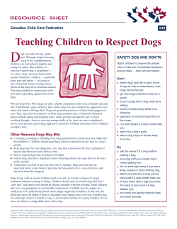


© Copyright 2026