
How to Catch a Membrane Protein in Action: A Review... Membrane Protein Immobilization Strategies and Their Applications
640 Chem. Rev. 2011, 111, 640–656 How to Catch a Membrane Protein in Action: A Review of Functional Membrane Protein Immobilization Strategies and Their Applications Virginie Fru¨h,†,‡ Ad P. IJzerman,† and Gregg Siegal*,‡ Division of Medicinal Chemistry, Leiden Amsterdam Center for Drug Research, Leiden University, Leiden, The Netherlands, and Leiden Institute of Chemistry, Leiden University, Leiden, The Netherlands Received March 4, 2009 Contents 1. Introduction 2. Noncovalent Interactions with the Whole Cell 2.1. Via Wheat Germ Lectin-Agarose for Whole-Cell ImmobilizationsMP: Glucose Transporter Glut1, P2Y1, and H1R 3. Noncovalent Interactions (Adsorption) with the Membrane 3.1. Via Adsorption to Polylysine-Ion Channel 5-HT3 Receptor and GPCR R1B-Adrenergic Receptor 3.2. Via Direct Adsorption of Native Attoliter Vesicles on Glass SlidessIon Channel: 5-HT3 Receptor 3.3. Via Native Membrane Hydrophobic Interactions on BeadssMP: Acetylcholine Receptor 3.4. Via Electrostatic Immobilization of ProteoliposomessMP: Glucose Transporter Glut1 3.5. Via Direct Lipid Distribution on GlasssMP: fd Coat and FXYD Proteins 3.6. Via Liposome Fusion into Lipid Bilayers on Platinum/Glass or Silicon SlidessGPCR: Bacteriorhodopsin 3.7. Via Microarrayed SurfacessGPCRs: Adrenergic Receptor, Neurotensin Receptor, and Dopamine Receptor 4. Covalent Interactions with Linker/Spacer to Membrane 4.1. Via Thiopeptide Tethered Membrane BilayerssMP: Cytochrome C Oxidase 4.2. Via Polymer Cushion-Supported Lipid BilayerssGPCR: Rhodopsin 4.3. In Vitro Synthesis in the Presence of a Tethered Lipid MembranesGPCR: OR5 4.4. Via Polymerized Lipid MonomerssGPCR: Rhodopsin 5. Covalent Interactions with Protein with Subsequent Lipid Reconstitution 5.1. Via Carbodiimide Coupling with Lipid ReconstitutionsGPCR: Rhodopsin 6. Specific Immobilization of Protein in the Native Membrane via Linker 640 642 642 642 642 645 645 646 646 647 647 648 648 648 649 649 649 649 650 * To whom correspondence should be addressed. Leiden Institute of Chemistry, Leiden University, Postbus 9502, 2300RA Leiden, The Netherlands. Phone: +31 (0)71 527 4543. Fax: +31-(0)71-5274609. E-mail: [email protected]. † Leiden Amsterdam Center for Drug Research, Leiden University. ‡ Leiden Institute of Chemistry, Leiden University. 6.1. Via Biotinylated Protein Bound to Streptavidin or AvidinsGPCR: Neurokinin-1 Receptor 7. Specific Immobilization of Detergent Reconstituted Protein via Linker 7.1. Via N-terminus of Protein on Sepharose ResinsMP: KcsA and DsbB 7.2. Via Tag and Biotinylated Antibody on BSA-Biotin SurfacessGPCR: β2-Adrenergic Receptor 7.3. Via HIS Tag on Quartz SurfacesMP: Ion Channel 5-HT3R 7.4. Via Biotinylated LigandsGPCR: Neurotensin Receptor-1 8. Specific Immobilization of Lipid Reconstituted Protein via Linker 8.1. Via Biotinylated Proteins in a Mixed Self-Assembled Monolayer (SAM)sGPCR: Rhodopsin 8.2. Via Lipid Bilayer Tethered through HIS-Tagged ProteinsMP: Cytochrome C Oxidase 9. Specific Immobilization of Alternatively Reconstituted Protein via Linker 9.1. Via Biotinylated AmphipolsVarious MPs 9.2. Via the N-terminus of Nanodisc Solubilized Protein MP: KcsA and DsbB 10. Perspectives 11. Abbreviations 12. Acknowledgments 13. References 650 650 650 651 651 652 652 652 653 653 653 653 654 654 655 655 1. Introduction Analysis of the results from the human proteome project suggests >30% of proteins are membrane bound.1 Membrane proteins (MPs) are in the core group responsible for signal transduction, including the G-protein coupled receptors (GPCRs), which represent 30-40% of targets for marketed drugs.2 It is needless to stress, therefore, the great commercial, industrial, and research value of MPs. There is growing interest in immobilizing MPs to various surfaces to create new biosensors or platforms enabling the study of their biological functions and identification of new leads for drug discovery. Soluble proteins have been readily immobilized through cross-linking aldehydes or thiols with protein amines or carboxyl groups for applications such as microarraying by printing.3-6 Functional immobilization of MPs requires consideration of their physiological needs, often dictated by the quality and components of the natural hydrophobic environment surrounding this class of proteins. 10.1021/cr900088s 2011 American Chemical Society Published on Web 09/10/2010 Functional Membrane Protein Immobilization Strategies Virginie Fru¨h carried out her undergraduate studies in Switzerland and in the U.K., where she graduated with a B.Sc. in coastal marine biology. She went on to work for Serono Laboratories in Switzerland, contributing to projects in both biochemistry and pharmacology. In 2009, she obtained her Ph.D. in biochemistry at the University of Leiden under the supervision of Dr. Ad. P. IJzerman and Dr. Gregg Siegal. She is currently a postdoctoral fellow at the Agricultural University of Bogor, Indonesia, working on the development of sustainable surfactants. Ad IJzerman is professor of medicinal chemistry at the Leiden/Amsterdam Center for Drug Research (Leiden University) and an expert on G proteincoupled receptors, in particular adenosine receptors. His team, together with the Stevens’ lab at the Scripps Institute, has recently achieved the structure elucidation of one of the adenosine receptor subtypes, the A2A receptor. He has a keen interest in novel concepts of intervening with receptor function, in particular allosteric modulation and residence time. The aim of this review is to focus on the immobilization chemistry applied to MPs and how these have enabled ever more complex assays from simple whole cell analyses to purified proteins on a chip, from the 1990s onward. With MPs, often the challenge is not the immobilization per se, but rather the requirements of the analysis and the protein itself with regards to protein availability and the type of lipid environment needed. Earlier studies of MPs, because of the difficulty in purifying them, involved whole cell or membrane fragment immobilization where native biological responses could be followed by “macroscopic” methods such as frontal chromatography (section 2). In these studies, immobilization was accomplished by simple techniques such as incorporation of membrane fragments upon swelling of gel beads during freeze-thawing7,8 or by coating beads with various cell-adhesive substances.9 Although these systems allow one to study the native environment, the immobilization itself is random and relatively unstable. Furthermore, reliable signal detection is often limited by high levels of nonspecific binding to the environment around the target MP. Chemical Reviews, 2011, Vol. 111, No. 2 641 Dr. Siegal obtained his Ph.D. at the University of Rochester in New York and pursued postdoctoral studies with Professor Kurt Wu¨thrich at the ETH in Zu¨rich, Switzerland, and Professor Paul Driscoll at the Ludwig Institute of Cancer Research in London, U.K. In 1997, he moved to Leiden University, where he received a Dutch Royal Society Fellowship to form his own research group. His current research interests include development of drug discovery technologies and their implementation in the drug discovery process. Recent efforts have focussed on application of biophysical ligand screening techniques to membrane proteins. Solubilized MPs may be either used as such in detergent micelles or reconstituted in various types of detergent or lipid formations.10-13 With recent advances in the development of mimics of the native membrane, however, the stability and formation of various lipid assemblies are becoming less problematic. However, one cannot emphasize enough how challenging these steps are. Removing the protein from its hydrophobic environment and reconstituting it into another is a difficult process that may damage the protein or result in less than optimal functionality.14,15 Furthermore, increasing complexity arises with increasing numbers of transmembrane segments.16 The most successful stories are currently based on the nicotinic acetylcholine receptor and rhodopsin, due to the availability of these MPs in large quantities and relatively good stabilities. Newer membrane mimics have allowed direct adsorption of appropriately prepared MPs to flat surfaces or chips made of glass, platinum-glass, and platinum-silicon to create monolayers of lipids that hold the protein onto the surface (section 2). Subsequent strategies have evolved to create even more stable lipid bilayers, as opposed to less stable monolayers, by fusing various types of solubilized MPs to lipid monolayers on surfaces. These methods provide more robust immobilization and are applicable to more sensitive studies, provided fluorescently or radioactively labeled components are available to specifically monitor MP activity. As a result, the highly sensitive detection of such signals does not require large amounts of solubilized MP. As these methods were applied to increasing numbers of MPs, it became clear that immobilizing membranes or mimics too close to a surface may restrict diffusion of solutes below the membrane and sterically hinder extramembranous protein dynamics. As a result, so-called solidsupported membranes have been developed, where the space below has been filled with hydrophilic polymers, combined with various linkers, to immobilize the MP in a manner that accommodates dynamic behavior. Solid-supported membranes are a better mimic of the natural environment because they create additional membrane fluidity and allow for mobility of large extramembranous protein domains17 (section 3). Many of these techniques have evolved in such a way that solubilization and purification of the protein may not be a prerequisite, but when this is the case, the 642 Chemical Reviews, 2011, Vol. 111, No. 2 solubilization from crude cell lysates or lipid reconstitution may be carried out in situ (section 4). On the detection side, earlier assays primarily used biological readouts. Although these techniques were sufficient for high-throughput or high-content assays of, for example, ligand binding and cell signaling activation, they typically required labeling of a protein or ligand to produce a signal. Recently, newer, so-called label-free technologies have emerged, such as surface plasmon resonance (SPR) and NMR,18 that are able to directly detect ligand binding, thereby rendering use of chemically, biologically, or radioactively labeled protein or ligands redundant. These technologies are sensitive to intermolecular interactions and capable of detecting weak binding affinities in the mM range (for NMR), provided the target protein is available in sufficient quantities and functionally solubilized. As opposed to soluble proteins, MP interactions with ligands, lipids, or other proteins are often specifically located on one side of the cellular membrane. Some strategies have therefore been taken one step further by using high-affinity interactions for specifically orienting MPs in the immobilized membrane on chips (sections 5-7). Techniques involving in vitro expression in the presence of the tethered lipid membrane in which the protein will be integrated directly upon expression19 are an elegant way to achieve oriented, immobilized MPs. As strategies to couple MPs become increasingly perceptive, a thorough summary of the numerous accomplishments is useful. Here, we present such a summary in the form of text and tables (see Tables 1 and 2) that focuses on the chemistries of various immobilization types, with descriptions of their applications and results. 2. Noncovalent Interactions with the Whole Cell 2.1. Via Wheat Germ Lectin-Agarose for Whole-Cell ImmobilizationsMP: Glucose Transporter Glut1, P2Y1, and H1R Fru¨h et al. The Kd of CB and D-glucose binding to Glut1 were comparable to values previously reported from studies on whole cells and membrane vesicles. This immobilization method also enabled comparison of the Kd of Glut1 for CB and D-glucose across different types of preparations of the reconstituted receptor, without any effects on the affinity. The advantage of this technique was that cells remained intact, in contrast to electrostatic adsorption, which causes hemolysis.15 The gel beads could be stored for one month but lost 40% of their capacity to bind cells. It was reported that it was not cell integrity but rather the capacity of cells to bind to the gel (and therefore the remaining functionality and density of membrane glycophorins) that determined the stability of the column, along with physical and chemical properties of the storage environment. Another advantage was that protein purification was not necessary, and manipulations involving column preparation and cell immobilization were simple. Nevertheless, the immobilization was sensitive to pressure changes, and gentle washing induced detachment of the cells, causing loss in reproducibility. Using this immobilization strategy with other applications will depend on conditions for cell homeostasis and functionality, as this technique was only optimized with respect to hematocyte cell lines. A variant of the whole cell immobilization technique involves immobilization of cytoplasmic membrane preparations on silica beads that have been derivatized with lipids. This socalled cellular membrane affinity chromatography has been used to noncovalently immobilize membrane suspensions from mammalian cells recombinantly expressing GPCRs. In particular, membrane preparations from a human astrocytoma cell line, 1321N1, expressing the P2Y1 purinergic receptor, wereimmobilizedonimmobilizedartificialmembrane-phosphotidylcholine (IAM-PC) columns.21 The authors first demonstrated specific binding of 2-MeSADP to the immobilized P2Y1 receptor and determined a Kd that compared well to literature values. Subsequently, they probed for the presence of endogenously expressed histamine H1 receptor by assessing the binding of Mepyramine. Mepyramine bound with a Kd of 2.9 nM, in good agreement with literature values, demonstrated that multiple GPCRs could be assayed by immobilization of membrane preps from a single cell line. The columns of immobilized membranes proved to be stable for 6-8 weeks. 3. Noncovalent Interactions (Adsorption) with the Membrane Frontal chromatography can be used to study interactions between a mobile phase and a stationary phase by regression analysis of the retention volumes. Furthermore, when whole cells are immobilized, this technique can allow the study of binding affinities of soluble ligands to MPs in their native environment. Immobilization can be achieved by adhering whole cells to agarose gel beads via wheat germ lectin (WGL).9,15 WGL is a derivative of wheat germ agglutinin and binds to N-acetylglucosamine and to N-acetylneuraminic acid on the extracellular face of red blood cell membranes.20 The Glut1 transporter targeted for these assays was indirectly immobilized to Sepharose 4B gel beads prederivatized with WGL by simply incubating cells overexpressing the receptor with gel beads.20 The interaction of Glut1 with D-glucose and cytochalasin B (CB, a fungal alkaloid that inhibits glucose transport) was measured by frontal chromatography. 3.1. Via Adsorption to Polylysine-Ion Channel 5-HT3 Receptor and GPCR r1B-Adrenergic Receptor Studying whole cells by fluorescence often leads to high background signals from the cytoplasm. This issue, combined 25 30 36, 37 1 43-45 46, 48 13, 56 61, 63 19 64 10 12, 73-75 79 12 88 89 93 94, 95 102 80 3.2 3.3 3.4 3.5 3.6 3.7 4.1 4.2 4.3 4.4 5 6 7.1 7.2 7.3 7.4 8.1 8.2 9.1 9.2 ref 9, 15 22-24 2.1 3.1 section R R O O O O O O O D R D R R R R O I R R I I I/R covalent coupling affinity capture affinity capture microprinting/affinity capture affinity capture affinity capture affinity capture covalent coupling in vitro expression/bilayer insertion cross-linking of bis-SorbPC by UV light microprinting/affinity capture affinity capture hydrophobic adsorbance hydrophobic tethers hydrophobic adsorption by printing freeze-thawing and hydrophobic glass adsorption hydrophobic, liposome adsorption onto monolayers freeze-thawing lectin-gel bead adsorption polylysine, silicon nitride interactions with native membrane native vesicle budding from whole cells hydrophobic adsorption method silver modified to bind nickel (NTA) biacore chip and polysterene beads sepharose resin with reactive aldehyde groups gold sensor chips streptavidin-coated chip glass modified to bind nickel (NTA) carboxymethyl-modified dextran glass slides chemically oxidized silicon sepharose resin with reactive aldehyde groups glass or gold surfaces cuvette gold-covered chromium layered glass carboxymethyl-modified dextran polymer hydrogel gold γ-aminopropylsilane (GAPS)-derivatized gold slides platinum/glass and platinum/silicon slides glass slides glass slides and/or functionalized glass polyvinyltoluene fluorophor microbeads superdex 200 gel beads Sepharose 4B gel beads polylysine coated glass or silicon nitride surfaces surface 5-carbon chainshydrophilic streptavidin-biotin protein HIS tag thiolipid and biotin streptavidin-biotin streptavidin-avidin + antibody + flag tag protein HIS tag streptavidin-avidin + antibody 5-carbon chainshydrophilic peptides polymer cushion thiopeptides tethers synthetic polymer Amphipol (Apol) nanodiscs nonaethyleneglycol monododecylether (C12E9) micelles CHAPS/DDM/CHS detergent self-assembled monolayers (SAMs) of w-hydroxyundecanethiol (HTA) reconstituted DPGPC DDM detergent DPC detergent reconstituted POPC lipid bilayer native membrane vesicles covalent peptidesDMPE fused with PC vesicles OG vesicles Triton X-114, Triton X-100, sodium cholate native membrane vesicles native vesicles or proteoliposomes reconstituted POPC/POPG synthetic bilayers formed by fusion of proteoliposomes to monolayers vesicles of DPPC/DMPC (4:1) egg-yolk PC native membrane vesicles native membrane native membrane native membrane membrane OmpA KcsA DsbB bacteriorhodopsin cytochrome C oxidase rhodopsin Neurotensin receptor-1 β2 adrenergic receptor with FLAG tag and fluorophore 5-HT3R biotinylated neurokinin-1 receptor, Gramicidin-A OmpA KcsA DsbB rhodopsin rhodopsin OR5 rhodopsin rhodopsin, acetylcholinesterase, cytochrome oxidase, nicotinic acetylcholine receptor adrenergic receptor, Neurotensin receptor, dopamine receptor cytochrome C oxidase Fd coat protein glucose transporter Glut1 acetylcholine receptor 5-HT3 receptor glucose transporter Glut1 5-HT3 receptor protein Table 1. Summary of Direct (D) or Indirect (I) Immobilization Methods with Random (R) or Oriented (O) Protein on the Surface, Including Type of Membrane and Protein Involved, In Order of Appearance in the Text Functional Membrane Protein Immobilization Strategies Chemical Reviews, 2011, Vol. 111, No. 2 643 22-24 25 30 36, 37 1 43-45 46, 48 13, 56 61, 63 19 64 10 12, 73-75 79 12 88 89 93 94, 95 102 80 3.1 3.2 3.3 3.4 3.5 3.6 3.7 4.1 4.2 4.3 4.4 5 6 7.1 7.2 7.3 7.4 8.1 8.2 9.1 9.2 ref 9, 15 2.1 section cytochrome C oxidase bacteriorhodopsin, OmpA, cytochrome b6f, bc1, NAChR OmpA, KcsA, DsbB Neurotensin receptor-1 rhodopsin β2 adrenergic receptor with FLAG tag and fluorophore ion channel 5-HT3R receptor rhodopsin biotinylated neurokinin-1 receptor OmpA, KcsA, DsbB rhodopsin OR5 rhodopsin cytochrome C oxidase rhodopsin, acetylcholinesterase, cytochrome oxidase, nicotinic acetylcholine receptor (nAChR) adrenergic receptor, Neurotensin receptor, dopamine receptor Fd coat protein glucose transporter Glut1 acetylcholine receptor ion channel 5-HT3 receptor NK1 receptor and ion channel 5-HT3 receptor glucose transporter Glut1 protein 2 100 µmol/mL 250 RU 100 nmol/mL 0.1 pmol/mm 100-400 receptors/um2 400 RU 50 ng/cm2 12.5-125 ng/mg 1750 receptors/µm2 44.6 µg/mL surface covered solubilized DPC DDM DDM, OTG, NMP, C8E4 nonaethylene glycol monododecyl ether detergent (C12E9) CHAPS/DDM/CHS no DDM DPC egg PC no no no Triton X-114, Triton X-100, sodium cholate, and saccharose no no OG, DPPA, DPPE, DPPC, and cholesterol cholate no no no no no one month several hours one month few months few months several weeks one month shelf life fragment-based drug discovery, binding assays binding binding studies, activation with agonist redox experiments binding experiments fragment-based drug discovery binding studies, activation under agonist binding potential for hybridization, kinetics, and binding binding studies, protein orientation isomerization and G-protein activation binding studies binding studies deorphanizing receptors, lead optimization and identification, bioassays, high-throughput screening binding studies and kinetics binding interactions, interactions with other components in the cell membrane binding studies, identification of orphan receptors binding, interactions, conditions structural solid-state, 15N labeled protein binding studies binding, interactions, conditions lipid and protein mobility in the native membrane, binding Interactions studies allowed NMR, fluorescence-based assays electrochemistry surface plasmon resonance SPR SPA total internal reflection fluorescence plasmon-waveguide resonance (PWR) spectroscopy SPR total internal reflection fluorescence NMR and fluorescence-based assays fluorescence microscopy SPR, impedance spectroscopy, AFM SEIRAS impedance spectroscopy fluorescence-based assays, radioligand binding assays* radioligand binding, electrochemistry, fluorescence solid-state NMR frontal chromatography fluorescence, fluorescence after photobleaching (FRAP), single-molecule imaging fluorescence, confocal microscopy, radioligand binding, fluorescence imaging radioligand binding frontal chromatography analysis Table 2. Summary of Protein Immobilization Solubilization Requirements, Shelf Life, Type of Analysis Obtained, And Type of Analytical Methods Applicable, In Order of Appearance in the Text 644 Chemical Reviews, 2011, Vol. 111, No. 2 Fru¨h et al. Functional Membrane Protein Immobilization Strategies with limited access to the intracellular compartment, obscures many of the dynamic processes between the membrane leaflets. To circumvent this, supported cell-membrane sheets allow one to observe target proteins from the intracellular side when both leaflets of a cell membrane are physically pulled apart.22,23 The ligand-gated ion channel 5-HT3 receptor and the R1B-adrenergic receptor were both overexpressed in HEK-293 cells, which were grown on poly-L-lysine (PLL)coated plastic. Another PLL surface was pressed down onto the cells and removed after several minutes of contact, ripping apart the cells and exposing the membrane’s intracellular face with endogenous lipids and proteins. To visualize the target lipids and proteins, the latter were expressed as fusion constructs with fluorescent proteins and the former were colocalized with fluorescent lipid markers. The supported polylysine membrane sheets have also been modified to allow access to the extracellular side by pressing down silicon nitride surfaces into which arrays have been cut out.24 Labeling with fluorescent probes from different sides of the membranes enabled the authors to study the mobility of lipids, GPCRs, and G proteins within and between the leaflets.23 Following the dynamics of heterotrimeric G protein partitioning into lipid anchor microdomains was also possible along with the mobility regimes of glycosylated transmembrane proteins. Competition binding studies using known, fluorescent ligands confirmed the functionality of the proteins studied. In comparison to wholecell analyses, the advantages of supported cell-membrane sheets are numerous. The mechanical stability is excellent, minimizing unwanted movement of the observed membrane upon application of stimuli, which can be problematic for, e.g., imaging applications. Although labeling is required, specific targets can be observed in their native surroundings, even maintaining the appropriate orientation, while all the time monitoring cellular processes such as signaling and protein translocation. Following populations of labeled proteins was therefore possible as a direct result of removing the intrinsically highly fluorescent cytosol during the preparation of these membrane sheets. This strategy is generally applicable to all cell membranes but is restricted to the use of labeled ligands or proteins. Adapting the immobilization to metallic surfaces may allow the strategy to be applied to surface plasmon resonance applications. Chemical Reviews, 2011, Vol. 111, No. 2 645 or via biotinylation of membrane components or target proteins.26,27 Attoliter vesicles can be obtained in one step, without changes in receptor function, orientation, or localization in the membrane. Vesicle formation is a simple process induced by incubating cells, in this case expressing the serotonin-gated mouse ion channel 5-hydroxytryptamine type-3 receptor (5-HT3R), with cytochalasin B (CB). This causes cells to produce tubular extensions that bud off when agitated. Native vesicles were thus formed that could be separated from whole cells by centrifugation. Usefully, the vesicles could be stored at 20 °C for weeks without causing loss of receptor function. Coexpression of cytosolic green fluorescent protein resulted in the possibility to monitor and characterize vesicle formation and the presence of cytosolic components. Specific binding of fluorescently labeled ligands could be used to locate the receptor in the vesicles, and radioligand binding assays revealed their appropriate orientation and function. Furthermore, agonist and antagonist ligands showed the same behavior with vesicles as with detergent-solubilized receptors. Finally, Ca2+ signaling within the vesicles upon agonist activation could also be detected, suggesting the functionality of the complete signaling pathway. These micrometer-sized vesicles are novel miniaturized reaction centers where receptor-based assays can be carried out in physiological conditions. These experiments not only revealed the native function of the receptors within these vesicles but also proved that other important players such as ion pumps are still present and functional. Vesicle formation yielded native behavior when tested with Chinese Hamster Ovary (CHO) or HEK293 cells, suggesting this procedure can be generally applied to other cell strains. When fluorescent ligands are available, these miniaturized and stabilized compartments can provide the basis for highthroughput assays where patterns of immobilization can reveal receptor function and binding affinities depending on various tailored reactive groups. 3.3. Via Native Membrane Hydrophobic Interactions on BeadssMP: Acetylcholine Receptor 3.2. Via Direct Adsorption of Native Attoliter Vesicles on Glass SlidessIon Channel: 5-HT3 Receptor Obtaining large amounts of MPs is often limiting, and therefore, immobilizing miniaturized vesicles containing the target protein can be useful for studying receptor-mediated cellular responses. In this particular case, Pick et al. have been able to produce vesicles straight from the native membrane of mammalian cells, with unprecedented attoliter volumes.25 The vesicles can be immobilized in different manners, either through direct lipid adsorption to glass slides Radioligand binding assays are often used to quantify binding affinities of ligands to MPs because only picomolar amounts of protein are required. However, this requires separation of bound from unbound ligands. To circumvent this, the scintillation proximity assay system (SPA)28,29 was developed, where receptors are immobilized on solid microspheres that contain scintillation fluid. This homogeneous assay provides specific information on radiolabeled ligand binding without the need for separation from unbound ligand, as scintillation is only stimulated by radioligand in close proximity to the microspheres.30,31 As a first attempt to apply SPA to MPs, membranes containing acetylcholine receptor were isolated from the electric organ of Torpedo californica 646 Chemical Reviews, 2011, Vol. 111, No. 2 and directly added to polyvinyltoluene fluorophore microbeads previously suspended in Triton X-100. Under these conditions, the membrane preparations spontaneously adsorbed to the beads. These beads contained scintillation fluid, and upon binding of the radiolabeled ligand to the receptor, the close proximity of receptor led to photon emission proportional to the amount of immobilized protein. In addition to obviating the requirement for separation of unbound from bound ligand, this method has less nonspecific binding than usually encountered in radioligand binding assays. Radioligand binding assays of 125I-R-bungarotoxin to the acetylcholine receptor enabled the study of various parameters such as dose response of acetylcholine and suberyldicholine with respect to radiolabeled R-bungarotoxin binding, providing apparent Ki values. The effects of different parameters such as concentrations of detergents and salts on the activity of the receptor were also studied, along with the detection of specific antigens by antibody binding. SPA has a number of other advantages for MPs. Because of the great sensitivity of scintillation counting, as little as 1 ng of receptor can be detected. Although SPA requires prior radiolabeling and modification of the protein or ligand, many receptor-SPA beads are available commercially. The method has been used in a broad range of studies, varying from identification and quantification of PCR products32 to highthroughput screening of inhibitors of DNA binding proteins33,34 to studying interactions between antibodies and antigens.35 Finally, because of the simplicity of the immobilization procedure, it should be generally applicable to all MPs. Fru¨h et al. kinetics were easily studied with such a setup due to lower nonspecific binding to the lipids in vesicles compared to whole cells. Immobilized membrane vesicles had 4 times more transport inhibitor (CB) binding capacity than proteoliposome gel beads, showing that the membrane vesicles contained a high density of Glut1. In the presence of dithioerythritol (DTE), the binding capacity of CB was >80% stable after 20-40 days in all types of columns studied. Furthermore, the activity could be controlled by inhibiting Glut1 with HgCl2 and restoring Glut1 activity by adding DTE, which has the effect of partially reversing the Hg2+ inactivation. Steric trapping should enable other applications such as studies of effects of ionic strength, pH, temperature, and lipid composition on ligand-binding characteristics. This methodology has the potential, as do other whole cell or membrane fragment-based applications, of enabling studies of interactions occurring within the native membrane because of the presence of other required membrane-bound components. However, applications of this immobilization strategy remain limited to the use of gel beads for steric trapping of the swelled vesicles, where high amounts of protein per mg of membrane are required for sensitive readouts. Furthermore, the noncovalent immobilization leads to degradation during use, and clearly, the protein to be studied needs to be stable to multiple freeze-thaw cycles. 3.5. Via Direct Lipid Distribution on GlasssMP: fd Coat and FXYD Proteins 3.4. Via Electrostatic Immobilization of ProteoliposomessMP: Glucose Transporter Glut1 Quantitative affinity chromatography used to be limited to studying interactions between soluble substances. By immobilizing a membrane protein on gel beads, however, binding of soluble substances to MPs could be quantified by regression analysis of the retention volumes for different concentrations of ligands. To study interactions between soluble substances and the glucose transporter Glut1, membrane vesicles containing the latter were immobilized onto gel beads.36,37 Membrane vesicles were obtained by stripping red blood cells of peripheral proteins and partially solubilizing them with octylglucoside (OG). In some cases, the vesicles were purified by ion-exchange chromatography for reconstitution of the Glut1 transporter into proteoliposomes.38 It has been reported that freeze-thawing39 and freeze-drying with subsequent rehydration40 can cause vesicles or proteoliposomes to fuse together and form larger particles. This approach was used to sterically trap vesicles after two cycles of freezing in ethanol/CO2 and thawing in the presence of gel beads.36 Using the sterically trapped liposomes, specific interactions between Glut1 and CB, and between Glut1 and D-glucose,41 were observed. Ligand binding affinities and The biological role of the protein coating filamentous fd bacteriophages (fd coat protein) involves interacting with the host’s cellular membrane. Three-dimensional structure determination is a powerful method to determine molecular mechanisms of protein function. However, in many cases, solution NMR studies of an MP in detergent micelles will not provide sufficient information because the dynamics and orientation of the protein in detergent micelles may be very different than in a membrane bilayer. Therefore, the fd coat protein was immobilized onto glass slides in phospholipid bilayers with one simple step for structural studies using solid-state NMR.1 For these studies, 15N-labeled fd coat protein was produced in E. coli and solubilized in detergent micelles. The solubilized fd coat protein was then reconstituted into phospholipid vesicles consisting of a mixture of palmitoyl oleoyl phosphatidylcholine/palmitoyl oleoyl phosphatidylglycerol (POPC/POPG).42 Glass slides were covered with the vesicle suspension containing the reconstituted protein, and the bulk water was left to evaporate. The slides were then stacked and bathed in saturated ammonium phosphate solution. Finally, the slides were placed in thin polyethylene tubing and inserted into a rotor for NMR studies, which allowed complete resolution of the amine resonances in a 3D magic angle spinning (MAS) NMR correlation spectrum. The choice of lipids determines the Functional Membrane Protein Immobilization Strategies level of bilayer orientation on the slides and allows control over the horizontal and vertical spacing between phospholipids.11 The presence of unsaturated chains leads to more fluid bilayers, and larger vertical spaces between bilayers can be obtained by incorporating negatively charged lipids.11 The noncovalent, hydrophobic interactions with the glass were strong enough to hold the vesicles immobile on the substrate for the duration of an NMR experiment, despite the considerable shear forces generated by spinning the sample. No details were given on the protein stability or functionality, but it was fully incorporated into phospholipid bilayers onto the glass support. This immobilization strategy allows a wider range of MPs to be studied by solid-state NMR and may be used in other applications compatible with glass surfaces. The extensive vesicle preparation was a complicated process, however, and may not be generally applicable to other MPs. Although fd coat protein could be obtained in relatively large quantities, this is rarely the case with other MPs. Further, it may prove difficult to adapt this method of orienting the protein to new, small-volume MAS rotors. 3.6. Via Liposome Fusion into Lipid Bilayers on Platinum/Glass or Silicon SlidessGPCR: Bacteriorhodopsin For an immobilized MP to remain functional, the fluid lipid or detergent environment must not become too rigid upon immobilization or it might impede the dynamic behavior of MPs. Lipid rigidification can be minimized when proteoliposomes are immobilized by fusion with monolayers on glass surfaces to form bilayers without any chemical cross-linking. To demonstrate the approach, bacteriorhodopsin from purple membranes of Halobacterium halobium was added to a lipid, cholesterol, and octylglucoside mixture, and upon removal of octylglucoside during gel filtration, proteoliposomes were formed by the so-called detergentdepletion technique.43 Langmuir-Blodgett (LB) lipid monolayers44 were used to treat platinum/glass surfaces and subsequently converted into bilayers when fused with the bacteriorhodopsin proteoliposomes.45 Bacteriorhodopsin photoactivity in the reconstituted bilayers, monitored by electrochemistry, was comparable to natural membrane fragments containing bacteriorhodpsin on platinum surfaces. Three other MPs were also studied, including acetylcholinesterase from bovine brain, cytochrome oxidase from bovine heart, and the nicotinic acetylcholine receptor from the electric organ of Torpedo. The purification and incorporation into proteoliposomes of the three MPs varied depending on suitable detergents, but they were all incorporated into stable lipid bilayers following the same protocol. Acetylcholinesterase enzymatic activity was easily studied by fluorescence measurements despite low immobilization yields, but only semiquantitative data was available for cytochrome C oxidase, whereas studies of the nicotinic acetylcholine receptor Chemical Reviews, 2011, Vol. 111, No. 2 647 with its ligand 125I-R-bungarotoxin were hindered by issues of nonspecific binding. Although this technique provided a surface with a fluid membrane environment, meeting criteria for efficient molecular assays and potentially providing access to printing arrays, the usual challenge of obtaining functional protein in proteoliposomes remains. The bilayers did remain immobilized to the supports for prolonged periods of time after storage, but no information was provided on the stability of the immobilized MPs. 3.7. Via Microarrayed SurfacessGPCRs: Adrenergic Receptor, Neurotensin Receptor, and Dopamine Receptor Reducing the demand for protein and reagents has been one of the strategies used to address the limitations faced when MP purification and solubilization result in low yields. This limitation is even more critical when MP immobilization is at the heart of high-throughput screening for drug discovery. An innovative way of addressing both limitations has been to immobilize GPCRs through microarray printing assays.46,47 A nice example of this technique has been developed at Corning. Membranes in vesicular solutions of dimyristylphoshatidylcholine (DMPC) and 1,2-dimyristoyl-sn-glycerophosphatidylethanolamine (DMPE) or egg-yolk PC were printed via a robotic-pin printer on γ-aminopropylsilane (GAPS)-derivatized gold surfaces.48,49 The details of the methods used with GAPS are proprietary to Corning. GAPS-coated slides exhibited high mechanical stability during printing, with no dependence on lipid phases, thereby overcoming the desorption encountered when withdrawing the slides through air.50 While covalently attaching membranes has been noted to impede lateral fluidity of membrane lipids, and consequently protein function, noncovalent printing techniques, through membrane adsorption onto glass slides, have proved to preserve GPCR and G protein functionality. Thus, binding constants of known ligands were readily obtained, as well as compound selectivity between and within GPCR families. However, the assay in this format could not be used to differentiate between agonists and antagonists. Arrays of the β1-adrenergic receptor, the Neurotensin receptor (NTR1), and the dopamine (D1) receptor have been created. As an example, upon incubation with fluorescently labeled neurotensin, fluorescence was restricted to areas in which NTR1 had been immobilized, as expected. Binding constants consistent with literature were also determined for known small-molecule ligands using the printed array.48,49 A recent exciting advance in printing is the adaptation to porous substrates onto which the membranes are transferred, allowing access to both sides of the membrane.51 Array printing is expensive because of the costs involved in acquiring such printers and also in time spent to optimize procedures that involve correct buffer compatibility, ligand specificities, 648 Chemical Reviews, 2011, Vol. 111, No. 2 affinities, and association and dissociation rates. However, although at present microarrays have been analyzed with fluorescent or radiolabeled46 ligands, they can in principle be adapted for other applications such as SPR and electrochemistry, providing the surfaces are compatible. Information on cell-surface interactions and ligands for orphan receptors might also be derived from these microarrays, potentially enabling discovery and validation of new lead molecules and high-throughput screening. One interesting variant of this idea is to generate a self-sorting array. Bailey and colleagues printed arrays in which oligonucleotides of differing sequence were localized onto appropriately prepared surfaces.52 Liposomes were generated from Sf9 cells recombinantly expressing either the H1 histamine or M2 muscarinic receptor. Subsequently, cholesterol conjugated to an oligonucleotide complementary to one of the immobilized oligonucleotides was allowed to bind the liposomes. Incubation of a mixture of liposomes with the printed surface resulted in sequencedependent immobilization of the GPCRs at the expected sites. The GPCRs immobilized in such a manner bound known ligands with expected affinities. 4. Covalent Interactions with Linker/Spacer to Membrane Fru¨h et al. been established before, but it was measured here for the first time using impedance spectroscopy. This immobilization strategy proved to be compatible with sensitive measurement of proton transport through the lipid bilayer. Furthermore, kinetics and binding interactions of cyanide with CcO could be studied. Because of the reported difficulty in preparing thiopeptide or thiolipid compounds in the bulk phase, this strategy allowed the simplified formation of a robust tethered bilayer in steps. Furthermore, the tethered bilayer allowed surface analytical techniques involving electric currents due to the metal surface used as a support, suggesting the technique may be adaptable to other applications with such requirements. Again, the challenge is to find the appropriate solubilization condition for the protein, but the space conferred between the surface and the bilayer environment of the protein by the tethered membrane bilayer can minimize steric hindrance of large, dynamic MPs and allow smallmolecule transport and diffusion. 4.2. Via Polymer Cushion-Supported Lipid BilayerssGPCR: Rhodopsin 4.1. Via Thiopeptide Tethered Membrane BilayerssMP: Cytochrome C Oxidase SPR applications require flat, metallic surfaces upon which proteins must be immobilized at very high densities and, ideally, as close to the surface as possible. However, studies of immobilized MPs have revealed the need to create space between the protein and the surface to reduce steric hindrance of MP dynamics. Therefore, molecules such as thiolipids53 and thiopeptides have been developed as linkers, also called tethers, because they couple the membrane in close proximity to the surface but allow the membrane to remain fluid. Thiopeptides have been used previously54,55 as a more hydrophilic alternative to thiolipids, for example, to immobilize cytochrome C oxidase (CcO).13,56 In this particular case, a thiopeptide-lipid monolayer was covalently linked to gold surfaces with subsequent incorporation of the MP within the lipid bilayer. An oriented peptide monolayer was created by covalently binding an N-terminally thiolated, oligoserine peptide to a gold surface.57 To complete the tethered membrane mimic, vesicles obtained by dialyzing Lipoid Egg PC58,59 were incubated with the monolayer surfaces to form the thiopeptide-lipid bilayer by adsorption. CcO, previously solubilized with Triton,56,60 was added to the cell containing the thiopeptide-lipid bilayer surface and spontaneously became incorporated into the bilayer with the micellar dilution technique. All steps of the process could be monitored by SPR, and final thicknesses of bilayers could be measured. Active proton transport driven by CcO had As discussed, previous studies revealed how immobilizing lipid bilayers containing MPs too close to the surface may hinder protein dynamics and membrane stability. An alternate solution to tethered membrane bilayers has been the introduction of a hydrophilic polymer cushion interspaced with hydrophobic anchors below the lipid bilayer. The cushion, formed by a hydrophilic polymer interspaced with long alkyl chains, provides a lubricating surface that allows the lipid bilayer to remain mobile and provides a better mimic of the native environment.61,62 The gold L1 SPR chip used for such polymer-supported lipid layers consisted of a covalently linked, carboxymethyl-modified, dextran polymer hydrogel where a large part of the sugar moieties were grafted with lipophilic alkyl side chains to which liposomes adhere.61,63 Whereas the preparation of small unilamellar vesicles (SUVs) containing rhodopsin used to be necessary,63 here, fusion of crude rhodopsinenriched membranes directly onto Biacore L1 chips was used instead of purification.61,63 SPR, atomic force microscopy (AFM), and electrochemical impedance spectroscopy (EIS) were used to follow receptor immobilization. The receptors remained mobile in the plane of the lipid, suggesting that this strategy has the potential to be used to study receptor dimerization or interaction with other proteins.63 Furthermore, the chips could be used directly, without prior treatment, and repeatedly with different receptors after washes with detergents to remove earlier immobilized proteins. This may be generally applicable to other systems as the strategy allows on-surface enrichment for MPs with low expression yields. Functional Membrane Protein Immobilization Strategies Chemical Reviews, 2011, Vol. 111, No. 2 649 4.3. In Vitro Synthesis in the Presence of a Tethered Lipid MembranesGPCR: OR5 Biophysical analysis requires pure protein preparations functionally immobilized to a surface, and obtaining such conditions for GPCRs remains challenging due to loss of functionality and refolding problems typically encountered with GPCR solubilization and purification. To address this limitation, a new strategy that uses in vitro GPCR synthesis in the presence of a previously established,54,55 solidsupported, peptide tethered lipid membrane (tBLM) has been developed.19 Using an approach similar to that described in section 3.1, a hydrophilic peptide spacer was covalently attached to gold slides through amino-terminal thiol groups. The carboxyl group of the peptide was subsequently activated and amino-coupled to dimyristoyl-l-R-phosphatidylethanolamine (DMPE), forming a lipid monolayer. The monolayer was subsequently fused with lipid vesicles to create the final bilayer. The odorant receptor, OR5 from Rattus norVegicus, was expressed in vitro directly on the sensor surface.19 This caused spontaneous integration of the GPCR into the tBLM upon biosynthesis. The procedure was monitored by surface plasmon enhanced fluorescence spectroscopy (SPFS) where fluorescent antibody binding to the GPCR created signals when the fluorophore was close to the surface. The OR5 receptor was successfully oriented on the surface in such a way that it mimics the orientation in the native endoplasmic reticulum, with the N-terminus facing the extracellular side. Ligand binding was monitored by surface-enhanced infrared reflection absorption spectroscopy (SERIAS) where absorbance differences were linked to effective binding of lilial, a small hydrophobic ligand for OR5. Details were not provided concerning the stability of such slides, and although this application requires fluorescently labeled ligands, the success with GPCRs, which tend to be among the most challenging MPs due to conformational heterogeneity, instability, and low expression yields, suggests that the procedure has potential to be generally applied to other MPs and to analytical methods that do not necessitate labeling, such as SPR. developed to provide a more stable environment.64 A planarsupported lipid bilayer (PSLB) composed of 1,2-bis[10-2′,4′(hexadienoyloxy)decanoyl]-sn-glycero-3-phosphocholine (bisSorbPC)65 was used for these studies. The bis-SorbPC can be covalently cross-linked by exposure to UV light, providing the necessary resistance to washing. Rhodopsin was solubilized in buffer containing OG for these experiments.66,67 Reconstitution of rhodopsin into the PSLBs was carried out in a plasmon-waveguide resonance (PWR) cell. The PWR cell was set up in such a way that UV light could be directed to it from a Mercury lamp, with a band-pass filter to remove all visible light, which would irreversibly photoactivate rhodopsin. More than 95% of the bis-SorbPC was polymerized, as monitored by UV absorbance. PWR spectroscopy was used to characterize rhodopsin immobilized in the crosslinked PSLBs. Within the PSLBs, the opsin isomerization and G protein activation could be monitored and rates were found to be similar to those obtained in a fluid dioleoylphosphatidylcholine (DOPC) bilayer. MP immobilization via this procedure has the advantage of being completed in a few hours, resulting in a system with intact and functional immobilized protein. Although no details were given on the reversibility of this polymerization, it rendered the lipids resistant to Triton X-100 treatment. Although this method has great potential for on-surface purification and immobilization of other GPCRs, preserving their functionality in such a cross-linked environment may not be as easy to achieve. 5. Covalent Interactions with Protein with Subsequent Lipid Reconstitution 5.1. Via Carbodiimide Coupling with Lipid ReconstitutionsGPCR: Rhodopsin 4.4. Via Polymerized Lipid MonomerssGPCR: Rhodopsin Rhodopsin has been used extensively to study efficient immobilization into lipid environments, but in general, lipids have been shown to be unstable when used for immobilizing MPs on chips. This is because the hydrophobic interactions that keep the lipids adsorbed to the glass are not strong enough to resist eventual desorption after extensive washing. To address this issue, cross-linked synthetic lipids were GPCR biosensor studies require high densities of pure and functional immobilized protein, and this is the limiting factor with regards to most GPCRs. To address this limitation, a technique was developed in a flow cell to allow stepwise addition and removal of mixed detergent and lipid micelles and protein to create an on-surface lipid bilayer reconstituted around a solubilized GPCR.10 In principle, large quantities 650 Chemical Reviews, 2011, Vol. 111, No. 2 of unfolded GPCR could be used as input for this approach. The gold L1 sensor chip was treated to create covalently linked carboxymethyl-modified dextran polymer with random glucose moieties substituted by alkyl groups.63 The alkyl groups hydrophobically bound to mixed micelles of OG detergent and POPC lipid as they were injected over the surface to create the initial lipid layer. OG-solubilized rhodopsin53 was injected and immobilized on the surface by both the amide chemistry and the strong hydrophobic interaction between the surface lipid layer and the OG micelles. The immobilized rhodopsin was immediately reconstituted in a POPC bilayer by injection of mixed OG and POPC micelles over the surface. The technique makes use of the high critical micellar concentration (CMC) of OG (25 mM). As buffer is injected over the flow cell, OG monomers detach much quicker than POPC, and as they detach and wash away, the remaining POPC micelles spontaneously fuse into a continuous lipid bilayer over the sensor surface and the immobilized rhodopsin. The functionality of rhodopsin after immobilization was tested by surface plasmon resonance with light activation and measurement of transducin dissociation from the membrane as it consumed guanosine 5′-triphosphate (GTP).53 Signaling was only detected in the sample flow cell containing rhodopsin-POPC. There was no signal in the absence of lipids, which proved that their presence68 and correct reconstitution69 was necessary for the correct function of rhodopsin.10,70 The ligand-binding capacity of the receptor was therefore preserved through this immobilization procedure and could be repeatedly measured. This technique resulted in reasonably high densities of up to 4 ng/mm2 of immobilized protein, while the use of a polymer hydrogel layer improved the stability of the immobilized bilayer. Although protein solubilization and lipid reconstitution were fast and straightforward via the commercially available Biacore systems, defining the correct lipid and detergent stoichiometries for new target proteins will require significant optimization.71 The technique, however, can be adapted to different immobilization chemistries, including high-affinity interactions with antibodies to capture the protein in a defined orientation72 (see section 7.2). 6. Specific Immobilization of Protein in the Native Membrane via Linker Fru¨h et al. ligand to its binding site. Specific interactions between the protein or membrane and the surface have been used in place of random ones for better control of the protein’s orientation. The most common strategies involve the biotin interaction with streptavidin or avidin.12,73-75 In one case, a biotinylated Neurokinin-1 receptor was overexpressed in CHO cell lines and immobilized through a streptavidin linker to a biotin covered slide.73 The procedure was very simple, consisting primarily of treating the quartz slides with biotinylated bovine serum albumin (BSA), which results in a surface resistant to salt and detergent washes. The multivalent streptavidin was then used to bridge the biotinylated GPCR to the biotinylated BSA for immobilization. The cell lysates containing overexpressed C-terminally biotinylated Neurokinin-1 receptor were then injected into the chamber directly to yield a specifically oriented immobilization of the receptor. Total internal reflection fluorescence (TIRF) was used to detect binding of streptavidin to the BSA-biotin surface and to detect binding of the fluorescently labeled substance P (SP), a Neurokinin-1 receptor agonist, to the oriented receptor. Binding studies revealed functionality of the receptor in accordance with previous studies76 but used as little as 1 attomol of receptor due to the controlled orientation and high sensitivity of TIRF. This simple immobilization was the first example of biotinylated MP on quartz surfaces directly from crude cell lysates without purification, thus allowing one to study, e.g., mechanisms involved in GPCR and G protein interactions in the native system. This technique has the potential to be applied to a wide array of applications, and if higher densities of proteins are needed for readouts, on-surface purification and enrichment is possible because of the high affinity of the immobilization reagents. It was also suggested that integration of biotinylated lipids into the plasma membrane of cells prior to homogenization could be an alternative, and in some cases, chemically oxidized silicon surfaces may be used to avoid interactions with the lipids.74 7. Specific Immobilization of Detergent Reconstituted Protein via Linker 7.1. Via N-terminus of Protein on Sepharose ResinsMP: KcsA and DsbB 6.1. Via Biotinylated Protein Bound to Streptavidin or AvidinsGPCR: Neurokinin-1 Receptor Many MPs have specific roles in binding extracellular ligands. Therefore, when immobilizing such proteins to study their biological activity, it is desirable to orient them in a controlled manner so as to maximize the accessibility of the Fragment-based drug discovery, an approach that consists of screening small molecules (<300 Da) with promising bioavailability properties that weakly interact with a pharmaceutical target,77 has gained considerable attention in the pharma industry. Sensitive techniques, such as crystallography and NMR, are required to detect the weak binding of fragments to the target. However, many biophysical techniques require large quantities of stable protein in a pure form, and that is generally not possible with many MPs. Although 60% of all drugs target MPs,78 it has not proven Functional Membrane Protein Immobilization Strategies possible to apply fragment methods to them. Target immobilized NMR screening (TINS)79 has addressed some of these limitations, such as the issue of nonspecific binding of fragments to surfactants or lipids, and shows promise as a fragment screening method on MPs. In TINS, 1D NMR spectra of the fragments are simultaneously acquired in the presence of immobilized reference and target MPs in order to detect specific binders above the nonspecific level of fragments interacting with detergents. All proteins in this case were solubilized in dodecylphosphocholine (DPC) during the purification procedure, which involved simple metal affinity chromatography followed by gel filtration in the presence of the detergent. The immobilization procedure used Schiff’s base chemistry between primary amines of the protein and aldehyde groups on the commercially available sepharose resin. The immobilization efficiencies were reported similar to previous studies on soluble proteins, with a final concentration equivalent of 100 µM of both the reference and target proteins on the resin. The functionality of immobilized DsbB was confirmed by an enzymatic assay, which indicated that immobilized DsbB retained 90% of its functionality. The functional immobilization of both KcsA and DsbB was further demonstrated by detecting binding of known ligands using TINS.79,80 TINS addresses the protein demand by reusing a single sample of the target to screen the entire library, thereby requiring <50 nmol of protein. Immobilization can be achieved via the N-terminus or a variety of other chemical strategies, making it potentially broadly applicable to MPs. OmpA has been noted to retain conformation in a variety of surfactants,81-85 making it a useful reference. Although the method enabled fragmentbased drug discovery on MPs, there remained several limitations. A full screen of 1000 fragments required a week to complete, and most MPs may not be stable in such conditions, although, in some cases, immobilization has proven to improve MP stability. Furthermore, the solubilization processes must still be tailored to each protein. However, alternative solubilization media (see section 9) such as Amphipols81 and Nanodiscs,14,86 which appear to be compatible with TINS, offer the possibility to generalize the procedure. Chemical Reviews, 2011, Vol. 111, No. 2 651 (β2AR) was specifically labeled with fluorescein at Cys265, a conformationally sensitive site. The specific labeling of only Cys265 by the sulfhydryl-reactive fluorescent probe fluorescein maleimide was possible because the other cysteines of the protein were either inaccessibly located in the transmembrane domains or nonreactive because of the presence of disulfide bonds.87 For exploitation of the biotin-avidin interaction, two strategies were used in conjunction with an avidin or streptavidin linker. In the first, the protein was modified with an N-terminal FLAG epitope and indirectly linked to the avidin surface through a biotinylated anti-FLAG antibody. In the second strategy, a second cysteine residue was biotinylated and linked to the surface through avidin or streptavidin as in section 5. Liganddependent activation of the β2AR was detected with fluorescence microscopy by monitoring changes in fluorescence intensity upon ligand binding and receptor translocation. The β2AR immobilized via the antibody displayed nearly identical responses to an agonist as the receptor in solution. The β2AR directly immobilized by the biotin tag yielded less consistent responses that were significantly smaller than for the receptor coupled via the antibody. Whether this was an artifact of the immobilization itself or due to biotinylation of the GPCR was not clear. This method did not require protein reconstitution into a lipid bilayer and could immobilize protein with minimal loss due to the high-affinity coupling. Although this detection method required the GPCR to be labeled with a fluorophore at a sensitive site, the immobilization strategy allowed the authors to study conformational changes of the protein and is compatible with array technologies involving high-throughput screening of MPs on chips for example, provided solubilization conditions have been found for a particular MP. 7.3. Via HIS Tag on Quartz SurfacesMP: Ion Channel 5-HT3R 7.2. Via Tag and Biotinylated Antibody on BSA-Biotin SurfacessGPCR: β2-Adrenergic Receptor High-affinity interactions have been used successfully to immobilize GPCRs in their native membrane. However, many applications require pure protein to provide results with highsignal-to-noiseratios.Here,thehigh-affinitystreptavidin-biotin interaction has been used to capture detergent-solubilized GPCRs in a controlled and oriented manner and monitor activity by fluorescence microscopy.12 To detect ligand binding, the detergent-solubilized β2-adrenergic receptor Many applications require pure protein immobilized at very high density. Immobilized metal affinity chromatography (IMAC) has been used to successfully immobilize the detergent-solubilized serotonin-gated ion channel 5-HT3R to quartz slides without the need for further lipid reconstitution.88 This approach may be generally applicable to all MPs that can be solubilized and functionally expressed with a HIS tag. Before immobilization, the glass slides were first modified to contain thiol groups by gas-phase silanization. The thiol groups were then covalently linked to a lysine derivative of nitriloacetic acid (NTA) using a bifunctional maleimide-succinimide cross-linker. The newly created surface was fused to a Teflon spacer to create a flow-through cell and was subsequently charged with Ni2+. The 5-HT3R ion channel, containing a HIS tag, was solubilized in nonaethyleneglycol monododecyl and immobilized via chelation to the Ni2+-NTA. The metal affinity-based immobilization procedure was reversible, allowing quantifica- 652 Chemical Reviews, 2011, Vol. 111, No. 2 tion of immobilized receptors by elution with imidazole. Total internal reflection fluorescence spectroscopy was used to carry out competition binding experiments using the nonlabeled competitor quipazine against GR-fluorescein ([1,2,3,9-tetrahydro-3-[(5-methyl-1H-imidazol-4-yl)methyl]9-(3-amino-(N-fluoresceinthiocarbamoyl)propyl)-4H-carbazol-4-one]). The affinity of GR-fluorescein for the receptor in solution was identical to the surface-bound protein, demonstrating the feasibility of the process for identifying potential new drugs and quantifying their affinities with dose-response curves. This procedure was extremely simple and could potentially be applied to a variety of analytical techniques, given the usual constraints that the MP can be functionally solubilized and expressed with a HIS tag. The current application is limited to fluorescent ligands, but the sensitivity of the methods results in signal detection with as little as 1.6 attomol of immobilized MP, corresponding to the yield of a single mammalian cell! The detection is realtime, mass-independent, and can be combined with microfluidic applications to further explore high-throughput analysis for drug discovery. Further, given the nature of the immobilization surface, it seems likely that this procedure could be readily adapted to SPR applications. 7.4. Via Biotinylated LigandsGPCR: Neurotensin Receptor-1 SPR analysis of GPCRs is often limited by the fact that low molecular weight ligands do not generate large signals. However, by immobilizing the ligand instead, the method allows one to study GPCR binding to biotinylated ligands immobilized to streptavidin-covered Biacore chips,89 without the need for lipid reconstitution. For these studies, the Neurotensin receptor-1 was expressed in E. coli as an N-terminal fusion product with the maltose binding protein and a C-terminal His tag for stability and purification purposes. Receptor purification by IMAC and neurotensin affinity chromatography preceded solubilization in detergent micelles containing (3-[(3-cholamidopropyl)dimethylammonio]-1-propanesulfonate), n-dodecyl-β-maltoside (DDM), and cholesteryl hemisuccinate (CHS). The peptide ligand Neurotensin (NT) was N-terminally biotinylated and immobilized onto the streptavidin-coated Biacore chips as the positive control. A ligand containing a scrambled version of the primary structure was also synthesized and immobilized on a second streptavidin-coated chip as a negative control. Binding was monitored by SPR by flushing the flow cells with the detergent-solubilized receptor, and highly specific interactions could be observed and confirmed. The chips could be regenerated by uncoupling bound receptors with high salt washes. The authors reported lower amplitude response than expected at higher levels of immobilized ligand, which they explained by potential ligand occlusion that could be prevented by the use of a longer linker between the surface and the ligand. Although no quantification of the binding affinities was provided, the technique has potential Fru¨h et al. in array technology provided small molecular weight ligands are available and biotinylation does not affect their binding affinities to the target protein studied. Further, the technique could also be adapted to imaging-mode SPR.90 Although this technique was possible with detergent-solubilized protein, the limitation of finding appropriate detergents for protein solubility remains a protein-specific issue. 8. Specific Immobilization of Lipid Reconstituted Protein via Linker 8.1. Via Biotinylated Proteins in a Mixed Self-Assembled Monolayer (SAM)sGPCR: Rhodopsin To study MPs, which specifically bind ligands from the extracellular side of the membrane, methods have tended toward developing controlled, oriented immobilization in order to expose the appropriate side of the protein to the ligands. For such a controlled immobilization, a specific chemistry reacting to the appropriate terminus of the protein is necessary. Here, carbohydrate-specific chemistry91,92 for N-terminal biotinylation of glycosylated proteins was combined with thiolipids to anchor rhodopsin to gold surfaces.93 Gold sensor chip surfaces were covered with a homogeneous self-assembled monolayer (SAM) of ω-hydroxyundecanethiol (HTA), interspaced with biotin attached to the surface through thiol groups, by micropatterned printing. The HTA layer was created to avoid protein immobilization in these areas. Subsequent addition of BSA blocked all nonspecific binding sites, and addition of streptavidin provided appropriate high-affinity binding sites for the biotinylated receptor. The glycosylated receptor was specifically biotinylated on the carbohydrate chains near the N-terminus by oxidizing the carbohydrate moieties with NaIO4 prior to adding biotin hydrazide.91 Because only the extracellular facing portions of the receptor are glycosylated, rhodopsin was immobilized with the intracellular side facing away from the surface, allowing for maximal interaction with G proteins. Surface plasmon resonance in the presence of GTP demonstrated the possibility of studying the receptor’s constitutive activity. Interaction with the G protein was directly observed after a flash of light, and the initial slope of the desorption signal was a good approximation of receptor density/unit area of surface. Relaxation of the activated G protein was also measured, and binding of agonists such as 11-cis- or 9-cisretinal was used to show that the immobilized rhodopsin was functional. This immobilization strategy stabilized the GPCR for several hours and multiple cycles of ligand addition and removal, for which activation could easily be monitored. Furthermore, due to micropatterns with and without the receptor, nonspecific binding was calculated as a localized reference, adding robustness to the data not otherwise available in SPR analyses. Although this particular biotiny- Functional Membrane Protein Immobilization Strategies Chemical Reviews, 2011, Vol. 111, No. 2 653 lation method may be limited to MPs with glycosylated regions near the targeted terminus, it is an attractive method for studying G protein mechanisms. However, when applying MPs in native environments, SPR often proves to be less sensitive than when used with the well-behaved and highly overexpressed proteins such as rhodopsin. properties are more compatible with modern techniques such as structural biology or biophysical MP-ligand characterization. Amphipols and nanodiscs, which have been used for MP immobilization, are discussed here, whereas a third, SMALPs,101 shows promise but has yet to be used for immobilization. 8.2. Via Lipid Bilayer Tethered through HIS-Tagged ProteinsMP: Cytochrome C Oxidase 9.1. Via Biotinylated AmphipolsVarious MPs Studying MPs by electrochemistry is often limited by the insulating properties of the lipids or detergents if they are applied directly onto the metallic surface. For detection of electron transfer, the protein therefore needs to be immobilized away from the surface. Here, a combination of lipid reconstitution and tethering the target protein through a HIS tag was used to immobilize cytochrome C oxidase (CcO) to a silver surface.94,95 The surfaces were roughened by electrochemical processes and functionalized with Nhydroxysuccinimide ester (NHS) groups by addition of dithiobis-(N-succinimidyl propionate). An ion-chelating layer of nitrilotriacetic acid groups (NTA) was finally created by binding the terminal amino groups of N-(5-amino-1-carboxypentyl) iminodiacetic acid to the existing NHS layer. Complexation of this new NTA monolayer layer with Cu2+ ions made it possible to reversibly immobilize HIS-tagged proteins. Immobilization was simply accomplished by flowmediated addition of C-terminally HIS-tagged CcO in DDM. Finally, the lipid bilayer was established around the receptor by incubation with a buffer solution of 1,2-diphytanoyl-snglycero-3-phosphocholine (DPGPC) with subsequent removal of DDM. The heme groups of CcO remained intact and functional upon immobilization, as the ferric and ferrous states of hemes a and a3 could be monitored repeatedly after electrode potential changes. The electron transfer rates of the protein were found to remain unchanged after immobilization, demonstrating an efficient electron exchange via the HIS tag. An additional benefit of the IMAC methodology is that it can be used to purify and enrich the protein in situ.94 This chemistry has been exploited by tagging C-termini of GPCRs96-99 and is being used to produce highthroughput screening platforms with flow cytometry.100 Details were not supplied regarding stability of the surfaces or of the immobilized target. Although protein solubilization remains a challenge, a particularly exciting possibility is the potential to use crude cell lysates with this approach.94 9. Specific Immobilization of Alternatively Reconstituted Protein via Linker Until very recently, only a limited number of options have been available to solubilize MPs, namely, detergents or various lipid assemblies. However, three new media have been proposed as exciting alternatives whose chemical Amphipols (Apols) are synthetic polymers with a hydrophilic backbone (in this case, polyacrylate) grafted with various hydrophobic side chains. The amphipathic nature of Apols enables them to stably bind the transmembrane portion of MPs to generate a soluble complex. A variety of MPs have been successfully reconstituted into Apols including GPCRs. In the present case, the authors reported functional solubilization of bacteriorhodopsin, the nicotinic acetylcholine receptor, the cytochrome b6f and bc1 complexes, and the transmembrane domain of E. coli outer membrane protein A.102 The Apol was chemically modified to include a biotin moiety prior to reconstitution. Subsequently, streptavidin was immobilized on one of two surfaces, and the Apol-MP complex was trapped via noncovalent binding. In one application, a streptavidin-coated sensor chip was used to immobilize the Apol-MP complex for SPR studies. Specific binding of antibodies to each protein was subsequently observed in a Biacore instrument. Binding of a fluorescent ligand for the nicotinic acetylcholine receptor, immobilized on streptavidin-coated beads, was detected by fluorescence microscopy. A particularly exciting aspect of the work is the general usefulness of Apols for functional solubilization of MPs, a point that does not escape the author’s attention. 9.2. Via the N-terminus of Nanodisc Solubilized Protein MP: KcsA and DsbB Nanodiscs (NDs) are lipid bilayers in which the exposed hydrophobic side chains at the edge of the disk-shaped complex are protected by binding of an amphipathic, R-helical protein. Nanodiscs are advantageous in that they are an excellent membrane mimetic and yet have a welldefined and relatively homogeneous structure. Recently, the bacterial proteins KcsA and DsbB were functionally reconstituted into NDs and immobilized via Schiff’s base chemistry.83 In this case, the immobilization was partially via the 654 Chemical Reviews, 2011, Vol. 111, No. 2 MP and partly via the ND, but in principle, an appropriate affinity tag could be placed or engineered on the ND such that immobilization would occur exclusively via the ND. The ND-solubilized protein was compared to micelle-solubilized protein for biophysical ligand-screening studies. It was shown that there is significantly more nonspecific binding of small molecules to micelles than to NDs and that this nonspecific binding occurs more frequently with more hydrophobic small molecules. The nonspecific binding can mask specific binding, thereby hindering the ability to assess whether a large portion of the library binds to the MP of interest. 10. Perspectives Biophysical assays of protein function and/or ligand binding are playing an ever-increasing role in both academic and industrial life science research. Applications in academic laboratories include determining and understanding protein interaction networks that lead to regulation of cell behavior (e.g., proteomics) while the primary use in industry has been for the discovery of ligands that modulate protein behavior. To scale these assays down and/or make efficient use of limited resources, the proteins are often immobilized on flat metallic or glass surfaces with little or no biocompatible characteristics. Although the array of biophysical techniques that have been successfully applied to soluble proteins is impressive, until recently similar applications to membranebound proteins were sparse. Here we have highlighted many of the success stories culled from the literature of the past few years. One common denominator of these success stories is the innovation and effort required to overcome the bioincompatibility of the surfaces. A second recurring theme is that each solution must be tailored to the individual protein being studied. Together these two remaining issues represent a bottleneck to widespread, high-throughput biophysical assays that could take advantage of, for example, printing techniques. We see two developments that, when combined with methodologies that have been described, offer exciting opportunities for more generic application of biophysical techniques. Interestingly, these developments come from both sides of the problem, that is, the protein itself and the media used to solubilize the protein, and therefore are potentially complementary. Approaching the problem from the protein point of view, the group of Christopher Tate at Cambridge University (U.K.) has developed a technique for selecting mutants that provide enhanced thermal and conformational stability while retaining desired ligand-binding properties.103 Importantly, these stabilized proteins are compatible with a much wider range of detergents than the wild-type protein and thus should be more amenable to large-scale immobilization studies. From the media point of view, important advances have been made in developing alternatives to detergentsforfunctionalsolubilizationofMPs.Nanodiscs,14,86,104 bilayer lipid assemblies surrounded by a stabilizing protein, and amphipols,105-107 synthetic amphipathic polymers, have successfully replaced detergents to solubilize a variety of MPs including GPCRs. Lipidic sponge phases represent an even more recent addition to this list, which, so far, shows promise for crystallization of MPs.108 The crucial advantage of these new media is that they represent a more or less “one size fits all” solution that holds the promise of eliminating, or at least greatly minimizing, the requirement to precisely tailor each solution to the MP. Fru¨h et al. Ultimately, applications that combine the high sensitivity of biophysical methods with the possibility of studying an MP in its native environment without the need for purification and reconstitution represent the Holy Grail for many research goals. While at present still out of reach, the growing body of information on production and immobilization strategies and the ever-increasing sophistication of biophysical methods will undoubtedly conspire to enable this in situ approach. The combination of such biophysical studies with increasing success in the crystallization and NMR analysis of MPs should prove a powerful approach to both reveal molecular mechanisms of membrane protein function and enable rational elaboration of small-molecule modulators of this function. 11. Abbreviations 5-HT3 Apol BSA DsbB CB CcO C8E4 CHAPS CHS DDM DMPC DMPE DPC DPGPC DPPA DPPC DPPE FRAP GAPS GR HTA IMAC KcsA LB nAChR ND NMP NMR NTA OG OmpA OTG PC POPC POPG PSLB PWR SAM SERIAS SPA SPFS SPR tBLM TIFR serotonin-gated mouse ion channel 5-hydroxytryptamine type-3 receptor Amphipol bovine serum albumin disulfide bond-forming protein B cytochalasin B cytochrome C oxidase n-octyltetraoxyethylene 3-[(3-cholamidopropyl)dimethylammonio]-1propanesulfonate cholesteryl hemisuccinate n-dodecyl-β-maltoside dimyristylphosphatidylcholine 1,2-dimyristoyl-sn-glycerophosphatidylethanolamine dodecylphosphocholine 1,2-diphytanoyl-sn-glycero-3-phosphocholine dipalmitoyl L-R-phosphatidic acid dipalmitoyl phosphatidylcholine 1,2-dipalmitoyl-sn-glycero-3-phosphoethanolamine fluorescence recovery after photobleaching γ-aminopropylsilane 1,2,3,9-tetrahydro-3-[(5-methyl-1H-imidazol-4yl)methyl]-9-[3-amino-(N-fluoresceinthiocarbamoyl)propyl]-4H-carbazol-4-one ω-hydroxyundecanethiol immobilized metal affinity chromatography K+ channel from Streptomyces liVidans Langmuir-Blodgett nicotinic acetylcholine receptor nanodisc N-methylpyrolidone nuclear magnetic resonance nitrilotriacetic acid octylglucoside outer membrane protein A n-octyl-D-thioglucopyranoside phosphatidylcholine palmitoyl oleoyl phosphatidylcholine palmitoyl oleoyl phosphatidylglycerol planar-supported lipid bilayers plasmon-waveguide resonance self-assembled monolayer surface-enhanced infrared reflection absorption spectroscopy scintillation proximity assay surface plasmon enhanced fluorescence spectroscopy surface plasmon resonance tethered bilayer lipid membrane total internal reflection fluorescence microscopy Functional Membrane Protein Immobilization Strategies 12. Acknowledgments The authors thank Prof. Karen Martinez for carefully reading the manuscript. 13. References (1) Marassi, F. M.; Ramamoorthy, A.; Opella, S. J. Proc. Natl. Acad. Sci. U. S. A. 1997, 94, 8551. (2) Leiefert, W. R.; Aloia, A. L.; Bucco, O.; Glatz, R. V.; McMurchie, E. J. J. Biomol. Screening 2005, 10, 765. (3) MacBeath, G.; Schreiber, S. L. Science 2000, 289, 1760. (4) Bertucci, C.; Cimitan, S. J. Pharm. Biomed. Anal. 2003, 32, 707. (5) Arenkov, P.; Kukhtin, A.; Gemmell, A.; Voloshchuk, S.; Chupeeva, V.; Mirzabekov, A. Anal. Biochem. 2000, 278, 123. (6) Guschin, D.; Yershov, G.; Zaslavsky, A.; Gemmell, A.; Shick, V.; Proudnikov, D.; Arenkov, P.; Mirzabekov, A. Anal. Biochem. 1997, 250, 203. (7) Lu, L.; Brekkan, E.; Haneskog, L.; Yang, Q.; Lundahl, P. Biochim. Biophys. Acta 1993, 1150, 135. (8) Yang, Q.; Lundahl, P. Anal. Biochem. 1994, 218, 210. (9) Gottschalk, I.; Li, Y. M.; Lundahl, P. J. Chromatogr., B 2000, 739, 55. (10) Karlsson, O. P.; Lofas, S. Anal. Biochem. 2002, 300, 132. (11) Franzin, C. M.; Gong, X. M.; Thai, K.; Yu, J. H.; Marassi, F. M. Methods 2007, 41, 398. (12) Neumann, L.; Wohland, T.; Whelan, R. J.; Zare, R. N.; Kobilka, B. K. ChemBioChem 2002, 3, 993. (13) Naumann, R.; Jonczyk, A.; Hampel, C.; Ringsdorf, H.; Knoll, W.; Bunjes, N.; Graber, P. Bioelectrochem. Bioenergy 1997, 42, 241. (14) Civjan, N. R.; Bayburt, T. H.; Schuler, M. A.; Sligar, S. G. BioTechniques 2003, 35, 556. (15) Gottschalk, I.; Lagerquist, C.; Zuo, S. S.; Lundqvist, A.; Lundahl, P. J. Chromatogr., B 2002, 768, 31. (16) Schmid, E. L.; Tairi, A. P.; Hovius, R.; Vogel, H. Anal. Chem. 1998, 70, 1331. (17) Fang, Y.; Frutos, A. G.; Lahiri, J. ChemBioChem 2002, 3, 987. (18) Vanwetswinkel, S.; Heetebrij, R. J.; van Duynhoven, J.; Hollander, J. G.; Filippov, D. V.; Hajduk, P. J.; Siegal, G. Chem. Biol 2005, 12, 207. (19) Robelek, R.; Lemker, E. S.; Wiltschi, B.; Kirste, V.; Naumann, R.; Oesterhelt, D.; Sinner, E. K. Angew. Chem., Int. Ed. 2007, 46, 605. (20) Adair, W. L.; Kornfeld, S. J. Biol. Chem. 1974, 249, 4696. (21) Moaddel, R.; Oliveira, R. V.; Hyppolite, P.; Juhaszova, M.; Wainer, I. W. J. Pharm. Biomed. Anal. 2010, 52, 416. (22) Perez, J. B.; Martinez, K. L.; Segura, J. M.; Vogel, H. AdV. Funct. Mater. 2006, 16, 306. (23) Perez, J. B.; Segura, J. M.; Abankwa, D.; Piguet, J.; Martinez, K. L.; Vogel, H. J. Mol. Biol. 2006, 363, 918. (24) Danelon, C.; Perez, J. B.; Santschi, C.; Brugger, J.; Vogel, H. Langmuir 2006, 22, 22. (25) Pick, H.; Schmid, E. L.; Tairi, A. P.; Ilegems, E.; Hovius, R.; Vogel, H. J. Am. Chem. Soc. 2005, 127, 2908. (26) Bayley, H.; Cremer, P. S. Nature 2001, 413, 226. (27) Stamou, D.; Duschl, C.; Delamarche, E.; Vogel, H. Angew. Chem., Int. Ed. Engl. 2003, 42, 5580. (28) Hart, H. E.; Greenwald, E. B. Mol. Immunol 1979, 16, 265. (29) Gruner, S. M.; Kirk, G.; Patel, L.; Kaback, H. R. Biochemistry 1982, 21, 3239. (30) Nelson, N. Anal. Biochem. 1987, 165, 287. (31) Bosse, R.; Garlick, R.; Brown, B.; Menard, L. J. Biomol. Screen. 1998, 3, 285. (32) Simpson, P. R.; Yu, X. H.; Redza, Z. M.; Anson, J. G.; Chan, S. H.; Lin, Y. J. Virol. Methods 1997, 69, 197. (33) Gevi, M.; Domenici, E. Anal. Biochem. 2002, 300, 34. (34) Lerner, C. G.; Saiki, A. Y. C. Anal. Biochem. 1996, 240, 185. (35) Trivedi, V.; Zhang, S. C.; Castoreno, A. B.; Stockinger, W.; Shieh, E. C.; Vyas, J. M.; Frickel, E. M.; Nohturfft, A. Proc. Natl. Acad. Sci. U. S. A. 2006, 103, 18226. (36) Lu, L.; Brekkan, E.; Haneskog, L.; Yang, Q.; Lundahl, P. Biochim. Biophys. Acta 1993, 1150, 135. (37) Yang, Q.; Lundahl, P. Anal. Biochem. 1994, 218, 210. (38) Sandberg, M.; Lundahl, P.; Greijer, E.; Belew, M. Biochim. Biophys. Acta 1987, 924, 185. (39) Kasahara, M.; Hinkle, P. C. J. Biol. Chem. 1977, 252, 7384. (40) Kirby, C.; Gregoriadis, G. Biotechnol. J. 1984, 2, 979. (41) Brekkan, E.; Lundqvist, A.; Lundahl, P. Biochemistry 1996, 35, 12141. (42) Bayer, R.; Feigenson, G. W. Biochim. Biophys. Acta 1985, 815, 369. (43) Puu, G.; Koch, M. Biochem. Pharmacol. 1990, 40, 2209. (44) Sellstrom, A.; Gustafson, I.; Ohlsson, P. A.; Olofsson, G.; Puu, G. Colloids Surf. 1992, 64, 289. Chemical Reviews, 2011, Vol. 111, No. 2 655 (45) Puu, G.; Gustafson, I.; Artursson, E.; Ohlsson, P. A. Biosens. Bioelectron. 1995, 10, 463. (46) Posner, B.; Hong, Y. L.; Benvenuti, E.; Potchoiba, M.; Nettleton, D.; Lui, L.; Ferrie, A.; Lai, F.; Fang, Y.; Miret, J.; Wielis, C.; Webb, B. Anal. Biochem. 2007, 365, 266. (47) Fang, Y.; Lahiri, J.; Picard, L. Drug DiscoVery Today 2003, 8, 755. (48) Fang, Y.; Frutos, A. G.; Lahiri, J. ChemBioChem 2002, 3, 987. (49) Fang, Y.; Frutos, A. G.; Lahiri, J. J. Am. Chem. Soc. 2002, 124, 2394. (50) Lahiri, J.; Kalal, P.; Frutos, A. G.; Jonas, S. T.; Schaeffler, R. Langmuir 2000, 16, 7805. (51) Carre, A. R. E.; Efrenov, A. M.; Fang, Y.; Hong, Y.; Lacarriere, V.; Lahiri, J.; Lai, F.; Mauro, J. C.; Raghavan, S.; Webb, B. L. Patent 20060147993 2006. (52) Bailey, K.; Bally, M.; Leifert, W.; Vo¨ro¨s, J.; McMurchie, T. Proteomics 2009, 9, 2052. (53) Heyse, S.; Ernst, O. P.; Dienes, Z.; Hofmann, K. P.; Vogel, H. Biochemistry 1998, 37, 507. (54) Wiltschi, B.; Schober, M.; Kohlwein, S. D.; Oesterhelt, D.; Sinner, E. K. Anal. Chem. 2006, 78, 547. (55) Sinner, E. K.; Reuning, U.; Kok, F. N.; Sacca, B.; Moroder, L.; Knoll, W.; Oesterhelt, D. Anal. Biochem. 2004, 333, 216. (56) Naumann, R.; Schmidt, E. K.; Jonczyk, A.; Fendler, K.; Kadenbach, B.; Liebermann, T.; Offenhausser, A.; Knoll, W. Biosens. Bioelectron. 1999, 14, 651. (57) Bunjes, N.; Schmidt, E. K.; Jonczyk, A.; Rippmann, F.; Beyer, D.; Ringsdorf, H.; Graber, P.; Knoll, W.; Naumann, R. Langmuir 1997, 13, 6188. (58) Schmidt, G.; Graber, P. Biochim. Biophys. Acta 1985, 808, 46. (59) Boekema, E. J.; Schmidt, G.; Graber, P.; Berden, J. A. Z. Naturforsch., C 1988, 43, 219. (60) Kadenbach, B.; Stroh, A.; Ungibauer, M.; Kuhnnentwig, L.; Buge, U.; Jarausch, J. Method Enzymol. 1986, 126, 32. (61) Minic, J.; Grosclaude, J.; Aioun, J.; Persuy, M. A.; Gorojankina, T.; Salesse, R.; Pajot-Augy, E.; Hou, Y. X.; Helali, S.; Jaffrezic-Renault, N.; Bessueille, F.; Errachid, A.; Gomila, G.; Ruiz, O.; Samitier, J. Biochim. Biophys. Acta 2005, 1724, 324. (62) Vidic, J.; Pla-Roca, M.; Grosclaude, J.; Persuy, M. A.; Monnerie, R.; Caballero, D.; Errachid, A.; Hou, Y. X.; Jaffrezic-Renault, N.; Salesse, R.; Pajot-Augy, E.; Samitier, J. Anal. Chem. 2007, 79, 3280. (63) Cooper, M. A.; Hansson, A.; Lofas, S.; Williams, D. H. Anal. Biochem. 2000, 277, 196. (64) Subramaniam, V.; Alves, I. D.; Salgado, G. F. J.; Lau, P. W.; Wysocki, R. J.; Salamon, Z.; Tollin, G.; Hruby, V. J.; Brown, M. F.; Saavedra, S. S. J. Am. Chem. Soc. 2005, 127, 5320. (65) Sisson, T. M.; Lamparski, H. G.; Kolchens, S.; Elayadi, A.; O’Brien, D. F. Macromolecules 1996, 29, 8321. (66) Botelho, A. V.; Gibson, N. J.; Thurmond, R. L.; Wang, Y.; Brown, M. F. Biochemistry 2002, 41, 6354. (67) Alves, I. D.; Salamon, Z.; Varga, E.; Yamamura, H. I.; Tollin, G.; Hruby, V. J. J. Biol. Chem. 2003, 278, 48890. (68) Brown, M. F. Chem. Phys. Lett. 1994, 73, 159. (69) Bubis, J. Biol. Res 1998, 31, 59. (70) Schmitt, J.; Danner, B.; Bayerl, T. M. Langmuir 2001, 17, 244. (71) Schurholz, T. Biol. Psychiatry 1996, 58, 87. (72) Stenlund, P.; Babcock, G. J.; Sodroski, J.; Myszka, D. G. Anal. Biochem. 2003, 316, 243. (73) Martinez, K. L.; Meyer, B. H.; Hovius, R.; Lundstrom, K.; Vogel, H. Langmuir 2003, 19, 10925. (74) Lei, S. B.; Tero, R.; Misawa, N.; Yamamura, S.; Wan, L. J.; Urisu, T. Chem. Phys. Lett. 2006, 429, 244. (75) Kada, G.; Riener, C. K.; Hinterdorfer, P.; Kienberger, F.; Stoh, C. M.; Gruber, H. J. Single Mol. 2002, 3, 119. (76) Tota, M. R.; Daniel, S.; Sirotina, A.; Mazina, K. E.; Fong, T. M.; Longmore, J.; Strader, C. D. Biochemistry 1994, 33, 13079. (77) Hann, M. M.; Leach, A. R.; Harper, G. J. Chem. Inf. Comp. Sci. 2001, 41, 856. (78) Zheng, C. J.; Han, L. Y.; Yap, C. W.; Xie, B.; Chen, Y. Z. Drug DiscoVery Today 2006, 11, 412. (79) Fru¨h, V.; Heetebrij, R.; Siegal, G. Fragment-Based Drug DiscoVery: A Practical Approach; John Wiley & Sons: Chichester, U.K., 2008; Chapter Ch. 6, p 135. (80) Fru¨h, V.; Zhou, Y.; Chen, D.; Loch, C.; AB, E.; Grinkova, Y. N.; Verheij, H.; Sligar, S. G.; Bushweller, J. H.; Siegal, G. Chem. & Bio. 2010, 17, 881. (81) Zoonens, M.; Giusti, F.; Zito, F.; Popot, J. L. Biochemistry 2007, 46, 10392. (82) Bond, P. J.; Sansom, M. S. P. J. Mol. Biol. 2003, 329, 1035. (83) Arora, A.; Abildgaard, F.; Bushweller, J. H.; Tamm, L. K. Nat. Struct. Biol. 2001, 8, 334. (84) Pautsch, A.; Schulz, G. E. Nat. Struct. Biol. 1998, 5, 1013. (85) Pautsch, A.; Schulz, G. E. J. Mol. Biol. 2000, 298, 273. 656 Chemical Reviews, 2011, Vol. 111, No. 2 (86) Bayburt, T. H.; Carlson, J. W.; Sligar, S. G. J. Struct. Biol 1998, 123, 37. (87) Ghanouni, P.; Steenhuis, J. J.; Farrens, D. L.; Kobilka, B. K. Proc. Natl. Acad. Sci. U.S.A. 2001, 98, 5997. (88) Schmid, E. L.; Tairi, A. P.; Hovius, R.; Vogel, H. Anal. Chem. 1998, 70, 1331. (89) Harding, P. J.; Hadingham, T. C.; McDonnell, J. M.; Watts, A. Eur. Biophys. J. Biophys. 2006, 35, 709. (90) Neumann, T.; Junker, H. D.; Schmidt, K.; Sekul, R. Curr. Top. Med. Chem. 2007, 7, 1630. (91) Oshannessy, D. J.; Dobersen, M. J.; Quarles, R. H. Immunol. Lett. 1984, 8, 273. (92) Spinke, J.; Liley, M.; Schmitt, F. J.; Guder, H. J.; Angermaier, L.; Knoll, W. J. Chem. Phys. 1993, 99, 7012. (93) Bieri, C.; Ernst, O. P.; Heyse, S.; Hofmann, K. P.; Vogel, H. Nat. Biotechnol. 1999, 17, 1105. (94) Friedrich, M. G.; Giess, F.; Naumann, R.; Knoll, W.; Ataka, K.; Heberle, J.; Hrabakova, J.; Murgida, D. H.; Hildebrandt, P. Chem. Commun. 2004, 2376. (95) Giess, F.; Friedrich, M. G.; Heberle, J.; Naumann, R. L.; Knoll, W. Biophys. J. 2004, 87, 3213. (96) Sklar, L. A.; Vilven, J.; Lynam, E.; Neldon, D.; Bennett, T. A.; Prossnitz, E. BioTechniques 2000, 28, 976. (97) Sklar, L. A.; Simons, P.; Waller, A.; Biggs, S.; Buranda, T.; Foutz, T.; Edwards, B. S.; Prossnitz, E. R. Cytometry, Part A 2004, 59A, 94. Fru¨h et al. (98) Waller, A.; Simons, P.; Prossnitz, E. R.; Edwards, B. S.; Sklar, L. A. Comb. Chem. High Throughput Screening 2003, 6, 389. (99) Waller, A.; Simons, P. C.; Biggs, S. M.; Edwards, B. S.; Prossnitz, E. R.; Sklar, L. A. Trends Pharmacol. Sci. 2004, 25, 663. (100) Sklar, L. A.; Carter, M. B.; Edwards, B. S. Curr. Opin. Pharmacol 2007, 7, 527. (101) Knowles, P. F.; Finka, R.; Smith, C.; Lin, Y. P.; Dafforn, T.; Overduin, M. J. Am. Chem. Soc. 2009, 131, 7484. (102) Charvolin, D.; Perez, J.-B.; Rouvie`re, F.; Giusti, F.; Bazzacco, P.; Abdine, A.; Martinez, K. L.; Popot, J.-L. Proc. Natl. Acad. Sci. U.S.A. 2009, 106, 405. (103) Serrano-Vega, M. J.; Magnani, F.; Shibata, Y.; Tate, C. G. Proc. Natl. Acad. Sci. U.S.A. 2008, 105, 877. (104) Leitz, A. J.; Bayburt, T. H.; Barnakov, A. N.; Springer, B. A.; Sligar, S. G. BioTechniques 2006, 40, 601. (105) Champeil, P.; Menguy, T.; Tribet, C.; Popot, J. L.; Le Maire, M. J. Biol. Chem. 2000, 275, 18623. (106) Zoonens, M.; Catoire, L. J.; Giusti, F.; Popot, J. L. Proc. Natl. Acad. Sci. U. S. A. 2005, 102, 8893. (107) Diab, C.; Tribet, C.; Gohon, Y.; Popot, J. L.; Winnik, F. M. Biochim. Biophys. Acta, Biomembr. 2007, 1768, 2737. (108) Wohri, A. B.; Johansson, L. C.; Wadsten-Hindrichsen, P.; Wahlgren, W. Y.; Fischer, G.; Horsefield, R.; Katona, G.; Nyblom, M.; Oberg, F.; Young, G.; Cogdell, R. J.; Fraser, N. J.; Engstrom, S.; Neutze, R. Structure 2008, 16, 1003. CR900088S

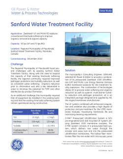

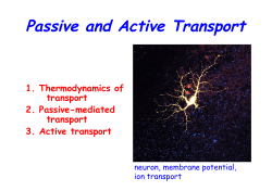


© Copyright 2026