
C M P
CIGNA MEDICAL COVERAGE POLICY
The following Coverage Policy applies to all plans administered by CIGNA Companies including plans
administered by Great-West Healthcare, which is now a part of CIGNA.
Subject Electrical Stimulators
Table of Contents
Coverage Policy .................................................. 2
General Background ........................................... 3
Coding/Billing Information ................................. 18
References ........................................................ 20
Policy History..................................................... 28
Effective Date ............................ 8/15/2010
Next Review Date……………….8/15/2011
Coverage Policy Number ................. 0160
Hyperlink to Related Coverage Policies
Bone Growth Stimulators: Electrical
(Invasive, Noninvasive), Ultrasound
Diaphragmatic/Phrenic Nerve Stimulation
Electrical Stimulation for Wound Healing
Extracorporeal Electromagnetic Stimulation
for Urinary Incontinence
Gastric Pacing/Gastric Electrical Stimulation
(GES)
Injectable Bulking Agents for Urinary
Conditions
Local Injection Therapy and Neurosurgery
for Cervicogenic Headache and Occipital
Neuralgia
Pulsed Electromagnetic Therapy
Reflex Sympathetic Dystrophy/Complex
Regional Pain Syndrome
Sacral Nerve Stimulation for Urinary Voiding
Dysfunction
Scoliosis Treatments, Idiopathic
Sensory Stimulation for Individuals in Coma
or Persistent Vegetative State
Speech Therapy for Swallowing and
Feeding Disorders
Spinal Cord Stimulation
Tinnitus Treatment Services and Devices
Transcranial Magnetic Stimulation
Transcutaneous Electrical Joint Stimulation
Devices
Vagus Nerve Stimulation (VNS)
INSTRUCTIONS FOR USE
Coverage Policies are intended to provide guidance in interpreting certain standard CIGNA HealthCare benefit plans as well as benefit
plans formerly administered by Great-West Healthcare. Please note, the terms of a participant’s particular benefit plan document [Group
Service Agreement (GSA), Evidence of Coverage, Certificate of Coverage, Summary Plan Description (SPD) or similar plan document] may
differ significantly from the standard benefit plans upon which these Coverage Policies are based. For example, a participant’s benefit plan
document may contain a specific exclusion related to a topic addressed in a Coverage Policy. In the event of a conflict, a participant’s
benefit plan document always supercedes the information in the Coverage Policies. In the absence of a controlling federal or state
coverage mandate, benefits are ultimately determined by the terms of the applicable benefit plan document. Coverage determinations in
each specific instance require consideration of 1) the terms of the applicable group benefit plan document in effect on the date of service; 2)
any applicable laws/regulations; 3) any relevant collateral source materials including Coverage Policies and; 4) the specific facts of the
particular situation. Coverage Policies relate exclusively to the administration of health benefit plans. Coverage Policies are not
recommendations for treatment and should never be used as treatment guidelines. Proprietary information of CIGNA. Copyright ©2010
CIGNA
Page 1 of 28
Coverage Policy Number: 0160
Coverage Policy
Coverage for electrical stimulation devices is subject to the terms, conditions and limitations of the
applicable benefit plan’s Durable Medical Equipment (DME) benefit and schedule of copayments. Please
refer to the applicable benefit plan document to determine benefit availability and the terms, conditions
and limitations of coverage. Under many benefit plans, coverage for DME is limited to the lowest-cost
alternative.
If coverage for electrical stimulation devices is available, the following conditions of coverage apply.
CIGNA covers neuromuscular electrical stimulation (NMES) as medically necessary when used as one
component of a comprehensive rehabilitation program for the treatment of disuse atrophy when the
nerve supply to the atrophied muscle is intact.
Some benefit plans have a specific limitation of coverage of transcutaneous electrical nerve stimulation
(TENS) units. Please refer to the applicable benefit plan document. If not specifically limited by the
benefit plan, CIGNA covers the use of a transcutaneous electrical nerve stimulator (TENS) as medically
necessary for EITHER of the following:
•
•
chronic pain when there is failure of at least a 30-day trial of conventional medical management
including medications (e.g., nonsteroidal anti-inflammatory drugs [NSAIDs], acetaminophen) and
physical therapy
as an adjunct to conventional post-operative pain management within 30 days of surgery
CIGNA covers a conductive garment as medically necessary when used in conjunction with medically
necessary NMES or TENS for ANY of the following clinical situations:
•
•
•
The use of conventional electrodes, tapes or lead wires is not feasible either because the individual has
a large area requiring treatment or a large number of sites requiring stimulation.
The site(s) requiring stimulation (i.e., back) is/are difficult to reach with conventional electrodes, tapes or
lead wires.
A co-existing medical condition (e.g., skin problems) precludes the use of conventional electrodes,
tapes, or lead wires.
CIGNA does not cover the use of any of the following electrical stimulation devices, because each is
considered experimental, investigational, or unproven for the treatment of any condition (this list may
not be all-inclusive):
•
•
•
•
•
•
•
•
•
•
•
•
•
•
•
bioelectric nerve block (electroceutical therapy)
cranial electrical stimulation (cranial electrotherapy stimulation)
electro therapeutic point stimulation (ETPSSM)
functional electrical stimulation (FES)
H-WAVE electrical stimulation
high-voltage galvanic stimulator (HVG)
interferential therapy (IFT)
microcurrent electrical nerve stimulation (MENS), including frequency-specific microcurrent (FSM)
stimulation
pelvic floor electrical stimulation (PFES)
percutaneous electrical nerve stimulation (PENS)
percutaneous neuromodulation therapy (PNT)
peripheral/posterior tibial nerve stimulation (PTNS)
PrimaBella™ nerve stimulation therapy
electrical sympathetic stimulation therapy
threshold/therapeutic electrical stimulation (TES)
Page 2 of 28
Coverage Policy Number: 0160
General Background
Electrical stimulators provide direct, alternating, pulsating and/or pulsed waveform forms of energy. The devices
are used to exercise muscles, demonstrate a muscular response to stimulation of a nerve, relieve pain, relieve
incontinence, and provide test measurements. Electrodes for such devices may be implanted, indwelling,
transcutaneous (needles) or surface. Electrical stimulators may have controls for setting the pulse length, pulse
repetition frequency, pulse amplitude, and triggering modes.
Neuromuscular Electrical Stimulation (NMES)
NMES is the application of electrical current through electrodes on the skin to targeted muscles to elicit muscle
contraction and relaxation. NMES is proposed to promote muscle restoration and to prevent or diminish muscle
atrophy and spasms and is an established treatment modality for disuse atrophy when the nerve supply to the
muscle is intact. NMES is typically used as a component of a comprehensive rehabilitation program. Protocols
in the literature recommend no more than two hours of NMES treatment within a 24-hour period and the
treatment plan is typically re-evaluated every 30 days. Compared to TENS, NMES delivers a stronger current
with a wider pulse width, and TENS is primarily indicated for the management of chronic pain.
U.S. Food and Drug Administration (FDA): Neuromuscular electrical stimulators are 510(k) FDA approved as
Class II devices. An example of a NMES device is the EMS 7500 (Koalaty Products, Ind., Roswell, GA). The
device is approved “for use for (1) relaxing muscle spasms, (2) increasing local blood circulation, (3) immediate
post-surgical stimulation of calf muscles to prevent venous thrombosis, (4) muscle re-education, (5) maintaining
or increasing range of motion, and (6) preventing or retarding disuse atrophy”.
Literature Review - Disuse Atrophy: Systematic reviews and randomized controlled trials support NMES for
the treatment of disuse atrophy and reported that NMES was as effective as, or more effective than, exercise
(Bax, 2005; Lieber, et al., 1996). NMES is a well-established treatment modality for disuse atrophy in patients
where the nerve supply to the muscle is intact.
Literature Review – Other Indications: There is insufficient evidence to support the effectiveness of NMES in
the prevention and/or management of multiple conditions including: congestive heart failure; chronic obstructive
pulmonary disease; dysphagia; knee rehabilitation; muscular dystrophy; muscle wasting and weakness
associated with cancers; cerebral palsy; stroke pain; toning, strengthening and firming of abdominal muscles;
rheumatoid arthritis; fecal incontinence; low back pain; Bell’s palsy; sensory stimulation for coma patients; motor
disorders; and chronic ulcers.
In a systematic review of randomized controlled trials, Monaghan et al. (2010) assessed the effectiveness of
NMES in strengthening quadriceps before and after total knee replacement. Two studies met inclusion criteria.
NMES plus exercise resulted in better quadriceps muscle activation compared to exercise alone (n=39), but was
not maintained at the 12-week follow-up. No significant differences were reported in either study for maximum
voluntary isometric torque or endurance between the NMES group and the control group.
Sillen et al. (2009) conducted a systematic review of randomized controlled trials to analyze the role of NMES in
strength, exercise capacity, and disease-specific health status in patients with congestive heart failure (n=9
studies) and chronic obstructive pulmonary disease (n=5 studies) with disabling dyspnea, fatigue, and exercise
intolerance. The limited number of studies, heterogeneous patient populations, and variability in NMES
methodology prohibited the use of meta-analysis. Although some of the studies reported significant
improvements with NMES compared to no exercise or usual care, outcomes, including adverse events, were
conflicting. Additional studies are indicated to provide sufficient evidence to establish the clinical utility of NMES
in this patient population.
In a 2008 systematic review of anterior cruciate ligament reconstruction (ACL) rehabilitation, Wright et al.
reported that 14 randomized controlled trials had evaluated postoperative NMES following ACL reconstruction.
Because of the variety of parameters in the studies, poor study quality, heterogeneous patient populations, and
the lack of randomization, blinding and independent observers, the authors noted that it was difficult to make
generalized conclusions regarding NMES, and it does not appear to be a requirement for successful ACL
reconstruction rehabilitation.
Page 3 of 28
Coverage Policy Number: 0160
Professional Societies/Organizations: In the evaluation of the literature regarding the use of electrical
stimulation for the treatment of spasticity in multiple sclerosis, the Multiple Sclerosis Council for Clinical Practice
Guidelines (2005) stated that “surface electrical stimulation may be of benefit in reducing spasticity in persons
with MS, but there is currently no evidence to support this supposition at this time”.
Transcutaneous Electrical Nerve Stimulation (TENS)
TENS devices consist of an electronic stimulus generator that transmits pulses of various configurations through
electrodes on the skin to stimulate the peripheral nerves for the purpose of pain management. Conventional
TENS or high frequency TENS delivers 40–150 hertz (Hz) compared to acupuncture-like TENS that delivers a
low frequency at 1–4 Hz. TENS has been used for a number of applications, including acute and chronic pain,
postoperative pain, obstetrical pain, and pain associated with medical procedures.
U.S. Food and Drug Administration (FDA): TENS are approved by the FDA 510(k) process as a Class II
device for the relief and management of chronic intractable pain. Examples of these devices include the Empi
Active Transcutaneous Nerve Stimulator (Empi, Inc., Clear Lake, SD) and the StimPad™ TENS System
(AEMED, Inc. West Palm Beach, FLA).
Literature Review - Acute Postoperative Pain and Chronic Pain: The evidence in the peer-reviewed
literature supports TENS for the treatment of pain in the acute post-operative period (i.e., within 30 days of
surgery) and as a secondary treatment option for patients with chronic pain when conventional therapies (e.g.,
nonsteroidal anti-inflammatory drugs, physical therapy) have failed. TENS is a well-established treatment
modality for these indications. Systematic reviews, meta-analysis and randomized controlled trials reported a
reduction in pain and analgesic use in the treatment of acute post-operative pain and chronic pain (e.g., back
pain, osteoarthritis of the knees) following TENS (Emmiler, et al., 2008; Cipriano, et al., 2008; Bjordal, et al.,
2007; Solak, et al., 2007; Khadilkar, et al., 2005; Bjordal, et al., 2003).
Literature Review - Other Indications: The evidence in the published peer-reviewed scientific literature has
not established the effectiveness of TENS for the treatment of any other indications including, but not limited to:
post-traumatic acute pain, pain associated with childbirth (i.e., labor), nausea and vomiting of pregnancy,
dysmennorrhea, suspected placental insufficiency, urinary incontinence, vestibulodynia, dental pain, mandibular
alignment (i.e., neuromuscular orthodontics), fracture healing, myofascial dysfunction pain, chronic leg ulcers,
unstable angina, hypertension, stroke, asthma, reflex sympathetic dystrophy or complex regional pain
syndrome, motion sickness, chemotherapy-induced pain, tinnitus, rheumatoid arthritis, and drug withdrawal
(e.g., opiate addiction). Overall, systematic reviews, randomized controlled trials and case series have reported
that there was no improvement with TENS for these indications. Studies have included small heterogeneous
patient populations with short-term follow-ups, data have been insufficient or conflicting, and the application of
TENS varied (e.g., physician applied vs. patient applied, location of electrodes). The clinical utility of TENS has
not been established for all other indications.
Acute Pain: Walsh et al. (2009) assessed the analgesic effectiveness of TENS in acute pain for adults (n=919)
in a systematic review of 12 randomized controlled trials. The types of acute pain included procedural pain (e.g.
cervical laser treatment, venipuncture, screening flexible sigmoidoscopy) and nonprocedural pain (e.g.
postpartum uterine contractions, rib fractures). The authors were unable to make any definitive conclusions due
to the insufficient extractable data.
Dysmenorrhea: In a systematic review of seven randomized controlled trials (n=164), Proctor et al. (2009)
evaluated the effectiveness of low-frequency TENS (acupuncture-like TENS, 1–4 hertz [Hz]) and high-frequency
TENS (conventional TENS, 50–120 Hz) for the treatment of primary dysmenorrhea. Studies compared TENS to
placebo, no treatment or medical treatment. Overall, high-frequency TENS was reported more effective than
placebo TENS for relief of pain. There was no difference in pain relief with low-frequency TENS compared to
placebo. There were conflicting results regarding whether high-frequency TENS was more effective than lowfrequency TENS. Due to the small patient populations, various methods of the application of TENS, and the lack
of precision in the comparisons, clear recommendations for clinical applications could not be made.
Labor: Dowswell et al. (2009) conducted a systematic review on the use of TENS during labor. A total of 19
randomized controlled trials (n=1671) comparing TENS to pharmacotherapy or placebo met inclusion criteria.
TENS was applied to the back (n=15), acupuncture points (n=2), and cranium (n=2). Overall, there were no
significant differences between pain ratings in the TENS group and the control groups. In cases where TENS
Page 4 of 28
Coverage Policy Number: 0160
was used as an adjunct to epidural analgesia, there was no evidence that it reduced pain. There was no
consistent evidence that TENS had any impact on interventions and outcomes of labor.
Low Back Pain: Khadilkar et al. (2008) conducted a systematic review to determine if TENS was more effective
than placebo for the management of chronic low back pain. Four “high-quality” randomized controlled trials
(n=585) met inclusion criteria. Due to conflicting evidence, the authors were unable to determine if TENS was
beneficial in reducing back pain intensity. Two trials involving 410 patients reported that TENS did not improve
back-specific functional status, the level of disability from the pain, the use of medical services or work status.
There were no significant differences in outcomes when conventional TENS was compared to acupuncture-like
TENS.
Phantom Pain and Stump Pain: Mulvey et al. (2010) conducted a systematic review of randomized controlled
trials to assess the effectiveness of TENS for the treatment of phantom pain and stump pain following
amputation in adults. No studies were identified.
Stroke: NG and Hui-Chan (2009) conducted a randomized controlled trial (n=109) to determine if TENS would
improve functional walking performance (i.e., gait velocity, walking endurance and functional mobility) in
hemiparetic stroke patients with spastic plantar flexors. In addition to a control group (n=29), patients were
assigned to one of three intervention groups: TENS only (n=28), TENS plus exercise (n=27) or placebo
stimulation plus exercise (n=25). Each patient self-administered 20 sessions, five days per week for four weeks.
Each group received 60 minutes of TENS and the exercise groups received an addition 60 minutes of exercise
following TENS or placebo stimulation. Final follow-up occurred four weeks after the treatment ended. At the
final follow-up compared to all other groups, significant improvements were seen in the TENS plus exercise
group in gait velocity (p<0.001) and reduction in timed up and go scores (P<0.01). The TENS plus exercise
group covered significantly more distance in the 6-minute walk test (6MWT) (p<0.01) compared to the control
group and the TENS only group. Additional studies with larger patient populations and long-term follow-up are
indicated to validate the results of this study. The generalizability of this study is limited to stroke patients with
moderate to severe spasticity in the ankle plantar flexors. The frequency, duration, and intensity of combined
rehabilitation programs have not been established.
Yan et al. (2009) conducted a randomized controlled trial (n=62) to investigate whether TENS, when applied to
acupuncture points in patients after acute stroke, decreased spasticity and/or increased muscle strength was
more effective than placebo stimulation and standard rehabilitation. Patients were randomized to TENS,
placebo-TENS, or standard rehabilitation. Stimulation was applied to four acupuncture points in the affected
lower leg for 60 minutes, five days a week for three weeks. Compared to placebo or rehabilitation, TENS
significantly increased the number of patients with normal tone and ankle dorsiflexor strength and decreased the
co-contraction ratio (p<0.05). Overall, the TENS patient walked two to four days earlier than the other patients,
but the difference was not significant between the three groups.
Urinary Incontinence: In a randomized controlled trial, Hagstroem et al. (2009) evaluated the effectiveness of
sacral TENS in 27 children, ages 5–14 years, with severe nonneuropathic daytime urge incontinence (i.e.,
median of 7 wet days per week) refractory to standard urotherapy and anticholinergics. Anticholinergics were
discontinued two weeks prior to the onset of the study and detrusor overactivity was confirmed by testing.
Patients were randomized to TENS (FemiScan™ Stim, Mega Electronics, Ltd., East Longmeadow, MA) or to
sham therapy. TENS was applied two hours per day for four weeks. Five children had no response to TENS and
eight children had a partial response. Two children in the sham group improved significantly. With the exception
of the number of wet days per week (p<0.05) there were no significant differences in pre- and post-treatment
parameters (e.g., incontinence scores, number of incontinence episodes, voiding frequency) in the two groups.
The change in scores in wet days per week and incontinence episodes per day were significantly greater in the
TENS group vs. the sham group (p<0.05 and p<0.01, respectively). The TENS group had a significant decrease
in incontinence scores, wet days per week, number of daily urge incontinence and improved response to
urgency (p<0.01 each) compared to no significant changes in the sham group. Bladder reservoir function did not
change in either group. Limitations of the study include the small patient population and short-term follow-up.
Vestibulodynia: Murina et al. (2008) assessed the efficacy of TENS in the treatment of 40 women with
vestibulodynia. The women were randomized to either TENS or sham and received treatment twice a week for
20 sessions. At the three month follow-up, visual analogue scale scores and short-form McGill-Melzack Pain
Questionnaire scores improved significantly (p=0.004, p=0.001, respectively) in the TENS group compared to
Page 5 of 28
Coverage Policy Number: 0160
the sham group. Three of 15 women in the TENS group relapsed three months following the end of the study.
No adverse events were reported.
Professional Societies/Organizations: In a 2010 technology assessment on the efficacy of TENS in the
treatment of pain in neurologic disorders, the American Academy of Neurology stated that based on the
available evidence, TENS is not recommended for the treatment of low-back pain, but is recommended for the
treatment of painful diabetic neuropathy.
In their chronic pain medical treatment guidelines, the Work Loss Data Institute (2009) recommended TENS as
a treatment option for acute post-operative pain in the first 30 days following surgery. They also recommended
TENS as a secondary treatment modality on a one-month trial bases for chronic intractable pain of at least three
months’ duration for conditions such as neuropathic pain (e.g., diabetic neuropathy, post-herpetic neuralgia),
multiple sclerosis, phantom limb pain, complex regional pain syndrome or spasticity. They noted that there is a
lack of evidence supporting the efficacy of TENS for chronic pain and stated that TENS should be used as an
adjunct to an “evidence-based restorative program”.
Following a systematic review of randomized controlled trials of 17 nonpharmacologic therapies for low back
pain, the American Pain Society and the American College of Physicians (2007) stated that TENS had not been
shown to be effective for acute, subacute or chronic low back pain (Chou and Huffman, 2007).
Conductive Garments
Conductive garments are fabric electrodes placed between an electrical stimulator and a patient’s skin for the
delivery of electrical stimulation. They are an established alternative to standard electrode to aid in the treatment
of patients with chronic pain who have large areas or a large numbers of sites to be stimulated or the frequency
is such that it is not feasible to use conventional electrodes, tapes, or lead wires; sites requiring stimulation that
are not accessible by the patient with conventional electrodes, tapes or lead wires (i.e., back); and medical
conditions (e.g., skin problems) that preclude the use of conventional electrodes, tapes or lead wires.
U.S. Food and Drug Administration (FDA): AG Garments (San Diego, CA) conductive electrodes are Class II,
510(k) approved by the FDA “as reusable (by a single patient), cutaneous, flexible, conductive garment/fabric
electrodes for interface between electrical stimulators and a patient’s skin for the delivery of electrical
stimulation” (FDA, 2002).
Bioelectric Nerve Block (Electroceutical Therapy)
Bioelectric therapy, also known as electromedicine, noninvasive neuron-blockade devices, electroceutical
neuron-blockade devices and bioelectric treatment systems, is proposed as a treatment for acute pain and
chronic pain (e.g., back pain, diabetic pain, joint pain, fibromyalgia, headache, and reflex sympathetic
dystrophy). Electroceutical treatments use much higher electrical frequencies than TENS units (ranging from
one to 20,000 Hz compared to 0.5 to 100 Hz used in TENS).
U.S. Food and Drug Administration (FDA): An example of a device used for bioelectric therapy is the Matrix
PRO ElecDT (Matrix Electromedical, Inc., Las Vegas, NV) was 510(k) approved by the FDA as an interferential
current therapy device.
Literature Review: There is insufficient evidence in the published peer-reviewed scientific studies to support
the safety and effectiveness of bioelectric therapy. Well-designed, randomized controlled clinical studies are
needed to determine the clinical utility of electroceutical therapy in the treatment of patients with acute or chronic
pain.
Cranial Electrical Stimulation
Cranial electrical stimulation (CES), also called electrotherapy, electrotherapeutic sleep, electrosleep, electric
cerebral stimulation, cranial transcutaneous electrical nerve stimulation, cerebral electrotherapy, transcranial
electrotherapy, transcranial electrical stimulation, transcranial direct electrical stimulation (tDCS), transcerebral
electrotherapy, neuroelectric therapy, and craniofacial electrostimulation, delivers low level electrical stimulation
(i.e., microcurrent) to the brain through electrodes that are attached to the ear lobes or behind the ears. It has
been proposed that CES’s direct effect on the brain’s limbic system, hypothalamus, reticular activation system,
and/or the autonomic nervous system can control the symptoms of various conditions. This therapy is not to be
confused with transcranial magnetic stimulation or vagus nerve stimulation. CES has been proposed for the
Page 6 of 28
Coverage Policy Number: 0160
treatment of anxiety, depression, insomnia, substance abuse, fibromyalgia, Alzheimer’s, attentiondeficit/hyperactivity disorder (ADHD), asthma, spastic colitis, tension headaches, hypertension, chemotherapy
symptoms in cancer patients, burn patients, and other pain-related disorders.
U.S. Food and Drug Administration (FDA): CES devices are approved under the FDA 510(k) class III process
for the treatment of insomnia, depression, or anxiety. Examples of these devices include the Cranial Electrical
Nerve Stimulator (Johari Digital Healtcare Ltd., Fall CITY, WA), Alpha-Stim® (Electromedical Products, Inc.,
Hawthorne, CA), and LISS Cranial Stimulator and Fisher-Wallace Cranial Stimulator by Medical Consultants Intl.
Ltd. (Glen Rock, NJ). Some devices are approved for use by the patient at home.
Literature Review: The evidence in the published peer-reviewed literature does not support the effectiveness of
CES for any indication. Studies consist of randomized trials with small patient populations, short-term followups, and conflicting outcomes.
Rose et al. (2009) conducted a randomized controlled trial to compare the short-term effects of CES (AlphaStim) (n=19) to sham stimulation (n=19) on sleep disturbance, depressive symptoms, and subjective appraisal
in individuals who were the primary caregivers for their spouses with Alzheimer’s disease. Subjects used CES
60 minutes per day for four weeks and completed a daily log. At the end of four weeks, there were no significant
differences in overall sleep disturbances, sleep quality, or sleep onset latency scores. The CES group did report
a nine-minute decrease in sleep onset latency compared to a one minute increase in the sham group. There
were no significant differences between the groups in depressive symptoms or in burden, mastery, impact or
satisfaction of the care giving situation.
In a randomized controlled trial, Tan et al. (2006) evaluated CES (Alpha-Stem) (n=18) and sham stimulation
(n=20) for the treatment of chronic pain in men with spinal cord injury. Patients administered their treatment at
home for 21 consecutive days for 60 minutes per day and kept a log of their pain immediately before and after
treatment. At the end of the 21 days, patients were interviewed in the clinic and the sham participants were
allowed to use active CES for an additional 21 days. There were no significant differences between the groups
in pain ratings following treatment, but the average change in daily pain intensity and interference prior to and
following treatment were significantly better for the CES group and the sham group (p=0.03, p=0.004,
respectively). A significant reduction in post-treatment pain was reported by the sham group subjects who
switched to CES (n=17) (p=0.003). The authors noted that a limitation of the study was the inability to generalize
the outcomes to a female population, and the lack of evaluation of CES on psychological distress, quality of life,
and analgesic consumption.
Lichtbroun et al. (2001) compared the outcomes of fibromyalgia patients randomly assigned to CES (AlphaStim) (n=20), sham (n=20), or a control group (n=20). Self-treatments were administered for 60 minutes per day
for three weeks. CES treatments resulted in a significant improvement in tender point scores (p<0.001), and
self-rated pain scores (p<0.02), quality of sleep (p<0.02), quality of life p<0.03), and feelings of well-being
(p<0.05). Similar significant gains were reported in the sham and control patients who switched to CES at the
end of the initial three weeks. No significant gains were reported by the sham or control groups.
Electro Therapeutic Point Stimulation (ETPSSM)
ETPS neuromechanical therapy or neuropathic acupuncture involves the detection and treatment of chronic
intractable neuromyofascial pain using the TENS US Unit (Acumed Medical Supplies, LTD, Stanford, CT). The
transcutaneous device detects treatment points on the skin and applies brief, concentrated electrical
microstimulation in short bursts. Traditional TENS units apply alternating current compared to the direct current
applied by ETPS. Depending on how the device is programmed, the therapy is also proposed to decrease
circulation and assist in resolution of swelling and pain or to increase circulation to enhance immune response
and neural regeneration. The treatments can be self-administered by the patient at home (Acumed 2010;
Hocking, 2002).
U.S. Food and Drug Administration (FDA): The TENS US Unit is approved by the FDA 510(k) Class II device
as the TENS Pro 900 for the treatment of chronic intractable pain.
Literature Review: There is insufficient evidence in the published peer-reviewed scientific literature to support
the safety and effectiveness of ETPS. The available studies are primarily in the form of case reports and case
series with small patient population and short-term follow-ups.
Page 7 of 28
Coverage Policy Number: 0160
Functional Electrical Stimulation (FES)
FES or functional neuromuscular stimulation (FNS) attempts to replace stimuli from destroyed nerve pathways
to assist neurologically impaired patients (e.g., spinal cord injury, stroke) with functional movement and to
suppress spasticity. FES is a high-intensity (25–100 milliamps), short duration therapy that may be delivered for
20 minutes to one hour, several times a week, for months. For the device to be effective, the peripheral nerve
must be intact.
FES is proposed to allow ambulation in paraplegics (e.g., Parastep® I System, Sigmedics, Inc., Fairborn, OH),
as a means of stationary exercise to prevent or reduce muscle atrophy in upper and lower extremities (e.g.,
ERGYS I; Therapeutic Alliance, Inc., Fairborn, OH), to improve ambulation in patients with gait disorders (e.g.,
drop foot) such as hemiplegia due to stroke, cerebral injury, or incomplete spinal cord injury (e.g., Walkaide™
stimulator; Neuromotion, Edmonton, Alberta, Canada; NESS L300 foot drop system, Bioness Inc., Valencia, CA)
or to provide range of motion and function in patients with upper limb paralysis or hemiplegia (NESS H200 hand
rehabilitation system [previously known as the Handmaster], Bioness Inc., Valencia, CA).
U.S. Food and Drug Administration (FDA): FES devices, such as the Parastep, that have been proposed for
restoring ambulation to paraplegics are regulated by the FDA’s premarket approval (PMA) process.
Functional electrical stimulators that are used to provide stationary exercise for paraplegics, to correct gait
disorders, or to provide range of motion and function are approved under the FDA 510(k) process as Class II
devices. The RT300 FES cycle ergometer (Restorative Therapies, Inc., Baltimore, MD) is approved as a
powered muscle stimulator for “general rehabilitation for relaxation of muscle spasms, prevention or retardation
of disuse atrophy, increasing local blood circulation and maintaining or increasing range of motion” (FDA, 2009).
Other Restorative FES devices include the RT300 Leg, RT300 Leg and Arm, RT300 Arm, RT300 for children.
Literature Review: Studies in the published peer-reviewed scientific literature have investigated FES in the
management of patients with cerebral palsy, foot drop, head injury, multiple sclerosis, and stroke. FES has also
been proposed as a means of sustaining muscle strength, re-establishing lost muscle atrophy, or improve
exercise function in chronic heart failure. Overall, the studies have included small heterogeneous patient
populations, short-term follow-ups, and various treatment protocols, outcome measures, and FES devices. The
evidence in the published peer-reviewed scientific literature does not support the clinical effectiveness of FES.
Cerebral Palsy (CP): Seifart et al. (2009) conducted a systematic review of the literature to “assess and
synthesize the existing evidence regarding the use of FES in children and adolescents with CP [cerebral palsy]
when applied to the lower limb”. Out of 37 citations, three case reports, one single subject and one crossover
design study met inclusion criteria (n=29). FES application to either the triceps sura muscle alone or in
combination with the tibialis anterior muscle was favorable. The wide variety of application methods, stimulation
protocol, and outcome measures made analysis difficult.
Foot Drop: In a multicenter nonrandomized prospective study, Stein et al. (2010) compared the orthotic and
therapeutic effects of FES (WalkAide; Innovative Neurotronics, Austin, TX) on straight line and figure-8 walking
performance of patients with chronic nonprogressive (i.e., stroke, spinal cord injury, head injury, cerebral palsy)
(n=41) and progressive (i.e., secondary progressive multiple sclerosis, familial spastic paraparesis) (n=32)
disorders resulting in foot drop. The patients were tested with FES on and off (orthotic effect) and before and
after (therapeutic effect) use. Follow-up ranged from 3–11 months. At the three-month follow-up, both groups
had a significant similar orthotic (p<0.03) and therapeutic effect (p<0.005) on figure-8 walking speed. Overall,
the physiological cost index (PCI) (i.e., difference in resting and active heart rate) showed a decreasing trend
(p=0.031). At 11 months, the overall therapeutic effect was significant for the nonprogressive group (n=13)
(p<0.001) and the progressive group (n=18) (p=0.021). The PCI significantly decreased over time in the
nonprogressive group (p=0.039), but not in the progressive group. The nonprogressive group continued to
increase speed with and without FES compared to the progressive group who reached a plateau of gait speed
with a tendency to a decline in speed. There was a significant difference in the use of the WalkAide by the
progressive group compared to the nonprogressive group (p=0.037). Limitations of the study include the small
heterogeneous patient population, short-term follow-up, and lack of randomization and a control group.
Everaert et al. (2010) conducted a study on a subgroup (n=36) of the Stein et al. (2010) patient population to
“determine the effect of long-term use of a foot-drop stimulator [Walkaide] on residual corticospinal connections
Page 8 of 28
Coverage Policy Number: 0160
in people with central nervous system disorders”. Patients with nonprogessive (n=10) and progressive (n=26)
disorders used FES for 3–12 months. The nonprogressive and progressive groups had a significant increase in
mean-rectified MEPmax (motor-evoked potentials) following FES (p=0.003 and p=0.046, respectively).The MEP
latencies were significantly smaller for the nonprogressive group than the progressive group (p=0.02). The mean
increases in maximum voluntary contraction (MVC) following FES were significantly different in the
nonprogressive group (p=0.008) and the progressive group (p=0.013), but not significantly different between the
two groups. The mean increases in MVC following FES were also significantly different in the nonprogressive
group (p=0.008) and the progressive group (p=0.013) and not significantly different between the two groups.
There was a significant positive correlation between MEPmax changes and MVC changes (p<0.001) indicating
that those with greater increases in MEPmax after WalkAide use also had greater increases in MVC. There was a
significant improvement in walking speed following FES in the nonprogressive group (p=0.008) and the
progressive group (p=0.014). The negative mean change in PCI indicated that the patients were walking with
less effort following FES. The increases in MVC and MEP indicated that the patients had better voluntary control
over the tibialis anterior muscle.
Hausdorff and Ring (2008) reported on the effects of the NESS L300 neuroprosthesis on the gait of 24 patients
with chronic hemiparesis (5.8 ± 5.2 years) from stroke or traumatic brain injury whose walking was impaired by
foot drop. In random order, the patients walked for six minutes while wearing force-sensitive insoles once with
and once without the neuroprosthesis. Patients were evaluated again at four and eight weeks. Outcome
measures included walking speed, swing, and stride time as well as a gait symmetry index and stride time
variability. Significant improvement from baseline was noted for all measures at all follow-up timeframes.
According to the authors, the use of FES for the correction of foot drop is not common and may be due to
insufficient evidence and the ergonomic and technical problems associated with the FES systems. The study is
limited by the small sample size and lack of comparison to standard treatment.
Multiple Sclerosis: Barrett et al. (2009) randomly assigned 20 secondary progressive multiple sclerosis
(SPMS) patients to FES (Odstock Dropped Foot Stimulator, Odstock Medical, Ltd., Salisbury, UK) and 24 MS
patients to home exercise program to evaluate the effects of FES on walking performance. Patients had a
predominantly unilateral dropped foot that was impairing mobility. Patients began with short walks the first two
weeks and were then allowed to walk as tolerated. The FES settings were adjusted as needed for each
individual throughout the study period. Follow-up occurred at six, 12, and 18 weeks. Statistically significant
improvements in the 10 meter (m) walking speed and distance walked in three minutes were seen in the
exercise group compared to no significant improvement in the FES group (p=0.028). Otherwise, there were no
significant differences between the groups. There were no mean changes from baseline and 18-week outcomes
within the FES group compared to a significant increase in both walking over 10 m (p=0.0001) and distance
walked in three minutes (p=0.005) in the exercise group. Within the FES group, there was a significant increase
in mean values for 10 m walking speed and distance in three min with stimulation compared to no stimulation.
Limitations of the study as noted by the authors included: the small patient population, patient fatigue that
resulted in reduced walking time from six to three minutes, no baseline walking analysis, the drop out rate, and
the inability to generalize the results due to the SPMS drop foot diagnosis of this MS group.
Paul et al. (2008) compared 12 patients with MS to 12 healthy controls to investigate the effects of FES
(Odstock Dropped Foot Stimulator) on speed and physiological cost of gait. The MS patients had used FES for
six or more months prior to the study (average use 3 years and nine months). MS patients completed the
protocols with and without FES. A significant increase in speed and decrease in physiological cost of walking
(i.e., oxygen uptake) were seen in the MS group with and without FES (p<0.001, p=0.017, respectively). The
walking speed of the controls was greater than the MS group with and without FES. When the controls walked
at the preferred walking speed of the MS patients with and without FES, there were no significant differences
between the two groups nor was there a signicant difference in oxygen uptake.
Spinal Cord Injury: Postans et al. (2004) reported the outcomes of a pilot study (n=14) using the application of
partial weight-bearing (PWB) supported treadmill gait training augmented by a customized FES device in
subjects with acute incomplete SCIs. The subjects, with American Spinal Injury Association class C or D
injuries, participated in training that consisted of treadmill walking with PWB support augmented by FES. The
study included walking on the treadmill for up to 25 minutes a day, five days a week, for four weeks. The
intervention was compared to a four-week control period during which standard physical therapy (PT) was
given. The authors reported a greater increase in overground walking endurance achieved after FES compared
to after standard PT. A similar pattern was observed for overground walking speed.
Page 9 of 28
Coverage Policy Number: 0160
Ladouceur et al. (2000) reported a prospective, uncontrolled study (n=14) to evaluate FES for correction of gait
disorder with incomplete SCI. All patients were enabled to ambulate assisted by FES surface electrodes.
Patients were trained in FES for four weeks and encouraged to use FES as much as possible. At final follow-up,
maximal therapeutic walking speed had increased 0.25 m/sec (p<0.005) and maximal combined walking speed
had increased 0.26 m/sec (p<0.002) compared to baseline.
Stroke: FES has also been investigated in randomized controlled trials as an adjunct therapy for rehabilitation in
stroke patients. Randomized controlled trials have evaluated FES in lower extremity muscle training using FES
cycling with standard rehabilitation (n=20) (Ferrante, et al., 2008), retraining of walking functions (n=40)
(Tanovic, 2009), and/or with arm and hand rehabilitation (n=23) (Mangold, et al., 2009). Due to the small patient
populations and short-term follow-ups (e.g., 4–8 weeks), the effectiveness of FES for the treatment of stroke
patients has not been established.
Professional Societies/Organizations: The National Institute for Health and Clinical Excellence (NICE)
(United Kingdom) (2009) published a guidance document on FES for foot drop of central neurological origin and
stated that the evidence on safety and efficacy “appears adequate to support” the use of FES for foot drop in
terms of improving gait, but the efficacy as it relates to quality of life and activities of daily living needs to be
further investigated.
H-Wave Electrical Stimulation
The H-WAVE electrical stimulation device generates a biphasic, exponentially decaying waveform with pulsewide widths. Its waveform distinguishes it from TENS and other forms of electrical stimulators. H-WAVE is
classified as a powered muscle stimulator. The large pulse width theoretically enables contraction in the muscle
for extended periods of time at a low fatigue rate and increases circulation, muscle relaxation, pain relief and
wound healing. H-wave stimulation has been used in the treatment of pain related to a variety of etiologies, such
as diabetic neuropathy, muscle sprains, temporomandibular joint dysfunctions, or reflex sympathetic dystrophy.
H-wave electrical stimulation must be distinguished from the H-waves that are a component of
electromyography. H-wave devices are available for self-administered home therapy.
U.S. Food and Drug Administration (FDA): The H-WAVE® Muscle Stimulator (Electronic Waveform
Laboratory, Inc., Huntington Beach, CA) is FDA 510(k) approved is a class II device.
Literature Review: There is insufficient evidence in the published peer reviewed scientific literature to support
the safety and effectiveness of the H-Wave electrical stimulators.
Blum et al. (2009) conducted a double-blind randomized controlled trial (n=22) to assess H-wave stimulation (HWave) in patients who had undergone open-reduction rotator cuff reconstruction. Twelve patients were
randomized to H-wave and ten to sham therapy. Starting the day of surgery, patients used the device for one
hour, twice a day, for 90 days. Physical therapy was initiated eight weeks following surgery. Patients treated
with H-wave advanced more quickly with physical therapy. Ninety days following surgery the H-wave group had
a loss of 11.67 degrees (p=0.007) in external rotation (arm at side) and the placebo group had a loss of 21.65
(p=0.007). Regarding internal rotation (arm at 90 degrees), the H-wave group had a loss of 13.33 degrees
(p=0.006) compared to a loss of 23.25 degrees (p=0.0062) in the placebo group. No statistical differences were
seen in all other range of motions. Limitations of this study include the small patient population and short-term
follow-up.
In a technology assessment evaluating the use of H-wave for pain management, ECRI (2009) concluded that
due to a lack of comparative effectiveness data it was not possible to determine how the efficacy of H-wave
compared to other therapies. A search of 22 databases provided two randomized controlled trials (n=54) that
met inclusion criteria, and although a statistically significant advantage overall was indicated, the difference was
not large enough to be clinically significant.
Professional Societies/Organizations: The Work Loss Data Institute (2009) stated that H-wave therapy may
be considered on a one-month trial basis as an adjunct to an “evidence-based functional restoration” program
for the treatment of diabetic neuropathic pain or chronic soft tissue inflammation. Its use should only be
considered following failure of conservative care (e.g., pharmacotherapy, physical therapy, TENS).
Page 10 of 28
Coverage Policy Number: 0160
High Voltage Galvanic Stimulation (HVG)
Galvanic stimulation is characterized by high voltage pulsed stimulation and is proposed primarily for local
edema reduction through muscle pumping and polarity effect. Edema is comprised of negatively charged
plasma proteins, which leak into the interstitial space. The theory of galvanic stimulation is that the high voltage
stimulus applies an electrical potential which disperses the negatively charged proteins away from the
edematous site, thereby helping to reduce edema. The high voltage and direct current used in HVG
differentiates it from the low voltage and alternating current used in TENS or NMES (Medi-Stem, 2009).
U.S. Food and Drug Administration (FDA): High Voltage Galvanic Stimulator (Control Solutions, Inc.,
Northbrook, IL) is 510(k) FDA approved as a class II device.
Literature Review: The few studies that were identified in the literature that addressed HVG were primarily
randomized clinical trials and case comparisons published prior to 1997 with small patient populations and
short-term follow-up. Patient selection criteria were lacking. There is insufficient evidence in the published peer
reviewed scientific literature to support the safety and efficacy of HVG stimulation.
Interferential Therapy (IFT)
IFT, also known as interferential stimulation (IFS), is a treatment modality that is proposed to relieve
musculoskeletal pain and increase healing in soft tissue injuries and bone fractures. Two medium-frequency,
pulsed currents are delivered via electrodes placed on the skin over the targeted area producing a lowfrequency current. IFT delivers a crisscross current at 4000–4150 pulses per second resulting in deeper muscle
penetration. These features are proposed to provide more effective pain control compared to TENS with less
pain. It is theorized that IFT prompts the body to secrete endorphins and other natural painkillers and stimulates
parasympathetic nerve fibers to increase blood flow and reduce edema.
U.S. Food and Drug Administration (FDA): Interferential stimulator instruments are approved as 510(k) Class
II devices. Examples of FDA-approved devices include the RSJ, RS JC (RS Medical, Vancouver, WA), IF 8000
(Biomotion, Madison, AL), RS-4i™ (RS Medical, Vancouver, WA).
Literature Review: The evidence in the published peer reviewed scientific literature does not support the safety
and effectiveness of IFT for the treatment of multiple conditions including: constipation, urinary incontinence,
pain associated with musculoskeletal disorders or injuries, osteoarthritis, stimulation of soft tissue healing, and
stimulation of bone fracture healing.
Rutjes et al. (2009) conducted a systematic review of randomized or quasi-randomized controlled trials of
electrical stimulation, including IFT (n=4 studies), for the treatment of osteoarthritis. Due to the poor
methodological and reporting quality of the studies, the effectiveness of IFT could not be confirmed.
In a randomized controlled trial, Demirturk et al. (2008) compared IFT (n=20) to Kegel exercises using a
biofeedback device (n=20) for the treatment of urinary stress incontinence in women. Treatments lasted 15
minutes per session, three times a week, for 15 sessions. Outcome criteria included pelvic floor muscle
strength, one-hour pad test and quality of life questionnaire. Following treatment, all parameters improved
significantly (p<0.5 each) in each group There were no significant differences in outcomes between the two
groups. No adverse events were reported.
The California Technology Assessment Forum (2005) evaluated the literature on IFT for the treatment of
musculoskeletal pain and concluded that this treatment modality has not been shown to be as beneficial as
alternative treatments such as nonsteroidal anti-inflammatory drugs and exercise therapy. Although IFT was
found to be a generally safe technique, it did not meet the CTAF technology assessment criteria for the
treatment of musculoskeletal pain.
In randomized controlled trials, Minder et al. (2003) found IFT to have no overall beneficial effect on delayedonset muscle soreness. In contrast, Jarit et al. (2003) found that home IFT therapy may help to reduce pain and
swelling in individuals who undergo knee surgery, and Hou et al. (2002) reported that IFT may have benefit for
relieving myofascial pain when used in conjunction with hot packs and range-of-motion exercises. AlvesGuerreiro et al. (2001) concluded that neither TENS nor action-potential stimulation therapy (APS) nor IFT
produced a significant hypoanalgesic effect in healthy test subjects.
Page 11 of 28
Coverage Policy Number: 0160
Professional Societies/Organizations: Regarding IFT, the Work Loss Data Institute (2009) stated that “there
is no quality evidence of effectiveness except in conjunction with recommended treatments, including return to
work, exercise and medications” and the evidence of improvement was limited. The reported results from trials
were negative or non-interpretable and study design and methodology were poor. There was a lack of
standardized protocol for IFT, and the therapies varied in electrode-placement technique, frequency of
stimulation, pulse duration, and treatment time.
Following a systematic review of randomized controlled trials of 17 nonpharmacologic therapies for low back
pain, the American Pain Society and the American College of Physicians (2007) stated that there was a limited
number of studies available on IFT, and there were no clear differences between the use of IFT and spinal
manipulation or traction for subacute or chronic back pain (Chou and Huffman, 2007).
Microcurrent Electrical Nerve Stimulation (MENS)
MENS involves the use of a device that delivers small amounts of electrical current (millionths of an amp) to
help relieve pain and heal soft tissues of the body. The application of microcurrent stimulation to an injured area
is proposed to realign the body’s electrical current and increase the production of adenosine triphosphate,
resulting in increased healing and recovery and blocking of perceived pain. The electrical current is subsensory
and usually not felt. MENS differs from TENS in that it uses a significantly reduced electrical stimulation. The
goal of TENS is to block pain, while MENS acts on naturally-occurring electrical impulses to decrease pain by
stimulating the healing process.
Frequency specific microcurrent (FSM) is a type of microcurrent therapy. The microcurrent device has two
separate channels that allow both the frequency and current to be set independently for each channel. FSM is
proposed as a treatment option for nerve and muscle pain, shingles, and herpes (Frequency Specific
Microcurrent, 2007).
U.S. Food and Drug Administration (FDA): The FDA categorizes microcurrent devices as TENS devices
intended for pain relief. The device is used to apply an electrical current to electrodes on a patient's skin to treat
pain. Precision Micro (Precision Microcurrent, Inc, Newberg, OR) is 510(k) FDA as a class II device equivalent
to predicate TENS devices.
Literature Review: There is insufficient evidence in the published peer-reviewed scientific literature to support
the safety and effectiveness of MENS including FSM. Rajpurohit et al. (2010) conducted a randomized
controlled trial to compare the effectiveness of TENS (n=30) to MENS (n=30) on masticatory muscle pain
secondary to bruxism (i.e., teeth grinding, clenching, clamping). Patients were treated for seven days.
Compared to the TENS group, the MENS group experienced a significant improvement in the Visual Analog
Scale (VAS) (p≤0.0001) and tenderness (p≤0.001). Limitations of the study include the small patient population,
short-term follow-up, and inclusion of patients who had previously been treated with TENS or MENS.
Zuim et al. (2006) compared four groups of patients (n=5 patients per group) with temporomandibular disorders
who were treated with MENS and/or occlusal splint therapy. Group 1 was treated with MENS plus splint. Group
2 was treated with placebo MENS plus splint. Group 3 was treated with MENS only, and group 4 was treated
with placebo MENS. Although there was a reduction of pain in all groups, there were no statistical differences
between the four groups or between MENS and occlusal splint therapy.
Pelvic Floor Electrical Stimulation (PFES): Although the exact mechanism is not fully understood, it is
postulated that electrical stimulation of the bladder floor activates the pudendal nerve, causing contraction of
smooth, striated urethral muscles and striated pelvic floor muscles. The electrical stimulation is transmitted via
vaginal or anal electrodes intending to improve urethral closure and strengthen the pelvic floor muscles.
U.S. Food and Drug Administration (FDA): All devices with surface electrodes used for bladder stimulation
are Class II devices. Examples of FDA 510(k) approved, nonimplantable electrical stimulators include the
Detrusan® 500 (Innovamed USA, Inc., Lehigh Acres, FL) and the Pathway™ CTS 2000 (Prometheus Group,
Duxbury, MA).
Literature Review: There is insufficient evidence in the published peer-reviewed scientific literature to support
electrical bladder stimulation for the treatment of urinary incontinence. In a Cochrane review of conservative
management for post-prostatectomy urinary incontinence, Hunter et al. (2007) reported that “analysis of other
Page 12 of 28
Coverage Policy Number: 0160
conservative interventions such as transcutaneous electrical nerve stimulation and anal electrical stimulation, or
combinations of these interventions were inconclusive. There were too few data to determine treatment effects
on incontinence after TURP. The findings should continue to be treated with caution, as most studies were of
poor to moderate quality”.
In a randomized controlled trial, Goode et al. (2003) studied the effect of behavioral training with and without
PFES on stress incontinence in 200 women. Patients were randomly assigned to eight weeks (four visits) of
behavioral training; eight weeks (four visits) of behavioral training plus home PFES; or eight weeks of selfadministered behavioral treatment using a self-help booklet. Significant improvement was seen in all groups on
the incontinence impact questionnaire, with no between-group differences. Treatment with PFES did not
increase effectiveness of a comprehensive behavioral program for women with stress incontinence. The authors
noted that there is no current consensus regarding the efficacy of bladder stimulation and clear patient selection
criteria are not available.
The California Technology Assessment Forum (2004) reviewed the evidence on the use of PFES as a treatment
for urinary incontinence in women. The report evaluated nine trials that compared electrical stimulation to
placebo. Of these studies, three found statistically significant results in favor of PFES. However, because of the
variations within the studies, conclusions could not be drawn as to the efficacy of this intervention. The authors
found insufficient evidence to conclude that PFES is as beneficial as behavioral or pharmacological therapies.
Professional Societies/Organizations: In a guidance document on the management of urinary incontinence in
women, the National Institute for Health and Clinical Excellence (NICE, 2006) (United Kingdom) recommended
that electrical stimulation not be routinely used in the treatment of women with overactive bladder or be used in
combination with pelvic floor muscle training. “While there is no evidence of effectiveness for electrical
stimulation” the consensus of the Guideline Development Group was that electrical stimulation may be
considered to aid motivation and adherence to therapy in women who cannot actively contract pelvic floor
muscles.
Percutaneous Electrical Nerve Stimulation (PENS) and Percutaneous Neuromodulation Therapy (PNT):
PENS involves the delivery of an electrical current through the insertion of a needle below the skin. PENS is
similar to TENS except that the needles are inserted one to four centimeters around or adjacent to the
applicable nerve. PENS is generally reserved for patients who fail to obtain pain relief from TENS (Hamza, et
al., 2000).
PNT is a variation of PENS which has been developed as a treatment for neck and back pain. This treatment
involves insertion of five pairs of needle-like electrodes into the skin of the neck or back to stimulate nerve fibers
in the deep tissues. The treatment regimen typically consists of two to three, 30-minute sessions per week, for
two to six weeks.
U.S. Food and Drug Administration (FDA): The Vertis PNT System (Vertis Neuroscience Inc., Seattle, WA)
was granted marketing approval by the FDA via the 510(k) process. The FDA stated that “PNT is indicated for
the symptomatic relief and management of chronic or intractable pain and/or as an adjunctive treatment in the
management of post-surgical pain and post-trauma pain” (FDA, 2002). The Vertis PNT Control Unit, with a
cervical electrode and cable, received 510(k) approval on September 11, 2002.
Literature Review: There is insufficient evidence in the published peer-reviewed literature to support the safety
and effectiveness of PENS or PNT as a treatment option for chronic pain. Weiner et al. (2008) conducted a
randomized controlled trial (n=200) to evaluate the efficacy of PENS in adults with chronic low back pain.
Patients were randomized to either 1) PENS, 2) brief electrical stimulation to control for treatment expectance
(control-PENS), 3) PENS plus general conditioning and aerobic exercise (GCAE) or to 4) control-PENS plus
GCAE. Treatment was delivered twice a week for six weeks to the 50 participants in each group. All groups
reported significantly reduced pain (McGill Pain Questionnaire short form) and disability and improved gait
velocity, which was sustained at six months. Significantly fewer fear avoidance beliefs were reported in the
CGAE group compared to the non-CGAE group. Comparable reduced pain and function were reported by the
PENS and control-PENS group, whether delivered for five minutes or 30 minutes. Thus, the exact dose of
electrical stimulation needed for analgesia could not be determined. PENS and GCAE were more effective than
PENS alone in reducing fear avoidance beliefs, but not in reducing pain or in improving physical function. There
Page 13 of 28
Coverage Policy Number: 0160
was a statistically significant improvement in chair rise time in the control-PENS plus CGAE compared to
control-PENS alone. The overall drop-out rate was 8%.
Kang et al. (2007) conducted a single-blinded, randomized study of 63 patients with knee pain secondary to
osteoarthritis. Twenty-eight patients were randomly assigned to the sham group and 35 to the live treatment
group. The study investigated the efficacy of PNT in reducing knee pain and medication consumption during the
first week following treatment. Pain levels were rated on a 100-mm visual analog pain scale. The live group had
greater efficacy than the sham group in all time periods; however, only in the immediate post-treatment period
did it reach statistical significance (p=0.0361). The overall median pain intensity difference over all periods was
14.5 for the live group and 6.5 for the sham group and reached statistical significance (p=0.0071). At one week
follow-up, the live group reported significantly less medication use (p<0.0001) than the sham group.
A Hayes report (2006) on the Vertis PNT system for the treatment of low back pain (LBP) examined four
randomized crossover trials (n=34–75). No complications were found to be associated with PNT. Potential
complications based on exclusion criteria of studies included heart disease and implanted cardiac pacemaker.
According to the Hayes brief, results of these studies suggested that PNT reduces LBP and the disability due to
this pain, but the randomized crossover trials provided evidence that the benefits are temporary. In these
studies, pain reoccurred between treatment sessions. It was concluded that the current evidence regarding the
safety and efficacy of PNT for LBP is promising but insufficient to support its use.
Professional Societies/Organizations: The Work Loss Data Institute (2009) stated that there is a “lack of high
quality evidence to prove long-term efficacy” of the use of PENS. They stated that PENS may be considered as
an adjunct to an “evidence-based functional restoration” program after all other non-surgical treatments have
been tried, failed, or are unsuitable or contraindicate (e.g., exercises, TENS).
Peripheral/Posterior Tibial Nerve Stimulation (PTNS): PTNS, also called peripheral tibial nerve stimulation or
percutaneous tibial nerve stimulation, is electrical stimulation of the posterior tibial nerve proposed as a
treatment option for urinary incontinence. PTNS involves neuromodulation of the sacral nerve which is thought
to inhibit bladder instability.
U.S. Food and Drug Administration (FDA): The Urgent® PC Neuromodulation System (Uroplasty, Inc.,
Minneapolis, MN) was granted marketing approval by the FDA via the 510(k) process. The Urgent PC
stimulation system delivers an electrical current to the sacral nerve from the tibial nerve via a needle electrode.
According to the FDA, the device is intended to treat patients suffering from urinary urgency, urinary frequency,
and urge incontinence (FDA, 2005).
Literature Review: PTNS has been proposed for the treatment of urological disorders including urinary
incontinence, overactive bladder, pelvic floor dysfunction, and chronic prostatitis. There is insufficient evidence
in the published peer-reviewed scientific literature to support the effectiveness of PTNS. Studies primarily
include small study populations and short-term follow-ups with self-reported outcomes and conflicting results.
Patient selection criteria and treatment protocol have not been established.
In a multicenter, randomized controlled trial (i.e., the Study of Urgent® PC vs. Sham Effectiveness in Treatment
of Overactive Bladder Symptoms [SUmiT]), Peters et al. (2010) compared the efficacy of PTNS (n=110) to
inactive sham therapy (n=110) in patients with overactive bladder symptoms. Patients received 30-minute
sessions, once a week, for 12 weeks. At 13 weeks, patient responses on the global response assessment
(GRA) showed a significant improvement in overall bladder symptoms and urinary frequency (p<0.001, each) in
the PTNS group compared to the sham group. Self-reported 3-day voiding diaries recorded statistically
significant improvements in frequency, nighttime voids, voids with moderate to severe urgency and urinary urge
incontinence episodes in the PTNS group. Significant improvements were reported in moderate to severe
urgency and urge incontinence in the sham patients. Following PTNS, statistically significant improvements
were seen on the condition specific OAB questionnaire (short form) symptom severity score and quality of life
scores compared to sham. In the PTNS group statistically significant improvements were reported on the SF-36
general health survey quality of life in the physical (p=0.002) and mental domain scales. (p=0.049). PTNS
adverse events included ankle bruising, needles site discomfort and bleeding, and tingling in the leg. No sham
adverse events were reported. A limitation of the study is the short-term follow-up.
Page 14 of 28
Coverage Policy Number: 0160
Peters et al. (2009) conducted a randomized controlled trial (i.e., the Overactive Bladder Innovative Therapy trial
[OrBIT]) to compare the effectiveness of PTNS (Urgent PC) (n=50) to extended-release tolterodine (n=50) for
the treatment of overactive bladder with or without a history of anticholinergic drug use. PTNS treatments were
delivered for 30 minutes, once per week, for 12 weeks. Tolterodine patients received 4 milligrams (mg) daily
with a subsequent decrease to 2 mg if intolerability occurred (n=2). Follow-up was conducted face-to-face with
PTNS patients and by phone for tolterodine patients. A statistically significant improvement or cure in the PTNS
group compared to the tolterodine group was reported by patient assessment (p=0.01) but was not significant
when reported by the investigators (p=0.05). Significant improvements in overactive bladder symptom episodes
were reported from baseline to 12 weeks in both groups, but the differences between the two groups were not
significant. The mean reduction in voids for 24 hours was not significant in either group. Although improvements
were seen in OAB questionnaire results and quality of life scores in both groups, the differences between
groups were not significant. No serious adverse events or device malfunctions were reported. Limitations of the
study noted by the authors included no placebo in the tolterodine arm, no sham in the PTNS arm, and no
blinding to treatment assignment. Sixteen patients did not complete the 12-week study.
MacDiarmid et al. (2010) reported the one-year outcome of 25 patients from the above Peters et al. (2010)
OrBIT study who volunteered to continue with PTNS. Compared to baseline, the benefits at 12-weeks were
sustained at one year as reported by the patients on the GRA and voiding diaries. Compared to baseline,
significant mean improvements were seen in frequency decreased by 2.8 voids daily (p<0.001), daily urge
incontinence episodes decreased by 1.6 (p<0.001), nocturia decreased by 0.8 voids (p<0.05), and voided
volume improved by 39 cubic centimeters (cc) (p<0.05). Author noted limitations of the one-year follow-up study
included “a potential placebo effect based on subject time spent with clinicians, no prescribed tapering schedule
to evaluate when treatment effects fail, no cost analysis, and no device sham effect”. It was also unknown if
patients were counseled on fluid management and if or how fluid management habits may have influenced the
results.
In a randomized controlled trial, Kabay et al. (2009) compared the efficacy of PTNS (Medtronic Keypoint® Net,
Alpine Biomed Corp. Fountain Valley, CA) (n=45) to sham therapy (n=44) for the treatment of therapy-resistant
category IIIB chronic non-bacterial prostatitis (i.e., chronic pelvic pain syndrome). Patients received a 30 minute
treatment, once a week, for 12 weeks. Compared to baseline, the 12-week mean National Institutes of Health
Chronic Prostatitis Symptom Index (NIH-CPSI) scores, the mean Visual Analog Score (VAS) for pain, and the
mean VAS for urgency significantly improved (p<0.001 each) in the PTNS group, but not in the sham group.
Due to the small patient population and short-term follow-up, long-term randomized controlled trials with large
patient populations are needed to support the outcomes of this study.
Kabay et al. (2009) conducted a case series to evaluate the acute effects of PTNS on urodynamic findings in
patients (n=32) with Parkinson’s disease. Patients had storage symptoms (e.g., urge incontinence, urgency,
frequency), overactive bladder (OAB), and involuntary detrusor contraction (confirmed on routine cystometry). A
significant improvement (p<0.001) was seen in the mean first involuntary detrusor (1st IDCV) recorded at
baseline, 145.2 ± 41.1 (55–265) milliliter (ml), compared to results during PTNS, 244.7 ± 51.7 (145–390) ml. In
20 cases, increments of 50% in the 1st IDCV volume were seen during PTNS. A statistically significant
improvement of 100 ml of maximum cystometric capacity was seen in 18 cases (p<0.001). Five of seven
patients (71.4%) with pseudodyssnergia had complete relief of symptoms. The authors cautioned that the data
should be verified in a prospective multicenter study before PTNS is introduced into routine clinical practice.
In a prospective observational study, Nuhoglu et al. (2006) investigated the efficacy of the UroSurge®
Percutaneous Stoller Afferent Nerve Stimulator (SANS™) (Urosurge, Inc., Coralville, IA) device in 35 patients
with OAB deemed unresponsive to pharmacotherapy. Treatment was given once a week for 30 minutes for a
total of 10 weeks. Quality of life questionnaires, three-day voiding diaries, and cystometry were completed
before and after SANS treatment and at one-year follow-up. The early success rate was 54%, with
improvements seen in voiding diary parameters, urodynamic parameters and quality of life scores. Efficacy was
maintained at one year in only 23% of 32 patients. It was concluded that SANS treatment had a positive shortterm effect in patients with resistant OAB; however, efficacy decreased in about three months.
In a randomized controlled trial, Karademir et al. (2005) compared outcomes in patients after treatment with
SANS alone (n=21) to SANS plus a low-dose anticholinergic medication (n=22). SANS was applied for 60
minutes, once weekly, for a total of eight weeks. The treatment response rate was 61.6% for the SANS group
and 83.2% in the SANS plus medication group. In both groups, the best symptomatic improvements were
Page 15 of 28
Coverage Policy Number: 0160
obtained in patients with urge incontinence. The authors commented that combination with a low-dose
anticholinergic increased the success rate of SANS without causing significant side effects.
Vandoninck et al. (2003) reported on 90 consecutive OAB patients treated with PTNS. Patients underwent 12
PTNS sessions. The primary outcome measure was a decrease in number of urinary leakage episodes of 50%
or more per 24 hours. Pre- and post-treatment urodynamic data were available from 46 participants. The
objective and subjective success rates were reported to be 56% and 64%, respectively. Frequency/volume chart
data and quality of life scores improved significantly (p<0.01). Detrusor instabilities (DI) could be eliminated in
only a few cases. It was noted that patients without DI at baseline were 1.7 times more prone to respond to
PTNS. It was concluded that patients without DI or with late-onset DI seemed to be the best candidates for
PTNS. According to the authors, PTNS is an emerging therapy that requires further research, assessing
predictive factors and optimal stimulation parameters.
Govier et al. (2001) conducted a prospective multicenter clinical trial to determine the safety and efficacy of
percutaneous peripheral afferent nerve stimulation for treatment of refractive OAB and/or pelvic floor
dysfunction. Patients were treated with 12 weekly SANS sessions. Urodynamic studies, detailed voiding diaries,
quality of life surveys, and incontinence impact questionnaires were completed before, during and after the
study. The 12-week study was completed by 47 of 53 patients. A total of 71% of patients were reported to be
treatment successes by the investigators and were started on long-term treatment. On average, patients noticed
a 25% reduction in mean daytime and 21% reduction in mean nighttime voiding frequencies (p<0.05). Urge
incontinence was reduced by an average of 35% (p<0.05). Statistically significant improvements were noted in
selective pain and quality of life indexes. No significant adverse events related to treatment were noted in any
patients.
Professional Societies/Organizations: In their recommendations on the management of urinary incontinence
in women, NICE (2006) stated that “there is a need for a robust evaluation for PTNS for the treatment of urinary
incontinence”. One 12-week randomized controlled trial (n=43), and two case series (n=103) with 1–3 weeks
follow-up met inclusion criteria.
PrimaBella™ Nerve Stimulation Therapy
PrimaBella (manufactured by Neurowave Medical Technologies, Chicago, IL and distributed by Alaven
Pharmaceutical, LLC., Marietta, GA) nerve stimulation therapy, also called acustimulation or transcutaneous
electrical acupoint stimulation, is proposed for the treatment of nausea and vomiting associated with pregnancy.
The device is worn on the wrist, delivers intermittent electrical pulses to the Neiguan point (P6) sending signals
via the median nerve to the emetic center in the brain. The stimulation is proposed to regulate the nausea
signaling process between the brain and stomach resulting in normal stomach rhythm and relief of nausea and
vomiting (Alaven Pharmaceutical, 2009).
U.S. Food and Drug Administration (FDA): PrimaBella is FDA approved as a 510(k) class II neuromodulation
device approved for nausea and vomiting due to pregnancy (NVP). The original approval of this same device
was the Reliefband NST Device (Woodside Biomedical, Inc., Lake Forest, CA).
Literature Review: There is insufficient evidence in the published peer-reviewed scientific literature to support
the effectiveness of PrimaBella for the treatment of NVP. Rosen et al. (2003) conducted a randomized
controlled trial with pregnant volunteers (n=187) to evaluate the effectiveness of acustimulation (ReliefBand now
called PrimaBella) in relieving nausea and vomiting in pregnancy. Patients were 6–12 weeks’ gestation and
randomized to acustimulation (n=95) or sham stimulation (n=92). The device was worn for 21 days, and the
patient kept a log of device usage and location, and occurrences of nausea and vomiting. The use of
medications for nausea was not controlled during the study. The time-averaged change in the self-reported
Rhodes Index of Nausea, Vomiting and Retching total experience was significantly better in the acustimulation
group compared to the sham group (p=0.02). At the end of the 21-day trial, weight gain was significantly greater
in the study group compared to the sham group (p=0.003). Occurrences of dehydration secondary to nausea
and vomiting were significantly fewer in the acustimulation group compared to the sham group (p=0.013). There
were no significant differences between the groups in medication usage or urinary ketones. The authors noted
that the study “did not have sufficient power to assess whether efficacy of the device was related to gestational
age at enrollment”. Other limitations of the study include the short-term use of the device, and a drop out rate of
18.6%.
Page 16 of 28
Coverage Policy Number: 0160
Electrical Sympathetic Stimulation Therapy
Electrical sympathetic stimulation therapy is a form of electrical stimulation of the peripheral nerves by applying
eight electrodes bilaterally to the lower legs, feet, arms and hands. The therapy is proposed for the treatment of
chronic, intractable pain. Bilateral application of the electrodes is proposed to target the autonomic nervous
system, and create a systemic effect of pain relief. Treatments are typically one hour in duration and may be
administered in a physician’s office or at home.
U.S. Food and Drug Administration (FDA): Sympathetic therapy devices are approved by the FDA 510(k)
process. Two such devices are the Dynatron STS and the Dynatron STS RX (Dynatronics Corp., Salt Lake city,
UT). The devices are indicated for “symptomatic relief of chronic intractable pain and/or management of posttraumatic or post-surgical pain” (FDA, 2001).
Literature Review: The evidence in the published peer-reviewed scientific literature does not support the safety
and effectiveness of sympathetic therapy. Studies are primarily in the form of case series with small patient
populations and short-term follow-ups (Guido, 2002).
Threshold/Therapeutic Electrical Stimulation (TES)
TES is the application of a low level current (2–10 milliamps) to the muscles in the body. It is typically applied at
home while the patient is sleeping, for 8–12 hours per night, for up to six nights a week, for years. Researchers
have proposed the use of TES for decreasing neuromuscular spasms that result from involuntary muscle
contractions in patients with motor disorders (e.g., cerebral palsy, spina bifida). Proposed outcomes of TES
include: improved muscle strength, decreased spasticity, increased joint mobility, and improved bowel and
bladder dysfunction. It is also proposed as a treatment option for scoliosis.
U.S. Food and Drug Administration (FDA): TES devices are approved as 510(k) FDA Class II devices. The
NT200-TES (Bio-Medical Research LTD, Laurel, MD) is an example of an approved device.
Literature Review: The exact mechanism by which threshold electrical stimulation (TES) might improve motor
function in children with cerebral palsy or other motor disorders is unclear. Study results are conflicting
regarding the potential benefit of TES. There is insufficient published peer-reviewed scientific literature to
support TES in the treatment of cerebral palsy or other motor disorders.
Kerr et al. (2006) conducted a randomized, placebo-controlled trial to assess the efficacy of NMES and TES in
strengthening quadriceps muscles of both legs in 60 children with cerebral palsy (CP) with diplegia. The children
were randomized into one of three groups: NMES (n=18), TES (n=20), or placebo (n=22). Outcome measures
included peak torque of the left and right quadriceps muscles, gross motor function, and impact of disability.
They were assessed at baseline, at a six week follow-up visit, and at end of treatment (16 weeks). No
statistically significant difference was noted for NMES or TES versus placebo for strength or function.
Statistically significant differences were noted between NMES and TES versus placebo for impact of disability at
the end of treatment, but only between TES and placebo at the six week follow-up. The authors noted that
further evidence is required to establish the role of NMES and TES as an adjunct therapy, to define patient
populations that would benefit from NMES and TES and to determine the appropriate dosing parameters.
Dali et al. (2002) conducted a randomized controlled trial to determine whether a group of stable children with
CP (i.e., 36 males, 21 females; mean age 10; age range 5–18) would improve their motor skills after 12 months
of TES. Two-thirds received active and one-third received inactive stimulators. Tests were videotaped and
assessed blindly to record qualitative changes that might not be reflected in performance measurements. Range
of motion, degree of spasticity, and muscle growth measured by computed tomography (CT) were evaluated.
Fifty-seven of 82 outpatients who were able to walk at least with a walker completed all 12 months of treatment
(hemiplegia [n= 25]); diplegia [n= 32]). There was no significant difference between active and placebo
treatment in any of the tested groups. Visual and subjective assessments favored TES, whereas objective
indices showed the opposite trend. The authors concluded that TES in these CP patients did not have any
significant clinical effect during the test period and that additional studies are needed to establish whether or not
TES causes improvement in children with other movement disorders than the children with hemiplegia and
diplegia in this study.
Page 17 of 28
Coverage Policy Number: 0160
Summary
Neuromuscular electrical stimulation (NMES) is an established therapy for the treatment of disuse atrophy when
the nerve supply to the atrophied muscle is intact and NMES is used as one component of a comprehensive
rehabilitation program.
There is evidence in the published peer-reviewed literature to support the use of transcutaneous electrical nerve
stimulator (TENS) for the treatment of patients with chronic pain when conventional therapy has failed. Tens is
also supported for the treatment of pain in the immediate post-operative period following surgery (i.e. within 30
days of surgery).
Conductive garments used in conjunction with TENS or NMES are appropriate when the patient has large areas
or numbers of sites to be stimulated; the frequency is such that conventional electrodes, tape, or lead wires are
not feasible; the patient is not able to access sites that require stimulation (i.e., back); or the patient has a
medical condition (e.g., skin problem) that precludes the use of conventional electrodes, tape, or lead wires.
There is insufficient evidence in the published peer-reviewed scientific literature to support the use of electrical
stimulation devices such as bioelectric nerve block (electroceutical therapy), cranial electrical stimulation (cranial
electrotherapy stimulation), electro therapeutic point stimulation (ETPSSM), functional electrical stimulation
(FES), H-WAVE electrical stimulation, high-voltage galvanic stimulation (HVG), interferential therapy (IFT),
microcurrent electrical nerve stimulation (MENS), including frequency-specific microcurrent (FSM), pelvic floor
electrical bladder stimulation (PFES), percutaneous electrical nerve stimulation (PENS), percutaneous
neuromodulation therapy (PNT), peripheral/posterior tibial nerve stimulation (PTNS), PrimaBella™ nerve
stimulation therapy, electrical sympathetic stimulation therapy, and threshold/therapeutic electrical stimulation
(TES).
Coding/Billing Information
Note: This list of codes may not be all-inclusive.
Covered when medically necessary when used to report neuromuscular electrical stimulation (NMES):
HCPCS
Codes
E0745
E0731
ICD-9-CM
Diagnosis
Codes
728.2
Description
Neuromuscular stimulator, electronic shock unit
Form fitting conductive garment for delivery of TENS or NMES (with conductive
fibers separated from the patient’s skin by layers of fabric)
Description
Muscular wasting and disuse atrophy, not elsewhere classified
Covered when medically necessary when used to report transcutaneous electrical nerve stimulator
(TENS):
HCPCS
Codes
E0720
E0730
E0731
ICD-9-CM
Diagnosis
Codes
Description
Tens, two lead, localized stimulation
Transcutaneous electrical nerve stimulation device, four or more leads, for
multiple nerve stimulation
Form fitting conductive garment for delivery of TENS or NMES (with conductive
fibers separated from the patient’s skin by layers of fabric)
Description
Page 18 of 28
Coverage Policy Number: 0160
338.12
338.18
338.21338.29
Acute post-thoracotomy pain
Other acute postoperative pain
Chronic pain
Experimental/Investigational/Unproven/Not Covered:
HCPCS
Codes
E0740
E0764
E0765
E0770
E1399†
Description
Incontinence treatment system, pelvic floor stimulator, monitor, sensor and/or
trainer
Functional neuromuscular stimulator, transcutaneous stimulation of muscles of
ambulation with computer control, used for walking by spinal cord injured, entire
system, after completion of training program
FDA approved nerve stimulator, with replaceable batteries, for treatment of
nausea and vomiting
Functional electrical stimulator, transcutaneous stimulation of nerve and/or
muscle groups, any type, complete system, not otherwise specified.
Durable medical equipment, miscellaneous
†
Note: Not covered when used to report any electrical stimulator device indicated in this coverage policy
as experimental, investigational or unproven.
Experimental/Investigational/Unproven/Not Covered for any electrical stimulator included within this
coverage policy:
ICD-9-CM
Diagnosis
Codes
292.0
296.20296.26
296.30296.36
300.00300.09
303.00–
303.93
304.00304.93
307.41
307.41
311
314.01
331.0
388.30388.32
401.0-401.9
413.0-413.9
428.0-428.9
438.82
493.00493.92
496
564.00564.09
Description
Drug withdrawal
Major depressive disorder, single episode
Major depressive disorder, recurrent episode
Anxiety states
Alcohol dependence syndrome
Drug dependence
Transient disorder of initiating or maintaining sleep
Persistent disorder of initiating or maintaining sleep
Depressive disorder, not elsewhere classified
Attention deficit disorder, with hyperactivity
Alzheimer’s disease
Tinnitus
Essential hypertension
Angina pectoris
Heart failure
Other late effects of cerebrovascular disease; dysphagia
Asthma
Chronic airway obstruction, not elsewhere classified
Constipation
Page 19 of 28
Coverage Policy Number: 0160
596.51
625.3
625.70625.79
643.00643.93
707.0-707.9
780.01
780.52
781.0
781.2
782.3
786.00786.90
787.2-787.29
787.6
788.30788.39
994.6
V11.3
Hypertonicity of bladder
Dysmenorrhea
Vulvodynia
Excessive vomiting in pregnancy
Chronic ulcer of skin
Coma
Insomnia, unspecified
Abnormal involuntary movements
Abnormality of gait
Edema
Dyspnea and respiratory abnormalities
Dysphagia
Incontinence of feces
Urinary incontinence
Motion sickness
Personal history of alcoholism
All other codes
*Current Procedural Terminology (CPT®) ©2010 American Medical Association: Chicago, IL.
References
1. Acumed. ETPSSM neuromechanical therapy. 2010. Accessed Jul 7, 2010. Available at URL address:
http://www.acumedmedical.ca/etps.html
2. Agency for Healthcare Research and Quality (AHRQ). Evidence Report/Technology Assessment
number 187. Treatment of overactive bladder in women. Aug 2009. Accessed Jul 19, 2010. Available at
URL address: http://www.ahrq.gov/clinic/epcindex.htm#dietsup
3. Agency for Healthcare Research and Quality (AHRQ). Evidence Report/Technology Assessment
number 161. Fecal and urinary incontinence in adults, prevention. Dec 2007. Accessed Jul 19, 2010.
Available at URL address: http://www.ahrq.gov/clinic/epcquick.htm#Ptopics
4. Alaven Pharmaceutical. PrimaBella™. 2009. Accessed July 6, 2010. Available at URL address:
http://www.primabellarx.com/
5. Alaven Pharmaceutical. PrimaBella™. AMCP-format dossier. 2009.
6. Alves-Guerreiro J, Noble J, Lowe A, Walsh D. The effect of three electrotherapeutic modalities upon
peripheral nerve conduction and mechanical pain threshold. Clin Physiol. 2001;21(6):704-11.
7. American Academy of Neurology. Technology assessment: efficacy of transcutaneous electric nerve
stimulation in the treatment of pain in neurologic disorders. Jan 2010. Accessed Jul 2, 2010. Available
at URL address:
http://www.aan.com/practice/guideline/index.cfm?fuseaction=home.welcome&Topics=13&Topics=14&T
opics=15&Topics=27&Topics=23&Topics=16&Topics=17&Topics=18&Topics=19&Topics=25&Topics=2
0&Topics=22&Topics=21&TopicsAll=1&keywords=&Submit=Search+Guidelines
8. Barrett CL, Mann GE, Taylor PN, Strike P. A randomized trial to investigate the effects of functional
electrical stimulation and therapeutic exercise on walking performance for people with multiple sclerosis.
Mult Scler. 2009 Apr;15(4):493-504.
Page 20 of 28
Coverage Policy Number: 0160
9. Bax L, Staes F, Verhagen A. Does neuromuscular electrical stimulation strengthen the quadriceps
femoris? A systematic review of randomised controlled trials. Sports Med. 2005;35(3):191-212.
10. Bioness, Inc. NESS L300 Foot Drop System. 2010. Accessed Jul 7, 2010. Available at URL address:
http://www.bioness.com/Home.php
11. Bjordal JM, Johnson MI, Lopes-Martins RAB, Bogen B, Chow R, Ljunggren AE. Short-term efficacy of
physical interventions in osteoarthritic knee pain. A systematic review and meta- analysis of randomised
placebo-controlled trials. BMC Musculoskelet Disord. 2007 Jun 22;8(1):51.
12. Bjordal JM, Johnson MI, Ljunggreen AE. Transcutaneous electrical nerve stimulation (TENS) can
reduce postoperative analgesic consumption. A meta-analysis with assessment of optimal treatment
parameters for postoperative pain. Eur J Pain. 2003;7(2):181-8.
13. Blum K, Chen AL, Chen TJ, Waite RL, Downs BW, Braverman ER, Kerner MM, Savarimuthu SM,
DiNubile N. Repetitive H-wave device stimulation and program induces significant increases in the
range of motion of post operative rotator cuff reconstruction in a double-blinded randomized placebo
controlled human study. BMC Musculoskelet Disord. 2009 Oct 29;10:132.
14. Blum K, Chen TJH, Martinez-Pons M, DiNubile NA, Waite RL, Schoolfield J, et al. The H-Wave small
muscle fiber stimulator, a nonpharmacologic alternative for the treatment of chronic soft- tissue injury
and neuropathic pain: an extended population observational study. Adv Ther. 2006 Sep-Oct;23(5):73949.
15. Brosseau L, Wells GA, Finestone HM, Egan M, Dubouloz CJ, Graham I, Casimiro L, Robinson VA,
Bilodeau M, McGowan J. Clinical practice guidelines for electrical stimulation. Top Stroke Rehabil 2006
Spring;13(2):54-60. Accessed Jul 7, 2010. Available at URL address:
http://www.guideline.gov/summary/summary.aspx?ss=15&doc_id=9918&nbr=005314&string=functional
+AND+electrical+AND+stimulation
16. California Technology Assessment Forum (CTAF). Electrical Stimulation for the Treatment of Urinary
Incontinence in Women. October 20, 2004. Accessed Jun 22, 2010. Available at URL address:
http://ctaf.org/content/general/detail/540
17. California Technology Assessment Forum (CTAF). Interferential Stimulation for the Treatment of
Musculoskeletal Pain. October 19, 2005. Accessed Jun 19, 2010. Available at URL address:
http://ctaf.org/content/general/detail/513
18. Cameron M, Lonergan E, Lee H. Transcutaneous Electrical Nerve Stimulation (TENS) for dementia.
Cochrane Database of Systematic Reviews 2003, Issue 3.
19. Capel ID, Dorrell HM, Spencer EP, Davis MW. The amelioration of the suffering associated with spinal
cord injury with subperception transcranial electrical stimulation. Spinal Cord. 2003 Feb;41(2):109-17.
20. Chou R, Huffman LH; American Pain Society; American College of Physicians. Nonpharmacologic
therapies for acute and chronic low back pain: a review of the evidence for an American Pain
Society/American College of Physicians clinical practice guideline. Ann Intern Med. 2007 Oct
2;147(7):492-504.
21. Chou R, Loeser JD, Owens DK, Rosenquist RW, Atlas SJ, Baisden J, Carragee EJ, Grabois M, Murphy
DR, Resnick DK, Stanos SP, Shaffer WO, Wall EM; American Pain Society Low Back Pain Guideline
Panel. Interventional therapies, surgery, and interdisciplinary rehabilitation for low back pain: an
evidence-based clinical practice guideline from the American Pain Society. Spine (Phila Pa 1976). 2009
May 1;34(10):1066-77.
Page 21 of 28
Coverage Policy Number: 0160
22. Cipriano G, de Camargo Carvalho AC, Bernardelli GF, Peres PA. Short-term transcutaneous electrical
nerve stimulation after cardiac surgery: effect on pain, pulmonary function and electrical muscle activity.
Interact Cardiovasc Thorac Surg. 2008 Apr 16.
23. Dali C, Hansen F, Pedersen S, Skov L, Hilden J, Bjornskov I, et al. Threshold electrical stimulation
(TES) in ambulant children with CP: a randomized double-blind placebo-controlled clinical trial. Dev Med
Child Neurol. 2002 Jun;44(6):364-9.
24. Demirtürk F, Akbayrak T, Karakaya IC, Yüksel I, Kirdi N, Demirtürk F, Kaya S, Ergen A, Beksac S.
Interferential current versus biofeedback results in urinary stress incontinence. Swiss Med Wkly. 2008
May 31;138(21-22):317-21.
25. Dowswell T, Bedwell C, Lavender T, Neilson JP. Transcutaneous electrical nerve stimulation (TENS) for
pain relief in labour. Cochrane Database of Systematic Reviews 2009, Issue 2. Art. No.: CD007214.
DOI: 10.1002/14651858.CD007214.pub2.
26. Dynatron System Solutions. Chronic pain products. Dynatron® STS™. Frequently asked questions.
2005. Accessed Jul 2, 2010. Available at URL address: http://dynatronsys.com/html/faqs.htm
27. ECRI Institute. H-wave device stimulation therapy for pain management. Plymouth Meeting (PA): ECRI
Institute; 2009 Apr. Accessed Jun 19, 2010. Available at URL address: http://www.ecri.org.
28. Embrey DG, Holtz SL, Alon G, Brandsma BA, McCoy SW. Functional electrical stimulation to
dorsiflexors and plantar flexors during gait to improve walking in adults with chronic hemiplegia. Arch
Phys Med Rehabil. 2010 May;91(5):687-96.
29. Emmiler M, Solak O, Kocogullari C, Dundar U, Ayva E, Ela Y, Cekirdekci A, Kavuncu V. Control of acute
postoperative pain by transcutaneous electrical nerve stimulation after open cardiac operations: a
randomized placebo-controlled prospective study. Heart Surg Forum. 2008;11(5):E300-3.
30. Everaert DG, Thompson AK, Chong SL, Stein RB. Does functional electrical stimulation for foot drop
strengthen corticospinal connections? Neurorehabil Neural Repair. 2010 Feb;24(2):168-77.
31. Ferrante S, Pedrocchi A, Ferrigno G, Molteni F. Cycling induced by functional electrical stimulation
improves the muscular strength and the motor control of individuals with post-acute stroke. Europa
Medicophysica-SIMFER 2007 Award Winner. Eur J Phys Rehabil Med. 2008 Jun;44(2):159-67.
32. Frequency specific microcurrent. 2007. Accessed Jul 7, 2010. Available at URL address:
http://www.frequencyspecific.com/
33. Goode P, Burgio K, Lovcher J, Roth D, Umlauf M, Richter H. et al., Varner R, Lloyd L. Effect of
behavioral training with or without pelvic floor electrical stimulation on stress incontinence in women: a
randomized controlled trial. JAMA. 2003 Jul;290(3):395-7.
34. Govier FE, Litwiller S, Nitti V, Kreder KJ Jr, Rosenblatt P. Percutaneous afferent neuromodulation for
the refractory overactive bladder: results of a multicenter study. J Urol. 2001 Apr;165(4):1193-8.
35. Guido EH. Effects of sympathetic therapy on chronic pain in peripheral neuropathy subjects. Am J Pain
Mgmt. 2002;12:31-34.
36. Hagstroem S, Mahler B, Madsen B, Djurhuus JC, Rittig S. Transcutaneous electrical nerve stimulation
for refractory daytime urinary urge incontinence. J Urol. 2009 Oct;182(4 Suppl):2072-8.
37. Hamza MA, White PF, Craig WF, Ghoname ES, Ahmed HE, Proctor TJ, Noe CE, Vakharia AS, Gajraj
N. Percutaneous electrical nerve stimulation: a novel analgesic therapy for diabetic neuropathic pain.
Diabetes Care. 2000 Mar;23(3):365-70.
Page 22 of 28
Coverage Policy Number: 0160
38. Hausdorff JM, Ring H. Effects of a new radio frequency-controlled neuroprosthesis on gait symmetry
and rhythmicity in patients with chronic hemiparesis. Am J Phys Med Rehabil. 2008 Jan;87(1):4-13.
39. HAYES Medical Technology Directory™. Health Technology Brief. Vertis PNT™ System (Vertis
Neuroscience Inc.) for Percutaneous Neuromodulation Therapy for Low Back Pain. ©2006 Winifred S.
Hayes, Inc.; Jan 10, 2006. Update search Aug 4, 2007.
40. Hocking B. ETPS neuropathic acupuncture. Ch 54. In: Weiner RS (editor). Pain management: a
practical guide for clinicians, sixth edition. CRC Press: Washington D.C. 2002:Pgs 667-690.
41. Hosker G, Cody JD, Norton CC. Electrical stimulation for faecal incontinence in adults. Cochrane
Database of Systematic Reviews 2007, Issue 3.
42. Hou C, Tsai L, Cheng K. Immediate effects of various physical therapeutic modalities on cervical
myofascial pain and trigger-point sensitivity. Arch Phys Med Rehabil. 2002;83(10):1406-14.
43. Hui-Chan CW, Ng SS, Mak MK. Effectiveness of a home-based rehabilitation programme on lower limb
functions after stroke. Hong Kong Med J. 2009 Jun;15(3 Suppl 4):42-6.
44. Hunter KF,Moore KN, Glazener CMA. Conservative management for postprostatectomy urinary
incontinence. Cochrane Database of Systematic Reviews 2007, Issue 2.
45. Hurley D, Minder P, McDonough S, Walsh D, Moore A, Baxter D. Interferential therapy electrode
placement technique in acute low back pain: a preliminary investigation. Arch Phys Med Rehabil.
2001;82(4):485-93.
46. Jarit G, Mohr K, Waller R. The effects of home interferential therapy on post-operative pain, edema and
range of motion of the knee. Clin J Sport Med. 2003;13(1):16-20.
47. Kabay SC, Kabay S, Yucel M, Ozden H. Acute urodynamic effects of percutaneous posterior tibial nerve
stimulation on neurogenic detrusor overactivity in patients with Parkinson's disease. Neurourol Urodyn.
2009;28(1):62-7.
48. Kabay S, Kabay SC, Yucel M, Ozden H. Efficiency of posterior tibial nerve stimulation in category IIIB
chronic prostatitis/chronic pelvic pain: a Sham-Controlled Comparative Study. Urol Int. 2009;83(1):33-8.
49. Kajbafzadeh AM, Sharifi-Rad L, Baradaran N, Nejat F. Effect of pelvic floor interferential
electrostimulation on urodynamic parameters and incontinency of children with myelomeningocele and
detrusor overactivity. Urology. 2009 Aug;74(2):324-9.
50. Kang RW, Lewis PB, Kramer A, Hayden JK, Cole BJ. Prospective randomized single-blinded controlled
clinical trial of percutaneous neuromodulation pain therapy device versus sham for the osteoarthritic
knee: a pilot study. Orthopedics. 2007 Jun;30(6):439-45.
51. Karademir K, Baykal K, Sen B, Senkul T, Iseri C, Erden D. A peripheric neuromodulation technique for
curing detrusor overactivity: Stoller afferent neurostimulation. Scand J Urol Nephrol. 2005;39(3):230-3.
52. Karavidas A, Parissis J, Arapi S, Farmakis D, Korres D, Nikolaou M, Fotiadis J, Potamitis N, Driva X,
Paraskevaidis I, Matsakas E, Filippatos G, Kremastinos DT. Effects of functional electrical stimulation
on quality of life and emotional stress in patients with chronic heart failure secondary to ischaemic or
idiopathic dilated cardiomyopathy: a randomised, placebo-controlled trial. Eur J Heart Fail. 2008
Jul;10(7):709-13.
53. Kerr C, McDowell B, Cosgrove A, Walsh D, Bradbury I, McDonough. Electrical stimulation in cerebral
palsy: a randomized controlled trial. Dev Med Child Neurol. 2006 Nov;48(11):870-6.
Page 23 of 28
Coverage Policy Number: 0160
54. Khadilkar A, Milne S, Brosseau L, Robinson V, Saginur M, Shea B, Tugwell P, Wells G. Transcutaneous
electrical nerve stimulation (TENS) for chronic low-back pain. Cochrane Database of Systematic
Reviews 2005, Issue 3.
55. Khadilkar A, Milne S, Brosseau L, Wells G, Tugwell P, Robinson V, et al. Transcutaneous electrical
nerve stimulation for the treatment of chronic low back pain: a systematic review. Spine. 2005 Dec
1;30(23):2657-66.
56. Khadilkar A, Odebiyi DO, Brosseau L, Wells GA. Transcutaneous electrical nerve stimulation (TENS)
versus placebo for chronic low-back pain. Cochrane Database of Systematic Reviews 2008, Issue 4.
Art. No.: CD003008. DOI: 10.1002/14651858.CD003008.pub3.
57. Kiratli BJ (author). Therapeutic Interventions. In: Lin VW, Cardenas DD, Cutter NC, Frost FS, Hammond
MC, Lindblom LB, et al. (editors). Spinal Cord Medicine: Principles and Practices. Demos Medical
Publishing, Inc. New York; 2003.
58. Ladouceur M, Barbeau H. Functional electrical stimulation-assisted walking for persons with incomplete
spinal injuries: longitudinal changes in maximal overground walking speed. Scand J Rehabil Med. 2000
Mar;32(1):28-36.
59. Lichtbroun AS, Raicer MM, Smith RB. The treatment of fibromyalgia with cranial electrotherapy
stimulation. J Clin Rheumatol. 2001 Apr;7(2):72-8; discussion 78.
60. Lieber RL, Silva PD, Daniel DM. Equal effectiveness of electrical and volitional strength training for
quadriceps femoris muscles after anterior cruciate ligament surgery. J Orthop Res. 1996 Jan;14(1):1318.
61. MacDiarmid SA, Peters KM, Shobeiri SA, Wooldridge LS, Rovner ES, Leong FC, Siegel SW, Tate SB,
Feagins BA. Long-term durability of percutaneous tibial nerve stimulation for the treatment of overactive
bladder. J Urol. 2010 Jan;183(1):234-40.
62. Mangold S, Schuster C, Keller T, Zimmermann-Schlatter A, Ettlin T. Motor training of upper extremity
with functional electrical stimulation in early stroke rehabilitation. Neurorehabil Neural Repair. 2009
Feb;23(2):184-90.
63. Medi-Stem, Inc. Frequently used modalities. 2009. Accessed Jul 7, 2010. Available at URL address:
http://www.medi-stim.com/overview.html
64. Meijer JW, Voerman GE, Santegoets KM, Geurts AC. Short-term effects and long-term use of a hybrid
orthosis for neuromuscular electrical stimulation of the upper extremity in patients after chronic stroke. J
Rehabil Med. 2009 Feb;41(3):157-61.
65. Minder P, Noble J, Alves-Guerreiro J. Interferential therapy: lack of effect upon experimentally induced
delayed onset muscle soreness. Clin Physiol Funct Imaging. 2002;22(5):339-47.
66. Monaghan B, Caulfield B, O’Mathúna DP. Surface neuromuscular electrical stimulation for quadriceps
strengthening pre and post total knee replacement. Cochrane Database of Systematic Reviews 2010,
Issue 1. Art. No.: CD007177. DOI:10.1002/14651858.CD007177.pub2.
67. Mulvey MR, Bagnall AM, Johnson MI, Marchant PR. Transcutaneous electrical nerve stimulation
(TENS) for phantom pain and stump pain following amputation in adults. Cochrane Database of
Systematic Reviews 2010, Issue 5. Art. No.: CD007264. DOI: 10.1002/14651858.CD007264.pub2.
68. Multiple Sclerosis Council for Clinical Practice Guidelines. Spasticity management in multiple sclerosis.
2005. Accessed Jun 11, 2010. Available at URL address: http://www.mscare.org/cmsc/SpecialGuidelines-and-Studies.html
Page 24 of 28
Coverage Policy Number: 0160
69. Murina F, Bianco V, Radici G, Felice R, Di Martino M, Nicolini U. Transcutaneous electrical nerve
stimulation to treat vestibulodynia: a randomised controlled trial. BJOG. 2008 Aug;115(9):1165-70.
70. National Institute for Health and Clinical Excellence (NICE). Functional electrical stimulation for drop foot
of central neurological origin. Jan 2009. Accessed Jun 11, 2010. Available at URL address:
http://guidance.nice.org.uk/IPG278
71. National Institute for Health and Clinical Excellence (NICE). Urinary incontinence: full guidance. Oct 23,
2006. Accessed Jun 11, 2010. Available at URL address:
http://guidance.nice.org.uk/CG40/Guidance/pdf/English
72. Ng SS, Hui-Chan CW. Does the use of TENS increase the effectiveness of exercise for improving
walking after stroke? A randomized controlled clinical trial. Clin Rehabil. 2009 Dec;23(12):1093-103.
73. Ng MF, Tong RK, Li LS. A pilot study of randomized clinical controlled trial of gait training in subacute
stroke patients with partial body-weight support electromechanical gait trainer and functional electrical
stimulation: six-month follow-up. Stroke. 2008 Jan;39(1):154-60.
74. Ngai SP, Jones AY, Hui-Chan CW, Ko FW, Hui DS. Effect of Acu-TENS on post-exercise expiratory
lung volume in subjects with asthma-A randomized controlled trial. Respir Physiol Neurobiol. 2009 Jul
31;167(3):348-53.
75. Nightingale EJ, Raymond J, Middleton JW, Crosbie J, Davis GM. Benefits of FES gait in a spinal cord
injured population. Spinal Cord. 2007 Oct;45(10):646-57.
76. Nnoaham KE, Kumbang J. Transcutaneous electrical nerve stimulation (TENS) for chronic pain.
Cochrane Database of Systematic Reviews 2008, Issue 3. Art. No.: CD003222. DOI:
10.1002/14651858.CD003222.pub2.
77. Nuhoglu B, Fidan V, Ayyildiz A, Ersoy E, Germiyanoglu C. Stoller afferent nerve stimulation in woman
with therapy resistant over active bladder; a 1-year follow up. Int Urogynecol J Pelvic Floor Dysfunct.
2006 May;17(3):204-7.
78. Paul L, Rafferty D, Young S, Miller L, Mattison P, McFadyen A. The effect of functional electrical
stimulation on the physiological cost of gait in people with multiple sclerosis. Mult Scler. 2008
Aug;14(7):954-61.
79. Peters KM, Carrico DJ, Perez-Marrero RA, Khan AU, Wooldridge LS, Davis GL, Macdiarmid SA.
Randomized trial of percutaneous tibial nerve stimulation versus Sham efficacy in the treatment of
overactive bladder syndrome: results from the SUmiT trial. J Urol. 2010 Apr;183(4):1438-43.
80. Peters KM, Macdiarmid SA, Wooldridge LS, Leong FC, Shobeiri SA, Rovner ES, Siegel SW, Tate SB,
Jarnagin BK, Rosenblatt PL, Feagins BA. Randomized trial of percutaneous tibial nerve stimulation
versus extended-release tolterodine: results from the overactive bladder innovative therapy trial. J Urol.
2009 Sep;182(3):1055-61.
81. Postans N, Hasler J, Granat M, Maxwell D. Functional electric stimulation to augment partial weightbearing supported treadmill training for patients with acute incomplete spinal cord injury: a pilot study.
Arch Phys Med Rehabil. 2004;85:604-10.
82. Proctor M, Farquhar C, Stones W, He L, Zhu X, Brown J. Transcutaneous electrical nerve stimulation
for primary dysmenorrhoea. Cochrane Database of Systematic Reviews 2002, Issue 1. Art. No.:
CD002123. DOI: 10.1002/14651858.CD002123. Updated May 3, 2009.
83. Rajpurohit B, Khatri SM, Metgud D, Bagewadi A. Effectiveness of transcutaneous electrical nerve
stimulation and microcurrent electrical nerve stimulation in bruxism associated with masticatory muscle
pain - A comparative study. Indian J Dent Res 2010;21:104-6.
Page 25 of 28
Coverage Policy Number: 0160
84. Rose KM, Taylor AG, Bourguignon C. Effects of cranial electrical stimulation on sleep disturbances,
depressive symptoms, and caregiving appraisal in spousal caregivers of persons with Alzheimer's
disease. Appl Nurs Res. 2009 May;22(2):119-25.
85. Rosen T, de Veciana M, Miller HS, Stewart L, Rebarber A, Slotnick RN. A randomized controlled trial of
nerve stimulation for relief of nausea and vomiting in pregnancy. Obstet Gynecol. 2003 Jul;102(1):12935.
86. Rutjes AWS, Nüesch E, Sterchi R, Kalichman L, Hendriks E, Osiri M, Brosseau L, Reichenbach S, Jüni
P. Transcutaneous electrostimulation for osteoarthritis of the knee. Cochrane Database of Systematic
Reviews 2009, Issue 4. Art. No.: CD002823. DOI: 10.1002/14651858.CD002823.pub2.
87. Seifart A, Unger M, Burger M. The effect of lower limb functional electrical stimulation on gait of children
with cerebral palsy. Pediatr Phys Ther. 2009 Spring;21(1):23-30.
88. Sillen MJ, Speksnijder CM, Eterman RM, Janssen PP, Wagers SS, Wouters EF, Uszko-Lencer NH,
Spruit MA. Effects of neuromuscular electrical stimulation of muscles of ambulation in patients with
chronic heart failure or COPD: a systematic review of the English-language literature. Chest. 2009
Jul;136(1):44-61.
89. Solak O, Turna A, Pekcolaklar A, Metin M, Sayar A, Solak O, Gurses A. Transcutaneous electric nerve
stimulation for the treatment of postthoracotomy pain: a randomized prospective study. Thorac
Cardiovasc Surg. 2007 Apr;55(3):182-5.
90. Stein RB, Everaert DG, Thompson AK, Chong SL, Whittaker M, Robertson J, Kuether G. Long-term
therapeutic and orthotic effects of a foot drop stimulator on walking performance in progressive and
nonprogressive neurological disorders. Neurorehabil Neural Repair. 2010 Feb;24(2):152-67.
91. Tan G, Rintala DH, Thornby JI, Yang J, Wade W, Vasilev C. Using cranial electrotherapy stimulation to
treat pain associated with spinal cord injury. J Rehabil Res Dev. 2006 Jul-Aug;43(4):461-74.
92. Tanovic E. Effects of functional electrical stimulation in rehabilitation with hemiparesis patients. Bosn J
Basic Med Sci. 2009 Feb;9(1):49-53.
93. Thompson AK. Doran B. Stein RB. Short-term effects of functional electrical stimulation on spinal
excitatory and inhibitory reflexes in ankle extensor and flexor muscles. Experimental Brain Research.
2006;170(2):216-26.
94. Thrasher TA, Flett HM, Popovic MR. Gait training regimen for incomplete spinal cord injury using
functional electrical stimulation. Spinal Cord. 2006;44:357-61.
95. U.S. Food and Drug Administration (FDA). Premarket approval (PMA) database: P900038 PARASTEPI, Sigmedics, Inc. Rockville, MD: 1994 Apr 20. Accessed Jun 22, 2010. Available at URL address:
http://www.fda.gov/MedicalDevices/ProductsandMedicalProcedures/DeviceApprovalsandClearances/P
MAApprovals/ucm119050.htm
96. U.S. Food and Drug Administration (FDA). 510(k) Summary of Safety and Effectiveness K023212. AG
Garments Conductive Electrodes. Dec 13, 2002. Accessed Jun 11, 2010. Available at URL address:
http://www.accessdata.fda.gov/cdrh_docs/pdf2/k023212.pdf
97. U.S. Food and Drug Administration (FDA). 510(k) Summary of Safety and Effectiveness K090052.
Cranial Electrical Nerve Stimulator. May 29, 2009. Accessed Jul 6, 2010. Available at URL address:
http://www.accessdata.fda.gov/scripts/cdrh/cfdocs/cfPMN/pmn.cfm?ID=29972
98. U.S. Food and Drug Administration (FDA). 510(k) Summary of Safety and Effectiveness K010565.
Dynatron STS. Accessed Jul 2, 2010. Available at URL address:
http://www.accessdata.fda.gov/scripts/cdrh/cfdocs/cfPMN/pmn.cfm?ID=3758
Page 26 of 28
Coverage Policy Number: 0160
99. U.S. Food and Drug Administration (FDA). 510(k) Summary of Safety and Effectiveness. EMS 7500.Mar
24, 2008. Accessed Jun 11, 2010. Available at URL address:
http://www.accessdata.fda.gov/cdrh_docs/pdf8/K080661.pdf
100. U.S. Food and Drug Administration (FDA). 510(k) Summary of Safety and Effectiveness K052025
Percutaneous Urgent PC Neuromodulator System. Accessed Jun 11, 2010. Available at URL address:
http://www.accessdata.fda.gov/cdrh_docs/pdf5/K052025.pdf
101. U.S. Food and Drug Administration (FDA). 510(k) Summary of Safety and Effectiveness. K022241.
Percutaneous Neuromodulation Therapy (PNT). Accessed Jul 7, 2010. Available at URL address:
http://www.accessdata.fda.gov/cdrh_docs/pdf2/k022241.pdf
102. U.S. Food and Drug Administration (FDA). 510(k) Summary of Safety and Effectiveness k994387.
ReliefBand® NST™. Accessed Jul 6, 2010. Available at URL address:
http://www.accessdata.fda.gov/scripts/cdrh/cfdocs/cfPMN/pmn.cfm?ID=125573
103. U.S. Food and Drug Administration (FDA). 510(k) Summary of Safety and Effectiveness K090750. RT
300. 2009. Accessed Jul 2, 2010. Available at URL address:
http://www.accessdata.fda.gov/cdrh_docs/pdf9/K090750.pdf
104. van Balken MR. Percutaneous tibial nerve stimulation: the Urgent PC device. Expert Rev Med Devices.
2007 Sep;4(5):693-8.
105. van der Linden ML, Hazlewood ME, Hillman SJ, Robb JE.Functional electrical stimulation to the
dorsiflexors and quadriceps in children with cerebral palsy. Pediatr Phys Ther. 2008 Spring;20(1):23-9.
106. van der Salm A, Nene A, Maxwell D, Veltink P, Hermens H, IJzerman M. Gait impairments in a group of
patients with incomplete spinal cord injury and their relevance regarding therapeutic approaches using
functional electrical stimulation. Artif Organs. 2005 Jan;1:8-14.
107. Vandoninck V, van Balken MR, Finazzi Agro E, Petta F, Micali F, Heesakkers JP, et al. Percutaneous
tibial nerve stimulation in the treatment of overactive bladder: urodynamic data. Neurourol Urodyn.
2003;22(3):227-32.
108. Walsh DM, Howe TE, Johnson MI, Sluka KA. Transcutaneous electrical nerve stimulation for acute pain.
Cochrane Database of Systematic Reviews 2009, Issue 2. Art. No.: CD006142. DOI:
10.1002/14651858.CD006142.pub2.
109. Washington State Department of Labor and Industries. Office of the Medical Director Technology
Assessment. Percutaneous Neuromodulation Therapy. January 13, 2004. Accessed Jul 7, 2010.
Available at URL address:
http://www.lni.wa.gov/ClaimsIns/Providers/Treatment/CovMedDev/SpecCovDec/PercutNeur.asp
110. Weiner DK, Perera S, Rudy TE, Glick RM, Shenoy S, Delitto A. Efficacy of percutaneous electrical
nerve stimulation and therapeutic exercise for older adults with chronic low back pain: a randomized
controlled trial. Pain. 2008 Nov 30;140(2):344-57.
111. White PF, Ghoname EA, Ahmed HE, Hamza MA, Craig WF, et al. The effect of montage on the
analgesic response to percutaneous neuromodulation therapy. Anesth Analg. 2001 Feb;92(2):483-7.
112. Work Loss Data Institute (WLDI), California. Medical treatment utilization schedule regulations. Chronic
pain medical treatment guidelines. May 2009. Accessed Jul 7, 2010. Available at URL address:
http://www.dir.ca.gov/dwc/DWCPropRegs/MTUS_Regulations/MTUS_Regulations.htm
113. Wright RW, Preston E, Fleming BC, Amendola A, Andrish JT, Bergfeld JA, Dunn WR, Kaeding C, Kuhn
JE, Marx RG, McCarty EC, Parker RC, Spindler KP, Wolcott M, Wolf BR, Williams GN. A systematic
review of anterior cruciate ligament reconstruction rehabilitation: part II: open versus closed kinetic chain
Page 27 of 28
Coverage Policy Number: 0160
exercises, neuromuscular electrical stimulation, accelerated rehabilitation, and miscellaneous topics. J
Knee Surg. 2008 Jul;21(3):225-34.
114. Yan T, Hui-Chan CW. Transcutaneous electrical stimulation on acupuncture points improves muscle
function in subjects after acute stroke: a randomized controlled trial. J Rehabil Med. 2009
Apr;41(5):312-6.
115. Zuim PR, Garcia AR, Turcio KH, Hamata MM. Evaluation of microcurrent electrical nerve stimulation
(MENS) effectiveness on muscle pain in temporomandibular disorders patients. J Appl Oral Sci. 2006
Jan;14(1):61-6.
Policy History
Pre-Merger
Organizations
Last Review
Date
Policy
Number
Title
CIGNA HealthCare
Great-West Healthcare
8/15/2008
8/23/2007
0160
07.355.01
7/28/2006
1/23/2008
04.239.02
06.335.02
Electrical Stimulators
Incontinence, Posterior Tibial Nerve
Stimulation (PTNS)
Interferential Therapy
Percutaneous Neuromodulation
Therapy (PNT)
“CIGNA” and the “Tree of Life” logo are registered service marks of CIGNA Intellectual Property, Inc., licensed for use by CIGNA Corporation
and its operating subsidiaries. All products and services are provided exclusively by such operating subsidiaries and not by CIGNA Corporation.
Such operating subsidiaries include Connecticut General Life Insurance Company, CIGNA Behavioral Health, Inc., Intracorp, and HMO or
service company subsidiaries of CIGNA Health Corporation and CIGNA Dental Health, Inc. In Arizona, HMO plans are offered by CIGNA
HealthCare of Arizona, Inc. In California, HMO plans are offered by CIGNA HealthCare of California, Inc. and Great-West Healthcare of
California, Inc. In Connecticut, HMO plans are offered by CIGNA HealthCare of Connecticut, Inc. In North Carolina, HMO plans are offered by
CIGNA HealthCare of North Carolina, Inc. In Virginia, HMO plans are offered by CIGNA HealthCare Mid-Atlantic, Inc. All other medical plans
in these states are insured or administered by Connecticut General Life Insurance Company.
Connecticut General Life Insurance Company has acquired the business of Great-West Healthcare from Great-West Life & Annuity
Insurance Company (GWLA). Certain products continue to be provided by GWLA (Life, Accident and Disability, and Excess Loss). GWLA is
not licensed to do business in New York. In New York, these products are sold by GWLA's subsidiary, First Great-West Life & Annuity
Insurance Company, White Plains, N.Y.
Page 28 of 28
Coverage Policy Number: 0160


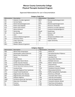


© Copyright 2026