
Giacomo Koch, Massimiliano Oliveri, Giovanni A. Carlesimo and Carlo Caltagirone 2002;59;1658 Neurology
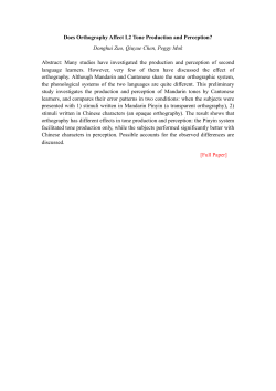
Selective deficit of time perception in a patient with right prefrontal cortex lesion
Giacomo Koch, Massimiliano Oliveri, Giovanni A. Carlesimo and Carlo Caltagirone
Neurology 2002;59;1658
This information is current as of January 3, 2010
The online version of this article, along with updated information and services, is
located on the World Wide Web at:
http://www.neurology.org/cgi/content/full/59/10/1658
Neurology® is the official journal of the American Academy of Neurology. Published continuously
since 1951, it is now a weekly with 48 issues per year. Copyright © 2002 by AAN Enterprises, Inc.
All rights reserved. Print ISSN: 0028-3878. Online ISSN: 1526-632X.
Downloaded from www.neurology.org at UNIV TEXAS on January 3, 2010
significantly changing BP and respiratory rate. Similar HR
changes with high frequency stimulation of the STN in patients
with PD were recently reported.4 The basal ganglia project to
several nuclei that may modify autonomic outflow5 as well as the
pediculopontine nuclei, part of the mesencephalic locomotor region, which when stimulated in cats increase HR and BP.2 High
frequency stimulation inhibits neuronal activity around the implanted electrode affecting fibers of passage as well as cell bodies;
thus, the neurons involved in the response cannot be ascertained.5
Interestingly, however, stimulation of the globus pallidum pars
interna (GPi), unlike the effect of STN stimulation, produced no
change in HR in patients with PD4; in anesthetized cats, GPi
stimulation produced bradycardia rather than tachycardia,6 suggesting that the tachycardia triggered by stimulation of the STN
may be a specific response.
Hyperactivity of STN neurons is a typical feature of PD5 that
impairs motor function and may also affect central command.
Abnormalities in central command may explain, at least in part,
some frequent autonomic deficits in patients with PD, particularly
their impaired cardiovascular adaptation to rapid assumption of
upright posture.7 High frequency electrical stimulation reduces
STN hyperactivity and improves motor performance. It also increases HR concomitantly with the reduction of akinesia, an appropriate autonomic response when initiating movement. Thus it
is tempting to speculate that in patients with PD STN stimulation
may also improve central command.
From the Department of Neurology, Mount Sinai School of Medicine, New
York, NY.
Selective deficit of time perception in a
patient with right prefrontal cortex lesion
the duration of events, judging them as shorter than they actually
were. He had difficulty evaluating how much time had elapsed
since the beginning of a determinate event. He said he was not
able to judge when the working day was over, leaving the office
earlier than the scheduled time.
Cranial MRI showed an ischemic lesion in the right frontal
lobe (dorsolateral prefrontal cortex, BA 46/9), and the patient was
admitted to a neurologic unit. Angio-MRI revealed occlusion of the
right internal carotid artery.
Methods. The patient obtained normal scores in a neuropsychological test battery evaluating short and long-term memory
(Rey Word List, Immediate and Delayed Recall; Figure of Rey-b,
Immediate and Delayed Recall; Digit Span; Corsi Span, Forward
and Backward; Verbal Supraspan; Immediate Visual Memory),
visuospatial abilities (Raven Progressive Matrices), attention (Trial Making Test), language (Verbal Fluency, Phrase Construction),
executive functions (Tower of London; Wisconsin Card Sorting
Test), praxia (copy of drawings). He did not present frontal signs,
such as impersistence and iterativity.
To evaluate the deficit in time estimation, we submitted the
patient and eight healthy control subjects (mean age: 45 ⫾ 5
years) to a verbal time estimation test in which they reported the
duration of a trial indicated by visual markers.6 Subjects sat fac-
Giacomo Koch, MD; Massimiliano Oliveri, MD, PhD;
Giovanni A. Carlesimo, MD, PhD; and Carlo Caltagirone, MD
The neural systems underlying subjective perception of time are of
increasing interest for neuroscientists. Previous studies in humans and in animals have documented a role of the cerebellum
and basal ganglia as an internal clock of discrete temporal units.1,2
Recent data from focal lesion investigations have suggested that
the frontal and the parietal lobes also are critical for time perception, especially for their role in attention and in maintaining the
representation of subjective time in the working memory.3,4 Neuropsychological and functional imaging studies have supported
the importance of the prefrontal cortex in the perception and in
the comparison of time intervals.5
We describe the case of a patient who had a selective impairment in the perception of events’ duration.
Case report. A 49-year-old man reported mental confusion
and difficulty with concentration. Neurologic examination showed
very mild left hemiparesis that regressed in a few days. After this
acute episode, the patient spontaneously had trouble estimating
Received April 30, 2002. Accepted in final form July 25, 2002.
Address correspondence and reprint requests to Dr. Horacio Kaufmann,
Mount Sinai School of Medicine, Box 1052, New York, NY 10029; e-mail:
[email protected]
Copyright © 2002 by AAN Enterprises, Inc.
References
1. Kaufman MP, Rybicki KJ. Discharge properties of group III and IV
muscle afferents: their responses to mechanical and metabolic stimuli.
Circ Res 1987;61:160 –165.
2. Eldridge FL, Millhorn DE, Kiley JP, Waldrop TG. Stimulation by central
command of locomotion, respiration and circulation during exercise. Respir Physiol 1985;59:313–337.
3. Angyan L. Somatomotor and cardiorespiratory responses to basal ganglia stimulation in cats. Physiol Behav 1994;56:167–173.
4. Thornton JM, Aziz T, Schlugman D, Paterson DJ. Electrical stimulation
of the midbrain increases heart rate and arterial blood pressure in
awake humans. J Physiol 2002;539:615– 621.
5. Beurrier C, Bioulac B, Audin J, Hammond C. High-frequency stimulation produces a transient blockade of voltage-gated currents in subthalamic neurons. J Neurophysiol 2001;85:1351–1356.
6. Angyan L. Cardiorespiratory effects of electrical stimulation of the globus pallidus in cats. Physiol Behav 1996;59:455– 459.
7. Senard JM, Brefel-Courbon C, Rascol O, Montastruc JL. Orthostatic
hypotension in patients with Parkinson’s disease: pathophysiology and
management. Drugs Aging 2001;18:495–505.
Figure. Mean perception of time intervals in patient (black circles) and control subjects (white squares). Values in
the two axes are expressed in milliseconds. Error bars indicate 1 SEM.
1658
NEUROLOGY 59
November (2 of 2) 2002
Downloaded from www.neurology.org at UNIV TEXAS on January 3, 2010
ing a computer screen in a quiet room. Number stimuli (1 through
9) were presented in a random sequence on the screen, each of
them for a period ranging from 200 to 2000 msec, until the selected time interval was completed. Time intervals were 5, 10, 30,
60 and 90, seconds. Each time interval was repeated randomly
four times, with a total sequence of 20 trials. At the end of each
trial the monitor presented the sentence, “How many seconds did
the trial last?” Participants were requested to report verbally the
duration of the interval.
During each trial, subjects were required to read the numbers
aloud to prevent subvocal counting and to divert attention to
timing. To reduce session length and to maintain constant cooperation, we did not test longer intervals.
Results. The patient was significantly less accurate as compared with control subjects in the evaluation of the longer interval
(90 seconds), showing a clear tendency to underestimate the real
time (time duration more than 2.5 SD shorter compared with the
controls’ mean) (figure). The patient’s performance at the other
time intervals did not significantly differ from that of the control
subjects.
Discussion. Results of recent research with patients with focal lesions suggest that the right frontal cortex is involved in time
perception. This report describes the case of a single patient in
which altered temporal processing emerges as a selective deficit
after lesion of the right dorsolateral prefrontal cortex.
The role of the dorsolateral prefrontal cortex in time perception
has been related to the encoding of temporal information into
memory, and some studies have considered time as a fourth dimension of the working memory.3 Our data indicate that the right
dorsolateral prefrontal cortex could also be involved in the evaluation of long time intervals, outside the working memory boundaries. One hypothesis is that the right frontal cortex could work as
an accumulator of a central internal clock, receiving inputs from
the basal ganglia and the cerebellum to form a conscious representation of time intervals. In addition, although neuropsychological
investigation failed to show signs of pathologic distractibility, we
cannot exclude a contribution of defective attentional control to
the patient’s poor time estimates.3
Receptive amelodia in a trained musician
aprosodia). He made errors of up to 30 degrees in attempting to
point to a sound source while blindfolded.
Carotid duplex revealed a hemodynamically significant stenosis of the right internal carotid artery. CAT scan and MRI of the
brain failed to show an infarction. SPECT scan revealed decreased
perfusion of the right temporal lobe (see the figure).
On follow-up examinations his ability to identify melodies improved to approximately 20% correct by 1 month after discharge,
and ultimately to 70% accuracy by 3 years. However, even when
he recognized a composition, recognition was not immediate.
Throughout this time period he remained unaware of his deficits
and denied any difficulty with musical perception.
Discussion. Our patient was found to have profound inability
to discern melody despite intact perception of pitch, rhythm, and
harmony. The cause was likely an ischemic injury of the right
temporal lobe.
Clinical case studies have reported patients who developed
receptive amelodia after unilateral ischemic1,2 or hemorrhagic3 injury of the right temporal lobe. None of these patients had formal
musical training. An amateur musician4 experienced distortions in
musical timbre and impaired recognition of the identity of voices
and environmental sounds after right temporal lobe infarction,
although he continued to recognize melodies. Another patient with
some musical training5 developed progressive loss of melody recog-
Steven A. Sparr, MD
Clinical case studies of patients with isolated disorders of musical
perception are exceedingly rare, and help elucidate the localization of various components of music in the brain. We report a
highly trained musician who experienced profound inability to
discern melody due to right temporal lobe injury.
Case history. A 91-year-old retired musicologist of great accomplishment with a history of diabetes mellitus and coronary
artery disease suddenly developed difficulty reading the left side
of his newspaper, unsteady gait, and difficulty dressing.
Initial examination showed no evidence of aphasia or dementia. He had a dense left homonymous hemianopia, mild left hemiparesis, and elements of left hemispatial neglect. On the evening
of admission he experienced auditory hallucinations of a choir
singing. All of these deficits cleared over the next 24 to 72 hours.
During his hospitalization his auditory abilities were studied
in detail. He was unable to identify the melody of any of a wide
variety of well-known musical pieces presented by recording or
live by piano. He did not recognize relatively simple tunes played
on a single instrument or vocal music, which contained the additional clues of verbal lyrics. He could repeat a series of three notes
or fewer, but consistently failed to reproduce a series of four notes
or more. He made errors in identifying instruments playing; at
one point he identified the horn section as a “harp.”
At the same time, he had no difficulty humming a tune from
memory. He was able to replicate the pitch of single notes. Given
two notes, he had no difficulty indicating which note was higher.
He readily distinguished consonant from dissonant chords. He
could replicate the rhythm of a series of handclaps. Indeed, with
many of the recorded pieces that he could not recognize, he was
able to establish the rhythm and accurately pretend to lead the
orchestra in time.
When presented with sheet music, he was immediately able to
discern the melody of the composition, and readily categorized its
style. He was able to explain the melodic lines and interactions of
various instruments in a Stravinsky score.
With respect to other nonlinguistic auditory functions, he was
unable to identify recorded sound effects, spoken or singing voices
of famous personalities, or the emotional tone of a voice (receptive
From the Department of Neuroscience (Drs. Koch, Carlesimo, and Caltagirone), University of Rome Tor Vergata; Fondazione Santa Lucia IRCCS
(Drs. Koch, Oliveri, Carlesimo, and Caltagirone), Rome; and Department of
Psychology (Dr. Oliveri), University of Palermo, Italy.
Received April 30, 2002. Accepted in final form July 24, 2002.
Address correspondence and reprint requests to Dr. Giacomo Koch, Fondazione Santa Lucia IRCCS, Laboratorio di Neurologia Clinica e Comportamentale, Via Ardeatina 306, 00179 Rome, Italy; e-mail: [email protected]
Copyright © 2002 by AAN Enterprises, Inc.
References
1. Gibbon J, Malapani C, Dale CL, and Gallistel CR. Toward a neurobiology
of temporal cognition: advances and challenges. Curr Opin Neurobiol
1997;7:170 –184.
2. Nichelli P, Alway D, and Grafman J. Perceptual timing in cerebellar
degeneration. Neuropsychologia 1996;34:863– 871.
3. Harrington DL, Haaland K, and Knight R. Cortical networks underlying
mechanism of time perception. J Neurosci 1998;18:1085–1095.
4. Rao SM, Mayer AR, and Harrington DL. The evolution of brain activation during temporal processing. Nat Neurosci 2001;4:317–323.
5. Mangels JA, Ivry RB, Shimuzu N. Dissociable contributions of the prefrontal and neocerebellar cortex to time perception. Cogn Brain Res
1998;7:15–39.
6. Mimura M, Kinsbourne M, and O’Connor M. Time estimation by patients with frontal lesions and by Korsakoff amnesics. J Int Neuropsychol Soc 2000;6:517–528.
Figure. SPECT scan shows diminished perfusion of the
right temporal lobe (left side of image).
November (2 of 2) 2002 NEUROLOGY 59 1659
Downloaded from www.neurology.org at UNIV TEXAS on January 3, 2010
Selective deficit of time perception in a patient with right prefrontal cortex lesion
Giacomo Koch, Massimiliano Oliveri, Giovanni A. Carlesimo and Carlo Caltagirone
Neurology 2002;59;1658
This information is current as of January 3, 2010
Updated Information
& Services
including high-resolution figures, can be found at:
http://www.neurology.org/cgi/content/full/59/10/1658
Subspecialty Collections
This article, along with others on similar topics, appears in the
following collection(s):
All Neuropsychology/Behavior
http://www.neurology.org/cgi/collection/all_neuropsychology_beh
avior Attention
http://www.neurology.org/cgi/collection/attention
Permissions & Licensing
Information about reproducing this article in parts (figures, tables)
or in its entirety can be found online at:
http://www.neurology.org/misc/Permissions.shtml
Reprints
Information about ordering reprints can be found online:
http://www.neurology.org/misc/reprints.shtml
Downloaded from www.neurology.org at UNIV TEXAS on January 3, 2010


© Copyright 2026