
Note: Large im ages and tables on this page m... Copyright © The McGraw-Hill Companies. A ll rights reserved. Print
essMedicine
1/6/13 | Print: Treatment and Prophy laxis of Bacterial Inf ections: Introduction
Print
C lose W indow
Note: Large im age s and table s on this page m ay ne ce ssitate printing in landscape m ode .
Copyright © The McGraw-Hill Companies. A ll rights reserved.
Harrison's Online > Part 8. Infe ctious Dise ase s > Se ction 4. Approach to The rapy for Bacte rial Dise ase s > C hapte r
133. Tre atm e nt and Prophylax is of Bacte rial Infe ctions >
Treatment and Prophylaxis of Bacterial Infections: Introduction
The development of vaccines and drugs that prevent and cure bacterial infections was one of the
twentieth century's major contributions to human longevity and quality of life. Antibacterial agents are
among the most commonly prescribed drugs of any kind worldwide. Used appropriately, these drugs
are lifesaving. However, their indiscriminate use drives up the cost of health care, leads to a plethora of
side effects and drug interactions, and fosters the emergence of bacterial resistance, rendering
previously valuable drugs useless. The rational use of antibacterial agents depends on an
understanding of (1) the drugs' mechanisms of action, spectra of activity, pharmacokinetics,
pharmacodynamics, toxicities, and interactions; (2) mechanisms underlying bacterial resistance; and (3)
strategies that can be used by clinicians to limit resistance. In addition, patient-associated parameters,
such as infection site, other drugs being taken, allergies, and immune and excretory status, are
critically important to appropriate therapeutic decisions. This chapter provides specific data required for
making an informed choice of antibacterial agent.
C opyright © The McGraw-Hill C ompanies. All rights reserved.
Privacy Notice. Any use is subject to the Terms of Use and Notice.
Your IP address is 24.97.224.18
0-www.accessmedicine.com.lilac.une.edu/popup.aspx?aID=9093788&print=y es
Print
C lose W indow
Note: Large im age s and table s on this page m ay ne ce ssitate printing in landscape m ode .
Copyright © The McGraw-Hill Companies. A ll rights reserved.
Harrison's Online > Part 8. Infe ctious Dise ase s > Se ction 4. Approach to The rapy for Bacte rial Dise ase s > C hapte r 133. Tre atm e nt and Prophylax is of Bacte rial Infe ctions >
MECHANISMS OF ACTION
Antibacterial agents, like all antimicrobial drugs, are directed against unique targets not present in mammalian cells. The goal is to limit toxicity to the host and
maximize chemotherapeutic activity affecting invading microbes only. Bactericidal drugs kill the bacteria that are within their spectrum of activity; bacteriostatic
drugs only inhibit bacterial growth. While bacteriostatic activity is adequate for the treatment of most infections, bactericidal activity may be necessary for cure
in patients with altered immune systems (e.g., neutropenia), protected infectious foci (e.g., endocarditis or meningitis), or specific infections (e.g., complicated
Staphylococcus aureus bacteremia). The mechanisms of action of the antibacterial agents to be discussed in this section are summarized in Table 133-1 and
are depicted in Fig. 133-1.
Table 133-1 Mechanisms of Action of and Resistance to Major Classes of Antibacterial Agents
Letter
for Fig.
133-1
Antibacterial Agenta
Major
Cellular
Target
Mechanism of Action
Major Mechanisms of Resistance
A
-Lactams (penicillins,
cephalosporins)
Cell wall
Inhibit cell-wall cross-linking
1. Drug inactivation ( -lactamase)
2. Insensitivity of target (altered penicillin-binding proteins)
3. Decreased permeability (altered gram-negative outermembrane porins)
4. Active efflux
B
C
Vancomycin
Cell wall
Interferes with addition of new cell-wall
subunits (muramyl pentapeptides)
Alteration of target (substitution of terminal amino acid of
peptidoglycan subunit)
Bacitracin
Cell wall
Prevents addition of cell-wall subunits by
inhibiting recycling of membrane lipid carrier
Not defined
Macrolides
(erythromycin)
Protein
synthesis
Bind to 50S ribosomal subunit
1. Alteration of target (ribosomal methylation and mutation of
23S rRNA)
2. Active efflux
D
E
F
Lincosamides
(clindamycin)
Protein
synthesis
Bind to 50S ribosomal subunit Block peptide
chain elongation
1. Alteration of target (ribosomal methylation)
Chloramphenicol
Protein
synthesis
Binds to 50S ribosomal subunit Blocks
aminoacyl tRNA attachment
1. Drug inactivation (chloramphenicol acetyltransferase)
Protein
synthesis
Binds to 30S ribosomal subunit Blocks
binding of aminoacyl tRNA
1. Decreased intracellular drug accumulation (active efflux)
Aminoglycosides
Protein
Bind to 30S ribosomal subunit Inhibit
1. Drug inactivation (aminoglycoside-modifying enzyme)
(gentamicin)
synthesis
translocation of peptidyl-tRNA
2. Decreased permeability through gram-negative outer
membrane
Tetracycline
2. Active efflux
2. Active efflux
2. Insensitivity of target
3. Active efflux
4. Ribosomal methylation
G
Mupirocin
Protein
synthesis
H
Streptogramins
Protein
[quinupristin/dalfopristin synthesis
(Synercid)]
Inhibits isoleucine tRNA synthetase
Mutation of gene for target protein or acquisition of new gene
for drug-insensitive target
Bind to 50S ribosomal subunit Block peptide
chain elongation
1. Alteration of target (ribosomal methylation: dalfopristin)
2. Active efflux (quinupristin)
3. Drug inactivation (quinupristin and dalfopristin)
I
Linezolid
Protein
synthesis
Binds to 50S ribosomal subunit Inhibits
initiation of protein synthesis
Alteration of target (mutation of 23S rRNA)
J
Sulfonamides and
trimethoprim
Cell
Competitively inhibit enzymes involved in
metabolism two steps of folic acid biosynthesis
Production of insensitive targets [dihydropteroate synthetase
(sulfonamides) and dihydrofolate reductase (trimethoprim)] that
bypass metabolic block
K
Rifampin
Nucleic acid Inhibits DNA-dependent RNA polymerase
synthesis
Insensitivity of target (mutation of polymerase gene)
L
Metronidazole
Nucleic acid Intracellularly generates short-lived reactive Not defined
synthesis
intermediates that damage DNA by electron
transfer system
M
Quinolones
(ciprofloxacin)
DNA
synthesis
Inhibit activity of DNA gyrase (A subunit) and 1. Insensitivity of target (mutation of gyrase genes)
topoisomerase IV
2. Decreased intracellular drug accumulation (active efflux)
1/6/13
AccessMedicine | Print: Mechanisms of Action
N
O
a
Novobiocin
DNA
synthesis
Inhibits activity of DNA gyrase (B subunit)
Not defined
Polymyxins (polymyxin
B)
Cell
membrane
Disrupt membrane permeability by charge
alteration
Not defined
Gramicidin
Cell
membrane
Forms pores
Not defined
Daptomycin
Cell
membrane
Forms channels that disrupt membrane
potential
Alteration of membrane charge
Compounds in parentheses are major representatives for the class.
Figure 133-1
Mechanisms of action of and resistance to antibacterial agents. Black lines trace the routes of drug interaction with bacterial cells, from entry to target site. The
letters in each figure indicate specific antibacterial agents or classes of agents, as shown in Table 133-1. The numbers correspond to mechanisms listed beneath each panel.
50s and 30s, large and small ribosome subunits; Ac, acetylation; Ad, adenylation; DHFR, dihydrofolate reductase; DHPS, dihydropteroate synthetase; IM, inner (cytoplasmic)
membrane; LPS, lipopolysaccharide; OM, outer membrane; P, phosphorylation; PBP, penicillin-binding protein; PG, peptidoglycan.
0-www.accessmedicine.com.lilac.une.edu/popup.aspx?aID=9093790&print=yes
2/5
1/6/13
AccessMedicine | Print: Mechanisms of Action
membrane; LPS, lipopolysaccharide; OM, outer membrane; P, phosphorylation; PBP, penicillin-binding protein; PG, peptidoglycan.
Inhibition of Cell-Wall Synthesis
One major difference between bacterial and mammalian cells is the presence in bacteria of a rigid wall external to the cell membrane. The wall protects
bacterial cells from osmotic rupture, which would result from the cell's usual marked hyperosmolarity (by up to 20 atm) relative to the host environment. The
structure conferring cell-wall rigidity and resistance to osmotic lysis in both gram-positive and gram-negative bacteria is peptidoglycan, a large, covalently linked
sacculus that surrounds the bacterium. In gram-positive bacteria, peptidoglycan is the only layered structure external to the cell membrane and is thick (20–80
nm); in gram-negative bacteria, there is an outer membrane external to a very thin (1-nm) peptidoglycan layer.
Chemotherapeutic agents directed at any stage of the synthesis, export, assembly, or cross-linking of peptidoglycan lead to inhibition of bacterial cell growth
and, in most cases, to cell death. Peptidoglycan is composed of (1) a backbone of two alternating sugars, N-acetylglucosamine and N-acetylmuramic acid; (2) a
chain of four amino acids that extends down from the backbone (stem peptides); and (3) a peptide bridge that cross-links the peptide chains. Peptidoglycan is
formed by the addition of subunits (a sugar with its five attached amino acids) that are assembled in the cytoplasm and transported through the cytoplasmic
membrane to the cell surface. Subsequent cross-linking is driven by cleavage of the terminal stem-peptide amino acid.
Virtually all the antibiotics that inhibit bacterial cell-wall synthesis are bactericidal. That is, they eventually result in the cell's death due to osmotic lysis.
However, much of the loss of cell-wall integrity following treatment with cell wall–active agents is due to the bacteria's own cell-wall remodeling enzymes
(autolysins) that cleave peptidoglycan bonds in the normal course of cell growth. In the presence of antibacterial agents that inhibit cell-wall growth, autolysis
proceeds without normal cell-wall repair; weakness and eventual cellular lysis occur. Antibacterial agents act to inhibit cell-wall synthesis in several ways, as
described below.
Bacitracin
Bacitracin, a cyclic peptide antibiotic, inhibits the conversion to its active form of the lipid carrier that moves the water-soluble cytoplasmic peptidoglycan
subunits through the cell membrane to the cell exterior.
Glycopeptides
Glycopeptides [vancomycin, teicoplanin, and telavancin (lipoglycopeptide)] are high-molecular-weight antibiotics that bind to the terminal D-alanine– D-alanine
component of the stem peptide while the subunits are external to the cell membrane but still linked to the lipid carrier. This binding sterically inhibits the
addition of subunits to the peptidoglycan backbone.
-Lactam Antibiotics
-Lactam antibiotics (penicillins, cephalosporins, carbapenems, and monobactams; Table 133-2) are characterized by a four-membered
-lactam ring and
prevent the cross-linking reaction called transpeptidation. The energy for attaching a peptide cross-bridge from the stem peptide of one peptidoglycan subunit
to another is derived from the cleavage of a terminal D-alanine residue from the subunit stem peptide. The cross-bridge amino acid is then attached to the
penultimate D-alanine by transpeptidase enzymes. The
-lactam ring of the antibiotic forms an irreversible covalent acyl bond with the transpeptidase enzyme
(probably because of the antibiotic's steric similarity to the enzyme's D-alanine– D-alanine target), preventing the cross-linking reaction. Transpeptidases and
similar enzymes involved in cross-linking are called penicillin-binding proteins (PBPs) because they all have active sites that bind
-lactam antibiotics.
Table 133-2 Classification of -Lactam Antibiotics
Route of Administration
Class
Parenteral
Oral
Narrow-spectrum
Penicillin G
Penicillin V
Enteric-active
Ampicillin
Amoxicillin, ampicillin
Enteric-active and
Ticarcillin, piperacillin
None
Oxacillin, nafcillin
Cloxacillin, dicloxacillin
Ticarcillin plus clavulanic acid, ampicillin plus sulbactam,
piperacillin plus tazobactam
Amoxicillin plus clavulanic acid
Cefazolin, cephapirin
Cephalexin, cefadroxil
Haemophilus-active
Cefuroxime, cefonicid, ceforanide
Cefaclor, cefuroxime axetil, ceftibuten, cefdinir, cefprozil,
cefditoren, cefpodoxime a
Bacteroides-active
Cefoxitin, cefotetan
None
Penicillins
-Lactamase-susceptible
antipseudomonal
-Lactamase-resistant
Antistaphylococcal
Combined with
inhibitors
-lactamase
Cephalosporins
First-generation
Second-generation
Third-generation
Extended-spectrum
Ceftriaxone, cefotaxime, ceftizoxime
None
Extended-spectrum and
antipseudomonal
Ceftazidime, cefepime
None
Extended-spectrum and antiMRSAb
Ceftobiprole
None
Carbapenems
Imipenem/cilastatin, meropenem, ertapenem, doripenem
None
Monobactams
Aztreonam
None
a
Some sources classify cefpodoxime as a third-generation oral agent because of a marginally broader spectrum.
0-www.accessmedicine.com.lilac.une.edu/popup.aspx?aID=9093790&print=yes
3/5
1/6/13
AccessMedicine | Print: Mechanisms of Action
a
Some sources classify cefpodoxime as a third-generation oral agent because of a marginally broader spectrum.
b Methicillin-resistant Staphylococcus aureus.
Inhibition of Protein Synthesis
Most of the antibacterial agents that inhibit protein synthesis interact with the bacterial ribosome. The difference between the composition of bacterial and
mammalian ribosomes gives these compounds their selectivity.
Aminoglycosides
Aminoglycosides (gentamicin, kanamycin, tobramycin, streptomycin, neomycin, and amikacin) are a group of structurally related compounds containing three
linked hexose sugars. They exert a bactericidal effect by binding irreversibly to the 30S subunit of the bacterial ribosome and inhibiting translocation of peptidyltRNA from the A to the P site. Uptake of aminoglycosides and their penetration through the cell membrane constitute an aerobic, energy-dependent process.
Thus, aminoglycoside activity is markedly reduced in an anaerobic environment. Spectinomycin, an aminocyclitol antibiotic, also acts on the 30S ribosomal
subunit but has a different mechanism of action from the aminoglycosides and is bacteriostatic rather than bactericidal.
Macrolides, Ketolides, and Lincosamides
Macrolide antibiotics (erythromycin, clarithromycin, and azithromycin) consist of a large lactone ring to which sugars are attached. Ketolide antibiotics, including
telithromycin, replace the cladinose sugar on the macrolactone ring with a ketone group. These drugs bind specifically to the 50S portion of the bacterial
ribosome and inhibit protein chain elongation. Although structurally unrelated to the macrolides, lincosamides (clindamycin and lincomycin) bind to a site on the
50S ribosome nearly identical to the binding site for macrolides.
Streptogramins
Streptogramins [quinupristin (streptogramin B) and dalfopristin (streptogramin A)], which are supplied as a combination in Synercid, are peptide macrolactones
that also bind to the 50S ribosomal subunit and block protein synthesis. Streptogramin B binds to a ribosomal site similar to the binding site for macrolides and
lincosamides, whereas streptogramin A binds to a different ribosomal site, blocking the late phase of protein synthesis. The two streptogramins act
synergistically to kill bacteria if the strain is susceptible to both components.
Chloramphenicol
Chloramphenicol consists of a single aromatic ring and a short side chain. This antibiotic binds reversibly to the 50S portion of the bacterial ribosome at a site
close to but not identical with the binding sites for the macrolides and lincosamides, inhibiting peptide bond formation by blocking attachment of the amino acid
end of aminoacyl-tRNA to the ribosome.
Linezolid
Linezolid is the only commercially available drug in the oxazolidinone class. Linezolid binds to the 50S ribosomal subunit and blocks the initiation of protein
synthesis.
Tetracyclines and Glycylcyclines
Tetracyclines (tetracycline, doxycycline, and minocycline) and glycylcyclines (tigecycline) consist of four aromatic rings with various substituent groups. They
interact reversibly with the bacterial 30S ribosomal subunit, blocking the binding of aminoacyl tRNA to the mRNA-ribosome complex. This mechanism is markedly
different from that of the aminoglycosides, which also bind to the 30S subunit.
Mupirocin
Mupirocin (pseudomonic acid) inhibits isoleucine tRNA synthetase by competing with bacterial isoleucine for its binding site on the enzyme and depleting cellular
stores of isoleucine-charged tRNA.
Inhibition of Bacterial Metabolism
The antimetabolites are all synthetic compounds that interfere with bacterial synthesis of folic acid. Products of the folic acid synthesis pathway function as
coenzymes for the one-carbon transfer reactions that are essential for the synthesis of thymidine, all purines, and several amino acids. Inhibition of folate
synthesis leads to cessation of bacterial cell growth and, in some cases, to bacterial cell death. The principal antibacterial antimetabolites are sulfonamides
(sulfisoxazole, sulfadiazine, and sulfamethoxazole) and trimethoprim.
Sulfonamides
Sulfonamides are structural analogues of p-aminobenzoic acid (PABA), one of the three structural components of folic acid (the other two being pteridine and
glutamate). The first step in the synthesis of folic acid is the addition of PABA to pteridine by the enzyme dihydropteroic acid synthetase. Sulfonamides compete
with PABA as substrates for the enzyme. The selective effect of sulfonamides is due to the fact that bacteria synthesize folic acid, while mammalian cells cannot
synthesize the cofactor and must use exogenous supplies. However, the activity of sulfonamides can be greatly reduced by the presence of excess PABA or by
the exogenous addition of end products of one-carbon transfer reactions (e.g., thymidine and purines). High concentrations of the latter substances may be
present in some infections as a result of tissue and white cell breakdown, compromising sulfonamide activity.
Trimethoprim
Trimethoprim is a diaminopyrimidine, a structural analogue of the pteridine moiety of folic acid. Trimethoprim is a competitive inhibitor of dihydrofolate
reductase; this enzyme is responsible for reduction of dihydrofolic acid to tetrahydrofolic acid—the essential final component in the folic acid synthesis pathway.
Like that of the sulfonamides, the activity of trimethoprim is compromised in the presence of exogenous thymine or thymidine.
Inhibition of Nucleic Acid Synthesis or Activity
Numerous antibacterial compounds have disparate effects on nucleic acids.
Quinolones
The quinolones, including nalidixic acid and its fluorinated derivatives (ciprofloxacin, levofloxacin, and moxifloxacin), are synthetic compounds that inhibit the
activity of the A subunit of the bacterial enzyme DNA gyrase as well as topoisomerase IV. DNA gyrase and topoisomerases are responsible for negative
supercoiling of DNA—an essential conformation for DNA replication in the intact cell. Inhibition of the activity of DNA gyrase and topoisomerase IV is lethal to
bacterial cells. The antibiotic novobiocin also interferes with the activity of DNA gyrase, but it interferes with the B subunit.
Rifampin
Rifampin, used primarily against Mycobacterium tuberculosis, is also active against a variety of other bacteria. Rifampin binds tightly to the B subunit of bacterial
0-www.accessmedicine.com.lilac.une.edu/popup.aspx?aID=9093790&print=yes
4/5
1/6/13
AccessMedicine | Print: Mechanisms of Action
DNA-dependent RNA polymerase, thus inhibiting transcription of DNA into RNA. Mammalian-cell RNA polymerase is not sensitive to this compound.
Nitrofurantoin
Nitrofurantoin, a synthetic compound, causes DNA damage. The nitrofurans, compounds containing a single five-membered ring, are reduced by a bacterial
enzyme to highly reactive, short-lived intermediates that are thought to cause DNA strand breakage, either directly or indirectly.
Metronidazole
Metronidazole, a synthetic imidazole, is active only against anaerobic bacteria and protozoa. The reduction of metronidazole's nitro group by the bacterial
anaerobic electron-transport system produces a transient series of reactive intermediates that are thought to cause DNA damage.
Alteration of Cell-Membrane Permeability
Polymyxins
The polymyxins [polymyxin B and colistin (polymyxin E)] are cyclic, basic polypeptides. They behave as cationic, surface-active compounds that disrupt the
permeability of both the outer and the cytoplasmic membranes of gram-negative bacteria.
Gramicidin A
Gramicidin A is a polypeptide of 15 amino acids that acts as an ionophore, forming pores or channels in lipid bilayers.
Daptomycin
Insertion of daptomycin, a bactericidal lipopeptide antibiotic, into the cell membrane of gram-positive bacteria forms a channel that causes depolarization of the
membrane by efflux of intracellular ions, resulting in cell death.
C opyright © The McGraw-Hill C ompanies. All rights reserved.
Privacy Notice. Any use is subject to the Terms of Use and Notice.
Your IP address is 24.97.224.18
0-www.accessmedicine.com.lilac.une.edu/popup.aspx?aID=9093790&print=yes
5/5
1/6/13
AccessMedicine | Print: Mechanisms of Resistance
Print
C lose W indow
Note: Large im age s and table s on this page m ay ne ce ssitate printing in landscape m ode .
Copyright © The McGraw-Hill Companies. A ll rights reserved.
Harrison's Online > Part 8. Infe ctious Dise ase s > Se ction 4. Approach to The rapy for Bacte rial Dise ase s > C hapte r
133. Tre atm e nt and Prophylax is of Bacte rial Infe ctions >
MECHANISMS OF RESISTANCE
Some bacteria exhibit intrinsic resistance to certain classes of antibacterial agents (e.g., obligate
anaerobic bacteria to aminoglycosides and gram-negative bacteria to vancomycin). In addition, bacteria
that are ordinarily susceptible to antibacterial agents can acquire resistance. Acquired resistance is a
major limitation to effective antibacterial chemotherapy. Resistance can develop by mutation of resident
genes or by acquisition of new genes. New genes mediating resistance are usually spread from cell to
cell by way of mobile genetic elements such as plasmids, transposons, and bacteriophages. The
resistant bacterial populations flourish in areas of high antimicrobial use, where they enjoy a selective
advantage over susceptible populations.
The major mechanisms used by bacteria to resist the action of antimicrobial agents are inactivation of
the compound, alteration or overproduction of the antibacterial target through mutation of the target
protein's gene, acquisition of a new gene that encodes a drug-insensitive target, decreased
permeability of the cell envelope to the agent, failure to convert an inactive prodrug to its active
derivative, and active efflux of the compound from the periplasm or interior of the cell. Specific
mechanisms of bacterial resistance to the major antibacterial agents are outlined below, summarized in
Table 133-1, and depicted in Fig. 133-1.
-Lactam Antibiotics
Bacteria develop resistance to
-lactam antibiotics by a variety of mechanisms. Most common is the
destruction of the drug by -lactamases. The
-lactamases of gram-negative bacteria are confined to
the periplasm, between the inner and outer membranes, while gram-positive bacteria secrete their lactamases into the surrounding medium. These enzymes have a higher affinity for the antibiotic than
the antibiotic has for its target. Binding results in hydrolysis of the
-lactam ring. Genes encoding
-
lactamases have been found in both chromosomal and extrachromosomal locations and in both grampositive and gram-negative bacteria; these genes are often on mobile genetic elements. Many
"advanced-generation"
-lactam antibiotics, such as ceftriaxone and cefepime, are stable in the
presence of plasmid-mediated
generation
-lactamases and are active against bacteria resistant to earlier-
-lactam antibiotics. However, extended-spectrum -lactamases (ESBLs), either acquired on
mobile genetic elements by gram-negative bacteria (e.g., Klebsiella pneumoniae and Escherichia coli) or
present as stable chromosomal genes in other gram-negative species (e.g., Enterobacter spp.), have
broad substrate specificity, hydrolyzing virtually all penicillins and cephalosporins. Carbapenems are
generally resistant to ESBL hydrolysis and are the drugs of choice for the treatment of infections
caused by ESBL-producing Enterobacteriaceae. However, Enterobacteriaceae (particularly K.
pneumoniae) that produce carbapenemases and are resistant to virtually all -lactam antibiotics have
now emerged. One strategy that has been devised for circumventing resistance mediated by lactamases is to combine the
-lactam agent with an inhibitor that avidly binds the inactivating enzyme,
preventing its attack on the antibiotic. Unfortunately, the inhibitors (e.g., clavulanic acid, sulbactam,
1/4
1/6/13
AccessMedicine | Print: Mechanisms of Resistance
and tazobactam) do not bind all chromosomal -lactamases (e.g., that of Enterobacter) or
carbapenemases and thus cannot be depended on to prevent the inactivation of -lactam antibiotics
by such enzymes. No
-lactam antibiotic or inhibitor has been produced that can resist all of the many
-lactamases that have been identified.
A second mechanism of bacterial resistance to
-lactam antibiotics is an alteration in PBP targets so
that the PBPs have a markedly reduced affinity for the drug. While this alteration may occur by
mutation of existing genes, the acquisition of new PBP genes (as in staphylococcal resistance to
methicillin) or of new pieces of PBP genes (as in streptococcal, gonococcal, and meningococcal
resistance to penicillin) is more important.
A final resistance mechanism is the coupling, in gram-negative bacteria, of a decrease in outermembrane permeability with rapid efflux of the antibiotic from the periplasm to the cell exterior.
Mutations of genes encoding outer-membrane protein channels called porins decrease the entry of lactam antibiotics into the cell, while additional proteins form channels that actively pump
-lactams out
of the cell. Resistance of Enterobacteriaceae to some cephalosporins and resistance of Pseudomonas
spp. to cephalosporins and piperacillin are the best examples of this mechanism.
Vancomycin
Clinically important resistance to vancomycin was first described among enterococci in France in 1988.
Vancomycin-resistant enterococci (VRE) have subsequently become disseminated worldwide. The
genes encoding resistance are carried on plasmids that can transfer themselves from cell to cell and on
transposons that can jump from plasmids to chromosomes. Resistance is mediated by enzymes that
substitute D-lactate for D-alanine on the peptidoglycan stem peptide so that there is no longer an
appropriate target for vancomycin binding. This alteration does not appear to affect cell-wall integrity,
however. This type of acquired vancomycin resistance was confined for 14 years to enterococci—more
specifically, to Enterococcus faecium rather than the more common pathogen E. faecalis. However, since
2002, S. aureus isolates that are highly resistant to vancomycin have been recovered from 11 patients
in the United States. All of the isolates contain vanA, the gene that mediates vancomycin resistance in
enterococci. In addition, since 1996, a few isolates of both S. aureus and Staphylococcus epidermidis
that display a four- to eightfold reduction in susceptibility to vancomycin have been found worldwide;
such S. aureus strains are termed vancomycin-intermediate-susceptibility S. aureus, or VISA. Many more
isolates may contain subpopulations with reduced vancomycin susceptibility (heteroVISA, or hVISA).
These isolates have not acquired the genes that mediate vancomycin resistance in enterococci but are
mutant bacteria with markedly thickened cell walls. These mutants were apparently selected in
patients who were undergoing prolonged vancomycin therapy. The failure of vancomycin therapy in
some patients infected with S. aureus or S. epidermidis strains exhibiting only intermediate susceptibility
to this drug is thought to have resulted from this resistance.
Daptomycin
In some S. aureus isolates with reduced susceptibility to daptomycin, a mutation in the mprF gene
leads to an increase in the net positive charge of the bacterial membrane, repelling the antibiotic.
Aminoglycosides
The most common aminoglycoside resistance mechanism is inactivation of the antibiotic.
Aminoglycoside-modifying enzymes, usually encoded on plasmids, transfer phosphate, adenyl, or acetyl
residues from intracellular molecules to hydroxyl or amino side groups on the antibiotic. The modified
antibiotic is less active because of diminished binding to its ribosomal target. Modifying enzymes that
can inactivate any of the available aminoglycosides have been found in both gram-positive and gram0-www.accessmedicine.com.lilac.une.edu/popup.aspx?aID=9093845&print=y es
2/4
negative bacteria. A second aminoglycoside resistance mechanism, which has been identified
predominantly in clinical isolates of Pseudomonas aeruginosa, is decreased antibiotic uptake, presumably
due to alterations in the bacterial outer membrane. A third, emerging form of resistance in gramnegative bacteria is methylation of the target 16S ribosomal RNA, which is mediated by plasmidencoded methylases.
Macrolides, Ketolides, Lincosamides, and Streptogramins
Resistance in gram-positive bacteria, which are the usual target organisms for macrolides, ketolides,
lincosamides, and streptogramins, can be due to the production of an enzyme—most commonly
plasmid-encoded—that methylates ribosomal RNA, interfering with binding of the antibiotics to their
target. Methylation mediates resistance to erythromycin, clarithromycin, azithromycin, clindamycin, and
streptogramin B. Resistance to streptogramin B converts quinupristin/dalfopristin from a bactericidal to
a bacteriostatic antibiotic. Streptococci can also actively cause the efflux of macrolides, and
staphylococci can cause the efflux of macrolides, clindamycin, and streptogramin A. Ketolides such as
telithromycin retain activity against most isolates of Streptococcus pneumoniae that are resistant to
macrolides. In addition, staphylococci can inactivate streptogramin A by acetylation and streptogramin
B by either acetylation or hydrolysis. Finally, mutations in 23S ribosomal RNA that alter the binding of
macrolides to their targets have been found in both staphylococci and streptococci.
Chloramphenicol
Most bacteria resistant to chloramphenicol produce a plasmid-encoded enzyme, chloramphenicol
acetyltransferase, that inactivates the compound by acetylation.
Tetracyclines and Tigecycline
The most common mechanism of tetracycline resistance in gram-negative bacteria is a plasmid-encoded
active-efflux pump that is inserted into the cytoplasmic membrane and extrudes antibiotic from the cell.
Resistance in gram-positive bacteria is due either to active efflux or to ribosomal alterations that
diminish binding of the antibiotic to its target. Genes involved in ribosomal protection are found on
mobile genetic elements. The parenteral tetracycline derivative tigecycline (a glycylcycline) is active
against tetracycline-resistant bacteria because it is not removed by efflux and can bind to altered
ribosomes.
Mupirocin
Although the topical compound mupirocin was introduced into clinical use relatively recently, resistance
is already becoming widespread in some areas. The mechanism appears to be either mutation of the
target isoleucine tRNA synthetase so that it is no longer inhibited by the antibiotic or plasmid-encoded
production of a form of the target enzyme that binds mupirocin poorly.
Trimethoprim and Sulfonamides
The most prevalent mechanism of resistance to trimethoprim and the sulfonamides in both grampositive and gram-negative bacteria is the acquisition of plasmid-encoded genes that produce a new,
drug-insensitive target—specifically, an insensitive dihydrofolate reductase for trimethoprim and an
altered dihydropteroate synthetase for sulfonamides.
Quinolones
The most common mechanism of resistance to quinolones is the development of one or more mutations
in target DNA gyrases and topoisomerase IV that prevent the antibacterial agent from interfering with
the enzymes' activity. Some gram-negative bacteria develop mutations that both decrease outer-
1/6/13
AccessMedicine | Print: Mechanisms of Resistance
membrane porin permeability and cause active drug efflux from the cytoplasm. Mutations that result in
active quinolone efflux are also found in gram-positive bacteria.
Rifampin
Bacteria rapidly become resistant to rifampin by developing mutations in the B subunit of RNA
polymerase that render the enzyme unable to bind the antibiotic. The rapid selection of resistant
mutants is the major limitation to the use of this antibiotic against otherwise-susceptible staphylococci
and requires that the drug be used in combination with another antistaphylococcal agent.
Linezolid
Enterococci, streptococci, and staphylococci can become resistant to linezolid in vitro by mutation of the
23S rRNA binding site. Clinical isolates of E. faecium and E. faecalis acquire resistance to linezolid readily
by this mechanism, often during therapy. A new plasmid-encoded resistance gene has been found in
staphylococci that methylates the linezolid ribosomal binding site. At least one outbreak of linezolidresistant S. aureus infections caused by isolates carrying this gene has been described.
Multiple Antibiotic Resistance
The acquisition by one bacterium of resistance to multiple antibacterial agents is becoming increasingly
common. The two major mechanisms are the acquisition of multiple unrelated resistance genes and the
development of mutations in a single gene or gene complex that mediate resistance to a series of
unrelated compounds. The construction of multiresistant strains by acquisition of multiple genes occurs
by sequential steps of gene transfer and environmental selection in areas of high-level antimicrobial
use. In contrast, mutations in a single gene can conceivably be selected in a single step. Bacteria that
are multiresistant by virtue of the acquisition of new genes include hospital-associated strains of gramnegative bacteria, enterococci, and staphylococci and community-acquired strains of salmonellae,
gonococci, and pneumococci. Mutations that confer resistance to multiple unrelated antimicrobial
agents occur in the genes encoding outer-membrane porins and efflux proteins of gram-negative
bacteria. These mutations decrease bacterial intracellular and periplasmic accumulation of -lactams,
quinolones, tetracyclines, chloramphenicol, and aminoglycosides. Multiresistant bacterial isolates pose
increasing problems in U.S. hospitals; strains resistant to all available antibacterial chemotherapy have
already been identified.
C opyright © The McGraw-Hill C ompanies. All rights reserved.
Privacy Notice. Any use is subject to the Terms of Use and Notice.
Your IP address is 24.97.224.18
0-www.accessmedicine.com.lilac.une.edu/popup.aspx?aID=9093845&print=y es
4/4
1/6/13
AccessMedicine | Print: Pharmacokinetics of Antibiotics
Print
C lose W indow
Note: Large im age s and table s on this page m ay ne ce ssitate printing in landscape m ode .
Copyright © The McGraw-Hill Companies. A ll rights reserved.
Harrison's Online > Part 8. Infe ctious Dise ase s > Se ction 4. Approach to The rapy for Bacte rial Dise ase s > C hapte r
133. Tre atm e nt and Prophylax is of Bacte rial Infe ctions >
PHARMACOKINETICS OF ANTIBIOTICS
The pharmacokinetic profile of an antibacterial agent refers to its concentrations in serum and tissue
versus time and reflects the processes of absorption, distribution, metabolism, and excretion.
Important characteristics include peak and trough serum concentrations and mathematically derived
parameters such as half-life, clearance, and distribution volume. Pharmacokinetic information is useful
for estimating the appropriate antibacterial dose and frequency of administration, for adjusting
dosages in patients with impaired excretory capacity, and for comparing one drug with another. In
contrast, the pharmacodynamic profile of an antibiotic refers to the relationship between the
pharmacokinetics of the antibiotic and its minimal inhibitory concentrations (MICs) for bacteria (see
"Principles of Antibacterial Chemotherapy," below). For further discussion of basic pharmacokinetic
principles, see Chap. 5.
Absorption
Antibiotic absorption refers to the rate and extent of a drug's systemic bioavailability after oral, IM, or IV
administration.
ORAL ADMINISTRATION
Most patients with infection are treated with oral antibacterial agents in the outpatient setting.
Advantages of oral therapy over parenteral therapy include lower cost, generally fewer adverse effects
(including complications of indwelling lines), and greater acceptance by patients. The percentage of an
orally administered antibacterial agent that is absorbed (i.e., its bioavailability) ranges from as little as
10–20% (erythromycin and penicillin G) to nearly 100% [amoxicillin, clindamycin, metronidazole,
doxycycline, trimethoprim-sulfamethoxazole (TMP-SMX), linezolid, and most fluoroquinolones]. These
differences in bioavailability are not clinically important as long as drug concentrations at the site of
infection are sufficient to inhibit or kill the pathogen. However, therapeutic efficacy may be
compromised when absorption is reduced as a result of physiologic or pathologic conditions (such as
the presence of food for some drugs or the shunting of blood away from the gastrointestinal tract in
patients with hypotension), drug interactions (e.g., of quinolones and metal cations), or noncompliance.
The oral route is usually used for patients with relatively mild infections in whom absorption is not
thought to be compromised by the preceding conditions. In addition, the oral route can often be used
in more severely ill patients after they have responded to parenteral therapy and can take oral
medications.
INTRAMUSCULAR ADMINISTRATION
Although the IM route of administration usually results in 100% bioavailability, it is not as widely used
in the United States as the oral and IV routes, in part because of the pain often associated with IM
injections and the relative ease of IV access in the hospitalized patient. IM injection may be suitable for
specific indications requiring an "immediate" and reliable effect (e.g., with long-acting forms of penicillin,
1/3
1/6/13
AccessMedicine | Print: Pharmacokinetics of Antibiotics
including benzathine and procaine, and with single doses of ceftriaxone for acute otitis media or
uncomplicated gonococcal infection).
INTRAVENOUS ADMINISTRATION
The IV route is appropriate when oral antibacterial agents are not effective against a particular
pathogen, when bioavailability is uncertain, or when larger doses are required than are feasible with
the oral route. After IV administration, bioavailability is 100%; serum concentrations are maximal at the
end of the infusion. For many patients in whom long-term antimicrobial therapy is required and oral
therapy is not feasible, outpatient parenteral antibiotic therapy (OPAT), including the use of convenient
portable pumps, may be cost-effective and safe. Alternatively, some oral antibacterial drugs (e.g.,
fluoroquinolones) are sufficiently active against many Enterobacteriaceae to provide potency equal to
that of parenteral therapy; oral use of such drugs may allow the patient to return home from the
hospital earlier or to avoid hospitalization entirely.
Distribution
To be effective, concentrations of an antibacterial agent must exceed the pathogen's MIC. Serum
antibiotic concentrations usually exceed the MIC for susceptible bacteria, but since most infections are
extravascular, the antibiotic must also distribute to the site of the infection. Concentrations of most
antibacterial agents in interstitial fluid are similar to free-drug concentrations in serum. However, when
the infection is located in a "protected" site where penetration is poor, such as cerebrospinal fluid
(CSF), the eye, the prostate, or infected cardiac vegetations, high parenteral doses or local
administration for prolonged periods may be required for cure. In addition, even though an
antibacterial agent may penetrate to the site of infection, its activity may be antagonized by factors in
the local environment, such as an unfavorable pH or inactivation by cellular degradation products. For
example, daptomycin's binding to pulmonary surfactant is believed to account for its poor efficacy in the
treatment of pneumonia. In addition, the abscess milieu reduces the penetration and local activity of
many antibacterial compounds, so that surgical drainage may be required for cure.
Most bacteria that cause human infections are located extracellularly. Intracellular pathogens such as
Legionella, Chlamydia, Brucella, and Salmonella may persist or cause relapse if the antibacterial agent
does not enter the cell. In general, -lactams, vancomycin, and aminoglycosides penetrate cells poorly,
whereas macrolides, ketolides, tetracyclines, metronidazole, chloramphenicol, rifampin, TMP-SMX, and
quinolones penetrate cells well.
Metabolism and Elimination
Like other drugs, antibacterial agents are disposed of by hepatic elimination (metabolism or biliary
elimination), by renal excretion of the unchanged or metabolized form, or by a combination of the two
processes. For most of the antibacterial drugs, metabolism leads to loss of in vitro activity, although
some agents, such as cefotaxime, rifampin, and clarithromycin, have bioactive metabolites that may
contribute to their overall efficacy.
The most practical application of information on the mode of excretion of an antibacterial agent is in
adjusting dosage when elimination capability is impaired (Table 133-3). Direct, nonidiosyncratic
toxicity from antibacterial drugs may result from failure to reduce the dosage given to patients with
impaired elimination. For agents that are primarily cleared intact by glomerular filtration, drug clearance
is correlated with creatinine clearance, and estimates of the latter can be used to guide dosage. For
drugs, the elimination of which is primarily hepatic, no simple marker is useful for dosage adjustment in
patients with liver disease. However, in patients with severe hepatic disease, residual metabolic
capability is usually sufficient to preclude accumulation and toxic effects.
0-www.accessmedicine.com.lilac.une.edu/popup.aspx?aID=9093876&print=y es
2/3
1/6/13
AccessMedicine | Print: Pharmacokinetics of Antibiotics
Table 133-3 Antibacterial Drug Dose Adjustments in Patients with Renal
Impairment
Antibiotic
Major Route of Excretion
Dosage Adjustment with Renal Impairment
Aminoglycosides
Renal
Yes
Azithromycin
Biliary
No
Cefazolin
Renal
Yes
Cefepime
Renal
Yes
Ceftazidime
Renal
Yes
Ceftriaxone
Renal/biliary
Modest reduction in severe renal impairment
Ciprofloxacin
Renal/biliary
Only in severe renal insufficiency
Clarithromycin
Renal/biliary
Only in severe renal insufficiency
Daptomycin
Renal
Yes
Erythromycin
Biliary
Only when given in high IV doses
Levofloxacin
Renal
Yes
Linezolid
Metabolism
No
Metronidazole
Biliary
No
Nafcillin
Biliary
No
Penicillin G
Renal
Yes (when given in high IV doses)
Piperacillin
Renal
Only with Clcr of <40 mL/min
Quinupristin/dalfopristin
Metabolism
No
Tigecycline
Biliary
No
TMP-SMX
Renal/biliary
Only in severe renal insufficiency
Vancomycin
Renal
Yes
Note: Clcr, creatinine clearance rate; TMP-SMX, trimethoprim-sulfamethoxazole.
C opyright © The McGraw-Hill C ompanies. All rights reserved.
Privacy Notice. Any use is subject to the Terms of Use and Notice.
Your IP address is 24.97.224.18
0-www.accessmedicine.com.lilac.une.edu/popup.aspx?aID=9093876&print=y es
1/6/13
AccessMedicine | Print: Principles of Antibacterial Chemotherapy
Print
C lose W indow
Note: Large im age s and table s on this page m ay ne ce ssitate printing in landscape m ode .
Copyright © The McGraw-Hill Companies. A ll rights reserved.
Harrison's Online > Part 8. Infe ctious Dise ase s > Se ction 4. Approach to The rapy for Bacte rial Dise ase s > C hapte r
133. Tre atm e nt and Prophylax is of Bacte rial Infe ctions >
PRINCIPLES OF ANTIBACTERIAL CHEMOTHERAPY
The choice of an antibacterial compound for a particular patient and a specific infection involves more
than just a knowledge of the agent's pharmacokinetic profile and in vitro activity. The basic tenets of
chemotherapy, to be elaborated below, include the following: When appropriate, material containing
the infecting organism(s) should be obtained before the start of treatment so that presumptive
identification can be made by microscopic examination of stained specimens and the organism can be
grown for definitive identification and susceptibility testing. Awareness of local susceptibility patterns is
useful when the patient is treated empirically. Once the organism is identified and its susceptibility to
antibacterial agents is determined, the regimen with the narrowest effective spectrum should be
chosen. The choice of antibacterial agent is guided by the pharmacokinetic and adverse-reaction profile
of active compounds, the site of infection, the immune status of the host, and evidence of efficacy from
well-performed clinical trials. If all other factors are equal, the least expensive antibacterial regimen
should be chosen.
Susceptibility of Bacteria to Antibacterial Drugs In Vitro
Determination of the susceptibility of the patient's infecting organism to a panel of appropriate
antibacterial agents is an essential first step in devising a chemotherapeutic regimen. Susceptibility
testing is designed to estimate the susceptibility of a bacterial isolate to an antibacterial drug under
standardized conditions. These conditions favor rapidly growing aerobic or facultative organisms and
assess bacteriostasis only. Specialized testing is required for the assessment of bactericidal
antimicrobial activity; for the detection of resistance among such fastidious organisms as obligate
anaerobes, Haemophilus spp., and pneumococci; and for the determination of resistance phenotypes
with variable expression, such as resistance to methicillin or oxacillin among staphylococci. Antimicrobial
susceptibility testing is important when susceptibility is unpredictable, most often as a result of
increasing acquired resistance among bacteria infecting hospitalized patients.
Pharmacodynamics: Relationship of Pharmacokinetics and In Vitro
Susceptibility to Clinical Response
Bacteria have historically been considered susceptible to an antibacterial drug if the achievable peak
serum concentration exceeds the MIC by approximately fourfold. Each antibiotic has a breakpoint
concentration that separates susceptible from resistant bacteria (Fig. 133-2). When a majority of
isolates of a given bacterial species are inhibited at concentrations below the breakpoint, the species is
considered to be within the spectrum of the antibiotic.
Figure 133-2
0-www.accessmedicine.com.lilac.une.edu/popup.aspx?aID=9093893&print=y es
1/6
1/6/13
Relationship between the pharmacokinetic-pharmacodynamic (PK-PD) properties of an antibiotic
and susceptibility. An organism is considered "susceptible" to an antibiotic if the drug's minimal inhibitory
concentration (MIC ) is below its "breakpoint" concentration (see text). PK-PD investigations explore various
pharmacodynamic indices and clinical responses, including the ratio of the maximal serum concentration to the
MIC (C max /MIC ), the ratio of the area under the serum concentration vs. time curve to the MIC (AUC /MIC ), and
the time during which serum concentrations exceed the MIC (T > MIC ). See Table 133-4.
The pharmacokinetic-pharmacodynamic (PK-PD) profile of an antibiotic refers to the quantitative
relationships between the time course of antibiotic concentrations in serum and tissue, in vitro
susceptibility (MIC), and microbial response (inhibition of growth or rate of killing). Three PK-PD
parameters quantify these relationships: the ratio of the area under the plasma concentration vs. time
curve to the MIC (AUC/MIC), the ratio of the maximal serum concentration to the MIC (C m ax /MIC), and
the time during a dosing interval that plasma concentrations exceed the MIC (T > MIC). The PK-PD
profile of an antibiotic class is characterized as either concentration dependent (fluoroquinolones,
aminoglycosides), such that an increase in antibiotic concentration leads to a more rapid rate of
bacterial death, or time dependent ( -lactams), such that the reduction in bacterial density is
proportional to the time that concentrations exceed the MIC. For concentration-dependent antibiotics,
the C m ax /MIC or AUC/MIC ratio correlates best with the reduction in microbial density in vitro and in
animal investigations. Dosing strategies attempt to maximize these ratios by the administration of a
large dose relative to the MIC for anticipated pathogens, often at long intervals (relative to the serum
half-life). Once-daily dosing of aminoglycoside antibiotics is one practical consequence of these
relationships. Another is the administration of larger doses of vancomycin than have been used in the
past (e.g., >2 g/d for an adult with normal renal function) to increase the AUC/MIC ratio in an effort to
improve the response rates of patients infected with methicillin-resistant S. aureus (MRSA). In contrast,
dosage strategies for time-dependent antibiotics emphasize the maintenance of serum concentrations
above the MIC for 30–50% of the dose interval. For example, some clinicians advocate prolonged—or
even constant—infusions of some
-lactam antibiotics, such as the carbapenems and the
-lactam/ -
lactamase inhibitors, to increase the T > MIC between doses. The clinical implications of these
pharmacodynamic relationships continue to be elucidated; their consideration has led to more rational
antibacterial dosage regimens. Table 133-4 summarizes the pharmacodynamic properties of the major
antibiotic classes.
Table 133-4 Pharmacodynamic Indices of Major Antimicrobial Classes
0-www.accessmedicine.com.lilac.une.edu/popup.aspx?aID=9093893&print=y es
2/6
1/6/13
AccessMedicine | Print: Principles of Antibacterial Chemotherapy
Parameter
Predicting
Response
Drug or Drug Class
Time above the
MIC
Penicillins, cephalosporins, carbapenems, aztreonam
24-h AUC/MIC
Aminoglycosides, fluoroquinolones, tetracyclines, vancomycin, macrolides,
clindamycin, quinupristin/dalfopristin, tigecycline, daptomycin
Peak to MIC
Aminoglycosides, fluoroquinolones
Note: MIC, minimal inhibitory concentration; AUC, area under the concentration curve.
Status of the Host
Various host factors must be considered in the devising of antibacterial chemotherapy. The host's
antibacterial immune function is of importance, particularly as it relates to opsonophagocytic function.
Since the major host defense against acute, overwhelming bacterial infection is the polymorphonuclear
leukocyte, patients with neutropenia must be treated aggressively and empirically with bactericidal
drugs for suspected infection (Chap. 86). Likewise, patients who have deficient humoral immunity (e.g.,
those with chronic lymphocytic leukemia and multiple myeloma) and individuals with surgical or
functional asplenia (e.g., those with sickle cell disease) should be treated empirically for infections with
encapsulated organisms, especially the pneumococcus.
Pregnancy increases the risk of toxicity of certain antibacterial drugs for the mother (e.g., hepatic
toxicity of tetracycline), affects drug disposition and pharmacokinetics, and—because of the risk of fetal
toxicity—severely limits the choice of agents for treating infections. Certain antibacterial agents are
contraindicated in pregnancy either because their safety has not been established (categories B and C)
or because they are known to be toxic (categories D and X). Table 133-5 summarizes antibacterial
drug safety in pregnancy.
Table 133-5 Antibacterial Drugs in Pregnancy
Antibacterial Drug
(Pregnancy Classa)
Toxicity in Pregnancy
Recommendation
Aminoglycosides (C/D)
Possible 8th-nerve toxicity
Caution b
Chloramphenicol (C)
Gray syndrome in newborn
Caution at term
Fluoroquinolones (C)
Arthropathy in immature animals
Caution
Clarithromycin (C)
Teratogenicity in animals
Contraindicated
Ertapenem (B)
Decreased weight in animals
Caution
Erythromycin estolate (B)
Cholestatic hepatitis
Contraindicated
Imipenem/cilastatin (C)
Toxicity in some pregnant animals
Caution
Linezolid (C)
Embryonic and fetal toxicity in rats
Caution
Meropenem (B)
Unknown
Caution
Metronidazole (B)
None known, but carcinogenic in rats
Caution
Nitrofurantoin (B)
Hemolytic anemia in newborns
Caution;
0-www.accessmedicine.com.lilac.une.edu/popup.aspx?aID=9093893&print=y es
3/6
1/6/13
AccessMedicine | Print: Principles of Antibacterial Chemotherapy
contraindicated at
termc
Quinupristin/dalfopristin (B) Unknown
Caution
Sulfonamides (C/D)
Caution;
contraindicated at
termc
Hemolysis in newborn with G6PDd deficiency;
kernicterus in newborn
Telavancin (C)
Unknown (adverse development in animals)
Pregnancy test before
use
Tetracyclines/tigecycline (D) Tooth discoloration, inhibition of bone growth in
fetus; hepatotoxicity
Contraindicated
Vancomycin (C)
Caution
Unknown
a Category
A: Controlled studies in women fail to demonstrate a risk to the fetus; the possibility of fetal
harm appears remote.
Category B: Either (1) animal reproduction studies have not demonstrated a fetal risk but there are no
controlled studies in pregnant women or (2) animal reproduction studies have shown an adverse effect
(other than a decrease in fertility) that was not confirmed in controlled studies of women in the first
trimester (and there is no evidence of risk in later trimesters).
Category C: Studies in animals have revealed adverse effects on the fetus (teratogenic, embryocidal,
or other), but no controlled studies of women have been conducted. Drug should be given only if the
potential benefit justifies the potential risk to the fetus.
Category D: There is positive evidence of human fetal risk, but the benefits from use in pregnant
women may nevertheless be acceptable (e.g., if the drug is needed in a life-threatening situation or for
a serious disease against which safer drugs cannot be used or are ineffective).
b
Use only for strong clinical indication in the absence of a suitable alternative.
c
See Crider et al, 2009.
d
G6PD, glucose-6-phosphate dehydrogenase.
In patients with concomitant viral infections, the incidence of adverse reactions to antibacterial drugs
may be unusually high. For example, persons with infectious mononucleosis and those infected with
HIV experience skin reactions more often to penicillins and folic acid synthesis inhibitors such as TMPSMX, respectively.
In addition, the patient's age, sex, racial heritage, genetic background, concomitant drugs, and
excretory status all determine the incidence and type of side effects that can be expected with certain
antibacterial agents.
Site of Infection
The location of the infected site may play a major role in the choice and dose of antimicrobial drug.
Patients with suspected meningitis should receive drugs that can cross the blood-CSF barrier; in
addition, because of the relative paucity of phagocytes and opsonins at the site of infection, the agents
should be bactericidal. -Lactam drugs are the mainstay of therapy for most of these infections, even
though they do not normally reach high concentrations in CSF. Their efficacy is based on the increased
permeability of the blood-brain and blood-CSF barriers to hydrophilic molecules during inflammation and
0-www.accessmedicine.com.lilac.une.edu/popup.aspx?aID=9093893&print=y es
4/6
1/6/13
AccessMedicine | Print: Principles of Antibacterial Chemotherapy
the low minimal bactericidal concentrations (MBCs) for most infectious organisms.
The vegetation, which is the major site of infection in bacterial endocarditis, is also a focus that is
protected from normal host-defense mechanisms. Antibacterial therapy needs to be bactericidal, with
the selected agent administered parenterally over a long period and at a dose that can eradicate the
infecting organism. Likewise, osteomyelitis involves a site that is resistant to opsonophagocytic removal
of infecting bacteria; furthermore, avascular bone (sequestrum) represents a foreign body that thwarts
normal host-defense mechanisms. Chronic prostatitis is exceedingly difficult to cure because most
antibiotics do not penetrate through the capillaries serving the prostate, especially when acute
inflammation is absent. Intraocular infections, especially endophthalmitis, are difficult to treat because
retinal capillaries lacking fenestration hinder drug penetration into the vitreous from blood.
Inflammation does little to disrupt this barrier. Thus, direct injection into the vitreous is necessary in
many cases. Antibiotic penetration into abscesses is usually poor, and local conditions (e.g., low pH or
the presence of enzymes that hydrolyze the drug) may further antagonize antibacterial activity.
In contrast, urinary tract infections (UTIs), when confined to the bladder, are relatively easy to cure, in
part because of the higher concentration of most antibiotics in urine than in blood. Since blood is the
usual reference fluid in defining susceptibility (Fig. 133-2), even organisms found to be resistant to
achievable serum concentrations may be susceptible to achievable urine concentrations. For drugs that
are used only for the treatment of UTIs, such as the urinary tract antiseptics nitrofurantoin and
methenamine salts, achievable urine concentrations are used to determine susceptibility.
Combination Chemotherapy
One of the tenets of antibacterial chemotherapy is that if the infecting bacterium has been identified,
the most specific chemotherapy possible should be used. The use of a single agent with a narrow
spectrum of activity against the pathogen diminishes the alteration of normal flora and thus limits the
overgrowth of resistant nosocomial organisms (e.g., Candida albicans, enterococci, Clostridium difficile, or
MRSA), avoids the potential toxicity of multiple-drug regimens, and reduces cost. However, certain
circumstances call for the use of more than one antibacterial agent. These are summarized below.
1. Prevention of the emergence of resistant mutants. Spontaneous mutations occur at a detectable
frequency in certain genes encoding the target proteins for some antibacterial agents. The use of
these agents can eliminate the susceptible population, select out resistant mutants at the site of
infection, and result in the failure of chemotherapy. Resistant mutants are usually selected when
the MIC of the antibacterial agent for the infecting bacterium is close to achievable levels in
serum or tissues and/or when the site of infection limits the access or activity of the agent.
Among the most common examples are rifampin for staphylococci, imipenem for Pseudomonas,
and fluoroquinolones for staphylococci and Pseudomonas. Small-colony variants of staphylococci
resistant to aminoglycosides also emerge during monotherapy with these antibiotics. A second
antibacterial agent with a mechanism of action different from that of the first is added in an
attempt to prevent the emergence of resistant mutants (e.g., imipenem plus an aminoglycoside
or a fluoroquinolone for systemic Pseudomonas infections). However, since resistant mutants
have emerged following combination chemotherapy, this approach clearly is not uniformly
successful.
2. Synergistic or additive activity. Synergistic or additive activity involves a lowering of the MIC or
MBC of each or all of the drugs tested in combination against a specific bacterium. In synergy,
each agent is more active when combined with a second drug than it would be alone, and the
drugs' combined activity is therefore greater than the sum of the individual activities of each
drug. In an additive relationship, the combined activity of the drugs is equal to the sum of their
individual activities. Among the best examples of a synergistic or additive effect, confirmed both
in vitro and by animal studies, are the enhanced bactericidal activities of certain 0-www.accessmedicine.com.lilac.une.edu/popup.aspx?aID=9093893&print=y es
5/6
1/6/13
AccessMedicine | Print: Principles of Antibacterial Chemotherapy
lactam/aminoglycoside combinations against enterococci, viridans streptococci, and P. aeruginosa.
The synergistic or additive activity of these combinations has also been demonstrated against
selected isolates of enteric gram-negative bacteria and staphylococci. The combination of
trimethoprim and sulfamethoxazole has synergistic or additive activity against many enteric
gram-negative bacteria. Most other antimicrobial combinations display indifferent activity (i.e., the
combination is no better than the more active of the two agents alone), and some combinations
(e.g., penicillin plus tetracycline against pneumococci) may be antagonistic (i.e., the combination
is worse than either drug alone).
3. Therapy directed against multiple potential pathogens. For certain infections, either a mixture of
pathogens is suspected or the patient is desperately ill with an as-yet-unidentified infection (see
"Empirical Therapy," below). In these situations, the most important of the likely infecting
bacteria must be covered by therapy until culture and susceptibility results become available.
Examples of the former infections are intraabdominal or brain abscesses and infections of limbs in
diabetic patients with microvascular disease. The latter situations include fevers in neutropenic
patients, acute pneumonia from aspiration of oral flora by hospitalized patients, and septic shock
or sepsis syndrome.
Empirical Therapy
In most situations, antibacterial therapy is begun before a specific bacterial pathogen has been
identified. The choice of agent is guided by the results of studies identifying the usual pathogens at
that site or in that clinical setting, by pharmacodynamic considerations, and by the resistance profile of
the expected pathogens in a particular hospital or geographic area. Situations in which empirical
therapy is appropriate include the following:
1. Life-threatening infection. Any suspected bacterial infection in a patient with a life-threatening
illness should be treated presumptively. Therapy is usually begun with more than one agent and
is later tailored to a specific pathogen if one is eventually identified. Early therapy with an
effective antimicrobial regimen has consistently been demonstrated to improve survival rates.
2. Treatment of community-acquired infections. In most situations, it is appropriate to treat non-lifethreatening infections without obtaining cultures. These situations include outpatient infections
such as community-acquired upper and lower respiratory tract infections, cystitis, cellulitis or local
wound infection, urethritis, and prostatitis. However, if any of these infections recurs or fails to
respond to initial therapy, every effort should be made to obtain cultures to guide re-treatment.
C opyright © The McGraw-Hill C ompanies. All rights reserved.
Privacy Notice. Any use is subject to the Terms of Use and Notice.
Your IP address is 24.97.224.18
0-www.accessmedicine.com.lilac.une.edu/popup.aspx?aID=9093893&print=y es
6/6
1/6/13
AccessMedicine | Print: Choice of Antibacterial Therapy
Print
C lose W indow
Note: Large im age s and table s on this page m ay ne ce ssitate printing in landscape m ode .
Copyright © The McGraw-Hill Companies. A ll rights reserved.
Harrison's Online > Part 8. Infe ctious Dise ase s > Se ction 4. Approach to The rapy for Bacte rial Dise ase s > C hapte r
133. Tre atm e nt and Prophylax is of Bacte rial Infe ctions >
CHOICE OF ANTIBACTERIAL THERAPY
Infections for which specific antibacterial agents are among the drugs of choice are detailed in Table
133-6. No attempt has been made to include all of the potential situations in which antibacterial
agents may be used. A more detailed discussion of specific bacteria and infections that they cause can
be found elsewhere in this volume.
Table 133-6 Infections for Which Specific Antibacterial Agents Are among the
Drugs of Choice
Agent
Infections
Common
Pathogen(s)
(Resistance
Rate,%) a
Penicillin G
Syphilis, yaws, leptospirosis, groups A and B streptococcal
infections, pneumococcal infections, actinomycosis, oral and
periodontal infections, meningococcal meningitis and
meningococcemia, viridans streptococcal endocarditis,
clostridial myonecrosis, tetanus, anthrax, rat-bite fever,
Pasteurella multocida infections, and erysipeloid (Erysipelothrix
rhusiopathiae)
Neisseria
meningitidis b
(intermediate,c
15–30;
resistant, 0;
geographic
variation)
Viridans
streptococci
(intermediate,
15–30;
resistant, 5–
10)
Streptococcus
pneumoniae
(intermediate,
23; resistant,
17)
Ampicillin, amoxicillin
Salmonellosis, acute otitis media, Haemophilus influenzae
Escherichia coli
meningitis and epiglottitis, Listeria monocytogenes meningitis, (37)
Enterococcus faecalis UTI
H. influenzae
(35)
Salmonella
0-www.accessmedicine.com.lilac.une.edu/popup.aspx?aID=9093922&print=y es
1/5
1/6/13
AccessMedicine | Print: Choice of Antibacterial Therapy
spp.b (30–50;
geographic
variation)
Enterococcus
spp. (24)
Nafcillin, oxacillin
Staphylococcus aureus (non-MRSA) bacteremia and
endocarditis
S. aureus (46;
MRSA)
Staphylococcus
epidermidis
(78; MRSE)
Piperacillin plus
tazobactam
Intraabdominal infections (facultative enteric gram-negative
bacilli plus obligate anaerobes); infections caused by mixed
flora (aspiration pneumonia, diabetic foot ulcers); infections
caused by Pseudomonas aeruginosa
P. aeruginosa
(6)
Cefazolin
E. coli UTI, surgical prophylaxis, S. aureus (non-MRSA)
bacteremia and endocarditis
E. coli (7)
Cefoxitin, cefotetan
Intraabdominal infections and pelvic inflammatory disease
Bacteroides
fragilis (12)
Ceftriaxone
Gonococcal infections, pneumococcal meningitis, viridans
streptococcal endocarditis, salmonellosis and typhoid fever,
hospital-acquired infections caused by nonpseudomonal
facultative gram-negative enteric bacilli
S. pneumoniae
(intermediate,
16; resistant,
0)
S. aureus (46;
MRSA)
E. coli and
Klebsiella
pneumoniae
(1; ESBL
producers)
Ceftazidime, cefepime
Hospital-acquired infections caused by facultative gramnegative enteric bacilli and Pseudomonas
P. aeruginosa
(16)
(See
ceftriaxone for
ESBL
producers)
Imipenem, meropenem
Intraabdominal infections, hospital-acquired infections (nonMRSA), infections caused by Enterobacter spp. and ESBLproducing gram-negative bacilli
P. aeruginosa
(6)
Aztreonam
Hospital-acquired infections caused by facultative gramnegative bacilli and Pseudomonas in penicillin-allergic
patients
P. aeruginosa
(16)
Vancomycin
Bacteremia, endocarditis, and other serious infections due to Enterococcus
MRSA; pneumococcal meningitis; antibiotic-associated
spp. (24)
0-www.accessmedicine.com.lilac.une.edu/popup.aspx?aID=9093922&print=y es
Acinetobacter
spp. (35)
AccessMedicine | Print: Choice of Antibacterial Therapy
pseudomembranous colitis d
Daptomycin
VRE infections; MRSA bacteremia
Rare
Gentamicin, amikacin,
tobramycin
Combined with a penicillin for staphylococcal, enterococcal, or Gentamicin: E.
viridans streptococcal endocarditis; combined with a -lactam coli (6)
antibiotic for gram-negative bacteremia; pyelonephritis
P. aeruginosa
(17)
Acinetobacter
spp. (32)
Erythromycin,
clarithromycin,
azithromycin
Legionella, Campylobacter, and Mycoplasma infections; CAP;
group A streptococcal pharyngitis in penicillin-allergic
patients; bacillary angiomatosis (Bartonella henselae); gastric
infections due to Helicobacter pylori; Mycobacterium aviumintracellulare infections
S. pneumoniae
(28)
Streptococcus
pyogenes b (0–
10;
geographic
variation)
H. pylorib (2–
20;
geographic
variation)
Clindamycin
Severe, invasive group A streptococcal infections; infections
caused by obligate anaerobes; infections caused by
susceptible staphylococci
S. aureus
(nosocomial =
58; CA-MRSA =
10 b )
Doxycycline, minocycline Acute bacterial exacerbations of chronic bronchitis,
granuloma inguinale, brucellosis (with streptomycin),
tularemia, glanders, melioidosis, spirochetal infections
caused by Borrelia (Lyme disease and relapsing fever;
doxycycline), infections caused by Vibrio vulnificus, some
Aeromonas infections, infections due to Stenotrophomonas
(minocycline), plague, ehrlichiosis, chlamydial infections
(doxycycline), granulomatous skin infections due to
Mycobacterium marinum (minocycline), rickettsial infections,
mild CAP, skin and soft tissue infections caused by grampositive cocci (CA-MRSA infections, leptospirosis, syphilis,
actinomycosis in the penicillin-allergic patient)
S. pneumoniae
(17)
Trimethoprimsulfamethoxazole
Community-acquired UTI; S. aureus skin and soft tissue
infections (CA-MRSA)
E. coli (19)
Sulfonamides
Nocardial infections, leprosy (dapsone, a sulfone), and
toxoplasmosis (sulfadiazine)
UNK
Ciprofloxacin,
levofloxacin,
moxifloxacin
CAP (levofloxacin and moxifloxacin); UTI; bacterial
gastroenteritis; hospital-acquired gram-negative enteric
infections; Pseudomonas infections (ciprofloxacin and
levofloxacin)
S. pneumoniae
(1)
0-www.accessmedicine.com.lilac.une.edu/popup.aspx?aID=9093922&print=y es
MRSA (5)
MRSA (3)
E. coli (13)
3/5
1/6/13
AccessMedicine | Print: Choice of Antibacterial Therapy
P. aeruginosa
(23)
Salmonella
spp. (10–50;
geographic
variation)
Neisseria
gonorrhoeaeb
(0–5, non–
West Coast
U.S.; 10–15,
California and
Hawaii; 20–
70, Asia,
England,
Wales)
Rifampin
Staphylococcal foreign body infections, in combination with
other antistaphylococcal agents; Legionella pneumonia
Staphylococci
rapidly
develop
resistance
during rifampin
monotherapy.
Metronidazole
Obligate anaerobic gram-negative bacteria (Bacteroides
spp.): abscess in lung, brain, or abdomen; bacterial
vaginosis; antibiotic-associated Clostridium difficile disease
UNK
Linezolid
VRE; staphylococcal skin and soft tissue infection (CA-MRSA)
Rare
Polymyxin E (colistin)
Hospital-acquired infection due to gram-negative bacilli
resistant to all other chemotherapy: P. aeruginosa,
Acinetobacter spp., Stenotrophomonas maltophilia
UNK
Quinupristin/dalfopristin VRE
Vancomycinresistant E.
faecalis b (100)
Vancomycinresistant E.
faecium (10)
Mupirocin
Topical application to nares to eradicate S. aureus carriage
UNK
a
Unless otherwise noted, resistance rates are based on all isolates tested in 2008 in the clinical
microbiology laboratory at Virginia Commonwealth University Medical Center. The rates are consistent
with those reported by the National Nosocomial Infections Surveillance System (Am J Infect Control
32:470, 2004).
b Data
from recent literature sources.
c
Intermediate resistance.
d
Drug is given orally for this indication.
0-www.accessmedicine.com.lilac.une.edu/popup.aspx?aID=9093922&print=y es
4/5
1/6/13
AccessMedicine | Print: Choice of Antibacterial Therapy
Abbrev iations: CA-MRSA, community-acquired methicillin-resistant S. aureus; CAP, community-acquired
pneumonia; MRSA, methicillin-resistant S. aureus; MRSE, methicillin-resistant S. epidermidis; UTI, urinary
tract infection; VRE, vancomycin-resistant enterococci; ESBL, extended-spectrum -lactamase; UNK,
resistance rates unknown.
The choice of antibacterial therapy increasingly involves an assessment of the acquired resistance of
major microbial pathogens to the antimicrobial agents available to treat them. Resistance rates are
dynamic (Table 133-6), both increasing and decreasing in response to the environmental pressure
applied by antimicrobial use. For example, increased fluoroquinolone use in the community is
associated with increasing rates of quinolone resistance in community-acquired strains of S.
pneumoniae, E. coli, Neisseria gonorrhoeae, and K. pneumoniae. Fluoroquinolone resistance has also
emerged rapidly among nosocomial isolates of S. aureus and Pseudomonas spp. as hospital use of this
drug class has increased. It is important to note that, in many cases, wide variations in worldwide
antimicrobial-resistance trends may not be reflected in the values recorded at U.S. hospitals. Therefore,
the most important factor in choosing initial therapy for an infection in which the susceptibility of the
specific pathogen(s) is not known is information on local resistance rates. This information can be
obtained from local clinical microbiology laboratories in the annual hospital "antibiogram," from state
health departments, or from publications of the Centers for Disease Control and Prevention (e.g.,
Antimicrobial Resistance in Healthcare Settings; www.cdc.gov/ncidod/dhqp/ar.html).
C opyright © The McGraw-Hill C ompanies. All rights reserved.
Privacy Notice. Any use is subject to the Terms of Use and Notice.
Your IP address is 24.97.224.18
0-www.accessmedicine.com.lilac.une.edu/popup.aspx?aID=9093922&print=y es
5/5
1/6/13
AccessMedicine | Print: Adv erse Reactions
Print
C lose W indow
Note: Large im age s and table s on this page m ay ne ce ssitate printing in landscape m ode .
Copyright © The McGraw-Hill Companies. A ll rights reserved.
Harrison's Online > Part 8. Infe ctious Dise ase s > Se ction 4. Approach to The rapy for Bacte rial Dise ase s > C hapte r
133. Tre atm e nt and Prophylax is of Bacte rial Infe ctions >
ADVERSE REACTIONS
Adverse drug reactions are frequently classified by mechanism as either dose-related ("toxic") or
unpredictable. Unpredictable reactions are either idiosyncratic or allergic. Dose-related reactions include
aminoglycoside-induced nephrotoxicity, linezolid-induced thrombocytopenia, penicillin-induced seizures,
and vancomycin-induced anaphylactoid reactions. Many of these reactions can be avoided by reducing
dosage in patients with impaired renal function, limiting the duration of therapy, or reducing the rate of
administration. Adverse reactions to antibacterial agents are a common cause of morbidity, requiring
alteration in therapy and additional expense, and they occasionally result in death. The elderly, often
those with the more severe infections, may be especially prone to certain adverse reactions. The most
clinically relevant adverse reactions to common antibacterial drugs are listed in Table 133-7. For
further discussion of adverse drug reactions, see Chap. 5.
Table 133-7 Most Clinically Relevant Adverse Reactions to Common Antibacterial
Drugs
Drug
-Lactams
Adverse Event
Comments
Allergies in 1–4% of
treatment courses
Cephalosporins cause allergy in 2–4% of
penicillin-allergic patients. Aztreonam is safe in
lactam-allergic patients.
Nonallergic skin
reactions
Ampicillin "rash" is common among patients with
Epstein-Barr virus infection.
-
Diarrhea, including
—
Clostridium difficile colitis
(Chap. 129)
Vancomycin
Anaphylactoid reaction
("red man syndrome")
Give as a 1- to 2-h infusion.
Nephrotoxicity,
ototoxicity, allergy,
neutropenia
Thought to be rare, but appear to be increasing
as larger dosages are used
Telavancin
Taste disturbance,
foamy urine,
gastrointestinal
distress
New drug; full spectrum of adverse reactions
unclear
Aminoglycosides
Nephrotoxicity
(generally reversible)
Greatest with prolonged therapy in the elderly or
with preexisting renal insufficiency. Monitor
serum creatinine every 2–3 days.
Ototoxicity (often
Risk factors similar to those for nephrotoxicity;
0-www.accessmedicine.com.lilac.une.edu/popup.aspx?aID=9093926&print=y es
1/3
1/6/13
AccessMedicine | Print: Adv erse Reactions
irreversible)
both vestibular and hearing toxicities
Gastrointestinal
distress
Most common with erythromycin
Ototoxicity
High-dose IV erythromycin
Cardiac toxicity
QTc prolongation and torsades de pointes,
especially when inhibitors of erythromycin
metabolism are given simultaneously
Hepatic toxicity
(telithromycin)
Warning added to prescribing information (July
2006)
Respiratory failure in
patients with
myasthenia gravis
(telithromycin)
Warning added to prescribing information (July
2006)
Clindamycin
Diarrhea, including C.
difficile colitis
—
Sulfonamides
Allergic reactions
Rashes (more common in HIV-infected patients);
serious dermal reactions, including erythema
multiforme, Stevens-Johnson syndrome, toxic
epidermal necrolysis
Hematologic reactions
Uncommon; include agranulocytosis and
granulocytopenia (more common in HIV-infected
patients), hemolytic and megaloblastic anemia,
thrombocytopenia
Renal insufficiency
Crystalluria with sulfadiazine therapy
Diarrhea, including C.
difficile colitis
—
Contraindicated for
general use in patients
<18 years old and
pregnant women
Appear safe in treatment of pulmonary infections
in children with cystic fibrosis
Macrolides/ketolides
Fluoroquinolones
Central nervous system —
adverse effects (e.g.,
insomnia)
Rifampin
Miscellaneous:
allergies, tendon
rupture, dysglycemias,
QTc prolongation
Rare, although warnings for tendon rupture have
been added to prescribing information
Hepatotoxicity
Rare
Orange discoloration of Common
urine and body fluids
Metronidazole
Miscellaneous: flu-like
symptoms, hemolysis,
renal insufficiency
Uncommon; usually related to intermittent
administration
Metallic taste
Common
Tetracyclines/glycylcyclines Gastrointestinal
distress
0-www.accessmedicine.com.lilac.une.edu/popup.aspx?aID=9093926&print=y es
Up to 20% with tigecycline
2/3
1/6/13
AccessMedicine | Print: Adv erse Reactions
Linezolid
Daptomycin
Esophageal ulceration
Doxycycline (take in A.M. with fluids)
Myelosuppression
Follows long-term treatment
Ocular and peripheral
neuritis
Follows long-term treatment
Distal muscle pain or
weakness
Weekly creatine phosphokinase measurements,
especially in patients also receiving statins
C opyright © The McGraw-Hill C ompanies. All rights reserved.
Privacy Notice. Any use is subject to the Terms of Use and Notice.
Your IP address is 24.97.224.18
0-www.accessmedicine.com.lilac.une.edu/popup.aspx?aID=9093926&print=y es
3/3
1/6/13
AccessMedicine | Print: Drug Interactions
Print
C lose W indow
Note: Large im age s and table s on this page m ay ne ce ssitate printing in landscape m ode .
Copyright © The McGraw-Hill Companies. A ll rights reserved.
Harrison's Online > Part 8. Infe ctious Dise ase s > Se ction 4. Approach to The rapy for Bacte rial Dise ase s > C hapte r
133. Tre atm e nt and Prophylax is of Bacte rial Infe ctions >
DRUG INTERACTIONS
Antimicrobial drugs are a common cause of drug-drug interactions. Table 133-8 lists the most common
and best-documented interactions of antibacterial agents with other drugs and characterizes the
clinical relevance of these interactions. Coadministration of drugs paired in the table does not
necessarily result in clinically important adverse consequences in all cases. The information in Table
133-8 is intended only to heighten awareness of the potential for an interaction. Additional sources
should be consulted to identify appropriate options. For further discussion of drug interactions, see
Chap. 5.
Table 133-8 Interactions of Antibacterial Agents with Other Drugs
Antibiotic
Interacts with
Erythromycin/clarithromycin/telithromycin Theophylline
Potential Consequence
(Clinical Significancea)
Theophylline toxicity (1)
Carbamazepine
CNS depression (1)
Digoxin
Digoxin toxicity (2)
Triazolam/midazolam
CNS depression (2)
Ergotamine
Ergotism (1)
Warfarin
Bleeding (2)
Cyclosporine/tacrolimus
Nephrotoxicity (1)
Cisapride
Cardiac arrhythmias (1)
Statins b
Rhabdomyolysis (2)
Valproate
Valproate toxicity (2)
Vincristine/vinblastine
Excess neurotoxicity (2)
Quinupristin/dalfopristin
Similar to erythromycin c
Fluoroquinolones
Theophylline
Theophylline toxicity (2)d
Antacids/sucralfate/iron
Subtherapeutic antibiotic
levels (1)
Tetracycline
Antacids/sucralfate/iron
0-www.accessmedicine.com.lilac.une.edu/popup.aspx?aID=9093929&print=y es
Subtherapeutic antibiotic
1/4
1/6/13
Tetracycline
AccessMedicine | Print: Drug Interactions
Trimethoprim-sulfamethoxazole
Metronidazole
Rifampin
Antacids/sucralfate/iron
Subtherapeutic antibiotic
levels (1)
Phenytoin
Phenytoin toxicity (2)
Oral hypoglycemics
Hypoglycemia (2)
Warfarin
Bleeding (1)
Digoxin
Digoxin toxicity (2)
Ethanol
Disulfiram-like reactions (2)
Fluorouracil
Bone marrow suppression (1)
Warfarin
Bleeding (2)
Warfarin
Clot formation (1)
Oral contraceptives
Pregnancy (1)
Cyclosporine/tacrolimus
Rejection (1)
HIV-1 protease inhibitors
Increased viral load,
resistance (1)
Nonnucleoside reversetranscriptase inhibitors
Increased viral load,
resistance (1)
Glucocorticoids
Loss of steroid effect (1)
Methadone
Narcotic withdrawal symptoms
(1)
Digoxin
Subtherapeutic digoxin levels
(1)
Itraconazole
Subtherapeutic itraconazole
levels (1)
Phenytoin
Loss of seizure control (1)
Statins
Hypercholesterolemia (1)
Diltiazem
Subtherapeutic diltiazem
levels (1)
Verapamil
Subtherapeutic verapamil
levels (1)
a
1 = a well-documented interaction with clinically important consequences; 2 = an interaction of
uncertain frequency but of potential clinical importance.
b
Lovastatin and simvastatin are most affected; pravastatin and atorvastatin are less prone to clinically
important effects.
c
The macrolide antibiotics and quinupristin/dalfopristin inhibit the same human metabolic enzyme,
CYP3A4, and similar interactions are anticipated.
d
Ciprofloxacin only. Levofloxacin and moxifloxacin do not inhibit theophylline metabolism.
Note: New interactions are commonly reported after marketing. Consult the most recent prescribing
information for updates. CNS, central nervous system.
Macrolides and Ketolides
Erythromycin, clarithromycin, and telithromycin inhibit CYP3A4, the hepatic P450 enzyme that
0-www.accessmedicine.com.lilac.une.edu/popup.aspx?aID=9093929&print=y es
2/4
1/6/13
AccessMedicine | Print: Drug Interactions
metabolizes many drugs. In
10% of patients receiving digoxin, concentrations increase significantly
when erythromycin or telithromycin is coadministered, and this increase may lead to digoxin toxicity.
Azithromycin has little effect on the metabolism of other drugs.
Many drugs, such as the azole antifungals, can also increase erythromycin serum concentrations,
leading to prolongation of the QT interval and a fivefold increase in mortality rate. This example serves
as a reminder that the true significance of drug-drug interactions may be subtle yet profound and that
close attention to the evolving safety literature is important.
Quinupristin/Dalfopristin
Quinupristin/dalfopristin is an inhibitor of CYP3A4. Its interactions with other drugs are similar to those
of erythromycin.
Linezolid
Linezolid is a monoamine oxidase inhibitor. Its concomitant administration with sympathomimetics (e.g.,
phenylpropanolamine) and with foods with high concentrations of tyramine should be avoided. Many
case reports describe serotonin syndrome following coadministration of linezolid with selective
serotonin reuptake inhibitors.
Tetracyclines
The most important interaction involving tetracyclines is reduced absorption when these drugs are
coadministered with divalent and trivalent cations, such as antacids, iron compounds, or dairy products.
Sulfonamides
Sulfonamides, including TMP-SMX, increase the hypoprothrombinemic effect of warfarin by inhibition of
its metabolism or by protein-binding displacement. This interaction is a relatively common cause of
bleeding in patients also taking warfarin, and the incidence may be increasing as more TMP-SMX is
used to treat community-acquired infections caused by MRSA.
Fluoroquinolones
Chelation of all fluoroquinolones with divalent and trivalent cations leads to a significant reduction in
absorption. Scattered case reports suggest that quinolones can also potentiate the effects of warfarin,
but this effect has not been observed in most controlled trials. Patients receiving glucocorticoids are at
increased risk of tendon rupture.
Rifampin
Rifampin is an excellent inducer of many cytochrome P450 enzymes and increases the hepatic clearance
of a large number of drugs. Before rifampin is prescribed for any patient, a review of concomitant drug
therapy is essential.
Metronidazole
Metronidazole has been reported to cause a disulfiram-like syndrome when alcohol is ingested. The
true frequency and significance of this reaction are unknown, and it is not well documented; however,
patients for whom metronidazole is prescribed are usually instructed to avoid alcohol. Inhibition of the
metabolism of warfarin by metronidazole leads to significant rises in prothrombin times.
C opyright © The McGraw-Hill C ompanies. All rights reserved.
Privacy Notice. Any use is subject to the Terms of Use and Notice.
0-www.accessmedicine.com.lilac.une.edu/popup.aspx?aID=9093929&print=y es
3/4
1/6/13
AccessMedicine | Print: Drug Interactions
Your IP address is 24.97.224.18
0-www.accessmedicine.com.lilac.une.edu/popup.aspx?aID=9093929&print=y es
4/4
1/6/13
Print
C lose W indow
Note: Large im age s and table s on this page m ay ne ce ssitate printing in landscape m ode .
Copyright © The McGraw-Hill Companies. A ll rights reserved.
Harrison's Online > Part 8. Infe ctious Dise ase s > Se ction 4. Approach to The rapy for Bacte rial Dise ase s > C hapte r
133. Tre atm e nt and Prophylax is of Bacte rial Infe ctions >
PROPHYLAXIS OF BACTERIAL INFECTIONS
Antibacterial agents are occasionally indicated for use in patients who have no evidence of infection but
who have been or are expected to be exposed to bacterial pathogens under circumstances that
constitute a major risk of infection. The basic tenets of antimicrobial prophylaxis are as follows: (1) The
risk or potential severity of infection should outweigh the risk of side effects from the antibacterial
agent. (2) The antibacterial agent should be given for the shortest period necessary to prevent target
infections. (3) The antibacterial agent should be given before the expected period of risk (e.g., within 1
h of incision before elective surgery) or as soon as possible after contact with an infected individual
(e.g., prophylaxis for meningococcal meningitis).
Table 133-9 lists the major indications for antibacterial prophylaxis in adults. The table includes only
those indications that are widely accepted, supported by well-designed studies, or recommended by
expert panels. Prophylaxis is also used but is less widely accepted for recurrent cellulitis in conjunction
with lymphedema, recurrent pneumococcal meningitis in conjunction with deficiencies in humoral
immunity or CSF leaks, traveler's diarrhea, gram-negative sepsis in conjunction with neutropenia, and
spontaneous bacterial peritonitis in conjunction with ascites. The use of antibacterial agents in children
to prevent rheumatic fever is also common practice.
Table 133-9 Prophylaxis of Bacterial Infections in Adults
Condition
Antibacterial Agent
Timing or
Duration of
Prophylaxis
Amoxicillin
Before and after
dental procedures
that manipulate
gingival tissue
Mupirocin
5 days (intranasal)
Rifampin
2 days
Fluoroquinolone
Single dose
Bite wounds a
Amoxicillin/clavulanic acid (alternatives:
amoxicillin, doxycycline, or moxifloxacin)
3–5 days
Recurrent cystitis
Trimethoprim-sulfamethoxazole or a
fluoroquinolone or nitrofurantoin
3 times per week
for up to 1 year or
after sexual
intercourse
Nonsurgical
Cardiac lesions highly susceptible to
bacterial endocarditis (prosthetic
valves, previous endocarditis,
congenital heart defects)
Recurrent S. aureus infections
Contact with patient with
meningococcal meningitis
0-www.accessmedicine.com.lilac.une.edu/popup.aspx?aID=9093949&print=y es
1/3
1/6/13
AccessMedicine | Print: Prophy laxis of Bacterial Inf ections
Surgical
Clean (cardiac, vascular, neurologic,
or orthopedic surgery)
Ocular
Cefazolin (vancomycin)b
Before and during
procedure
Topical combinations and subconjunctival
cefazolin
During and at end
of procedure
Clean-contaminated (head and neck, Cefazolin (or clindamycin for head and
high-risk gastroduodenal or biliary
neck)
tract surgery; high-risk cesarean
section; hysterectomy)
Before and during
procedure
Clean-contaminated (vaginal or
abdominal hysterectomy)
Cefazolin or cefoxitin or cefotetan or
ampicillin-sulbactam
Before and during
procedure
Clean-contaminated (high-risk
genitourinary surgery)
Fluoroquinolone
Before and during
procedure
Clean-contaminated (colorectal
surgery or appendectomy)
Oral: neomycin plus erythromycin or
metronidazole
Before and during
procedure
Parenteral: cefoxitin or cefotetan or
cefazolin plus metronidazole or ampicillinsulbactam
Dirtya (ruptured viscus)
Cefoxitin or cefotetan ± gentamicin,
clindamycin + gentamicin, or another
appropriate regimen directed at
anaerobes and gram-negative aerobes
Before and for 3–5
days after
procedure
Dirtya (traumatic wound)
Cefazolin
Before and for 3–5
days after trauma
a
In these cases, use of antibacterial agents actually constitutes treatment of infection rather than
prophylaxis.
b
Vancomycin is recommended only in institutions that have a high incidence of infection with methicillinresistant staphylococci.
The major use of antibacterial prophylaxis is to prevent infections following surgical procedures.
Antibacterial agents are administered just before the surgical procedure—and, for long operations,
during the procedure as well—to ensure high drug concentrations in serum and tissues during surgery.
The objective is to eradicate bacteria originating from the air of the operating suite, the skin of the
surgical team, or the patient's own flora that may contaminate the wound. In all but colorectal surgical
procedures, prophylaxis is predominantly directed against staphylococci and cefazolin is the drug most
commonly recommended. Prophylaxis is intended to prevent wound infection or infection of implanted
devices, not all infections that may occur during the postoperative period (e.g., UTIs or pneumonia).
Prolonged prophylaxis (beyond 24 h) merely alters the normal flora and favors infections with
organisms resistant to the antibacterial agents used. National efforts to reduce surgical-site infections
were begun in 2002 by the Surgical Infection Prevention Project (SIPP) sponsored by the Centers for
Medicare and Medicaid Services. Additional initiatives by the American College of Surgeons–National
Surgical Quality Improvement Program have been undertaken to further characterize best practices
and to reduce surgical-site infections.
0-www.accessmedicine.com.lilac.une.edu/popup.aspx?aID=9093949&print=y es
2/3
C opyright © The McGraw-Hill C ompanies. All rights reserved.
Privacy Notice. Any use is subject to the Terms of Use and Notice.
Your IP address is 24.97.224.18
1/6/13
AccessMedicine | Print: Duration of Therapy and Treatment Failure
Print
C lose W indow
Note: Large im age s and table s on this page m ay ne ce ssitate printing in landscape m ode .
Copyright © The McGraw-Hill Companies. A ll rights reserved.
Harrison's Online > Part 8. Infe ctious Dise ase s > Se ction 4. Approach to The rapy for Bacte rial Dise ase s > C hapte r
133. Tre atm e nt and Prophylax is of Bacte rial Infe ctions >
DURATION OF THERAPY AND TREATMENT FAILURE
Until recently, there was little incentive to establish the most appropriate duration of treatment;
patients were instructed to take a 7- or 10-day course of treatment for most common infections. A
number of recent investigations have evaluated shorter durations of therapy than have been used in
the past, including treatment of patients with community-acquired pneumonia (5 days) and those with
ventilator-associated pneumonia (7 or 8 days). Table 133-10 lists common bacterial infections for
which treatment duration guidelines have been established or for which there is sufficient clinical
experience to establish treatment durations. The ultimate test of cure for a bacterial infection is the
absence of relapse when therapy is discontinued. Relapse is defined as a recurrence of infection with
the identical organism that caused the first infection. In general, therefore, the duration of therapy
should be long enough to prevent relapse yet not excessive. Extension of therapy beyond the limit of
effectiveness will increase the medication's side effects and encourage the selection of resistant
bacteria. The art of treating bacterial infections lies in the ability to determine the appropriate duration
of therapy for infections that are not covered by established guidelines. Re-treatment of serious
infections for which therapy has failed usually requires a prolonged course (>4 weeks) with
combinations of antibacterial agents.
Table 133-10 Duration of Therapy for Bacterial Infections
Duration Infections
of
Therapy
Single
dose
Gonococcal urethritis, streptococcal pharyngitis (penicillin G benzathine), primary and
secondary syphilis (penicillin G benzathine)
3 days
Cystitis in young women, community- or travel-acquired diarrhea
3–10
days
Community-acquired pneumonia (3–5 days), community-acquired meningitis (pneumococcal
or meningococcal), antibiotic-associated diarrhea (10 days), Giardia enteritis, cellulitis,
epididymitis
2 weeks Helicobacter pylori–associated peptic ulcer, neurosyphilis (penicillin IV), penicillin-susceptible
viridans streptococcal endocarditis (penicillin plus aminoglycoside), disseminated gonococcal
infection with arthritis, acute pyelonephritis, uncomplicated S. aureus catheter-associated
bacteremia
3 weeks Lyme disease, septic arthritis (nongonococcal)
4 weeks Acute and chronic prostatitis, infective endocarditis (penicillin-resistant streptococcal)
>4
weeks
Acute and chronic osteomyelitis, S. aureus endocarditis, foreign-body infections (prostheticvalve and joint infections), relapsing pseudomembranous colitis
0-www.accessmedicine.com.lilac.une.edu/popup.aspx?aID=9093954&print=y es
1/2
1/6/13
AccessMedicine | Print: Duration of Therapy and Treatment Failure
C opyright © The McGraw-Hill C ompanies. All rights reserved.
Privacy Notice. Any use is subject to the Terms of Use and Notice.
Your IP address is 24.97.224.18
0-www.accessmedicine.com.lilac.une.edu/popup.aspx?aID=9093954&print=y es
2/2
1/6/13
AccessMedicine | Print: Strategies to Optimize Antimicrobial Use
Print
C lose W indow
Note: Large im age s and table s on this page m ay ne ce ssitate printing in landscape m ode .
Copyright © The McGraw-Hill Companies. A ll rights reserved.
Harrison's Online > Part 8. Infe ctious Dise ase s > Se ction 4. Approach to The rapy for Bacte rial Dise ase s > C hapte r
133. Tre atm e nt and Prophylax is of Bacte rial Infe ctions >
STRATEGIES TO OPTIMIZE ANTIMICROBIAL USE
Antibiotic use is often not "rational," and it is easy to understand why. The diagnosis of bacterial
infection is often uncertain, patients may expect or demand antimicrobial agents in this tenuous
situation, and clinicians wish to provide effective therapy even when the cause remains uncertain.
Furthermore, the rates of resistance for many bacterial pathogens are ever-changing, and even
experts may not agree on the appropriate therapy or the clinical significance of resistance in some
pathogens. Consequently, investigators report that
50% of antibiotic use is in some way
"inappropriate." Aside from the monetary cost of using unnecessary or expensive antibiotics, there are
the more serious costs associated with excess morbidity from superinfections such as C. difficile
disease, adverse drug reactions, drug interactions, and selection of resistant organisms. It is
increasingly recognized that these costs add substantially to the overall costs of medical care.
At a time when fewer new antimicrobial drugs are entering the worldwide market than in the past,
much has been written about the continued rise in rates of resistant microorganisms, its causes, and
the solutions. The message seems clear: the use of existing and new antimicrobial agents must be
more judicious and infection control efforts more effective if we are to slow or reverse trends in
resistance. The phrase antimicrobial stewardship is used to describe the new attitude toward
antibacterial agents that must be adopted to preserve their usefulness, and hospitals have been
encouraged by professional organizations to implement multidisciplinary antimicrobial stewardship
programs. These programs are designed to improve the quality of patient care by adopting best
practices at the local level to ensure that antimicrobial drugs are used only when necessary, at the
most appropriate dosage, and for the most appropriate duration. While some newer antibacterial
drugs undeniably represent important advances in therapy, many offer no advantage over older, less
expensive agents. With rare exceptions, newer drugs are usually found to be no more effective than
the comparison antibiotic in controlled trials, despite the "high prevalence of resistance" often touted
to market the advantage of the new antibiotic over older therapies.
The following suggestions are intended to provide guidance through the antibiotic maze. First,
objective evaluation of the merits of newer and older drugs is available. Online references such as the
Johns Hopkins website (www.hopkins-abxguide.org) offer current and practical information regarding
antimicrobial drugs and treatment regimens. Evidence-based practice guidelines for the treatment of
most infections are available from the Infectious Diseases Society of America (www.idsociety.org).
Furthermore, specialty texts such as Principles and Practice of Infectious Diseases are available online.
Second, clinicians should become comfortable using a few drugs recommended by independent experts
and professional organizations and should resist the temptation to use a new drug unless the merits
are clear. A new antibacterial agent with a "broader spectrum and greater potency" or a "higher serum
concentration-to-MIC ratio" will not necessarily be more clinically efficacious. Third, clinicians should
become familiar with local bacterial susceptibility profiles available via annual "antibiograms" published
by hospital clinical microbiology laboratories. It may not be necessary to use a new drug with
0-www.accessmedicine.com.lilac.une.edu/popup.aspx?aID=9093957&print=y es
1/2
1/6/13
AccessMedicine | Print: Strategies to Optimize Antimicrobial Use
"improved activity against P. aeruginosa" if that pathogen is rarely encountered or if it retains full
susceptibility to older drugs. Fourth, a skeptical attitude toward manufacturers' claims is still
appropriate. For example, rising rates of penicillin resistance in S. pneumoniae have been used to
promote the use of broader-spectrum drugs, notably the fluoroquinolones. However, except in patients
with meningitis, amoxicillin is still effective for infections caused by these "penicillin-resistant" strains.
Finally, with regard to inpatient treatment with antibacterial drugs, a number of efforts to improve use
are under study. The strategy of antibiotic "cycling" or rotation has not proved effective, but other
strategies, such as reductions in the duration of therapy, hold promise. Adoption of other evidencebased strategies to improve antimicrobial use may be the best way to retain the utility of existing
compounds. For example, appropriate empirical treatment of the seriously ill patient with one or more
broad-spectrum agents is important for improving survival rates, but therapy may often be simplified by
switching to a narrower-spectrum agent or even an oral drug once the results of cultures and
susceptibility tests become available. While there is an understandable temptation not to alter effective
empirical broad-spectrum therapy, switching to a more specific agent once the patient's clinical
condition has improved does not compromise outcome. A promising and active area of research
includes the use of shorter courses of antimicrobial therapy, perhaps guided by markers of infection
such as serum concentrations of procalcitonin. Many antibiotics that once were given for 7–14 days can
be given for 3–5 days with no apparent loss of efficacy and no increase in relapse rates (Table 133-10).
Shorter durations of therapy, once proven as effective as longer durations, offer an opportunity to
decrease overall drug use and may result in decreased resistance. Adoption of new guidelines for
shorter-course therapy will not undermine the care of patients, many unnecessary complications and
expenses will be avoided, and the useful life of these valuable drugs will perhaps be extended.
C opyright © The McGraw-Hill C ompanies. All rights reserved.
Privacy Notice. Any use is subject to the Terms of Use and Notice.
Your IP address is 24.97.224.18
0-www.accessmedicine.com.lilac.une.edu/popup.aspx?aID=9093957&print=y es
2/2
Print
C lose W indow
Note: Large im age s and table s on this page m ay ne ce ssitate printing in landscape m ode .
Copyright © The McGraw-Hill Companies. A ll rights reserved.
Harrison's Online > Part 8. Infe ctious Dise ase s > Se ction 4. Approach to The rapy for Bacte rial Dise ase s > C hapte r
133. Tre atm e nt and Prophylax is of Bacte rial Infe ctions >
FURTHER READINGS
Arias CA, Murray BE: Antibiotic-resistant bugs in the 21st century—a clinical super-challenge. N Engl J
Med 360:439, 2009
Campbell DA et al: Surgical site infection prevention: The importance of operative duration and blood
transfusion—results of the first American College of Surgeons–National Surgical Quality Improvement
Program Best Practices Initiative. J Am Coll Surg 207:810, 2008
Centers for Disease Control and Prevention: Guidance for control of infections with carbapenem-resistant
or carbapenemase-producing Enterobacteriaceae in acute care facilities. MMWR Morb Mortal Wkly Rep
58:256, 2009
Crider KS et al: Antibacterial medication use during pregnancy and risk of birth defects. National Birth
Defects Prevention Study. Arch Pediatr Adolesc Med 163:978, 2009
Dellit TH et al: Infectious Diseases Society of America and the Society for Healthcare Epidemiology of
America guidelines for developing an institutional program to enhance antimicrobial stewardship. Clin
Infect Dis 44:159, 2007
Nishimura RA et al: ACC/AHA 2008 guideline update on valvular heart disease: Focused update on
infective endocarditis. J Am Coll Cardiol 10:1016, 2008
Roberts RR et al: Hospital and societal costs of antimicrobial-resistant infections in a Chicago teaching
hospital: Implications for antibiotic stewardship. Clin Infect Dis 49:1175, 2009
Rupnik M et al: Clostridium difficile infection: New developments in epidemiology and pathogenesis. Nat
Rev Microbiol 7:526, 2009
Sánchez García M et al: Clinical outbreak of linezolid-resistant Staphylococcus aureus in an intensive
care unit. JAMA 303:2260, 2010
Spellberg B et al: The epidemic of antibiotic-resistant infections: A call to action for the medical
community from the Infectious Diseases Society of America. Clin Infect Dis 46:155, 2008
C opyright © The McGraw-Hill C ompanies. All rights reserved.
Privacy Notice. Any use is subject to the Terms of Use and Notice.
Your IP address is 24.97.224.18


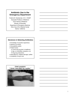


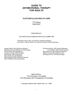

© Copyright 2026