
D IMEN SIONS
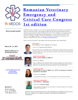
www.dvmpulse.com – Southern California Veterinary Medical Association’s Official Magazine
DIMEN
SIONS
IN
SURGERY
by
SCOTT ANDERSON, DVM, Diplomate of the American College of Veterinary
Surgeons, Diplomate of the American College of Veterinary Emergency and
Critical Care, Diplomate of the American Board of Veterinary Practitioners
PHIL GILL, DVM, Diplomate of the American College of Veterinary Surgeons
LARRY LIPPINCOTT, DVM, Diplomate of the American College of
Veterinary Surgeons
MARY SOMERVILLE, DVM, Staff Surgeon
SHARON SHIELDS, DVM, Staff Surgeon
RAVIV J. BALFOUR, DVM, Diplomate
of the American College of Veterinary Surgeons
ERIN WILSON, DVM, Staff Surgeon
Surgical Case Report:
Prostatic Omentalization
EMPHASIS:
In the past, numerous techniques have been used in the surgical
treatment of prostatitis, prostatic abscesses and prostatic cysts.
Partial or complete prostatectomy, marsupialization, or debridement and drainage have all been advocated; unfortunately all of
these techniques are associated with complications such as incontinence, cyst or abscess recurrence, etc.
More recently, prostatic omentalization has been described. In
this procedure, after opening and draining any cystic cavities or
abscesses within the prostate, a portion of the omentum is passed
through the prostate by blunt dissection. This improves the vascular supply to the affected tissue and prevents re-formation of a
closed cystic/abscess cavity. It is very effective in preventing
recurrence of disease. In this paper, we will describe omentalization of the prostate.
ureter
ureter
bladder
PREOPERATIVE DIAGNOSTICS:
1. Physical examination.
AXIOM: During rectal palpation to evaluate the prostate, be sure
to also evaluate the sublumbar lymph nodes.
2. Minimum database: CBC, serum chemistry profile and
urinalysis.
prostate
3. Radiography:
a. Two-view abdominal radiographs.
b. Contrast cystourethrogram: to rule-out concurrent
abnormalities such as urethral or cystic neoplasia
(which could cause similar signs to those produced by
prostatitis).
4. Abdominal ultrasonography.
urethra
multilobulated
prostatic
cyst or
abscess
5. Aspirate or biopsy: these are generally not performed
when a cyst or abscess is present, since the resulting
leakage of fluid into the abdomen could cause peritonitis.
6. Urine bacterial culture and sensitivity.
Figure One: This schematic drawing depicts a ventral view of the
canine urinary tract showing the prostatic pathology.
www.dvmpulse.com – Southern California Veterinary Medical Association’s Official Magazine
© 2001 Southern California Veterinary Medical Association
November 2001 1
www.dvmpulse.com – Southern California Veterinary Medical Association’s Official Magazine
DIMENSIONS IN SURGERY
continued from page 13
8. Cefalexin 20 mg/kg IV and enrofloxacin 7.5 mg/kg IV
immediately preoperatively.
9. Intravenous fluids to maintain renal perfusion intra-op.
SURGICAL TECHNIQUE:
1. Parapreputial incision, extending from the pubis to just
ahead of the prepuce.
AXIOM: The caudal superficial epigastric vessels will be transected, requiring cautery or ligation. The protractor preputii muscle
will also be transected.
2. Midline laparotomy, placing Gelpi self-retaining retractors
to maintain exposure.
3. Place stay sutures in the bladder to facilitate manipulation.
4. Blunt and sharp dissection through the periprostatic fat
along the ventral midline, to expose the prostate.
AXIOM: By approaching the prostate ventrally rather than from a
ventrolateral aspect, the degree of hemorrhage and the risk of nerve
damage are minimized.
5. Identify any cysts or abscesses (See Figure 1).
AXIOM: Of course, not all abscesses will be externally visible on the
prostate.
6. Make a stab incision and, using suction, drain the
contents of the cyst/abscess.
7. Culture the exudate present within the cavity.
8. Make a 1-2 cm incision in the ventrolateral aspect of the
prostate, on each side (See Figure 2).
Figure Two: This schematic drawing depicts two incisions into the prostatic cyst or abscess after the contents ahve been aspirated.
PREOPERATIVE CARE:
AXIOM: If sepsis and shock are present, standard medical management to stabilize the patient should be immediately instituted.
Surgery should be performed as soon as the clinician feels that the
medical management has achieved its maximum efficacy. Often it
is necessary to proceed with surgery even in a compromised patient,
since the underlying disease process (i.e. a ruptured abscess and peritonitis) cannot be resolved by nonsurgical means.
1. Indwelling cephalic catheter.
2. Intravenous anesthetic induction protocol
3. Endotracheal intubation and inflate cuff.
4. Isoflurane inhalant anesthesia to effect.
5. Lead II ECG and pulse oximetry monitoring during prep
and surgery.
6. Place an indwelling urinary catheter.
9. Using a digit or a hemostat, bluntly probe through the
entire parenchyma on each side to open any smaller cystic
or abscess cavities.
AXIOM: The urinary catheter facilitates palpation of the prostatic urethra; avoid this structure during dissection.
10. Take biopsy samples from several sites.
11. Using a warm balanced isotonic solution, liberally flush
the surgical field.
12. Pass a curved forceps through one of the incisions in the
prostate, ventral to the urethra, and exiting the opposite
prostatic incision (See Figure 3).
13. Grasp a portion of the omentum and draw it through the
prostate gland (see Figure 3).
AXIOM: If the friable omental tissue tears, enlarge the prostatic
incisions and then try again.
14. Using forceps, draw the omental pedicle back through the
prostate, dorsal to the urethra (see Figure 4).
AXIOM: The omentum now enters the prostate, passes circum- ferentially around the urethra, and exits through the same incision as
it entered. This places omentum throughout the parenchyma.
7. Clip and prep the ventral abdomen for aseptic surgery.
www.dvmpulse.com – Southern California Veterinary Medical Association’s Official Magazine
© 2001 Southern California Veterinary Medical Association
November 2001 2
www.dvmpulse.com – Southern California Veterinary Medical Association’s Official Magazine
DIMENSIONS IN SURGERY
continued from page 14
3A
Figure Three: This schematic drawing
depicts a cross-sectional view of the
prostate showing:
3C
3B
3A) A curved forceps is used to pull a
segment of omentum into the
prostatic cavity ventral to the
urethra.
3B) The omentum is mobilized through
the cavity and pulled out of the
opposite incision.
3C) The forceps are introduced through the original incision and
the tip of the mobilized omentum is grasped and pulled.
3D) The omental tip is pulled out othe original incision.
3D
15. Using 2-0 monofilament absorbable suture material,
suture the omentum to itself (see Figure 4).
16. Place a suction drain (i.e. a Jackson-Pratt drain) into the
prostate.
17. Routine abdominal, subcutaneous, and skin closure.
AXIOM: If the patient is intact, castration should always be performed.
POSTOPERATIVE CARE:
1. Broad spectrum antibiotics for 5-7 days postoperatively.
Figure Four: This schematic drawing depicts the completed prostatic
omentalization. The omentum has been sutured to itself as it exits the
prostate.
2. In most cases the urinary catheter is removed immediately
postop. It may be maintained, with a closed collection set,
in dogs that are severely dysuric.
3. Drain removal 3-4 days postoperatively.
www.dvmpulse.com – Southern California Veterinary Medical Association’s Official Magazine
© 2001 Southern California Veterinary Medical Association
continued on page 16
November 2001 3
www.dvmpulse.com – Southern California Veterinary Medical Association’s Official Magazine
DIMENSIONS IN SURGERY
continued from page 15
4. Suture removal 2 weeks postoperatively.
5. Pain management as warranted using injectable, oral, or
transdermal analgesics.
PROGNOSIS:
In patients that are not septic, the prognosis is optimistic with
only a small risk of recurrent cyst formation. Septic patients have
a 25-50% mortality.
AXIOM: With this procedure, postoperative persistent urinary
incontinence is very uncommon (as opposed to prostatectomy which
causes incontinence in almost all cases, and partial prostatectomy
which carries a significant risk of incontinence.
AUTHOR’S NOTE
If you have any questions concerning this paper, additional
references, surgical supplies or sources of products mentioned or used in this protocol, please FAX us at 1-310479-8976. We will answer your questions promptly.
Coming Attractions
When persistent pressure is present over a bony
prominence, ischemic necrosis of the soft tissues my occur,
resulting in a pressure sore (decubital ulcer). Most
commonly this occurs in patients who are recumbent due to
trauma or neurological conditions such as a disc rupture. In
particular, patients who are deep pain negative in the
hindquarters are at risk. In some thin dogs, the ulcers may
occur with no predisposing cause, merely by the pressure
generated when the patient is sitting or lying down.
The most effective option is to resect the underlying
bony prominence.
Next week we shall present our protocol for resection of
the ischial tuberosity.
See you then!
A Free Continuing Education
Service Available:
• To obtain a free bound book containing recent
“DIMENSIONS IN SURGERY” articles, merely mail
your business card to us, and on the back write: “YEARLY SUMMARIES.”
• Mail Your Card To:
Larry Lippincott, Scott Anderson, and Phil Gill
1736 South Sepulveda Blvd., Suite A
Los Angeles, California 90025.
• We will send you a binder containing the “DIMENSIONS IN SURGERY” articles from the past two years,
indexed and ready for quick office reference.
• Please be patient with the mailing of your articles.
• All first time “YEARLY SUMMARIES” requests
received after January 2001 will receive the last two
years’ articles in one bound book.
• 24 of the most requested articles from the first three
years of publication are still available and are contained
in the Practical Guide For Small Animal Surgery book
which can obtained from the SCVMA office.
• The SCVMA now publishes Dimensions In Surgery
articles and drawings on the Internet. Please visit us
at: www.DVMPulse.com
www.dvmpulse.com – Southern California Veterinary Medical Association’s Official Magazine
© 2001 Southern California Veterinary Medical Association
November 2001 4


© Copyright 2026