
Optimizing Chlamydia Someone once said: Assumptions are the mother
Optimizing
Chlamydia
trachomatis
Treponema
pallidum
and
diagnostics
Laura van Dommelen
OPTIMIZING CHLAMYDIA TRACHOMATIS AND TREPONEMA PALLIDUM DIAGNOSTICS
Proefschrift
ter verkrijging van de graad van doctor aan de Universiteit Maastricht,
op gezag van de Rector Magnificus, Prof dr. L.L.G Soete
volgens het besluit van het College van Decanen,
in het openbaar te verdedigen
op donderdag 31 oktober om 14.00
door
Laura van Dommelen
Geboren te Eindhoven
PROMOTOREN
Prof. dr. C.J.P.A. Hoebe
Prof. dr. C.A. Bruggeman
COPROMOTOR
Dr. F.H. van Tiel
BEOORDELINGSCOMMISSIE
Prof. dr. N.K. de Vries (voorzitter)
Prof. dr. J.E.A.M. van Bergen (AMC-UvA)
Prof. dr. P.C. Dagnelie
Prof. dr. G.J. Dinant
SPONSORS
Financial support by Stichting PAMM, MSD BV, Mediphos Group BV, apDia BV,
Roche Diagnostics Nederland BV, Pfizer Nederland, BD Diagnostics, Check-Points BV,
DiaSorin SA/NV, bioMerieux Benelux BV is gratefully acknowledged.
ABBREVATIONS
CDC
Centers for Disease Control and Prevention
CI
Confidence interval
Ct/CT
Chlamydia trachomatis
EB
Elementary bodies
ELISA
Enzyme-linked immunosorbent assay
FCU
First-catch urine
HBV
Hepatitis B virus
HIV
Human immunodeficiency virus
HSV
Herpes simplex virus
IFU
Inclusion forming units
LGV
Lymphogranuloma venereum
MSM
Men who have sex with men
NA
Not assessed
NAAT
Nucleic acid amplification test
ND
Not detected
Ng
Neisseria gonorrhoeae
NPV
Negative predictive value
nvCT
New variant Chlamydia trachomatis
Omp A
Outer membrane protein A
PCR
Polymerase strain reaction
PID
Pelvic inflammatory disease
POC(T)
Point-of-care (test)
PPV
Positive predictive value
qNAAT
Quantitative nucleic acid amplification test
RB
Reticulate bodies
SOA
Seksueel overdraagbare aandoeningen
SDA
Strand displacement amplification
STD
Sexually Transmitted Diseases
STI
Sexually Transmitted Infections
SVS
Self-taken vaginal swab
swCT
Swedisch variant Chlamydia trachomatis
Tp
Treponema pallidum
Tv
Trichomonas vaginalis
VDRL
Venereal Disease Research Laboratory test
Part A. Introduction
1.
Introduction on sexually transmitted infections
with a focus on Chlamydia trachomatis and Treponema pallidum.
10
Part B. Different Sample Types and Chlamydia trachomatis Detection
2.
Evaluation of one-sample testing of self-obtained vaginal swabs
and first catch urine samples separately and in combination for
the detection of Chlamydia trachomatis by two amplified DNA
assays in women visiting an sexually transmitted disease clinic.
30
»» Sex Transm Dis. 2011 Jun;38(6):533-5
Part C. Newly Developed Nucleic Acid Amplification Tests to Detect Chlamydia trachomatis
3.
TaqMan assay for Swedish Chlamydia trachomatis variant.
38
»» Emerg Infect Dis. 2007 Sep;13(9):1432-4
4.
High concordance of test results of the rapid and easy
Chlamydia trachomatis Detection and genoTyping Kit compared to
the COBAS Amplicor CT/NG test in females visiting an STD clinic.
42
»» Submitted
Part D. Point-of-Care Tests to Detect Sexually Transmitted Infections
5.
Alarmingly poor performance in
Chlamydia trachomatis point-of-care testing.
50
»» Sex Transm Infect. 2010 Oct;86(5):355-9
6.
Evaluation of a rapid one-step immunochromatographic test
and two immunoenzymatic assays for the detection of
anti-Treponema pallidum antibodies.
60
»» Sex Transm Infect. 2008 Aug;84(4):292-6
Part E.
Validation of Methodology Used in Sexually Transmitted Infections Research
7.
Confirmation of high specificity of an automated ELISA test for serological
diagnosis of syphilis - results from confirmatory testing after syphilis
screening and sensitivity analysis in the absence of a gold standard.
74
»» Submitted
8.
Chlamydia trachomatis DNA stability independent of
preservation temperature, type of medium en storage duration.
82
»» J Clin Microbiol. 2013 Mar;51(3):990-2
Part F.
Discussion and Summary
9.
10.
Discussion and Summary
Samenvatting
90
106
Part G. Addendum
11.
12.
13.
14.
Dankwoord
Co-authors (in alphabetical order)
About the author
List of (peer reviewed) Publications
116
120
122
123
Introduction
1. Introduction on sexually transmitted infections with
a focus on Chlamydia trachomatis and Treponema
pallidum.
Laura van Dommelen
GLOBAL BURDEN OF SEXUALLY TRANSMITTED INFECTIONS WITH A FOCUS ON CHLAMYDIA
TRACHOMATIS AND TREPONEMA PALLIDUM
Sexually transmitted infections (STI) are a major medical and public health challenge, due to
their high incidence and emerging threat of drug resistance. In 2008, WHO estimated 498.9
million curable STI, caused by Treponema pallidum (Tp), Neisseria gonorrhoeae (Ng), Chlamydia
trachomatis (Ct) and Trichomonas vaginalis (Tv). Moreover, millions of viral STI occur every year,
mainly caused by human immunodeficiency virus (HIV), herpes simplex viruses (HSV), human
papilloma viruses en hepatitis B virus (HBV). The number of newly acquired infections was 105.7
million for Ct and 10.6 million for syphilis according to WHO. Most curable STI occur in subSaharan Africa (incidence 92.6 million) and the Americas (incidence 125.7 million). All figures
however are hampered by the lack of good prevalence studies, limited healthcare seeking and
inadequate access to healthcare, among others.1
Syphilis infection, caused by Tp, during pregnancy contributes to 650,000 fetal and neonatal
deaths in developing countries, while prevention of congenital syphilis only costs $1.50
per person. Large scale screening in pregnant females in high burden countries is however
not practised yet.2 With Ct infection, under-reporting is huge because most people are not
aware of their infections and do not seek help. People with genital chlamydia may experience
symptoms of genital tract inflammation including urethritis and cervicitis, but the majority
remains asymptomatic. Ct is a significant public health problem because untreated Ct may
lead to pelvic inflammatory disease, subfertility and poor reproductive outcomes in some
women.3 Concurrent STI facilitates the transmission of HIV, which is the case in 40% of all
newly acquired HIV infections, which is a concern in countries with high HIV prevalence.
Furthermore, when STI coexist with HIV, higher loads of HIV are shed into genital fluids,
compared to individuals who do not have a coexisting STI. All STI together cause 17% of the
economic loss due to health related causes in developing countries. In developed countries,
the costs for STI screening, HIV treatment, managing infertility due to STI and cervical cancer
treatment are substantial.4
Sexually Transmitted Infections in The Netherlands
STI’s continue to be a serious health problem in the Netherlands. Certain ethnic minorities
(especially (former) inhabitants of Surinam and the former Netherlands Antilles), young
persons (<25 years of age) and men having sex with men (MSM) are important risk groups
for STI.5 Ct is the most prevalent STI in the Netherlands and the incidence is highest in the
heterosexual population under 25 years of age. At the different STI centres, the test positivity
rate was 11.5% in the year 2011. Lymphogranuloma venereum (LGV) was only detected in MSM
and 79% is known to be also HIV positive. The test positivity rate for Ng was much lower, 3.2%,
and was mostly detected in MSM. Syphilis was rare: only 476 cases were detected in 2011.
Almost 90% of the cases concerned MSM and the positivity rate has decreased since 2007 (from
4.3% to 2.0% in 2011). The prevalence of syphilis among pregnant women was estimated at
0.2% in 2009, which is a slight increase compared to previous years.5
Introduction on STI with a focus on Chlamydia trachomatis and Treponema pallidum
11
Most first-line STI related consultations are handled by the general practitioner (63%) 6;
the remaining consultations are mainly managed in STI clinics (e.g. Public Health Service).
Standard STI testing includes Ct, Ng and syphilis. HIV opting out is now widely practiced,
which means HIV testing is performed unless the client refuses.7 HBV, hepatitis C virus, genital
herpes and Tv are only tested when deemed necessary.5 Pregnant women are all screened for
syphilis, HBV and HIV before the 13th week of pregnancy.
The prevention and control of STI is based on five major strategies described by the Centers for
Disease Control and Prevention (CDC): education and counselling of persons at risk in order to
achieve changes in sexual behaviour; identification of asymptomatically infected persons and
of symptomatic persons unlikely to seek diagnostic and treatment services; effective diagnosis
and treatment of infected persons; evaluation, treatment, and counselling of sexual partners
of persons who are infected with an STI and pre-exposure vaccination of persons at risk for
vaccine-preventable STI.8 In general, the Dutch population is well informed concerning sexual
health, although this is not the case in adolescents, lower educated individuals and men with a
Turkish or Moroccan background. National campaigns on sexual health are effective in creating
awareness and condom use is considered important to prevent STI, but its use is not always
practiced. The number of STI consultations are increasing every year and the percentage of
positive STI tests has increased from 12% to 14% between 2004 and 20105 (Figure 1). MSM have
a higher percentage of positive STI tests compared with heterosexuals.
CHLAMYDIA TRACHOMATIS
Ct infection is the most prevalent bacterial STI worldwide and causes most cases of infectionrelated female infertility worldwide.4
Organism
Ct is an obligatory intracellular, non-motile, Gram negative organism. The developmental
cycle consists of an extracellular and an intracellular part (Figure 2). The extracellular form of
Ct consists of elementary bodies (EB). The cell wall of EB does not contain peptidoglycan, but
has a rigid wall as a result of a highly cross-linked outer-membrane complex.9 EB are able
to survive outside the cell and stay dormant until entering the host cell. Cell entry consists
of two stages; in the first stage, the EB attaches through reversible electrostatic interactions
12
Introduction on STI with a focus on Chlamydia trachomatis and Treponema pallidum
between the outer membrane protein A (OmpA), expressed on the Ct cell surface and the
heparan sulphate containing glycosaminoglycans on the cell. During the second, irreversible
binding stage the EB probably interact with protein disulfide isomerase, although this has
not yet been fully elucidated.9 After this step, translocated actin-recruiting phosphoprotein is
released into the host cell via type three secretions system, leading to actin remodelling, which
in turn allows EB to enter the host cell and inhibits apoptosis.10 Intracellularly, EB transform
into metabolically active, non infectious, reticulate bodies (RB). RB replicate and in the end of
the intracellular cycle transform back into EB. The EB are again released into the extracellular
space by exocytosis or cell lysis, able to infect other epithelial cells. This replication cycle takes
about 48-72 hours.
Figure 1 Number of consultations and percentage of positive STI tests (chlamydia, gonorrhoea,
infectious syphilis, HIV , infectious hepatitis B) in the national STI surveillance in the
Netherlands, 1995-2011 5
120000
110000
100000
9000
8000
7000
6000
5000
4000
3000
2000
1000
0
nr of consultations
% positive STI
15
14
13
12
11
10
9
9
6
7
0
8
5
1
2
3
4
5
6
7
8
9
0 1
199 199 199 199 199 200 200 200 200 200 200 200 200 200 200 201 201
Introduction on STI with a focus on Chlamydia trachomatis and Treponema pallidum
8
13
Figure 2 Chlamydia trachomatis developmental cycle 11
Entry into
host cells
EB sequestered
in inclusion
Exit to infect
other host cells
RB
Differentiation
into RBs
EB
Replication
Figure is placed by courtesy of Nature Publishing Group
The family Chlamydiaeceae consists of three genera: Chlamydia, Chlamydophila and
Clavochlamydia. The genus Chlamydia can be subdivided in three species: C. trachomatis
(human), C. suis (swine) and C. muridarum (mice). The human variant comprises of biovar
trachoma and lymphogranuloma venereum (LVG). The biovar trachoma consists of 14 serovars
(A, B, Ba, C, D, Da, E, F, G, H, I, Ia, J, K) of which A-C have most affinity to conjunctivae and D-K
to urogenital epithelium; LGV consists of serovar L1, L2, L2a and L3. Serovars E and F are most
prevalent in The Netherlands.12 This thesis will focus on C. trachomatis biovar trachoma
serovars D-K.
14
Introduction on STI with a focus on Chlamydia trachomatis and Treponema pallidum
Pathogenesis
Histological studies on Ct infection show both signs of acute and chronic inflammation
in the affected tissue. Epithelial cells are the primary target of Ct and initiate the immune
response.13 Secondly, neutrophils infiltrate the vagina and subsequently the uterus during
the first week after infection.14 Subsequently, epithelial cells and innate immune cells
produce proinflammatory cytokines and chemokines which further activate the innate and
adaptive immune cells. Ultimately, resolution of infection occurs, but sometimes leaving
a destroyed oviduct. Tissue damage can be the result of chronic infection (continues low
grade tissue damage) and/or reinfections which leads to the predominance of CD8 T-cell,
associated with scarring and fibrosis.15 The mechanisms behind chronic infection due to Ct
infections are not clear. The degree of infection is host dependent and may be dependent on
genetic polymorphisms.16-18 Cervical lymphocytes of females with or females without fertility
problems, for instance, produce different cytokine profiles when stimulated with Ct elementary
bodies.16 Also, a study on monozygotic and dizygotic twins on the influence of genetic trait on
the immune response to Ct (serovar A), suggested that genetic factors contribute for 39% to the
variation in lymphoproliferative responses.19
Transmission and natural course
Ct can be transmitted via sexual contact or perinatally. The incubation period for acute
infection is 7-14 days. Infection concordance for Ct between sexual partners is approximately
70%.20-22 The transmission and natural course of Ct infection depends on Ct virulence
factors, host immune response and the presence of co-infections. Approximately 50% of all
uncomplicated Ct infections in females resolves without therapy during the first year after
acquisition.23 Almost 20% of all infections is already cleared within the interval between
testing and treatment, which is on average 13 days.24
Clinical manifestations and complications
Infection with Ct is asymptomatic in the majority of the women and in more than 50% of the
men. When symptomatic, women can experience abnormal vaginal discharge, abdominal pain
or post-coital bleeding for instance. Other clinical manifestations in women include cervicitis,
urethritis, conjunctivitis, endometritis and pelvic inflammatory disease (PID). The incidence
of PID is heavily debated, since methodology and results vary between different studies and
is suggested to be between 5 and 20%.24-29 PID due to Ct can progress subclinically and may
eventually might lead to tubal infertility.30, 31 It is also associated with chronic abdominal pain.
32, 33 Another important complication is adverse pregnancy outcome. In neonates, perinatally
Introduction on STI with a focus on Chlamydia trachomatis and Treponema pallidum
15
acquired infection may result in for instance pneumonia and/or conjunctivitis.34 In men, Ct
can cause urethritis, prostatitis, epididymitis, conjunctivitis and Reiter’s syndrome (arthritis,
conjunctivitis and urethritis).
Diagnosis
The mainstay for the diagnosis of Ct are nucleic acid amplification tests (NAAT). Table 1 gives
an overview of the different, commercially available, NAAT used to detect Ct. NAAT are usually
performed on self-taken vaginal swabs (SVS) in females and on urine in men. When indicated,
NAAT can also be performed on many other materials, for instance pharyngeal, rectal of
corneal swabs and peritoneal fluid – although these samples are not always officially validated
and approved by the manufacturer.
Table 1
System
Currently most used commercially available NAAT to detect Chlamydia trachomatis
Assay
Company
Technique
Target
Validated material
ProbeTec
ProbeTec
ET or VIPER- CT/GC
XTR
Beckton
Dickinson
(BD)
SDA
Cryptic plasmid
Endocervical swabs, vaginal swabs,
male urethra swabs, urine
COBAS
Taqman
COBAS
CT/NG
Roche
Real-time PCR
Cryptic plasmid
and CT genome
(MOMP gene)
Endocervical swabs, urine
COBAS 4800 COBAS
CT/NG
Roche
Real-time PCR
Cryptic plasmid
and CT genome
(MOMP gene)
Endocervical swabs, urine
M2000
RealTime
RealTime
CT/NG
Abbott
Real-time PCR
Cryptic plasmid
Endocervial, male urethra swabs,
vaginal swab, urine
Versant
kPCR
Versant
CT/GC
DNA 1.0
Siemens
Real-time PCR
Cryptic plasmid
Endocervical swab, urethral swabs,
urine
TIGRIS or
Panther
Aptima
Combo
AC2
Hologic/
Gen-Probe
TMA
CT rRNA NG rRNA
Endocervical swabs, male urethra
swabs, urine
GeneXpert
Xpert®
CT/NG
Cepheid
Real-time PCR
CT genomic DNA
NG genomic DNA
Urine (male and female),
endocervical swab, and patientcollected vaginal swab
SDA = strand displacement amplification, TMA = transcription mediated amplification
16
Introduction on STI with a focus on Chlamydia trachomatis and Treponema pallidum
The overall sensitivity and specificity of NAAT for Ct detection in SVS ranges between 97-99%
and 95-100% 36,37 and between 96-100% and 99-100% for urine, respectively.38 Ct detection
in SVS is equally sensitive as detection in endocervical specimens.39 Furthermore, SVS is an
acceptable and feasible specimen in women.40
Before NAAT became available, Ct culture was considered as the gold standard. The sensitivity
of culture however is low and is no longer practised in clinical laboratories.41 Another
technique to diagnoses an infection due to Ct is direct fluorescent antibody testing, but as
with Ct culture, this technique is laborious, requires experienced technicians, is less sensitive
than NAAT and not necessarily detects acute infection. Also, enzyme immunoassays to detect
chlamydial lipopolysaccharide are widely available, but these again are less sensitive than
NAAT. Point of care tests (POCT) are upcoming and are currently being evaluated. However,
results obtained thus far do not warrant their use, instead of NAAT, in clinical practice
yet.35,42,43
For research purposes, Ct typing methods are available. Results can be used to reveal
transmission patterns, can be useful to detect associations with clinical manifestations, to
study pathogenicity and can help to differentiate between persistent or new infections.44
Variants in the major outer membrane protein (MOMP), encoded by the omp1 (ompA) gene, are
used for decades to serotype, or genotype, Ct. Several typing methods have been developed,
with different capabilities and limitations and variation in laboriousness.44 Also, typing can be
useful in identifying new Ct variants. In 2006, an unexpected drop in Ct prevalence was noticed
in Halland County, Sweden.45 Thorough analysis revealed the existence of a new variant of Ct,
the so-called Swedish variant of Ct (swCt), which was not detected by the most commonly used
NAAT. This was due to a deletion on the Ct plasmid which is the target of the used NAAT.
Treatment, follow-up, antibiotic resistance and prevention
According to the guidelines of the Dutch Society of Dermatology and Venereology, urogenital
infections due to Ct are treated with azithromycin 1g orally, once only (www.soaaids.nl). Anal
infections (non-LGV) are treated with doxycycline 100mg twice daily for 7 days. Azithromycin
is also first choice in pregnant women. Patients are advised not to have sexual contact during
and one week after treatment.46 Re-testing after treatment is not advised, except when
amoxicillin has been used in pregnant women (46, www.soaaids.nl). It is important to trace and
test all sexual contacts, since at least 1 out of 2 of these contacts on average are Ct positive
and can again lead to additional cases and repeat cases.47 Additionally, re-testing within
one year after the first positive Ct test is advisable, since the percentage of positive patients is
much higher in case of a previously positive Ct test compared to patients with a negative Ct
test (10.4% vs 2.9% respectively).48 The challenges of repeat and persistent infections after
Introduction on STI with a focus on Chlamydia trachomatis and Treponema pallidum
17
single dose treatment with azithromycin have emerged over the past 10 years. Treatment
failures might occur due to reduced effectiveness of azithromycin and doxycycline in a hypoxic
environment.49,50 Increasing number of studies suggest that more than 5% treatment failure
can occur during use of azithromycin.51
Although inexpensive and effective treatment of symptomatic patients is available, control
of Ct is challenging since most infected persons are asymptomatic. Despite the fact that over
the past two decades huge resources were put into attempts to reach these asymptomatic
patients, the impact of widespread screening for Ct has been disappointing. The reason for this
is that screening has not been shown to reach sufficient numbers of participants and thereby
reduce either transmission of infection 52 or the incidence of reproductive complications.
Besides screening and early treatment, effective prevention obviously relies on having safe
sex, e.g. using a condom. Another potentially effective measure may be offered by vaccination.
A good vaccine against Ct could eliminate Ct within 20 years after its introduction and high
coverage.10 Vaccination could potentially prevent Ct to pass the cervix and cause damage to
the oviduct. Many candidate proteins have been explored for their vaccine capacity, but no
vaccine is yet available nor expected in the near future.10,53
TREPONEMA PALLIDUM
Early syphilis infections during pregnancy cause 25% of stillbirths and 14% of neonatal deaths.4
Organism
Treponema pallidum (Tp) belongs to the family Spirochaetaceae which is characterized by its
typical spiral configuration. Other species belonging to the Spirochaetaceae are for instance
Leptospira spp. and Borrelia spp. Tp can be subdivided in the subspecies pallidum, endemicum,
pertenue and carateum which cause the disease syphilis, bejel, yaws and pinta, respectively.
In this thesis, the primarily focus is on Treponema pallidum subsp. pallidum.54 Tp has an
cytoplasmic membrane which is surrounded by an outer membrane. Within these membranes,
there is a thin layer of peptidoglycan. At both ends of the spirochete, flagellar motors are
present. Tp has only few intergral proteins in its outer membrane.55 The genome of Tp is
very small compared to other bacteria, e.g. Escherichia coli: 1.14 Mb vs. 4.6 Mb. The metabolic
activity of Tp is very limited.54
18
Introduction on STI with a focus on Chlamydia trachomatis and Treponema pallidum
Pathogenesis
Tp is capable of attachment to a variety of cell types, including epithelial and endothelial
cells.56 It is very motile, even in less fluid material, due to its corkscrew-like movements using
its endoflagella.55 Compared with other bacteria, the flagellar construction of Tp is well
developed and comprises several proteins instead of just one. These proteins have strong
antigenic properties.
Tp is extremely invasive. It induces the production of matrix metalloproteinase-1 which is
involved in collagen breakdown, which facilitates penetration and Tp is found in deeper
tissues, just hours after (mucosal) inoculation.54 Endothelial cells are promoted to express
adhesion molecules (intercellular adhesion molecule 1, vascular cell adhesion protein 1 and
E-selectin) which cause migration and adhesion of leukocytes.56 Although polymorphnuclear
lymphocytes are able to reduce the infectivity of Tp 57, these do not halt disease progression.
TLR-2 (toll like receptor 2; present on human cell surface) is essential in recognising Tp,
for instance by immature dendritic cells (DC’s ).58 Immature DC’s subsequently activate
T-cells by taking up Tp and migrating to the lymphnodes. After maturation, the DC’s produce
inflammatory cytokines (interleukin 12 and tumor necrosis factor alfa) and express maturation
markers. A delay in this process, facilitates Tp dissemination. Macrophages opsonise and kill
Tp, although it has been shown that a subpopulation of organisms is resistant to ingestion,
a phenomenon which is unexplained thus far.59 The most important lipoproteins for the
immune response, are TpN47, TpN17 and TpN15. At approximately day 10 after infection, the
number of T-cells and macrofages reaches its peak, after which the Tp load decreases.54
After dissemination, Tp is also capable of causing chronic infection, as will be discussed in the
next sections. The latent phase is possibly due to the small numbers of organisms present
at different anatomical sites and (very) slow replication during this stage and its relatively
non-antigenic surface. There are, however, outer membrane proteins on its cell surface which
are encoded by the tpr gene family. Gene conversion occurs and gene expression can vary
depending on the human immune response. This variable gene expression is called phase
variation and helps Tp to constantly evade the immune system.54
Introduction on STI with a focus on Chlamydia trachomatis and Treponema pallidum
19
Transmission and natural course
Tp can only infect humans and can be transmitted via contact between lesions in the primary
and secondary stage and mucosal membranes. The incubation period is between 10-90 days.
At least 60 organisms are needed to cause an infection 54 and approximately 60% of the sexual
partners will become infected.60
Clinical manifestations
In primary syphilis, a lesion, so called chancre, develops at the inoculation site and is usually
accompanied by regional lymphadenopathy. The chancre usually occurs 3 weeks after
exposure, is painless and heals spontaneously within 4-6 weeks. Due to is painless presentation
and rapid resolution, diagnosis of primary syphilis is hampered.
Secondary syphilis presents approximately 3 months after initial infection. The most classical
finding is maculopapular rash which can also be present on palms of hand and soles of
feet. Other accompanying symptoms are sore throat, muscle aches and lymphadenopathy.
Sometimes the dermal lesions can become necrotic, which is called lues maligna.
Condylomata lata can be present in the anal region in 10% of the patients and are highly
infectious. The kidneys and liver can be involved. Neurosyphilis occurs in 40% of patients
with early syphilis and in 25% of individuals with late syphilis. Most patients can resolve the
infection by themselves and never become (noticeably) symptomatic.
After the secondary stage, the latent phase starts which is variable in duration. In the first year
after secondary syphilis, it is considered the early latent phase and afterwards the late latent
phase (also if duration is unknown). Sexual transmission is not likely during the latent phase,
although foetal infection can occur. Before the use of antibiotics, approximately 30% of the
patients with latent syphilis, subsequently developed tertiary syphilis. Symptoms can occur as
long as 40 years after initial infection and can include gummata, cardiovascular infection and
neurological complications.
The probability of congenital infection is highest when the mother is in her first year of
infection. Treatment in the first or second trimester usually prevents the newborn from
being symptomatic. In the third trimester, primary syphilis can result in abortion or stillbirth.
Symptoms of congenital syphilis resemble those in adults only worse, and become apparent
2-10 weeks after birth. The baby can suffer from chronic rhinitis (snuffles), skin lesions,
condylomata lata, osteochondritis, among other symptoms. Late manifestations, occurring
after 2 years, are for instance keratitis, damage of the vestibulocochlear nerve, neurosyphilis,
arthropathy and gummata.54
20
Introduction on STI with a focus on Chlamydia trachomatis and Treponema pallidum
Diagnosis
Treponema pallidum cannot be cultured in vitro. Darkfield microscopy is still practised, but can
only visualize spirochetes in fresh lesions. NAAT is not currently available in most laboratories,
thus, diagnosis relies on serology. IgM antibodies can be detectable as early as 2 weeks postinfection and are produced in response to Tp surface lipids, flagellar proteins and lipoproteins,
among others. IgG is formed 2 weeks hereafter and is capable of blocking the binding capacity
of Tp and causes organism to be unable to produce dermal lesions, but do not kill Tp nor
prevent infection. The IgM response wanes when infection persists for longer periods of time
or when a patient has been adequately treated.61 The attributes of the serological assays are
displayed in table 2.11
The first serological assay for diagnosing syphilis was developed in the beginning of the
20th century and was called the Wasserman test. It detects antigens in the Tp membrane
which is originally derived from the host. These antigens comprise of lecithin, cholesterol
and cardiolopin, are not is not unique for Tp, therefore the Wasserman test is called a nontreponemal assay. Other non-treponemal tests are the rapid plasma reagin (RPR) and Venereal
Disease Research Laboratory (VDRL) which utilize the same antigens and are still used to
determine disease activity.61 Unfortunately, these antigens are also released in different other
(infectious) diseases, causing false positive results.62-64
Specific Tp immunoglobulins (Ig), IgM and IgG, are detected in the fluorescent treponemal
antibody absorption test (FTA-ABS) and Treponema pallidum hemagglutination assay (TPHA)
and Treponema pallidum particle-agglutination assay (TPPA). The FTA-ABS utilize anti-human
Ig to detect antibodies attachted to Tp fixed on slides. The TPHA is based on red blood cells
sensitized with Tp antigen which agglutinate in the presence of specific antibodies. TPPA uses
gel particles instead of red blood cells. During recent years, enzyme-linked immunosorbent
assays (ELISA) are upcoming. ELISA are designed to detect IgG or IgM antibodies, or both. These
assays have the great advantage that these can be performed on an automated system.61
Introduction on STI with a focus on Chlamydia trachomatis and Treponema pallidum
21
Table 2
Sensitivity and specificity of serological tests for syphilis 11
Test
characteristic
Non-treponema l tests
Treponemal tests
RPR
VDRL
EIA
TPHA / TPPA
FTA-ABS
Specimen
Serum or plasma
Serum or plasma
Serum or plasma
Serum or plasma
Serum or plasma
Sensitivity
86-100%
78-100%
82-100%
85-100%
70-100%
Specificiry
93-98%
98-100%
97-100%
98-100%
94-100%
Ease of use
Easy
Easy
Moderate
Complex
Complex
Level ofuse
Examination
room, on-site
laboratory
Examination
room,on-site
laboratory
lntermediate
laboratory,
reference
laboratory
Reference
laboratory
Reference
laboratory
Equipment
Rotator,
refrigerator
light microscope,
refrigerator
Incubator,
microwell plate
washer and
reader
Incubator
Fluorescence
microscope
Training
Minimal
Minimal
Moderate
Extensive
Extensive
Average cost
US$ 0,5
US$ 0,5
US$ 3
US$3
US$ 3
Comments
Most RPR
reagents require
refrigeration
Reagents require
refrigeration
Allows highthroughput
screening; does
not distinguish
between prior
treated and
active infection
Confirmatory
test, so does
not distinguish
between prior
treated and
active infection
Confirmatory
test, so does
not distinguish
between prior
treated and
active infection
EIA, enzyme immunoassay; FTA-ABS, fluorescent treponemal antibody-absorption test; RPR, rapid plasma reagin
test; TPHA/TPPA, Treponema pallidum haemagglutination assay/T. pallidum particle agglutination assay; VDRL,
venereal disease research laboratory test.)
22
Introduction on STI with a focus on Chlamydia trachomatis and Treponema pallidum
Treatment, follow-up, antibiotic resistance and prevention
Penicillin is the antibiotic of choice for treatment of all known syphilis stages. It can be
given intramuscularly for the early and latent stage and should be given intravenously for
neurosyphilis. In case of penicillin intolerance or allergy, doxycycline can be used. Partner
notification is essential to prevent secondary cases. After treatment of primary, secondary or
early latent syphilis, the VDRL must be checked every quarter for the first year and every half
year for the second year. In all other cases, follow-up should be guided by an experienced
physician (www.saoaids.nl)
Individuals with syphilis are mainly infectious -when having dermal lesions- during the first year
of infection. Contact with lesions and blood should be avoided. Mother to child transmission,
however, can also occur beyond this period, albeit rarely. To prevent symptomatic congenital
syphilis, all females in the Netherlands are serologically tested before their 13th week of
pregnancy.65 No vaccine against syphilis is presently available, nor expected to become
available in the near future.
AIMS AND OUTLINE OF THIS THESIS
The subject of this thesis is STI diagnostics in a broad sense, with a focus on syphilis and
chlamydia. Several hypotheses have been formulated. For instance what kind of samples can
be used in clinical practice and for research purposes? What are the assays of choice and how
should we interpret results? All chapters are designed to give answers to these questions.
Currently, Ct detection by NAAT is performed on SVS or urine in females, since dual testing
is not considered cost effective. Since SVS is considered the specimen of choice in women,
urinary tract only Ct infection could be missed. Chapter 2 explores the possible sensitivity gain
when using NAAT on combined swab and urine samples when testing for Ct in females.
As mentioned in this introduction, a new Ct variant (swCt) was discovered in 2006 which could
not be detected by the most commonly used NAAT.45 Rapid exploration of the extent of this
problem was needed. Chapter 3 describes a newly developed assay to specifically detect the
swCt, which can be used to monitor the spread of swCt.
Introduction on STI with a focus on Chlamydia trachomatis and Treponema pallidum
23
Different genotyping assays are developed to distinguish between different CT serovars
to understand more about Ct. Genotyping can provide information that can be helpful in
understanding epidemiology, transmission and pathogenicity. Chapter 4 evaluates a rapid and
easy Ct genotyping assay.
Another ‘hot’ issue in Ct research are the POCT. POCT can usually be performed within 30
minutes and treatment can be given instantly when positive. This could potentially mean a
great step forward in limiting Ct spread in the population compared to the current standard
(e.g. NAAT). POCT also could be very useful in remote areas in which STI diagnostics are
currently not available due to absence of facilities and financial means. The World Health
Organisation advices practising syndromic management if STI diagnostics are unavailable, but
its sensitivity and specificity is poor.66,67 Chapter 5 discusses the evaluation of three Ct rapid
antigen tests in comparison to NAAT.
POCT are also upcoming in the field of syphilis diagnostics. As with Ct, early syphilis can be
treated with single dose of penicillin and instant treatment would mean an improvement
compared to the current situation. Also, ELISA are being introduced in favour of the TPPA/TPHA
or a non-treponemal assay due to the ability to perform the assay on an automated system.
Chapter 6 discusses the evaluation of a rapid test and two immunoenzymatic assays to detect
antibodies against Tp.
As a follow-up to chapter 6, chapter 7 discusses the validity of using a selected sample set to
evaluate diagnostic assays. In case of syphilis, the prevalence in The Netherlands is very low
as mentioned before, but for the project described in chapter 6, a high number of syphilis
seropositive samples were used. Does the evaluation of a diagnostic assay using this sample
selection result in data which can be used in the general population?
In the afore mentioned chapters, many of the samples tested to answer a research question
were stored, frozen samples. Literature regarding the use of stored samples in the evaluation
of Ct NAAT is lacking. Do stored samples generate the same result as ‘fresh’ samples, in other
words can storage of samples lead to false negative results of NAAT testing? In chapter 8 the
stability of Ct DNA is explored by using NAAT under different circumstances.
Finally, chapter 9 gives an overview of the results of this thesis and how they relate to recent
literature.
24
Introduction on STI with a focus on Chlamydia trachomatis and Treponema pallidum
REFERENCES
1. Global incidence and prevalence of selected curable sexually transmitted infections - 2008 World Health
Organization, 2012.
2. Advancing MDG 4, 5 and 6: impact of congenital syphilis elimination: World Health Organization, 2010.
3. Carey AJ, Beagley KW. Chlamydia trachomatis, a hidden epidemic: effects on female reproduction and options
for treatment. Am J Reprod Immunol 2010;63(6):576-86.
4. Global strategy for the prevention and control of sexually transmitted infections: 2006-2015: WHO, 2007.
5. S.C.M. Trienekens FDHK, I.V.F. van den Broek, H.J. Vriend, E.L.M. Op de Coul, M.G. van Veen, A.I. van Sighem, I.
Stirbu-Wagner, M.A.B. van der Sande. Sexually transmitted infections, including HIV, in the Netherlands in 2011:
National Institute for Public Health and the Environment, 2012.
6. van Bergen JE, Kerssens JJ, Schellevis FG, Sandfort TG, Coenen TJ, Bindels PJ. Prevalence of STI related
consultations in general practice: results from the second Dutch National Survey of General Practice. Br J Gen
Pract 2006;56(523):104-9.
7. Dukers-Muijrers NH, Niekamp AM, Vergoossen MM, Hoebe CJ. Effectiveness of an opting-out strategy for HIV
testing: evaluation of 4 years of standard HIV testing in a STI clinic. Sex Transm Infect 2009;85(3):226-30.
8. Workowski KA, Berman S. Sexually transmitted diseases treatment guidelines, 2010. MMWR Recomm Rep
2010;59(RR-12):1-110.
9. Abdelrahman YM, Belland RJ. The chlamydial developmental cycle. FEMS Microbiol Rev 2005;29(5):949-59.
10. Howie SE, Horner PJ, Horne AW, Entrican G. Immunity and vaccines against sexually transmitted Chlamydia
trachomatis infection. Curr Opin Infect Dis 2011;24(1):56-61.
11. Peeling RW, Mabey D, Herring A, Hook EW, 3rd. Why do we need quality-assured diagnostic tests for sexually
transmitted infections? Nat Rev Microbiol 2006;4(12):909-21.
12. Spaargaren J, Verhaest I, Mooij S, Smit C, Fennema HS, Coutinho RA, et al. Analysis of Chlamydia trachomatis
serovar distribution changes in the Netherlands (1986-2002). Sex Transm Infect 2004;80(2):151-2.
13. Rasmussen SJ, Eckmann L, Quayle AJ, Shen L, Zhang YX, Anderson DJ, et al. Secretion of proinflammatory
cytokines by epithelial cells in response to Chlamydia infection suggests a central role for epithelial cells in
chlamydial pathogenesis. J Clin Invest 1997;99(1):77-87.
14. Morrison SG, Su H, Caldwell HD, Morrison RP. Immunity to murine Chlamydia trachomatis genital tract
reinfection involves B cells and CD4(+) T cells but not CD8(+) T cells. Infect Immun 2000;68(12):6979-87.
15. Van Voorhis WC, Barrett LK, Sweeney YT, Kuo CC, Patton DL. Repeated Chlamydia trachomatis infection of
Macaca nemestrina fallopian tubes produces a Th1-like cytokine response associated with fibrosis and scarring.
Infect Immun 1997;65(6):2175-82.
16. Agrawal T, Gupta R, Dutta R, Srivastava P, Bhengraj AR, Salhan S, et al. Protective or pathogenic immune
response to genital chlamydial infection in women-a possible role of cytokine secretion profile of cervical
mucosal cells. Clin Immunol 2009;130(3):347-54.
17. den Hartog JE, Ouburg S, Land JA, Lyons JM, Ito JI, Pena AS, et al. Do host genetic traits in the bacterial sensing
system play a role in the development of Chlamydia trachomatis-associated tubal pathology in subfertile
women? BMC Infect Dis 2006;6:122.
18. Lyons JM, Morre SA, Airo-Brown LP, Pena AS, Ito JI. Comparison of multiple genital tract infections with
Chlamydia trachomatis in different strains of female mice. J Microbiol Immunol Infect 2005;38(6):383-93.
19. Bailey RL, Natividad-Sancho A, Fowler A, Peeling RW, Mabey DC, Whittle HC, et al. Host genetic contribution to
the cellular immune response to Chlamydia trachomatis: Heritability estimate from a Gambian twin study. Drugs
Today (Barc) 2009;45 Suppl B:45-50.
20. Markos AR. The concordance of Chlamydia trachomatis genital infection between sexual partners, in the era of
nucleic acid testing. Sex Health 2005;2(1):23-4.
21. Rogers SM, Miller WC, Turner CF, Ellen J, Zenilman J, Rothman R, et al. Concordance of chlamydia trachomatis
infections within sexual partnerships. Sex Transm Infect 2008;84(1):23-8.
22. Quinn TC, Gaydos C, Shepherd M, Bobo L, Hook EW, 3rd, Viscidi R, et al. Epidemiologic and microbiologic
correlates of Chlamydia trachomatis infection in sexual partnerships. Jama 1996;276(21):1737-42.
Introduction on STI with a focus on Chlamydia trachomatis and Treponema pallidum
25
23. Geisler WM. Duration of untreated, uncomplicated Chlamydia trachomatis genital infection and factors associated
with chlamydia resolution: a review of human studies. J Infect Dis 2010;201 Suppl 2:S104-13.
24. Geisler WM, Wang C, Morrison SG, Black CM, Bandea CI, Hook EW, 3rd. The natural history of untreated
Chlamydia trachomatis infection in the interval between screening and returning for treatment. Sex Transm Dis
2008;35(2):119-23.
25. Risser WL, Risser JM. The incidence of pelvic inflammatory disease in untreated women infected with Chlamydia
trachomatis: a structured review. Int J STD AIDS 2007;18(11):727-31.
26. Aghaizu A, Atherton H, Mallinson H, Simms I, Kerry S, Oakeshott P, et al. Incidence of pelvic inflammatory disease in
untreated women infected with Chlamydia trachomatis. Int J STD AIDS 2008;19(4):283.
27. Simms I, Horner P. Has the incidence of pelvic inflammatory disease following chlamydial infection been
overestimated? Int J STD AIDS 2008;19(4):285-6.
28. Boeke AJ, van Bergen JE, Morre SA, van Everdingen JJ. [The risk of pelvic inflammatory disease associated with
urogenital infection with Chlamydia trachomatis; literature review]. Ned Tijdschr Geneeskd 2005;149(16):878-84.
29. Herzog SA, Althaus CL, Heijne JC, Oakeshott P, Kerry S, Hay P, et al. Timing of progression from Chlamydia
trachomatis infection to pelvic inflammatory disease: a mathematical modelling study. BMC Infect Dis 2012;12:187.
30. Haggerty CL, Gottlieb SL, Taylor BD, Low N, Xu F, Ness RB. Risk of sequelae after Chlamydia trachomatis genital
infection in women. J Infect Dis 2010;201 Suppl 2:S134-55.
31. den Hartog JE, Land JA, Stassen FR, Kessels AG, Bruggeman CA. Serological markers of persistent C. trachomatis
infections in women with tubal factor subfertility. Hum Reprod 2005;20(4):986-90.
32. Kortekangas-Savolainen O, Makinen J, Koivusalo K, Mattila K. Hospital-diagnosed late sequelae after female
Chlamydia trachomatis infections in 1990-2006 in Turku, Finland. Gynecol Obstet Invest 2012;73(4):299-303.
33. Oakeshott P, Kerry S, Aghaizu A, Atherton H, Hay S, Taylor-Robinson D, et al. Randomised controlled trial of
screening for Chlamydia trachomatis to prevent pelvic inflammatory disease: the POPI (prevention of pelvic
infection) trial. Bmj 2010;340:c1642.
34. Rours IG, Hammerschlag MR, Ott A, De Faber TJ, Verbrugh HA, de Groot R, et al. Chlamydia trachomatis as a cause
of neonatal conjunctivitis in Dutch infants. Pediatrics 2008;121(2):e321-6.
35. Bebear C, de Barbeyrac B. Genital Chlamydia trachomatis infections. Clin Microbiol Infect 2009;15(1):4-10.
36. Schachter J, Chernesky MA, Willis DE, Fine PM, Martin DH, Fuller D, et al. Vaginal swabs are the specimens of choice
when screening for Chlamydia trachomatis and Neisseria gonorrhoeae: results from a multicenter evaluation of the
APTIMA assays for both infections. Sex Transm Dis 2005;32(12):725-8.
37. Van der Pol B. COBAS Amplicor: an automated PCR system for detection of C. trachomatis and N. gonorrhoeae.
Expert Rev Mol Diagn 2002;2(4):379-89.
38. Gaydos CA, Theodore M, Dalesio N, Wood BJ, Quinn TC. Comparison of three nucleic acid amplification tests for
detection of Chlamydia trachomatis in urine specimens. J Clin Microbiol 2004;42(7):3041-5.
39. Keane FE, Bendall R, Saulsbury N, Haddon L. A comparison of self-taken vulvovaginal and cervical samples for the
diagnosis of Chlamydia trachomatis infection by polymerase chain reaction. Int J STD AIDS 2007;18(2):98-100.
40. Hoebe CJ, Rademaker CW, Brouwers EE, ter Waarbeek HL, van Bergen JE. Acceptability of self-taken vaginal swabs
and first-catch urine samples for the diagnosis of urogenital Chlamydia trachomatis and Neisseria gonorrhoeae
with an amplified DNA assay in young women attending a public health sexually transmitted disease clinic. Sex
Transm Dis 2006;33(8):491-5.
41. Livengood CH, 3rd, Wrenn JW. Evaluation of COBAS AMPLICOR (Roche): accuracy in detection of Chlamydia
trachomatis and Neisseria gonorrhoeae by coamplification of endocervical specimens. J Clin Microbiol
2001;39(8):2928-32.
42. Steingrimsson O, Pawlak C, Van Der Pol B, Turner BP, Hjaltalin Olafsson J, Dolphin L, et al. Multicenter comparative
evaluation of two rapid immunoassay methods for the detection of Chlamydia trachomatis antigen in endocervical
specimens. Clin Microbiol Infect 1997;3(6):663-667.
43. Rani R, Corbitt G, Killough R, Curless E. Is there any role for rapid tests for Chlamydia trachomatis? Int J STD AIDS
2002;13(1):22-4.
44. Pedersen LN, Herrmann B, Moller JK. Typing Chlamydia trachomatis: from egg yolk to nanotechnology. FEMS
Immunol Med Microbiol 2009;55(2):120-30.
45. Ripa T, Nilsson P. A variant of Chlamydia trachomatis with deletion in cryptic plasmid: implications for use of PCR
diagnostic tests. Euro Surveill 2006;11(11):E061109 2.
26
Introduction on STI with a focus on Chlamydia trachomatis and Treponema pallidum
46. Kalwij S, Macintosh M, Baraitser P. Screening and treatment of Chlamydia trachomatis infections. Bmj
2010;340:c1915.
47. Forbes G, Clutterbuck DJ. How many cases of chlamydial infection would we miss by not testing partners for
infection? Int J STD AIDS 2009;20(4):267-8.
48. Veldhuijzen IK, Van Bergen JE, Gotz HM, Hoebe CJ, Morre SA, Richardus JH. Reinfections, persistent infections,
and new infections after general population screening for Chlamydia trachomatis infection in the Netherlands.
Sex Transm Dis 2005;32(10):599-604.
49. Hong KC, Schachter J, Moncada J, Zhou Z, House J, Lietman TM. Lack of macrolide resistance in Chlamydia
trachomatis after mass azithromycin distributions for trachoma. Emerg Infect Dis 2009;15(7):1088-90.
50. Shima K, Szaszak M, Solbach W, Gieffers J, Rupp J. Impact of a low-oxygen environment on the efficacy of
antimicrobials against intracellular Chlamydia trachomatis. Antimicrob Agents Chemother 2011;55(5):2319-24.
51. Horner P. The case for further treatment studies of uncomplicated genital Chlamydia trachomatis infection. Sex
Transm Infect 2006;82(4):340-3.
52. van den Broek IV, van Bergen JE, Brouwers EE, Fennema JS, Gotz HM, Hoebe CJ, et al. Effectiveness of yearly,
register based screening for chlamydia in the Netherlands: controlled trial with randomised stepped wedge
implementation. Bmj 2012;345:e4316.
53. Finco O, Frigimelica E, Buricchi F, Petracca R, Galli G, Faenzi E, et al. Approach to discover T- and B-cell antigens
of intracellular pathogens applied to the design of Chlamydia trachomatis vaccines. Proc Natl Acad Sci U S A
2011;108(24):9969-74.
54. Lafond RE, Lukehart SA. Biological basis for syphilis. Clin Microbiol Rev 2006;19(1):29-49.
55. Liu J, Howell JK, Bradley SD, Zheng Y, Zhou ZH, Norris SJ. Cellular architecture of Treponema pallidum:
novel flagellum, periplasmic cone, and cell envelope as revealed by cryo electron tomography. J Mol Biol
2010;403(4):546-61.
56. Riley BS, Oppenheimer-Marks N, Radolf JD, Norgard MV. Virulent Treponema pallidum promotes adhesion of
leukocytes to human vascular endothelial cells. Infect Immun 1994;62(10):4622-5.
57. Cox DL, Sun Y, Liu H, Lehrer RI, Shafer WM. Susceptibility of Treponema pallidum to host-derived antimicrobial
peptides. Peptides 2003;24(11):1741-6.
58. Salazar JC, Pope CD, Moore MW, Pope J, Kiely TG, Radolf JD. Lipoprotein-dependent and -independent immune
responses to spirochetal infection. Clin Diagn Lab Immunol 2005;12(8):949-58.
59. Cruz AR, Ramirez LG, Zuluaga AV, Pillay A, Abreu C, Valencia CA, et al. Immune evasion and recognition
of the syphilis spirochete in blood and skin of secondary syphilis patients: two immunologically distinct
compartments. PLoS Negl Trop Dis 2012;6(7):e1717.
60. Singh AE, Romanowski B. Syphilis: review with emphasis on clinical, epidemiologic, and some biologic features.
Clin Microbiol Rev 1999;12(2):187-209.
61. Sena AC, White BL, Sparling PF. Novel Treponema pallidum serologic tests: a paradigm shift in syphilis screening
for the 21st century. Clin Infect Dis 2010;51(6):700-8.
62. I. Hernandez-Aguado FB, R. Moreno, F.J. Pardo, N. Torres, J. Belda, A. Espacio, and the Valencian Study Group on
HIV Epidemiology. False-positive tests for syphilis associated with human immunodeficiency virus and hepatitis
B virus infection among intravernous drug abusers. Eur J Clin Microbiol Infect Dis 1998;17(11):784-787.
63. Geusau A, Kittler H, Hein U, Dangl-Erlach E, Stingl G, Tschachler E. Biological false-positive tests comprise a high
proportion of Venereal Disease Research Laboratory reactions in an analysis of 300,000 sera. Int J STD AIDS
2005;16(11):722-6.
64. Rompalo AM, Cannon RO, Quinn TC, Hook EW, 3rd. Association of biologic false-positive reactions for syphilis
with human immunodeficiency virus infection. J Infect Dis 1992;165(6):1124-6.
65. Op de Coul EL, Hahne S, van Weert YW, Oomen P, Smit C, van der Ploeg KP, et al. Antenatal screening for HIV,
hepatitis B and syphilis in the Netherlands is effective. BMC Infect Dis 2011;11:185.
66. Yin YP, Wu Z, Lin C, Guan J, Wen Y, Li L, et al. Syndromic and laboratory diagnosis of sexually transmitted
infection: a comparative study in China. Int J STD AIDS 2008;19(6):381-4.
67. Vuylsteke B. Current status of syndromic management of sexually transmitted infections in developing
countries. Sex Transm Infect 2004;80(5):333-4.
Introduction on STI with a focus on Chlamydia trachomatis and Treponema pallidum
27
Different Sample Types
and Chlamydia trachomatis Detection
2. Evaluation of one-sample testing of self-obtained
vaginal swabs and first catch urine samples separately
and in combination for the detection of Chlamydia
trachomatis by two amplified DNA assays in women
visiting an sexually transmitted disease clinic.
»» Sex Transm Dis. 2011 Jun;38(6):533-5
Laura van Dommelen, Nicole Dukers-Muijrers,
Frank H. van Tiel, Elfi E. H. G. Brouwers, Christian J. P. A. Hoebe
This study evaluates the performance of self-obtained vaginal swabs (SVS)/first-catch
urine (FCU) combination samples in comparison to testing FCU or SVS alone. The
Chlamydia trachomatis detection rate for the SVS, FCU, and SVS/FCU combination
were 94%, 90%, and 94%, respectively. Self-obtained vaginal swabs are therefore
the specimen of choice for Chlamydia trachomatis Nucleic Acid Amplification Tests in
females.
30
One-sample testing of SVS and FCU separately and in combination for the detection of Ct
Chlamydia trachomatis (CT) represents the most common bacterial sexually transmitted
disease (STD) in women with major public health consequences due to its frequent
asymptomatic nature and its part in reproductive morbidity.1–3 However, willingness to
undergo traditional gynecologic STD testing is limited, and therefore efforts to enhance
compliance with testing among at-risk women are needed.4 Prior studies have shown that
testing self-obtained vaginal swabs (SVS) is equivalent in sensitivity and reliability to testing
traditional endocervical swabs for the diagnosis of CT by Nucleic Acid step forward, because
internal pelvic examination was an im-portant reason for females to be hesitant to visit an STD
clinic.4
Genital swabs and FCU have been compared numerous times when it comes to CT detection
by NAAT.10–14 The results with SVS are usually slightly better than with FCU, and testing both
SVS and FCU results in highest sensitivity. However, in most laboratories, 1-sample testing is
performed for reasons of cost efficiency. To further improve 1-sample testing, we hypothesized
that combining SVS with FCU in a single test will result in better performance than using
either SVS or FCU alone as sample. The objective of this study was to assess the laboratory
performance of 3 different testing approaches to find the most sensitive 1-sample test
procedure: SVS versus FCU versus a combined specimen of FCU/SVS.
All women visiting our STD clinic were asked to participate in the study. Before visiting the
clinic, each participant was instructed by telephone not to urinate within 2 hours before the
FCU collection. During the consultation, clients received instructions from educated STDnurses on how to take SVS and FCU. The FCU was taken before SVS. In order to have 2 SVS, a
dual swab was used, which was separated at the laboratory. The specimens were stored and
transported at 2°C to 8°C until processing.
The samples were tested for the presence of CT at 2 medical microbiology laboratories using
the following 2 different NAAT: (1) Strand Displacement Amplification (SDA) assay of Becton
Dickinson (ProbeTec ET system, MD) and (2) polymerase chain reaction (PCR) by Roche
Diagnostics Inc. (Cobas Amplicor system, CA); both assays are commercially available and
widely used. The SDA and PCR were performed according to the manufacturer’s instructions.
Clients with at least 1 of 3 sample types (SVS, FCU, SVS/FCU combination) tested as positive for
CT by NAAT were regarded as CT positive (comparison standard). CT detection rate (including
95% confidence interval [CI]) per sample type was calculated using the number of positive
samples, divided by the total number of clients in which this sample type was tested by
NAAT. The detection rates were compared between testing SVS alone, FCU alone, and SVS/
FCU combination in 1 assay and in a 2-test algorithm. Concordance was evaluated using
the McNemar test. Analyses were performed with the SPSS package version 17.0 (SPSS, Inc.,
Chicago, IL).
One-sample testing of SVS and FCU separately and in combination for the detection of Ct
31
Table 1
Overview of Chlamydia trachomatis NAAT Results for the Different Sample Types
SVS
FCU
SVS / FCU
SDA
PCR
Total
+
+
+
+
+
-
+
+
+
+
-
+
+
+
+
+
-
38
36
74
0
1
1
0
3
3
2
2
4
0
0
0
1
1
2
4
2
6
6
0
6
44
0
44
302
349
651
397
394
791
NA
NA
-
NAAT indicates nucleic acid amplification tests; SVS, self-obtained vaginal swabs; FCU, first-catch urine; SDA, strand
displacement amplification; PCR, polymerase chain reaction.
Between 2006 and 2007, 791 females were included. In all, 96 of 791 (12%) females tested
positive for CT by NAAT in SVS, FCU, and/or SVS/FCU combination. Results are presented
in Table 1. The CT detection rate (CI) for SVS, FCU, and SVS/FCU combination were 94%
(89%–99%), 90% (84%–96%), and 94% (89%–99%), respectively, if results of NAAT by SDA and
by PCR were analyzed together. If SVS and FCU would be tested in separate assays (2-test
algorithm), all CT-positive clients would have been detected (100%), which was not the case
when using SVS alone (P = 0.031, data not shown). The detection rate was not significantly
different between any of the sample types, when tested solely. The concordance rates between
results for SVS and for FCU, SVS, and SVS/FCU combination and FCU and SVS/FCU combination were 98.0% (P = 0.61), 99.0% (P = 1.00), and 98.8% (P = 0.51), respectively. Discordance in
NAAT results within the different sample types was found in 16 of 96 CT-positive results. If we
choose “CT positive in at least 2 out of 3 samples” as the comparison standard (instead of 1 of
3), CT prevalence was 11.5% (91/791). In this case, none of the aforementioned results changed
significantly (data not shown).
Our results show that the detection rate of SVS/FCU combination is equal to that of SVS
alone. In single testing, the possibility of false-positive results exists, but when changing the
comparison standard in “CT positive in at least 2 out of 3 samples,” the performance results
remained the same. A few limitations to our study should be mentioned. First, not all FCU
32
One-sample testing of SVS and FCU separately and in combination for the detection of Ct
samples were tested for CT (93%). Theoretically, “urinary tract only” CT-positive clients could
therefore have been missed. Second, CT DNA degradation could occur during the transport
of dry swabs, but in our experience this will not result in false-negative results.15 Lastly, 2
different NAAT have been used in this study (PCR and SDA), but every individual sample has
only been tested by a single NAAT. No head-to-head comparison of performance between the
2 assays can therefore be made. The overall reported performance of NAAT by PCR and by SDA
is however comparable, as was illustrated by our study, and results can therefore be analyzed
as 1 group.5,16,17 In light of these facts, using 2 different NAAT also represents a strength of
our study, because our data can be extrapolated even better to other STD populations. Other
strengths of our study are the large, representative STD population with high CT prevalence
(12%) and prospective inclusion of clients without knowledge of STD status, which allows
adequate comparison of different diagnostic methods. Moreover, our study is based on
well-standardized and documented laboratory procedures. Also, extensive instructions and
guidance regarding sampling have been given to the clients, with results in highly reliable data.
In a recent study, Falk et al evaluated the sensitivity of CT NAAT on SVS, FCU, and SVS/
FCU combination.18 Many women were already known to be CT positive before inclusion,
which resulted in a CT positivity rate of 54%. This selection makes it difficult to extrapolate
their results to another population, because previous knowledge of CT status and the CT
“prevalence” of 54% are not representative for an STD clinic population. Nevertheless, their
results are surprisingly similar to the results of our study. No significant difference in sensitivity
was found between SVS, endocervical specimen, and SVS/FCU combination. These authors
however found a significantly poorer sensitivity for FCU (95% CI of 81.8%–92.2%) compared
with the endocervical specimen and SVS (95% CI of 93.3%–99.0% and 92.5%–98.7%,
respectively). If SVS and FCU results would have been combined, sensitivity in this study
would have been 98.2% (95% CI of 96.3%–100%). Our results confirm that SVS and SVS/FCU
combination perform equally well. Although FCU did not perform significantly poorer in our
sample set, our results were similar to those found by Falk et al. The percentage of discrepant
results was also comparable: 15.6% (27/171) in their study and 16.6% (16/96) in our study. Also,
no significant difference in sensitivity for the tested sample types between symptomatic and
asymptomatic females in both studies was found.
Our results are the “missing link” in the ongoing discussion on whether a single SVS is the
appropriate specimen to test for CT by NAAT in females. SVS is an acceptable and feasible10
specimen for females. When using CT NAAT, the detection rate is not significantly different
between using SVS/ FCU combination or testing SVS plus FCU separately in 2 assays, in
symptomatic nor in asymptomatic females. Moreover, SVS is the most cost-effective sample
type for an STD clinic population.19 We can therefore conclude that SVS is the specimen of
choice to detect CT in females.
One-sample testing of SVS and FCU separately and in combination for the detection of Ct
33
REFERENCES
1. Boeke AJ, van Bergen JE, Morre SA, et al. The risk of pelvic inflammatory disease associated with urogenital
infection with Chlamydia trachomatis; literature review [in Dutch]. Ned Tijdschr Geneeskd 2005; 149:878 –884.
2. Mardh PA. Tubal factor infertility, with special regard to chlamydial salpingitis. Curr Opin Infect Dis 2004; 17:49–52.
3. van de Laar MJ, Morre SA. Chlamydia: A major challenge for public health. Euro Surveill 2007; 12:E1–E2.
4. Arkell J, Osborn DP, Ivens D, et al. Factors associated with anxiety in patients attending a sexually transmitted
infection clinic: Qualitative survey. Int J STD AIDS 2006; 17:299–303.
5. Cosentino LA, Landers DV, Hillier SL. Detection of Chlamydia trachomatis and Neisseria gonorrhoeae by strand
displacement amplification and relevance of the amplification control for use with vaginal swab specimens. J Clin
Microbiol 2003; 41:3592– 3596.
6. Hook EW III, Smith K, Mullen C, et al. Diagnosis of genitourinary Chlamydia trachomatis infections by using the
ligase chain reaction on patient-obtained vaginal swabs. J Clin Microbiol 1997; 35:2133–2135.
7. Knox J, Tabrizi SN, Miller P, et al. Evaluation of self-collected samples in contrast to practitioner-collected samples
for detection of Chlamydia trachomatis, Neisseria gonorrhoeae, and Trichomonas vaginalis by polymerase chain
reaction among women living in remote areas. Sex Transm Dis 2002; 29:647–654.
8. Schachter J, McCormack WM, Chernesky MA, et al. Vaginal swabs are appropriate specimens for diagnosis of
genital tract infection with Chlamydia trachomatis. J Clin Microbiol 2003; 41:3784–3789.
9. Stary A, Najim B, Lee HH. Vulval swabs as alternative speci-mens for ligase chain reaction detection of genital
chlamydial infection in women. J Clin Microbiol 1997; 35:836 –838.
10. Hoebe CJ, Rademaker CW, Brouwers EE, et al. Acceptability of self-taken vaginal swabs and first-catch urine
samples for the diagnosis of urogenital Chlamydia trachomatis and Neisseria gonorrhoeae with an amplified
DNA assay in young women attending a public health sexually transmitted disease clinic. Sex Transm Dis 2006;
33:491–495.
11. Schachter J, Chernesky MA, Willis DE, et al. Vaginal swabs are the specimens of choice when screening for
Chlamydia trachomatis and Neisseria gonorrhoeae: Results from a multicenter evaluation of the APTIMA assays for
both infections. Sex Transm Dis 2005; 32:725–728.
12. Michel CE, Sonnex C, Carne CA, et al. Chlamydia trachomatis load at matched anatomic sites: Implications for
screening strat-egies. J Clin Microbiol 2007; 45:1395–1402.
13. Skidmore S, Horner P, Herring A, et al. Vulvovaginal-swab or first-catch urine specimen to detect Chlamydia
trachomatis in women in a community setting? J Clin Microbiol 2006; 44:4389 – 4394.
14. Shafer MA, Moncada J, Boyer CB, et al. Comparing first-void urine specimens, self-collected vaginal swabs,
and endocervical specimens to detect Chlamydia trachomatis and Neisseria gon-orrhoeae by a nucleic acid
amplification test. J Clin Microbiol 2003; 41:4395–4399.
15. Catsburg A, van Dommelen L, Smelov V, et al. TaqMan assay for Swedish Chlamydia trachomatis variant. Emerg
Infect Dis 2007; 13:1432–1434.
16. Van Dyck E, Ieven M, Pattyn S, et al. Detection of Chlamydia trachomatis and Neisseria gonorrhoeae by enzyme
immunoassay, culture, and three nucleic acid amplification tests. J Clin Microbiol 2001; 39:1751–1756.
17. Templeton K, Roberts J, Jeffries D, et al. The detection of Chlamydia trachomatis by DNA amplification methods in
urine samples from men with urethritis. Int J STD AIDS 2001; 12:793– 796.
18. Falk L, Coble BI, Mjornberg PA, et al. Sampling for Chlamydia trachomatis infection—a comparison of vaginal, firstcatch urine, combined vaginal and first-catch urine and endocervical sampling. Int J STD AIDS 2010; 21:283–287.
19. Blake DR, Maldeis N, Barnes MR, et al. Cost-effectiveness of screening strategies for Chlamydia trachomatis using
cervical swabs, urine, and self-obtained vaginal swabs in a sexually trans-mitted disease clinic setting. Sex Transm
Dis 2008; 35:649 –655.
34
One-sample testing of SVS and FCU separately and in combination for the detection of Ct
One-sample testing of SVS and FCU separately and in combination for the detection of Ct
35
Newly Developed
Nucleic Acid Amplification Tests to
Detect Chlamydia trachomatis
3. TaqMan assay for Swedish Chlamydia trachomatis
variant.
»» Emerg Infect Dis. 2007 Sep;13(9):1432-4
Arnold Catsburg, Laura van Dommelen, Vitaly Smelov, Henry J.C. de Vries,
Alevtina Savitcheva, Marius Domeika, Björn Herrmann, Sander Ouburg,
Christian J.P.A. Hoebe, Anders Nilsson, Paul H.M. Savelkoul, Servaas A. Morré
38
TaqMan assay for Swedish Chlamydia trachomatis variant
Chlamydia trachomatis (CT) is the most prevalent bacterial sexually transmitted infection
worldwide. Recently, a new variant of CT (swCT) has been reported in Halland County, Sweden.
A total of 12 swCT specimens were sequenced and found to have the same deletion, a 377bp deletion in the cryptic plasmid.1 Because the deletion was found in the target area of
2 commercial CT nucleic acid amplification tests (Roche, Basel, Switzerland, and Abbott
Laboratories, Abbott Park, IL, USA), screening tests have produced false-negative results for
patients infected with this new Swedish variant.1 In specific regions of Sweden, the proportion
of all detected CT cases attributable to swCT ranges from 13% to 39%; a considerable number
of chlamydia infections have escaped detection by commonly used test systems1.
Although the first 2 studies to monitor potential spread of the swCT variant outside Sweden
(Ireland and the Netherlands) did not detect swCT, a third study (Norway) did identify this
variant .2-4 Subsequently, the European Surveillance of Sexually Transmitted Infections
network and the European Center for Disease Prevention and Control launched an initiative,
consisting of a short questionnaire, to learn more about this swCT variant problem outside
Sweden.5
However, quick monitoring of the spread of the swCT variant has been hampered by lack
of a direct test to detect this swCT variant and by lack of a readily available positive control.
We therefore constructed a positive control by using a clinical specimen of the swCT variant
in which the deletion was present (forward swCT 5′-TCC GGA TAG TGA ATT ATA GAG ACT ATT
TAA TC-3′ reverse swCT 5′GGT GTT TGT ACT AGA GGA CTT ACC TCT TC-3′).2 The specimen was
obtained in Sweden (by B.H.) and confirmed as swCT by the method described by Ripa and
Nilsson.6 The obtained 98-bp amplicon was subsequently cloned in a pGEM-T Easy Vector
(Promega Benelux b.v., Leiden, the Netherlands) and transformed in Escherichia coli DH5 α.
After extraction the plasmid was verified for the correct insert by sequencing and quantified as
described.7 This positive control is available for researchers and clinicians free of charge.
Subsequently, we developed a real-time PCR (TaqMan assay) that specifically detects the swCt
variant by using a probe that spans the 377-bp left and right gap border sequences: probeswCT 5′-FAM GGA TCC GTT TGT TCT GG MGB -3′. One copy of cloned positive swCT control could
be detected in our swCT assay. We selected 10 copies per PCR as positive swCT control for
each run. A total of 239 recent samples known to be CT positive and identified with techniques
detecting the swCT variant were retrospectively analyzed with our new swCT real-time PCR for
3 cohorts: 1) 30 real-time PCR CT-positive clinical samples (CT prevalence in the population,
1.8%) from the Department of Medical Microbiology and Infection Prevention, VU University
Medical Center, Amsterdam, the Netherlands; 2) 57 Becton Dickinson (Franklin Lakes, NJ, USA)
CT-positive samples (CT prevalence in the sexually transmitted disease population, 7.3%) from
the Department of Infectious Diseases, South Limburg Public Health Service, Heerlen, the
Netherlands; and 3) 152 CT-positive culture samples (CT prevalence in the population, average
15% 8) from the Faculty of Medicine, St. Petersburg State University, St. Peters-burg, Russia, and
from the Laboratory of Microbiology, D.O. Ott Research Institute of Obstetrics and Gynaecology, St. Petersburg, Russia.
TaqMan assay for Swedish Chlamydia trachomatis variant
39
Cohort 1 consisted of cervical swabs in 2-sucrose-phosphate (2SP) transport medium, stored
at –80°C. Cohort 2 consisted of frozen dry swabs that had been shaken for 10 s in 1 mL 2SP
transport medium before sample preparation. Cohort 3 consisted of positive cultured samples.
DNA extraction used 200 μL 2SP and was performed with the NucliSens easyMAG (bioMérieux,
Boxtel, the Netherlands); the DNA was eluted in 110 μL 2SP.7 Presence of CT DNA was
reconfirmed for all samples with our in-house PCR. Sensitivity of this assay was determined by
using a previously described serial dilution of lymphogranuloma venereum (LGV) strain L2 and
was assessed at 0.01 inclusion-forming units.9 Amplification and detection were performed
with an ABI Prism 7000 sequence detection system (Applied Biosystems, Foster City, CA, USA)
by standard PCR conditions of the manufacturer, with 45 cycles. The Swedish variant was found
in none of the 3 cohorts tested. Sensitivity and specificity were confirmed by using 12 swCT
variant samples from Sweden, which were all positive according to our swCT TaqMan assay.
Our new swCT TaqMan assay, combined with the positive control (which can be obtained
by contacting S.M.), will be a helpful tool for determining whether this Swedish CT variant is
present outside Sweden, other than in the 2 case-patients identified in Norway. We did not
find any evidence of the swCT variant in the Netherlands or St. Petersburg, Russia, each of
which is near Scandinavia (Table). Recently, the C. trachomatis LGV strain was discovered in
the Netherlands in a population of men who have sex with men. In this instance, the real-time
TaqMan assay also proved helpful in determining spread.10
Table.
Published studies and the current study on screening for the swCT variant*
Location
Ct+, no. detected
swCT variant, no. detected
Reference
Amsterdam, the Netherlands
75
ND
3
Dublin, Ireland
750
ND
4
Oslo, Norway†
47
2
5
St. Petersburg, Russia
152
ND
This study
Heerlen, the Netherlands
57
ND
This study
Amsterdam, the Netherlands
30
ND
This study
*swCT,
Swedish
Chlamydia trachomatis
variant
identified
in
Halland
County,
Sweden;
Ct+, C. trachomatis DNA; ND, not detected.
†2 female patients: 1 originally from Sweden, 1 from Norway.
40
TaqMan assay for Swedish Chlamydia trachomatis variant
REFERENCES
1. Soderblom T, Blaxhult A, Fredlund H, Herrmann B. Impact of a genetic variant of Chlamydia trachomatis on
national detection rates in Sweden. Euro Surveill. 2006;11:E061207.1.
2. de Vries HJC, Catsburg A, van der Helm JJ, Beukelaar EC, Morré SA, Fennema JSA, et al. No indication of Swedish
Chlamydia trachomatis variant among STI clinic visitors in Amsterdam. Euro Surveill. 2007;12:E070208.3.
3. Lynagh Y, Crowley B, Walsh A. Investigation to determine if newly-discovered variant of Chlamydia trachomatis is
present in Ireland. Euro Surveill. 2007;12: E070201.2.
4. Moghaddam A, Reinton N. Identification of the Swedish Chlamydia trachomatis variant among patients attending a
STI clinic in Oslo, Norway. Euro Surveill. 2007;12:E070301.3.
5. de Laar V, Ison C. Europe-wide investigation to assess the presence of new variant of Chlamydia trachomatis in
Europe. Euro Surveill. 2007;12:E070208.4.
6. Ripa T, Nilsson PA. A Chlamydia trachomatis strain with a 377-bp deletion in the cryptic plasmid causing falsenegative nucleic acid amplification tests. Sex Transm Dis. 2007;34:255–6.
7. Catsburg A, van der Zwet WC, Morré SA, Ouburg S, Vandenbroucke-Grauls CM, Savelkoul PH. Analysis of multiple
single nucleotide polymorphisms (SNP) on DNA traces from plasma and dried blood samples. J Immunol Methods.
2007;321:135–41.
8. Savitcheva A, Smirnova T, Pavlova N, Bashmakova M, Shishkina O, Novikov B, et al. Diagnosis and treatment of
genital Chlamydia trachomatis infection in St. Petersburg and Leningradskaya Oblastj. In: Domeika M., Hallen A.,
editors. Chlamydia trachomatis infection in Eastern Europe. Uppsala (Sweden): Uppsala University; 2000.
9. Morré SA, Sillekens P, Jacobs MV, van Aarle P, de Blok S, van Gemen B, et al. RNA amplification by nucleic acid
sequence-based amplification with an internal standard enables reliable detection of Chlamydia trachomatis in
cervical scrapings and urine samples. J Clin Microbiol. 1996;34:3108–14.
10. Morré SA, Spaargaren J, Fennema JSA, de Vries HJC, Peña AS. Real-time PCR for the rapid one-step diagnosis of
Chlamydia trachomatis LGV infection to help manage and contain the current outbreak in Europe and the USA.
Emerg Infect Dis. 2005;11:1311–2
TaqMan assay for Swedish Chlamydia trachomatis variant
41
4. High concordance of test results of the rapid and easy
Chlamydia trachomatis Detection and genoTyping
Kit compared to the COBAS Amplicor CT/NG test in
females visiting an STD clinic.
»» Submitted
Laura van Dommelen, Antoinette A.T.P. Brink, Frank H. van Tiel, Wim G.V. Quint,
Servaas A. Morré, Petra F. Wolffs, Christian J.P.A. Hoebe
We have compared Chlamydia trachomatis (Ct) detection in 672 self obtained
vaginal swab samples by the DNA enzyme immunoassay, part of Ct Detection and
genotyping Kit (Ct-DT), with the COBAS Amplicor CT/NG. Detection results were
highly concordant between the two tests. Furthermore, information on Ct load was
available to further analyze results. In addition, the Ct-DT proved to be a reliable
and user-friendly genotyping method. These results are essential for researchers
interested in large scale epidemiological Ct projects.
42
High concordance Ct Detection and genoTyping Kit compared to COBAS Amplicor CT/NG
INTRODUCTION
The prevalence of genital Chlamydia trachomatis (Ct) infections has increased in the last
decade. Improving diagnostic methods and increased knowledge of the epidemiology,
transmission and pathogenicity, can contribute to a reduction in Ct infections. Typing Ct
plays an important role in this process and several typing methods have been published in
recent years, as reviewed by Pedersen et al. 5 In the present study, we have compared the
performance of the polymerase chain reaction (PCR) based Ct Detection and genoTyping
Kit 9(Ct-DT; Labo Bio-medical Products B.V., Rijswijk, The Netherlands) with the COBAS
Amplicor CT/NG (Roche Diagnostics Systems, Basel, Switzerland) for the detection of Ct in a
well described female population consulting a sexually transmitted diseases (STD) clinic.13
Contrary to the COBAS Amplicor CT/NG, the Ct-DT is directed at two targets (on the cryptic
plasmid and Omp1 gene) to detect Ct, which potentially reduces the number of false negative
results.1
Although several studies have been published concerning the Ct-DT 2, 6-9, none addressed the
sensitivity in a representative population using a commercially available screening method.
With our study, we aim at providing data to validate the use of the Ct-DT as a Ct screening
method for large scale epidemiologic Ct research.
MATERIAL AND METHODS
Between September 2007 and April 2008, self obtained vaginal swabs (SVS) were collected
from females visiting an STD clinic, as described previously.13 In short, every client was asked
to take several SVS (pre-numbered 1-6). The presence of Ct DNA in the original study was
primarily determined on SVS 2 by the COBAS Amplicor CT/NG, which was performed according
to the manufacturer’s instructions (reference standard). SVS 1 and 6 were subsequently used
to determine the Ct load using a quantitative PCR (qPCR).13 The COBAS Amplicor CT/NG is
not licensed for SVS, but previous studies have not demonstrated a significant difference in
performance between SVS and practioner-collected endocervical swabs. 4,11 For the current
study, 200 μL of COBAS Amplicor CT/NG medium from SVS 2 was used for DNA extraction , using
the Qiagen DNA mini kit (Qiagen GmbH, Hilden, Germany). COBAS Amplicor medium was used
in agreement with Labo Bio-medical Products. All samples were previously stored at -80°C and
only thawed once for the current study. DNA was eluted in 100 μL, of which 10 μL was used
for the Ct-DT PCR-DNA enzyme immunoassay (DEIA). Ct genotyping was performed using the
Ct-DT reverse hybridization assay (RHA) on all samples showing a DEIA optical density (OD)
of at least 0.75 times the OD of the borderline DEIA control. An OD between 0.75-1.00 of the
borderline DEIA control is considered a borderline result and anything above 1.00 a positive
result. The Ct-DT was performed according to the manufacturer’s instruction as described
previously.9 Discrepant samples were retested by the DEIA and COBAS Amplicor CT/NG and,
if the discrepancy was still present, by the COBAS TaqMan CT Test v2.0 (Roche Diagnostics
Systems, Basel, Switzerland). A sample was considered true Ct positive (comparison standard)
High concordance Ct Detection and genoTyping Kit compared to COBAS Amplicor CT/NG
43
if the primary COBAS Amplicor CT/NG was positive and if Ct plasmids were detected with the
Ct-DT RHA. Statistical analyses were performed with the SPSS package version 14.0 (SPSS, IBM
corp., New York, USA) and www.statpages.org.3,10
RESULTS
In all, 772 patients were included in the original study.13 COBAS Amplicor medium was
available from 672 cases, resulting in a Ct prevalence of 10.9% (original study Ct prevalence
10.9%) when using the COBAS Amplicor CT/NG as a single assay. With the Ct-DT, 70 out of 73
COBAS Amplicor CT/NG Ct positive samples (96%) tested positive or borderline (2 samples),
leaving three discrepant results (Table 1 and 2). In both samples with borderline results,
the plasmid could be detected using RHA and therefore these samples were considered
true positives. In the original study, one borderline sample was also tested using the qPCR,
indicating a low Ct load (269 infection forming units (IFU)/SVS).
Table 1
Performance characteristics of Ct-DT compared with Cobas Amplicor CT/NG.3,10
Ct-DT
Cobas Amplicor CT/NG (Ct)
Positive
Negative
Total
Positive
70
0
70
PPV
100%
94.9-100%
Negative
3
599
602
NPV
99.5%
98.6-99.9%
Total
73
599
672
Se
95.9%
Sp
100%
95% CI
88.5-99.1%
95% CI
99.4-100%
95% CI
Ct-DT = Chlamydia trachomatis Detection and genoTyping Kit, CI = confidence interval,
PPV = positive predictive value, NPV = negative predictive value, Se = sensitivity, Sp = specificity
Retesting of the discrepant samples using the COBAS Amplicor CT/NG resulted in one positive
and two negative results. Repeated DEIA assays of discrepant samples were all negative. The
COBAS TaqMan also gave a positive result in the repeated COBAS Amplicor CT/NG positive
sample. In the original study the average qPCR result (average Ct load of SVS 1 and 6) in this
sample was 630 IFU/SVS. The qPCR was also performed on one of the (repeated) COBAS
Amplicor CT/NG negative samples, which resulted in an average Ct load of 38 IFU/SVS. The
average IFU/SVS measured in Ct positive samples (performed on 58/73 Ct positive samples)
44
High concordance Ct Detection and genoTyping Kit compared to COBAS Amplicor CT/NG
in the original study was 38110 IFU/SVS. All COBAS Amplicor Ct negative samples were also
negative with the Ct-DT, suggesting that plasmidless Ct strains were not present in this
population. The agreement between the Ct-DT and COBAS Amplicor CT results was 99.6% (κ =
0.98) and the sensitivity of the Ct-DT relative to COBAS Amplicor CT was 96% (95% confidence
interval 88.5-99.1%).
Genotyping results are presented in Table 2. In 97% (68/70) of the DEIA positive/borderline
samples, the serogroup could be determined; the serovar could be determined in 93% of these
samples (65/70). The average Ct loads in 3 out of 5 non-typable samples were 269, 6007 and
2197 IFU/SVS. Serovars E and F were most prevalent: 57% (37/65) and 28% (18/65), respectively.
Two patients were infected with two different serogroups/serovars (I/F plus C/? and B/E plus
C/K).
Table 2
Nucleic acid amplification test results, including serovar distribution
COBAS Amplicator
CT/NG
DEIA
Positive
Positive
RHA
N
Serogroup
Serovar
B complex
D/Da
4
E
37*
Not detected
1
Intermediate
complex
F
18**
G/Ga
3
C complex
I/Ia
2
J
1
Not detected
1
Only plasmid detected
Borderline
B complex
1
Not detected
Only plasmid detected
Negative
1
1
Negative
NA
NA
3
Negative
NA
NA
599
672
* ** NA
One double infection with serogroup C, serovar K.
One double infection with serogroup C (no serovar detected)
Not assessed
High concordance Ct Detection and genoTyping Kit compared to COBAS Amplicor CT/NG
45
DISCUSSION
We have shown that the Ct-DT is a sensitive and highly specific assay to detect Ct compared to
the COBAS Amplicor CT/NG and serovars could be determined in almost all Ct positive samples.
As described above, in two out of three discrepant samples the Ct load was shown to be low
in the original study (average of 38 and 630 IFU/SVS). Moreover, the repeated COBAS Amplicor
CT/NG was negative in one of these two samples, as well as in the third discrepant sample.
Therefore the limits of sensitivity of the Ct-DT become apparent in these samples with a low Ct
load. Regarding the non-typable samples, again the average Ct load, in the 3 out of 5 samples
for which the data were available, was much lower than the group average (2824 vs 38110 IFU/
SVS, respectively) which could explain the results. Also, sequence variation in the primer target
region of the omp1 gene could result in non-typable samples.7
A limitation of the study lies in the fact that retesting was only performed on discrepant
samples. This could lead to overestimation of test performance. However, considering the
previously reported agreement and specificity of the Ct-DT with other assays, this seems
unlikely.7,9 Strengths of this study are the well described population and the thorough
evaluation according to standardized clinical and laboratory procedures. Moreover, in the
large group of Ct positive females, quantitative Ct PCR data were available to make excellent
performance analysis.
Although assessing genotyping performance of the Ct-DT kit was not the primary goal of
this study, we have shown that the assay was able to determine serogroup/serovar in most
Ct positive samples. Quint et al. have already compared the Ct-DT genotyping with Omp1
sequencing and found a very good agreement (κ = 0.875)8 and therefore we expect our
results to be valid. The Ct-DT is a more rapid and easier to perform method to detect the most
commonly detected serovars than PCR-RFLP typing. Since the sensitivity and specificity for Ct
detection is comparable with the COBAS Amplicor CT/NG, the Ct-DT is an excellent assay for
large, epidemiological studies or diagnostic purposes. Moreover, it can detect multiple serovars
in one sample, which is not the case in Omp1 sequencing (using standard protocol sequencing
methods), unless the load difference between the two mixed serovars is small. The serovar
distribution we found in our study, is comparable to previously published Dutch data in an STD
clinic population.12 We did not find L1, L2 or L3 (LGV serovars) in our population, since these
serovars are most prevalent in men who have sex with men.
In conclusion, we have found a high agreement between the Ct-DT and COBAS Amplicor CT/NG
and were able to determine the serovar in 93% of the Ct positive samples. Due to its excellent
performance, we believe this rapid and easy to perform assay can play a major role in future
epidemiological studies.
46
High concordance Ct Detection and genoTyping Kit compared to COBAS Amplicor CT/NG
REFERENCES
1. An Q, Radcliffe G, Vassallo R, Buxton D, O’Brien WJ, et al. (1992) Infection with a plasmid-free variant Chlamydia
related to Chlamydia trachomatis identified by using multiple assays for nucleic acid detection. J Clin Microbiol 30:
2814-2821.
2. Bax CJ, Quint KD, Peters RP, Ouburg S, Oostvogel PM, et al. (2011) Analyses of multiple-site and concurrent
Chlamydia trachomatis serovar infections, and serovar tissue tropism for urogenital versus rectal specimens in
male and female patients. Sex Transm Infect 87: 503-507.
3. Clopper CJ, Pearsen ES. (1934) The use of confidence or fiducial limits illustrated in the case of the binomial.
Biometrika 26: 404-413 (http://statpages.org). Accessed October 7th 2012
4. Knox J, Tabrizi SN, Miller P, Petoumenos K, Law M, et al. (2002) Evaluation of self-collected samples in contrast to
practitioner-collected samples for detection of Chlamydia trachomatis, Neisseria gonorrhoeae, and Trichomonas
vaginalis by polymerase chain reaction among women living in remote areas. Sex Transm Dis 29: 647-654.
5. Pedersen LN, Herrmann B, Moller JK. (2009) Typing Chlamydia trachomatis: from egg yolk to nanotechnology. FEMS
Immunol Med Microbiol 55: 120-130.
6. Porras C, Safaeian M, Gonzalez P, Hildesheim A, Silva S, et al. (2008) Epidemiology of genital Chlamydia trachomatis
infection among young women in Costa Rica. Sex Transm Dis 35: 461-468.
7. Quint K, Porras C, Safaeian M, Gonzalez P, Hildesheim A, et al. (2007) Evaluation of a novel PCR-based assay for
detection and identification of Chlamydia trachomatis serovars in cervical specimens. J Clin Microbiol 45: 39863991.
8. Quint KD, Bom RJ, Bruisten SM, van Doorn LJ, Nassir Hajipour N, et al.(2010) Comparison of three genotyping
methods to identify Chlamydia trachomatis genotypes in positive men and women. Mol Cell Probes 24: 266-270.
9. Quint KD, van Doorn LJ, Kleter B, de Koning MN, van den Munckhof HA, et al. (2007) A highly sensitive, multiplex
broad-spectrum PCR-DNA-enzyme immunoassay and reverse hybridization assay for rapid detection and
identification of Chlamydia trachomatis serovars. J Mol Diagn 9: 631-638.
10. Rosner, B. (2006) Fundamentals of Biostatistics (http://statpages.org). Accessed October 7th 2012
11. Skidmore S, Kaye M, Bayliss D, Devendra S. (2008) Validation of COBAS Taqman CT for the detection of Chlamydia
trachomatis in vulvo-vaginal swabs. Sex Transm Infect 84: 277-278
12. Spaargaren J, Verhaest I, Mooij S, Smit C, Fennema HS, et al. (2004) Analysis of Chlamydia trachomatis serovar
distribution changes in the Netherlands (1986-2002). Sex Transm Infect 80: 151-152.
13. van Dommelen L, van Tiel FH, Ouburg S, Brouwers EE, Terporten PH, et al. (2010) Alarmingly poor performance in
Chlamydia trachomatis point-of-care testing. Sex Transm Infect 86: 355-359.
High concordance Ct Detection and genoTyping Kit compared to COBAS Amplicor CT/NG
47
Point-of-Care Tests to Detect
Sexually Transmitted Infections
5.
Alarmingly poor performance in Chlamydia
trachomatis point-of-care testing.
»» Sex Transm Infect. 2010 Oct;86(5):355-9
Laura van Dommelen, Frank H van Tiel, Sander Ouburg, Elfi E H G Brouwers,
Peter HW Terporten, Paul H M Savelkoul, Servaas A Morré, Cathrien A Bruggeman,
Christian J P A Hoebe
Background Infection by Chlamydia trachomatis (CT) is the most prevalent sexually
transmitted infection (STI) world wide. The most frequently used diagnostic test for
CT is a nucleic acid amplification test (NAAT), which is highly sensitive and specific.
To further shorten time delay until diagnosis has been made, in order to prevent CT
spread, the use of point-of-care (POC) tests may be the way forward. Objectives
The diagnostic performance of three POC tests, Handilab-C, Biorapid CHLAMYDIA
Ag test and QuickVue Chlamydia test, was evaluated and compared with NAAT.
Methods All women, above the age of 16 years, attending for a consultation at
an STI clinic between September 2007 and April 2008, were asked to participate.
Women were asked to complete a short questionnaire and to collect six self-taken
vaginal swabs (SVS). SVS 2 was used for NAAT and SVS 3 to 5 were randomised for the
different POC tests. SVS 1 and 6 were used for determining quantitative CT load to
validate the use of successive SVS. All POC tests were performed without knowledge
of NAAT results. NAAT was used as the ‘gold standard’.
Results 772 women were
included. CT prevalence was 11% in our population. Sensitivities of the Biorapid
CHLAMYDIA Ag test, QuickVue Chlamydia and Handilab-C test were 17%, 27% and
12%, respectively. Conclusions The evaluated POC tests, owing to their very low
sensitivities, are not ready for widespread use. These results underline the need for
good-quality assurance of POC tests, especially in view of the increased availability
of these tests on the internet.
50
Alarmingly poor performance in Chlamydia trachomatis point-of-care testing
INTRODUCTION
World wide, Chlamydia trachomatis (CT) remains the most prevalent bacterial sexually
transmitted infection (STI), with increased incidence in Europe over the past decade.1 CT
infection is a major cause of reproductive morbidity,2,3 bacterial conjunctivitis in neonates,4
and may facilitate HIV transmission.5 The use of nucleic acid amplification tests (NAAT) with
self-taken vaginal swabs (SVS) or urine have made CT testing more sensitive, specific and
acceptable.6 Nevertheless, case finding and case recognition is hampered first by the limited
willingness of patients at risk to undergo STI testing because of fear of pelvic examination
and stigmatisation, and second owing to the frequently asymptomatic nature of these
infections.7 Moreover, with the use of NAAT, there is still a time delay between first consultation
and treatment, usually around 1-2 weeks.8 Although some infections may resolve during
this period, secondary transmission can take place and infection can progress. Therefore, a
point-of-care (POC) test with proven diagnostic accuracy may well help limit the spread of and
morbidity associated with CT.
Over the past few years, an increase in the availability of POC tests in drug stores and on
the internet has been noticeable. In general, there appears to be a trend towards producing
diagnostics, which are faster and easier to use. WHO has formulated criteria for a POC test
which is adequate9: a new STI diagnostic test should be affordable by those at risk, sensitive
(sensitivity between 43% and 65%), specific (specificity of 98%), user-friendly, rapid and
robust, equipment-free and deliverable to those in need (ASSURED criteria; http://www.who.
int/std_diagnostics, accessed 14 June 2010). We have selected three widely available POC
CT diagnostic tests, which might meet these criteria but which have not yet been evaluated
thoroughly. We assessed laboratory performance and the potential acceptability, when used
in optimal conditions compared with NAAT, to maximise POC test results before evaluation
in non-laboratory and/or less developed settings. Moreover, the use of successive SVS was
validated using a quantitative CT NAAT.
METHODS
Study setting, specimen collection and population
Women above the age of 16 years applying for STI consultation between September 2007
and April 2008 were included in the study. The medical ethics committee of Maastricht
University Medical Centre approved this study (MEC LLL06srs) and all participants signed a
written consent form. At the STI clinic, each patient was asked to take six number-marked SVS
in the order of number (SVS 1 to 6). Patients were shown how to insert the vaginal swab by
approximately 4-5 cm and with 10 s vaginal rotation and rubbing time and how to position
the swab into each capped tube. During the consultation, demographic and behavioural data
were collected and, if indicated, samples were collected for other STI diagnostics. All data and
Alarmingly poor performance in Chlamydia trachomatis point-of-care testing
51
SVS were anonymised and transported to the hospital while refrigerated. Patients who tested
positive for CT were treated with a single dose of 1000 mg azithromycin. CT prevalence was
expected to be 11% in this population with no loss to follow-up.6
Point-of-care tests
SVS 3 to 5 were used for the POC tests. The three POC tests that were validated were the
Handilab-C (Zonda, Dallas, USA), Biorapid CHLAMYDIA Ag test (Biokit, S.A., Barcelona, Spain)
and QuickVue Chlamydia test (Quidel Corporation, San Diego, USA). All POC tests had a CE
mark and were commercially available. In order to control for possible differences in CT load
in successively taken SVS, the POC tests were randomised before distribution, into SVS groups
(named A, B and C) with Handilab-C, Biorapid CHLAMYDIA Ag test and QuickVue Chlamydia
tests being performed on SVS 3-4-5 in group A, SVS 4-5-3 in group B and SVS 5-3-4 in group C,
respectively. The Handilab-C is an enzymatic test with a detection limit of 16 inclusion bodies/
test (package insert). The Biorapid CHLAMYDIA Ag test and QuickVue Chlamydia test are antigen
tests; the detection limit of the Biorapid CHLAMYDIA Ag test is 57-570 elementary bodies/test
and the QuickVue Chlamydia should have a sensitivity of 81% when <100 inclusion forming
units (IFU)/ml are present (package inserts).
All POC tests were stored and performed under optimal conditions in the medical microbiology
laboratory, after training provided by the suppliers, and according to the manufacturers’
instructions. One exception was the use of an SVS instead of an endocervical specimen with
the Biorapid CHLAMYDIA Ag test and QuickVue Chlamydia test. The POC tests were performed
in the medical microbiology laboratory, but the Handilab-C test was started at the STI clinic:
‘fluid A’ was allowed to mix with the specimen and left standing for 10 min. After transportation
to the laboratory, the swab was pushed through the foil in order to make a short contact with
‘fluid B’. This procedure was discussed and supported by the manufacturer. The Handilab-C
cannot be used during menstruation and the second step of the test performance must be
completed within 24 h (definition of an ‘on time’ result). Both the Biorapid CHLAMYDIA Ag
test and QuickVue Chlamydia test had to be performed within 72 h after collecting the SVS
(definition of an ‘on time’ result). POC tests were performed and read by LvD and three fully
qualified microbiological technicians. NAAT results and clinical data were linked to the POC
test results no sooner than at the end of the study. Stratification by menstruation and time to
test performance was therefore done retrospectively.
52
Alarmingly poor performance in Chlamydia trachomatis point-of-care testing
NAAT tests
The COBAS Amplicor CT/NG (Roche Diagnostics Systems, Basel, Switzerland) on SVS 2 was
used as ‘gold standard’ for determining CT presence. Although the COBAS Amplicor CT/
NG is not licensed for SVS, previous studies have demonstrated no significant difference in
performance between the use of SVS and that of endocervical swabs.10 11 SVS 2 was placed
in 1 ml lysis buffer and after rotation for 10 s the swab was squeezed by pressing against the
plastic tube and then removed. Next, 1 ml diluent was added, mixed, centrifuged and 50 ml
of the supernatant was added to 50 ml PCR Mix. The sample was processed further according
to the standard operating procedure for CT PCR. A result of more than 10 000 DNA copies was
considered positive. All low positive samples between 2000 and 9999 copies of CT DNA were
retested to confirm the presence of CT. Samples with repeatedly borderline (n=1) or inhibited
(n=8) NAAT results were excluded from analysis.
For quantitative CT load determination, a real-time PCR (TaqMan assay) targeting the cryptic
plasmid of CT (sensitivity of 0.01 IFU as compared with 1 IFU for the COBAS Amplicor and able
to detect the recently reported Swedish variant of CT) or the human HLA was developed with
Primer Express v2.0 (Applied Biosystems, Foster City, California, USA), described previously by
Catsburg et al.12 Real-time PCR reactions were performed in a volume of 30 ml PCR volume,
consisting of TaqMan Mastermix (Applied Biosystems), 300 nM of each primer, 150 nM of each
probe and 5 ml prepared sample. Amplification and detection was performed with an ABI
Prism 7000 sequence detection system (Applied Biosystems) using standard PCR conditions
of the manufacturer, with 45 cycles. By using a chlamydial and a human target, the average
chlamydial/ human cell load ratio, and IFU/swab were calculated. All samples were spiked with
an optimal amount of internal control to validate the sample preparation as well as the RT-PCR
procedure.
Statistical analysis
Sensitivity, specificity, negative (NPV) and positive (PPV) predictive values of the different POC
tests compared with ‘gold standard’ PCR were calculated. Categorical variables were analysed
with the Pearson χ2 test for independence and with Fisher’s exact test where appropriate.
Binary logistic regression was used to determine the influence of different variables (including
randomisation) on the outcome of NAAT and POC tests. A p value <0.1 was used for selecting
variables and a p value <0.05 was used to determine significant adjusted OR. Quantitative CT
results were compared using the t test for paired samples. A p value <0.05 was considered
statistically significant. Analyses were performed with the SPSS package version 14.0.
Alarmingly poor performance in Chlamydia trachomatis point-of-care testing
53
Role of POC test providers
None of the POC test providers had any role in the study design, collection or interpretation of
the data or writing of the manuscript.
RESULTS
Population and questionnaire
Between September 2007 and April 2008, 772 women were included with a median age of 23
years (range 16-64). Over 95% of all clients filled in the questionnaire. The median age of first
sexual contact was 16 years (range 6-36). The median lifetime number of sexual partners was
nine (range 1->99) and almost half of these contacts were considered as unsafe sexual contact.
During the past 6 months, the median number of newly acquired sexual partners was three
(mean 4; range 0->99). Only two out of 772 women were coinfected with Neisseria gonorrhoeae.
No cases of syphilis or HIV were detected. In the month before visiting the outpatient clinic,
13% (99/772) of the clients had used antibiotics, five of whom were CT positive with NAAT. The
CT-positive clients could not recall which antibiotic they had used.
POC tests compared with NAAT
C trachomatis testing by COBAS Amplicor resulted in a CT prevalence of 11% in our population
(84/772 clients). Sensitivities, specificities, NPV and PPV of the different POC tests compared
with NAAT are presented in table 1. Results are presented according to time between collecting
the SVS and performance of the POC test and subdivided for women with self-reported
symptoms. Owing to logistical limitations, 49% of the Handilab-C results were performed in
time. On time Handilab-C results are depicted for non-menstruating clients, since this test
is not validated in the case of menstruation. Sensitivities of the Biorapid CHLAMYDIA Ag test,
QuickVue Chlamydia test and Handilab-C were 17%, 27% and 12%, respectively. The failure
rate (meaning an invalid or missing test result) of 5% when including all Handilab-C results is
mainly caused by presence of blood on the SVS, which hinders interpretation of the test result;
self-reported menstruation was the probable cause of 85% (23/27) of the bloody samples. If
all POC tests were included, sensitivity only decreased significantly in the QuickVue Chlamydia
test. Binary logistic regression was performed using all POC test results, taking into account
factors that might influence diagnostic test results 13 14 (details on the binary logistic regression
are available in the supplementary online table). This assessment suggested no relevant
influences.
54
Alarmingly poor performance in Chlamydia trachomatis point-of-care testing
Table 1
Performance of the different point-of-care tests
N
Sensitivity,
% (95% CI)
Specificity,
% (95% CI)
PPV (%)
NPV (%) Failure (%)
Performed within 72 h
737
17,3 (8,8 to 25,9)
93,5 (91,6 to 95,4) 23,2
90,9
1,2
Clients with symptoms
359
17,0 (6,3 to 27,8)
92,6 (89,7 to 95,5) 25,8
88,1
0,8
All results
763
17,1 (8,9 to 25,2)
93,7 (91,9 to 95,5) 24,6
90,4
1,2
Performed within 72 h
737
27,3 (17,3 to 37,2) 99,7 (99,3 to 100)
91,3
92,2
1,2
Clients with symptoms
357
28,6 (15,9 to 41,2) 99,7 (99,0 to 100)
93,9
89,8
1,4
All results
763
25,0 (15,7 to 34,3) 99,7 (99,3 to 100)
91,3
91,5
1,2
Biorapid CHLAMYDIA Ag test
QuickVue Chlamydia test
Handilab-C
Performed within 24h in
378
non-menstruating women
11,6 (2,0 to 21,2)
91,9 (89,0 to 94,9) 15,6
89
1
Clients with symptoms
180
11,1 (0,0 to 23,0)
91,5 (87,1 to 95,9) 18,8
85,4
0,6
All results
735
22,5 (133 to 31,7)
88,9 (86,4 to 91,3) 19,8
90,4
4,8
Quantitative CT NAAT results
Quantitative CT NAAT (qNAAT) was used on 70/84 positive CT samples. The qNAATwas inhibited
in six paired samples and in a single SVS 6; all other samples tested CT positive. Almost 30%
of the bacterial loads were identical between the first and sixth swab taken. Higher bacterial
loads were seen in SVS 1 (mean: 445678 IFU/swab, median: 19410 IFU/swab: this is excluding
extreme values with the Grubb test for outlier detection15) compared with SVS6 (mean: 29963
IFU/swab, median: 12180 IFU/swab: excluding extreme values). The CT load was <100 (but >20
CFU/ml) in one paired sample and in three single SVS 1 and two single SVS 6. Statistical analysis
demonstrated no significant difference in POC test performance in relation to CT load for the
different tests (data not shown). On average 14.6×10⁶ HLA targets per swab were seen in SVS 1
(median: 5.0×10⁶ HLA targets/swab), compared with an average of 706.7×10⁶ HLA targets/swab
in SVS 6 (median: 167.9×10⁶ HLA targets/swab). The Grubb test was used to detect and remove
outliers. The average bacterial load per cell was higher in SVS 1 than in SVS 6, probably owing
to mucus removal by the immediately preceding five SVS.
Alarmingly poor performance in Chlamydia trachomatis point-of-care testing
55
DISCUSSION
The development and marketing of POC tests for CT has taken place in response to the
demand for more rapid diagnosis, with the obvious goal of earlier treatment and prevention
of secondary cases. In this study, three POC tests were evaluated under optimal laboratory
conditions, in a population with a high CT prevalence (11%). Overall, our data show that all
POC tests perform alarmingly poorly.
A few limitations in our study should be mentioned. First, choosing patients solely from a
western laboratory setting, limits direct translation of our results to other settings. However,
the poor performance of POC tests in our setting is unlikely to improve under conditions with
lower resources. Second, reproducibility of POC tests could not be assessed, since each swab
could only be used for one POC test. Third, the COBAS Amplicor does not detect the Swedish
variant of CT (swCT or new variant nvCT) and POC test results might therefore be worse since
some CT-positive samples might have been missed. The swCT, however, has been detected
in The Netherlands in only one case so far and directly linked to a swCT-positive Swedish
women (Morré SA, personal correspondence).16 17 Finally, for the Biorapid CHLAMYDIA Ag test
and QuickVue Chlamydia test a SVS was used instead of an endocervical swab as stated in the
package insert. As we have shown, the CT loads in the SVS were almost all above the detection
limit of the different POC tests and statistical analysis demonstrated no significant influence of
CT load on test performance. Moreover, the bacterial loads found in our study using SVS, are
comparable to results found for endocervical swabs in a previous study.18
The strengths of our study are the large study population, the comparison of three POC tests
in one and the same study, the experiments performed to control for CT load differences in
successively taken SVS and, finally, the use of the ASSURED criteria as a reference enabling
objective reviewing of results.
In our experience, all POC tests were easy to perform with respect to laboratory handling, but
the Handilab-C was difficult to interpret, even after multiple tests had been carried out. In a
previously published evaluation, a small-scale Norwegian study 19 has already raised questions
about the performance of the Handilab-C. In this study, 50% of all participating women, who
were asked to perform the test themselves, were not certain how to interpret their Handilab-C
result. Sixteen out of 157 participating women were CT positive with NAAT (used as ‘gold
standard’). The Handilab-C result was interpreted as positive by only four, and as uncertain by
nine clients, which resulted in sensitivity between 25% and 57%. Michel et al recently evaluated
the Handilab-C in a group of 231 women (38/ 231 CT NAAT positive), again demonstrating a low
sensitivity, and discussed this in view of the value of a CE mark.20
56
Alarmingly poor performance in Chlamydia trachomatis point-of-care testing
The QuickVue Chlamydia has been evaluated twice thus far. In a 1997 publication, the
QuickVue Chlamydia was evaluated in a population of 724 women divided into two high-risk
and one low-risk population.21 Sensitivity and specificity were on average 90.1% and 99.5%,
respectively, in the high-risk populations (n=366, CT prevalence 14.1%). Performance of the
QuickVue Chlamydia in this study was compared with culture. Samples with false-positive
QuickVue Chlamydia results, however, were retested with NAAT and added to the true positive
results if found positive with NAAT. In contrast, culture-negative samples, with a negative
QuickVue Chlamydia result, were not retested with NAAT despite a sensitivity of culture of
only 65%.22 Therefore, false-negative QuickVue Chlamydia test results would not have been
detected, and performance of the QuickVue Chlamydia in this study has been overestimated.
In 2002, a second evaluation was published comparing QuickVue Chlamydia with NAAT in
two groups of 100 women.23 In the high-risk population, sensitivity and specificity were 65%
and 100%, respectively, with 16 women being positive with NAAT. In the low-risk population
however, the sensitivity was only 25% (1/4). If both groups in the study by Rani et al are taken
into account, the CT prevalence in their study is 10% (20/200), which is comparable to the
CT prevalence of 11% in our population. Recalculating sensitivity and specificity when using
both populations of Rani et al, rendered a sensitivity of 55.0% (95% CI 33.3% to 76.8%) and a
specificity of 100%, which is not significantly different from our results.
As can be extrapolated from our results, a POC test with excellent performance may make a
difference; assuming a primary CT transmission of 65% (without further transmission) when
having sexual contact,24 a treatment delay of 2 weeks 8, 25 and a POC test sensitivity of 100%,
eight additional new CT cases would have been avoided compared with NAAT. In contrast,
when applying the same calculation to our data, the result is negative compared with NAAT
and owing to false-positive results, participants would have been treated unnecessary,
especially in case of the Biorapid CHLAMYDIA Ag and Handilab-C test. In a recent evaluation,
the Chlamydia Rapid Test showed promising results26; this POC test primarily would have
detected fewer CT cases than NAAT, but owing to instant treatment prevent more CT cases,
resulting in equal outcome in our model. A sensitivity of 83.5% is not sufficient to replace NAAT
in a setting with minimal loss to follow-up; costebenefit analysis therefore may determine if
combining NAAT and a POC test is beneficial to avert additional CT cases.
In summary, results of this study, performed in a large population, show poorer laboratory
performance of the different POC tests than has previously been described. The ASSURED
criteria for POC testing including a sensitivity 43-65% and a specificity 98%,9 are not met by any
of the POC tests. The poor performance of all POC tests evaluated in our study has implications
Alarmingly poor performance in Chlamydia trachomatis point-of-care testing
57
for public health, since the Handilab-C test remained commercially available via the internet
(€29.95) during the entire inclusion period. The distributor has claimed a reliability of 98.15%
(not further specified) on his website while, for instance, sensitivity in our study population was
only 12%. Our results underline the need for good quality assurance of POC tests, especially
in view of their availability on the internet.27 Although excellent guidelines on CT POC test
evaluation exist,28 these guidelines are regularly ignored, and thus tighter regulations are
urgently needed to prevent unrestrained marketing.9 In our opinion, the CT POC tests we have
evaluated, are not ready for widespread use.
REFERENCES
1. van de Laar MJ, Morre SA. Chlamydia: a major challenge for public health. Euro Surveill 2007;12:E1e2.
2. Boeke AJ, van Bergen JE, Morre SA, et al. The risk of pelvic inflammatory disease associated with urogenital
infection with Chlamydia trachomatis; literature review. Ned Tijdschr Geneeskd 2005;149:878e84.
3. Mardh PA. Tubal factor infertility, with special regard to chlamydial salpingitis. Curr Opin Infect Dis 2004;17:49e52.
4. Rours IG, Hammerschlag MR, Ott A, et al. Chlamydia trachomatis as a cause of neonatal conjunctivitis in Dutch
infants. Pediatrics 2008;121:e321e6.
5. Laga M, Manoka A, Kivuvu M, et al. Non-ulcerative sexually transmitted diseases as risk factors for HIV-1
transmission in women: results from a cohort study. Aids 1993;7:95e102.
6. Hoebe CJ, Rademaker CW, Brouwers EE, et al. Acceptability of self-taken vaginal swabs and first-catch urine
samples for the diagnosis of urogenital Chlamydia trachomatis and Neisseria gonorrhoeae with an amplified DNA
assay in young women attending a public health sexually transmitted disease clinic. Sex Transm Dis 2006;33:491e5.
7. Arkell J, Osborn DP, Ivens D, et al. Factors associated with anxiety in patients attending a sexually transmitted
infection clinic: qualitative survey. Int J STD AIDS 2006;17:299e303.
8. Geisler WM, Wang C, Morrison SG, et al. The natural history of untreated Chlamydia trachomatis infection in the
interval between screening and returning for treatment. Sex Transm Dis 2008;35:119e23.
9. Peeling RW, Holmes KK, Mabey D, et al. Rapid tests for sexually transmitted infections (STIs): the way forward. Sex
Transm Infect 2006;82(Suppl 5):v1e6.
10. Knox J, Tabrizi SN, Miller P, et al. Evaluation of self-collected samples in contrast to practitioner-collected samples
for detection of Chlamydia trachomatis, Neisseria gonorrhoeae, and Trichomonas vaginalis by polymerase chain
reaction among women living in remote areas. Sex Transm Dis 2002;29:647e54.
11. Skidmore S, Kaye M, Bayliss D, et al. Validation of COBAS Taqman CT for the detection of Chlamydia trachomatis in
vulvo-vaginal swabs. Sex Transm Infect 2008;84:277e8; discussion 8e9.
12. Catsburg ASPHM, Vliet A, Algra J, et al. Development and evaluation of an internally controlled Real-Time
quantitative PCR assay for the detection of Chlamydia trachomatis. In: Chernesky M, Caldwell H, Christiansen G,
et al, Eds. Eleventh International Symposium on Human Chlamydial Infections. Niagara-on-the-Lake, Ontario,
Canada, 2006:521e4.
13. Marrazzo JM, Johnson RE, Green TA, et al. Impact of patient characteristics on performance of nucleic acid
amplification tests and DNA probe for detection of Chlamydia trachomatis in women with genital infections. J Clin
Microbiol 2005;43:577e84.
14. Ghanem KG, Johnson RE, Koumans EH, et al. Cervical specimen order and performance measures of Chlamydia
trachomatis diagnostic testing. J Clin Microbiol 2005;43:5295e7.
15. Grubbs F. Procedures for detecting outlying observations in samples. Technometrics 1969;11:1e21.
58
Alarmingly poor performance in Chlamydia trachomatis point-of-care testing
16. Morre SA, Catsburg A, de Boer M, et al. Monitoring the potential introduction of the Swedish Chlamydia
trachomatis variant (swCT) in the Netherlands. Euro Surveill 2007;12:E9e10.
17. de Vries HJ, Catsburg A, van der Helm JJ, et al. No indication of Swedish Chlamydia trachomatis variant among
STI clinic visitors in Amsterdam. Euro Surveill 2007;12:E070208 3.
18. Michel CE, Sonnex C, Carne CA, et al. Chlamydia trachomatis load at matched anatomic sites: implications for
screening strategies. J Clin Microbiol 2007;45:1395e402.
19. Moi H. Handilab C Chlamydia for home testing is not what it claims. Tidsskr Nor Laegeforen 2007;127:2083e5.
20. Michel CE, Saison FG, Joshi H, et al. Pitfalls of internet-accessible diagnostic tests: inadequate performance of a
CE-marked Chlamydia test for home use. Sex Transm Infect 2009;85:187e9.
21. Steingrimsson O, Pawlak C, Van Der Pol B, et al. Multicenter comparative evaluation of two rapid immunoassay
methods for the detection of Chlamydia trachomatis antigen in endocervical specimens. Clin Microbiol Infect
1997; 3:663e7.
22. Livengood CH 3rd, Wrenn JW. Evaluation of COBAS AMPLICOR (Roche): accuracy in detection of Chlamydia
trachomatis and Neisseria gonorrhoeae by coamplification of endocervical specimens. J Clin Microbiol
2001;39:2928e32.
23. Rani R, Corbitt G, Killough R, et al. Is there any role for rapid tests for Chlamydia trachomatis? Int J STD AIDS
2002;13:22e4.
24. Lin JS, Donegan SP, Heeren TC, et al. Transmission of Chlamydia trachomatis and Neisseria gonorrhoeae among
men with urethritis and their female sex partners. J Infect Dis 1998;178:1707e12.
25. Fernando I, Oroz C, Steedman N, et al. Factors affecting time to treatment following diagnosis of genital
Chlamydia trachomatis infection in Scottish genitourinary medicine clinics. Int J STD AIDS 2007;18:819e22.
26. Mahilum-Tapay L, Laitila V, Wawrzyniak JJ, et al. New point of care Chlamydia Rapid Testebridging the gap
between diagnosis and treatment: performance evaluation study. BMJ 2007;335:1190e4.
27. Owens SL, Arora N, Quinn N, et al. Utilising the internet to test for sexually transmitted infections: results of a
survey and accuracy testing. Sex Transm Infect 2010;86:112e16.
28. Herring A, Ballard R, Mabey D, et al. Evaluation of rapid diagnostic tests: chlamydia and gonorrhoea. Nat Rev
Microbiol 2006;4(Suppl 12):S41e8.
Alarmingly poor performance in Chlamydia trachomatis point-of-care testing
59
6.
Evaluation of a rapid one-step immunochromatographic test and two immunoenzymatic assays
for the detection of anti-Treponema pallidum antibodies.
»» Sex Transm Infect. 2008 Aug;84(4):292-6
L van Dommelen, A Smismans, V J Goossens,
J Damoiseaux, C A Bruggeman, F H van Tiel, C J P A Hoebe
Background The control of syphilis depends on screening of the population at
risk and is usually performed using the Treponema pallidum particle agglutination
test (TPPA). Outside Europe the rapid plasma reagin test (RPR) or Venereal Disease
Research Laboratory Test is most often used for screening purposes. Because of the
drawbacks in current diagnostic procedures, ie, long turnaround time, the need is
felt for a rapid and simple test that can potentially be performed on whole blood.
Objective and study design In this study a one-step immunochromatographic
test (Biorapid Syphilis) and two ELISA, the Bioelisa Syphilis 3.0 and ETI-Treponema
Plus, were evaluated. Methods Serum samples were collected between February
2000 and May 2006 at the University Hospital in Maastricht, The Netherlands. 145
TPPA-positive sera, confirmed by fluorescent treponemal antibody absorption (FTAAbs, treponemal test) and/or RPR (non-treponemal) were included. Furthermore,
41 sera from healthy controls and 144 TPPA-negative sera from controls with
underlying conditions that might interfere with T pallidum serology were collected.
Results The sensitivity and specificity of the Biorapid Syphilis, Bioelisa Syphilis
3.0 and ETI-Treponema Plus were 92% and 79%, 100% and 100% and 100% and
100%, respectively, with our selected sera. Conclusions The performance of both
ELISA was excellent in our study and is favoured over the TPPA because of its ability
to be run on an automated system. The sensitivity and specificity of the Biorapid
Syphilis were considered too low to implement the test in a hospital laboratory
in a developed country but it might be useful in primary healthcare settings in
developing countries.
60
Evaluation of rapid assays for the detection of anti-Treponema pallidum antibodies
INTRODUCTION
Syphilis, a sexually transmitted disease caused by the spirochete Treponema pallidum,
constitutes a major public health problem. According to the World Health Organisation
estimate of 1999, approximately 12 million new cases of syphilis occur worldwide every year,
with a wide variation in prevalence between countries.1,2 In Europe the incidence of syphilis
infections declined in the early 1990s as a result of the global health campaigns related to
the HIV pandemic. By the end of the 1990s, however, the incidence had started to rise again
because of an increase in unsafe sex.3 In developing countries, in sub-Saharan Africa for
example, syphilis in pregnant women is of particular concern because congenital syphilis
causes 26% of all stillbirths and 11% of neonatal deaths.4 In The Netherlands, most of the new
syphilis cases were attributed to men having unsafe sex with men, with an increase of 340%
between 2000 and 2004.5
Serological testing for treponemal antibodies is the cornerstone for the diagnosis and control
of syphilis. In general, the diagnosis of syphilis can be made by different types of diagnostics: (1)
direct microscopic examination, which has many logistical disadvantages; (2) non-treponemal
serological tests, which lack sensitivity in some stages of syphilis and specificity; (3) treponemal
serological tests, which are highly sensitive and specific and can remain positive for life
and (4) PCR, under evaluation for T pallidum.6 In The Netherlands, the T pallidum particle
agglutination test (TPPA; also known as TPHA: T pallidum haemagglutinin agglutination test), a
treponemal serological test, is most frequently used for screening purposes. Positive screening
results are confirmed by fluorescent treponemal antibody absorption (FTA-Abs; a treponemal
serological test) and the rapid plasma reagin test (RPR; a non-treponemal serological test).
The TPHA screening test is a manually performed test with a turnaround time of two hours and
requires laboratory facilities. Its performance, including interpretation, depends on the skills
and experience of the laboratory technician. The sensitivity and specificity of the TPHA are 76–
100% and 98–100%, respectively, depending on disease stage.6 Considering the drawbacks of
a long turnaround time, the need for human serum instead of whole blood and the availability
of a laboratory with experienced personnel, new tests without one or more of these drawbacks
in current diagnostic procedures are needed.
Several rapid syphilis tests have been developed that may enhance active case finding,
provided the test is easy to perform, robust and affordable. Used alone, they would be unable
to distinguish active from non-active disease because only antibodies against T pallidum are
detected, similar to the TPPA. Nevertheless, these tests may prove to be an effective tool in
the control of syphilis in difficultto-reach risk groups in field settings where they can facilitate a
crucial intervention. Several rapid tests have been evaluated, resulting in sensitivities ranging
between 40% and 100% in different settings.7–12 An overview of the evaluations of various
ELISA-based detection methods for T pallidum antibodies is given in table 1. In a recent article
by Cole et al,13 different ELISA were tested on a panel of 114 serum and plasma samples from
Evaluation of rapid assays for the detection of anti-Treponema pallidum antibodies
61
syphilis patients and 249 samples from blood donors. No significant differences in sensitivity or
specificity were detected between these ELISA. To process large numbers of samples, ELISAbased methods are still preferred to rapid tests.
Table 1
Overview of different ELISA evaluated for detecting T pallidum antibodies in serum
Author
Year
Ebel et al21
T pallidum
antibodies
Sample
total /
positive
Sensitivity
(%)
Specificity Remarks
(%)
1998 Bioelisa Syphilis IgG
824/434
99,5
99,4
Positives are TPHA
and FTA-Abs positive
Gutiérrez et
al22
2000 Enzygnost
Syphilis
IgG / IgM
821/401
100
99,5
13 Positive samples
were only RPR
positive, remainder
are MHA-TP positive
Sambri et al23
2001 RecomWell
Treponema
IgG
122/122
98,2
Castro et al24
2003 ETI-Syphilis-G
IgG
441/313
100
Aktas et al25
2005 Syphilis ICE
IgG / IgM
124/100
100
Enzywell T
pallidum
IgG / IgM
124/100
100
2006 Enzygnost
Syphilis
IgG / IgM
3055/102
100
97,9
Syphilis EIA 480
3055/102
100
99,6
ICE* Syphilis
3055/102
99,1
99,8
Viriyataveekul
et al26
ELISA
Positives are MHA-TP
positive
93
Positives are MHA-TP
positive
Positives are TPHA
positive
Positives are positive
in two out of three
ELISA and TPHA and /
or FTA-Abs
FTA-Abs, fluorescent treponemal antibody absorption; MHA-TP, microhaemagglutination assay for T pallidum; RPR,
rapid plasma reagin test; TPHA, T pallidum haemagglutinin agglutination test.
*ICE is a trademark of Abbott Murex.
The aim of this study was to assess the validity and reproducibility of a one-step
immunochromatographic rapid test, the Biorapid Syphilis (Biokit SA, Barcelona, Spain) and
two commercial ELISA, the Bioelisa Syphilis 3.0 (Biokit) and the ETI-Treponema Plus (DiaSorin
SpA, Saluggia, Italy), which can be run on an automated system. All three tests detect specific T
pallidum antibodies. To the best of our knowledge, an evaluation of the Biorapid Syphilis and
62
Evaluation of rapid assays for the detection of anti-Treponema pallidum antibodies
current versions of both ELISA has not been published so far. Furthermore, we have included a
broad range of sera from patients with underlying pathological and physiological conditions,
which are known to interfere with syphilis serology.14–20
MATERIALS AND METHODS
Patient samples
All TPPA-positive samples collected between February 2000 and May 2006 in the laboratory of
Medical Microbiology at the University Hospital in Maastricht, The Netherlands, were selected
for this study. The gold standard for a ‘‘true TPPA-positive sample’’ was defined as a sample
with a TPPA titre of 1 : 80 or above, confirmed by a positive FTA-Abs and/or a positive RPR and/
or a positive immunoblot at a reference centre (National Institute of Public Health and the
Environment, Bilthoven, The Netherlands). Ultimately 145 true TPPA-positive serum samples
were included, with no more than one sample per patient. RPR was positive in 80 of these
samples (55%), ranging from 1 : 1 to 1 : 256; all other samples were negative. A positive RPR
result, especially values above 1 : 8, suggests active disease. The FTA-Abs was positive in 141
of the 145 TPPA-positive samples; two samples were negative and two had a borderline result.
The immunoblot was performed and found positive in all four of these samples. The exact
clinical status and possible drug usage of the patients was not known because most samples
were collected at an anonymous venereal disease clinic. We know from clinical experience that
in the TPPA-positive population approximately 70% is in the latent phase; primary and tertiary
syphilis are rare and occasionally a rash is seen (secondary syphilis; data not published). In our
study the control group consisted of 41 healthy TPPA-negative controls and 144 TPPA-negative
controls with known underlying conditions that may interfere with T pallidum serology:
pregnancy (n = 21); high antistreptodornase titre (n = 10); high anti-cardiolipin antibodies (n
= 10); systemic lupus erythematosus (n = 10); diabetes mellitus (n = 7); rheumatoid factor IgM
positive (n = 10); leptospirosis (n = 10); borreliosis (n = 10); recent Epstein–Barr virus infection (n
= 10); recent cytomegalovirus infection (n = 10); current hepatitis B virus infection (n = 8); current
hepatitis C virus infection (n = 9) or current HIV infection (n = 19). Samples were unlinked from
any possible patient identifiers and kept at -20°C until evaluation.
T pallidum particle agglutination test, RPR and FTA-Abs
The TPPA (MHA-TP; Fujirebio, Tokyo, Japan) utilises gelatin particle carriers sensitised with
purified pathogenic T pallidum (Nichols strain), which agglutinate with antibodies against
T pallidum, if present, in serum. The RPR (Syfacard-R*; Abbott Murex, Dartford, UK) utilises
tissue lipid cardiolipin (antigen) to detect ‘‘reagin’’, ie, antibodies, directed against tissue
Evaluation of rapid assays for the detection of anti-Treponema pallidum antibodies
63
components, which appear in serum as a reaction to tissue damage caused by T pallidum.
Finally, in the FTA-Abs (Trepo Spt IF; BioMerieux SA, Marcy l’Etoile, France), human anti-T
pallidum immunoglobulins bind to the T pallidum on the slide, which in turn binds fluoresceinlabelled goat anti-human immunoglobulins, which can be seen with a fluorescence
microscope. All tests were performed according to the manufacturers’ instructions.
One-step immunochromatographic test
The one-step immunochromatographic test Biorapid Syphilis (Biokit) combines anti-human
immunoglobulin dyed conjugate and p15 and p17 T pallidum recombinant antigens to detect
anti-T pallidum IgG, IgA and IgM antibodies in plasma, serum or whole blood. Briefly, after
inserting 25 ml human serum in zone A, the T pallidum-specific antibodies, if present, will bind
to the anti-human immunoglobulin dyed conjugate to form an antigen–antibody complex.
This complex will fix the recombinant protein on the band in zone B (test zone). When no
human T pallidum antibodies are present, no antigen–antibody complexes will form and no
fixation will take place in zone B. The superfluous conjugate will flow to zone C and bind to the
reactions in zone B should be considered positive, according to the manufacturer. The test is
invalid without a reaction in zone C.
Enzyme-linked immunosorbent assays
Two ELISA were evaluated in this study: the Bioelisa Syphilis 3.0 (Biokit) and the ETI-Treponema
Plus (DiaSorin). Both ELISA are able to detect IgG and IgM antibodies, separately or together,
against T pallidum in serum or plasma. In both assays, human antibodies against T pallidum,
if present, will bind to p15, p17 and p47 T pallidum recombinant antigens in the microtitre
plate coating. Next, the conjugate containing the enzyme peroxidase is added, which will
bind to the T pallidum recombinant antigen–human antibody complex. Subsequently, the
enzyme substrate, chromogen and after incubation sulphuric (stop) reagents are added.
While performing the ETI-Treponema Plus the chromogen can be inserted into the automated
system at the beginning of the assay, whereas for the Bioelisa Syphilis 3.0 it should be prepared
and inserted 5– 10 minutes before usage. The absorbance value for the sample tested was
divided by the mean absorbance value of the low positive control (cut-off), if this ratio equalled
or was above 1.0, the result was considered positive in both ELISA. A result below 0.9 was
considered negative. Both ELISA were performed according to the manufacturers’ instructions,
using an automated system (DSX automated system; Dynex Technologies, Inc, USA).
The laboratory characteristics, reported by the manufacturers, of the TPPA and the evaluated
tests, are compared in table 2.
64
Evaluation of rapid assays for the detection of anti-Treponema pallidum antibodies
Table 2
An overview of the diagnostic characteristics of all tests used
TTPA
Biorapid Syphilis
Bioelisa Syphilis 3.0
ETI-Treponema Plus
Turnaround time
minutes/sample
130
20
150
150
Material required
Serum/plasma
Whole blood/serum/
plasma
Serum/plasma
Serum/plasma
Storage temperature
2–10°C
2–25°C
2–8°C
2–8°C
Automated
No
No
Yes
Yes
Additional laboratory Yes
equipment needed
No
Yes
Yes
Interpretation
Visual
Automated reader
Automated reader
Visual
TPPA, T pallidum particle agglutination test.
Statistics
A true positive result in either the immunochromatographic test or one of both ELISA was
defined as a positive result in a true TPPA-positive sample. A false-positive result was defined
as a positive result in a TPPA-negative sample and a false-negative result as a negative result in
a true TPPA-positive sample. A true negative result was defined as a negative result in a TPPAnegative sample. The sensitivity of a test was defined as the number of true positives divided
by the number of true positives plus false negatives. Specificity was defined as the number of
true negatives divided by the number of true negatives plus false positives.
RESULTS
One-step immunochromatographic test
Out of the 145 true TPPA-positive samples included, 12 samples were false negative in the
Biorapid Syphilis, resulting in an overall sensitivity of 92% in our sample collection (table 3).
The TPPA titre was below or equal to 1 : 320 in eight out of 12 false-negative samples (67%),
two out of 12 had an RPR of 1 : 1 and one an RPR of 1 : 16. In selected samples with a TPPA
titre above or equal to 1 : 2560 (n = 47), sensitivity increased to 100% but decreased to 83%
Evaluation of rapid assays for the detection of anti-Treponema pallidum antibodies
65
in selected samples with a TPPA titre below or equal to 1 : 320 (n = 46). The specificity of the
Biorapid Syphilis was 79%; all false-positive results are categorised in table 4. Although the
Biorapid Syphilis uses a one-step procedure, interpretation was difficult in some cases (fig 1).
Table 3
A comparison of Biorapid Syphilis, Bioelisa Syphilis and ETI-Treponema Plus test results
TPPA
Biorapid Syphilis
Bioelisa Syphilis
ETI-Treponema
Plus
Positive
Negative
Total
Validity (95% CI)
Positive
133
39
172
Sensitivity 92% (87 to 96)
Negative
12
146
158
Specificity 79% (75 to 83)
Positive
145
0
145
Sensitivity 100% (96 to 104)
Negative
0
185
185
Specificity 100% (96 to 104)
Positive
143*
0
143
Sensitivity 100% (96 to 104)
Negative
0
171
171
Specificity 100% (96 to 104)
TPPA, T pallidum particle agglutination test.
*Including two borderline results.
Figure 1 Biorapid Syphilis. (A) Sample insertion. (B) Test
result. (C) Internal control; 1. Positive result (strong);
2. Positive result (weak); 3. Negative result.
66
Evaluation of rapid assays for the detection of anti-Treponema pallidum antibodies
Table 4
Biorapid Syphilis false-positive results per category
False positives (%)
Healthy controls
8/41 (17)
Pregnancy
2/21 (10)
Cytomegalovirus
5/10 (50)
Epstein–Barr virus
2/10 (20)
Hepatitis B virus
2/8 (25)
Hepatitis C virus
1/9 (11)
HIV
6/19 (32)
Leptospirosis
4/10 (40)
Borreliosis
0/10 (0)
Diabetes mellitus
3/7 (43)
Rheumatoid factor IgM
5/10 (50)
Anti-cardiolipin antibodies IgG
1/5 (20)
Anti-cardiolipin antibodies IgM
0/5 (0)
Systemic lupus erythematosus
0/10 (0)
Elevated antistreptodornase
0/10 (0)
Total
39/185 (21)
Enzyme-linked immunosorbent assays
The sensitivity of the Bioelisa Syphilis 3.0 was 100%. As a result of too low a volume of some
samples, the ETI-Treponema Plus was performed on samples from 143 out of 145 patients and
171 out of 185 controls. The missing control samples include 10 samples from leptospirosis
patients. The sensitivity of the ETI-Treponema Plus was 100%, if two borderline results
(absorbance/cut-off ratio 0.988 and 0.936, respectively) are considered positive. Specificity was
100% for both ELISA.
Evaluation of rapid assays for the detection of anti-Treponema pallidum antibodies
67
DISCUSSION
In this study, we report on the sensitivity and specificity of a one-step immunochromatographic
rapid test, the Biorapid Syphilis and two ELISA, the Bioelisa Syphilis 3.0 and ETI-Treponema
Plus, when compared with the TPPA. Regarding the Biorapid Syphilis, the 12 false-negative
reactions resulted in a sensitivity of 92%, which we considered insufficient in our setting. In
particular, having a negative Biorapid Syphilis when the RPR is 16 (0.7%), which suggests active
disease, is worrisome. Although very easy to perform, the Biorapid Syphilis was often difficult
to interpret, which renders the test less suitable for use by non-laboratory personnel at, for
example, clinics specialising in sexually transmitted diseases. According to the manufacturer,
every visible reaction should be considered positive. As can be seen in fig 1, differentiating
between a positive, a weak positive and a negative result is subjective.
In this study, the specificity of the Biorapid Syphilis was determined to be 79%, based on the
selection of samples used for the negative control test panel in this study. This specificity may,
however, actually be higher when tested on a population at risk. Six out of 19 HIV-positive
sera were false positive in the Biorapid Syphilis, which is unfortunate because most syphilis
cases occur in areas with the highest HIV prevalence.1,2,7 Such false-positive reactions in
syphilis serology in HIV-infected patients have been described before.15, 16,18,28 Five out of
10 cytomegalovirus-positive control samples showed false-positive results with the Biorapid
Syphilis, which has not been described before in syphilis serological studies. Furthermore,
four out of 10 leptospirosis samples also gave false-positive reactions. Surprisingly, no
samples from patients with borreliosis gave false-positive reactions, although these have been
described in the literature.29,30 False-positive reactions in syphilis serology have also been
described in several autoimmune disorders and indeed were found in our study, especially in
the rheumatoid factor IgM-positive group.6,31,32
The largest multicentre evaluation of point-of-care tests with archived sera so far has recently
been published by Herring et al.12 Nine rapid point-of-care syphilis tests were evaluated at
eight laboratories on different continents. Sensitivities and specificities in that trial ranged
between 84.5–97.7% and 92.8– 98%, respectively. The sensitivity confidence interval of the
Biorapid Syphilis was comparable with the rapid tests used by Herring et al but the specificity
confidence interval was somewhat lower. This could be explained by the difference in sample
collection. Although the Biorapid Syphilis is not sufficiently sensitive to be used in our hospital
laboratory, it might be useful in primary healthcare settings in developing countries, as
suggested by Herring et al, although further testing (using whole blood) is needed.
68
Evaluation of rapid assays for the detection of anti-Treponema pallidum antibodies
The ELISA tested in our study have excellent sensitivities and the results were equal or better
when compared with the results of other ELISA (table 1).13 The specificity of both tests (100%)
is especially good, considering the fact that serum samples from patients with most known
possible cross-reacting pathological and physiological conditions were included in our
population.6,15–18,20,28,29,33 The turnaround time of the ELISA was comparable with that of the
TPPA. Both ELISA can be performed on an automated system, which may reduce the handling
time and because of the electronic transfer of information, fewer administrative errors will be
made. One small disadvantage of the Bioelisa Syphilis 3.0 is that the substrate-chromogen
solution must be prepared and inserted in the automated system 5–10 minutes before usage,
requiring additional handling by the laboratory technician. This is not the case with the
ETI-Treponema Plus substrate-chromogen, which is ready to use and can be inserted at the
beginning of the assay.
In conclusion, the sensitivity and specificity of the Biorapid Syphilis are considered too low
to implement the test for screening purposes in a hospital laboratory but it might be useful
in primary healthcare settings in developing countries. In contrast, both the Bioelisa Syphilis
3.0 and the ETI-Treponema Plus were found to have excellent sensitivities in this study and
no false-positive reactions were detected in the control groups. As the ELISA can be run on an
automated system, these have a clear advantage over the TPPA as a screening test.
REFERENCES
1. Peeling RW, Mabey DC. Syphilis. Nat Rev Microbiol 2004;2:448–9.
2. Singh AE, Romanowski B. Syphilis: review with emphasis on clinical, epidemiologic, and some biologic features.
Clin Microbiol Rev 1999;12:187–209.
3. Fenton KA, Lowndes CM. Recent trends in the epidemiology of sexually transmitted infections in the European
Union. Sex Transm Infect 2004;80:255–63.
4. Walker DG, Walker GJ. Forgotten but not gone: the continuing scourge of congenital syphilis. Lancet Infect Dis
2002;2:432–6.
5. Laar MJW van de, Boer IM de, Koedijk FDH, Op de Coul ELM. HIV and Sexually transmitted infections in the
Netherlands in 2004. An update: November 2005. Bilthoven, The Netherlands: National Institute for Public Health
and the Environment (RIVM). RIVM rapport 441100022 2005.
6. Larsen SA, Steiner BM, Rudolph AH. Laboratory diagnosis and interpretation of tests for syphilis. Clin Microbiol Rev
1995;8:1–21.
7. West B, Walraven G, Morison L, et al. Performance of the rapid plasma reagin and the rapid syphilis screening tests
in the diagnosis of syphilis in field conditions in rural Africa. Sex Transm Infect 2002;78:282–5.
8. Campos PE, Buffardi AL, Chiappe M, et al. Utility of the Determine Syphilis TP rapid test in commercial sex venues
in Peru. Sex Transm Infect 2006;82(Suppl 5):v22–5.
Evaluation of rapid assays for the detection of anti-Treponema pallidum antibodies
69
9. Montoya PJ, Lukehart SA, Brentlinger PE, et al. Comparison of the diagnostic accuracy of a rapid
immunochromatographic test and the rapid plasma reagin test for antenatal syphilis screening in Mozambique.
Bull WHO 2006;84:97–104.
10. Siedner M, Zapitz V, Ishida M, et al. Performance of rapid syphilis tests in venous and fingerstick whole blood
specimens. Sex Transm Dis 2004;31:557–60.
11. Diaz T, Almeida MG, Georg I, et al. Evaluation of the Determine Rapid Syphilis TP assay using sera. Clin Diagn Lab
Immunol 2004;11:98–101.
12. Herring AJ, Ballard RC, Pope V, et al. A multi-centre evaluation of nine rapid, point-of-care syphilis tests using
archived sera. Sex Transm Infect 2006;82(Suppl 5):v7–12.
13. Cole MJ, Perry KR, Parry JV. Comparative evaluation of 15 serological assays for the detection of syphilis infection.
Eur J Clin Microbiol Infect Dis 2007;26:705–13.
14. Marangoni A, Sambri V, Accardo S, et al. Evaluation of LIAISON Treponema Screen, a novel recombinant
antigen-based chemiluminescence immunoassay for laboratory diagnosis of syphilis. Clin Diagn Lab Immunol
2005;12:1231–4.
15. Rompalo AM, Cannon RO, Quinn TC, et al. Association of biologic false-positive reactions for syphilis with human
immunodeficiency virus infection. J Infect Dis 1992;165:1124–6.
16. Hernandez-Aguado FBI, Moreno R, Pardo FJ, et al. False-positive tests for syphilis associated with human
immunodeficiency virus and hepatitis B virus infection among intravernous drug abusers. Eur J Clin Microbiol Infect
Dis 1998;17:784–7.
17. Brauner A, Carlsson B, Sundkvist G, et al. False-positive treponemal serology in patients with diabetes mellitus. J
Diabetes Complicat 1994;8:57–62.
18. Geusau A, Kittler H, Hein U, et al. Biological false-positive tests comprise a high proportion of Venereal Disease
Research Laboratory reactions in an analysis of 300,000 sera. Int J STD AIDS 2005;16:722–6.
19. Rodriguez I, Alvarez EL, Fernandez C, et al. Comparison of a recombinant-antigen enzyme immunoassay with
Treponema pallidum hemagglutination test for serological confirmation of syphilis. Mem Inst Oswaldo Cruz
2002;97:347–9.
20. Tramont ED. Treponema pallidum (syphilis). In: Mandell GL, Bennett JE, Dolin R. Principles and practice of
infectious diseases. London, UK: Churchill Livingstone. 2005;2:2780.
21. Ebel A, Bachelart L, Alonso JM. Evaluation of a new competitive immunoassay (BioElisa Syphilis) for screening for
Treponema pallidum antibodies at various stages of syphilis. J Clin Microbiol 1998;36:358–61.
22. Gutierrez J, Vergara MJ, Soto MJ, et al. Clinical utility of a competitive ELISA to detect antibodies against
Treponema pallidum. J Clin Lab Anal 2000;14:83–6.
23. Sambri V, Marangoni A, Simone MA, et al. Evaluation of recomWell Treponema, a novel recombinant antigen-based
enzyme-linked immunosorbent assay for the diagnosis of syphilis. Clin Microbiol Infect 2001;7:200–5.
24. Castro R, Prieto ES, Santo I, et al. Evaluation of an enzyme immunoassay technique for detection of antibodies
against Treponema pallidum. J Clin Microbiol 2003;41:250–3.
25. Aktas G, Young H, Moyes A, et al. Evaluation of the serodia Treponema pallidum particle agglutination, the Murex
Syphilis ICE and the Enzywell TP tests for serodiagnosis of syphilis. Int J STD AIDS 2005;16:294–8.
26. Viriyataveekul R, Laodee N, Potprasat S, et al. Comparative evaluation of three different treponemal enzyme
immunoassays for syphilis. J Med Assoc Thai 2006;89:773–9.
27. CDC. The Global HIV/AIDS pandemic, 2006. MMWR Morb Mortal Wkly Rep 2006;55:841–4.
28. Augenbraun MH, DeHovitz JA, Feldman J, et al. Biological false-positive syphilis test results for women infected
with human immunodeficiency virus. Clin Infect Dis 1994;19:1040–4.
29. Raoult D, Hechemy KE, Baranton G. Cross-reaction with Borrelia burgdorferi antigen of sera from patients with
human immunodeficiency virus infection, syphilis, and leptospirosis. J Clin Microbiol 1989;27:2152–5.
30. Magnarelli LA, Anderson JF, Johnson RC. Cross-reactivity in serological tests for Lyme disease and other spirochetal
infections. J Infect Dis 1987;156:183–8.
70
Evaluation of rapid assays for the detection of anti-Treponema pallidum antibodies
31. Contreras MA, Andreu JL, Isasi C, et al. False positive treponemal test result in a patient with active systemic
lupus erythematosus. J Rheumatol 2000;27:2059.
32. Henquet CJ, de Vries RR. [Problems in the interpretation of serological results of syphilis]. Ned Tijdschr
Geneeskd 1994;138:1705–8.
33. Thomas DL, Rompalo AM, Zenilman J, et al. Association of hepatitis C virus infection with false-positive tests for
syphilis. J Infect Dis 1994;170:1579–81.
Evaluation of rapid assays for the detection of anti-Treponema pallidum antibodies
71
Validation of Methodology
Used in Sexually Transmitted Infections Research
7. Confirmation of high specificity of an automated
ELISA test for serological diagnosis of syphilis - results
from confirmatory testing after syphilis screening and
sensitivity analysis in the absence of a gold standard.
»» Submitted
Laura van Dommelen, Christian J.P.A. Hoebe, Frank H. van Tiel, Carel Thijs,
Valère J. Goossens, Cathrien A. Bruggeman, Inge H.M. van Loo
In clinical microbiology laboratories, serological diagnostic assays are usually
implemented after evaluation using a selected sample collection. In the current
study we have compared the specificity of Bioelisa Syphilis 3.0 after clinical
implementation as a syphilis screening test with the specificity found in a previous
evaluation using a selected sample collection. We included 14,622 sera (positivity
rate 1.4%) sent to the laboratory for syphilis serology in the period between
October 2007 and February 2010. We confirm the initially reported specificity and
further narrow down its confidence interval (specificity 99.5%, 95%CI 99.4-99.6%),
and show that this high specificity is valid across diverse patient categories. Here
we demonstrate that differences in positive predictive values between patient
categories reflect the prevalence of syphilis in these categories, and are not due to
differences in specificity. In addition, in a sensitivity analysis we show that these
conclusions are robust for several assumptions. Our re-evaluation shows that the
use of a selected serum sample collection is validated in the evaluation of syphilis
serological diagnostic assays. Confirmatory syphilis testing is mandatory in low
prevalence populations, even when the screening test has a very high specificity.
74
Confirmation specificity of an ELISA for serological diagnosis of syphilis
In clinical microbiology laboratories, serological diagnostic assays are usually implemented
after evaluation using a selected sample collection. We have previously evaluated the
performance of the Bioelisa Syphilis 3.0 compared with the Treponema pallidum Particle
Agglutination (TPPA) in a selected collection of serum samples.1 This enzyme-linked
immunosorbent assay (ELISA) detects T. pallidum antibodies. The sample collection included
145 sera from syphilis patients with active or latent disease, 41 sera from healthy controls and
144 sera from patients with underlying conditions which might influence T. pallidum antibody
testing. The sensitivity and specificity were both 100% (95% confidence interval (95%CI)
sensitivity 97.5-100%, specificity 98.0-100%, table 1, A). Since the Bioelisa Syphilis 3.0 can be
used on an automated system, this ELISA was implemented in our laboratory to replace the
TPPA as a screening method.
Since the initial evaluation was performed on a selected sample collection with an artificially
high syphilis prevalence, it was not surprising that we noticed a high number of false positive
results in the first year after implementation: a positive Bioelisa with confirmative testing with
TPPA, immunofluorescense, rapid plasma reagin and/or additional testing with an immunoblot
at a reference laboratory, being negative. In the current study we have therefore retrospectively
evaluated the Bioelisa Syphilis 3.0, to assess whether the high specificity would stand up in
clinical practice.
The Bioelisa Syphilis 3.0 (Biokit SA, Barcelona, Spain) was introduced in our laboratory on
October 1st 2007. If the Bioelisa was positive, the TPPA (MHA-TP, Fujirebio, Tokyo, Japan),
Rapid Plasma Reagin Test (RPR; Syfacard-R*, Abbott Murex, Dartford, UK) and/or fluorescent
treponemal antibody-absorption test (FTA-Abs; Trepo Spot IF, BioMerieux SA, Marcy l’Etoile,
France) were used for confirmation. If the previously mentioned serological tests gave an
inconclusive serological profile, according to the microbiologist on duty, the serum was sent
to the national reference laboratory (National Institute of Public Health and the Environment,
Bilthoven, The Netherlands) for additional testing with an immunoblot. The microbiologist
in duty decided if a sample was a true positive or negative sample. The diagnostic assays
were performed according to the manufacturers’ instructions, as previously described1. Data
were unlinked from possible patient identifiers and each patient was included only once in
the database. Results were analyzed using the 2-way contingency table analysis and exact
binominal confidence intervals calculation programs from www.statpages.org. 2,3
The Bioelisa was performed on 14622 sera sent to the laboratory in the period between
October 2007 and February 2010. In 1.4% (n=209) the Bioelisa was positive or borderline.
Since only 6 out of these 209 samples were tested borderline positive and further work-up was
similar to the Bioelisa positive samples, these samples were considered positive in further
analysis. The main reasons for syphilis screening were sexually transmitted diseases (STD)
screening (n=4519, 31%), screening during pregnancy (n=3169, 22%) and work-up for infertility
(n=1168, 8%). In another 7% (n=998) of the cases, syphilis screening was requested for known
reasons not previously mentioned (for example suspicion of neurosyphilis), leaving 33%
(n=4768) of the patients for whom no clinical data were available. The TPPA, VDRL and FTA
Confirmation specificity of an ELISA for serological diagnosis of syphilis
75
were performed in 99%, 100% and 89% of Bioelisa positive samples, respectively and 30% of
positive samples was sent to the reference laboratory. Ultimately, 65% of the Bioelisa positive
or borderline samples was reported as syphilis positive. Confirmative assays (e.g. TPPA)
were also performed on 55 samples which were Bioelisa negative (TPPA was used in serum/
cerebrospinal fluid to exclude neurosyphilis), resulting in 1 true positive sample.
Taking into account results of all performed diagnostic assays, the specificity of the Bioelisa
was 99.5% (95%CI 99.4-99.6%) and positive predictive value (PPV) was 65.1% (95%CI 58.271.5%) (table 1, B).2,3 The overall specificity of 99.5% is very consistent with the specificity
(100%) found in the initial study, when taking in account the confidence interval (95%CI was
98.0-100% in the initial study). Moreover, our results further narrow down the confidence
interval of specificity, presently 95%CI 99.4-99.6%.
When analyzing the results of the largest patient categories according to available clinical data,
the values of specificity were very similar (table 1, B1-3). Since Bioelisa negative samples were
not confirmed by additional testing, we have analyzed the data assuming a Bioelisa sensitivity
of 95% or 90% and again the specificity of the Bioelisa does not drop below 99.5% (sensitivity
analysis, table 1, C1-2). In the absence of an ideal comparison standard we also analyzed the
data assuming a comparison standard sensitivity of 90% or 80% and this again did not change
specificity significantly (table 1, D1, D2, respectively). This was also true in subgroup analyses
(data not shown).
Performing a prospective evaluation is time consuming and costly. It took us over 2 years to
gather 137 serologically confirmed syphilis cases. In our previous evaluation study, we have
therefore used a selection of archived sera. In the present study, we have compared the
results of the previous study with the performance in practice, after implementation of this
assay in our laboratory. In the initial evaluation, all syphilis serologically positive samples
comprehended 44% of the total number of sera. The prevalence of syphilis in The Netherlands,
however, is only 0.2% among pregnant women and 2.3% in men having sex with men.4 Our
current analysis shows that the high specificity found in the initial study, stands up after
implementation in a population with a low syphilis prevalence (0.9%). The specificity was
similar in all subgroups of patients, and hence differences in PPV are solely due to differences
in the prevalence of syphilis, as can be seen in table 1 (B1-3), with PPVs ranging from 21.1% (in
the low risk group of pregnancy screening) to 75.0% (in the group without clinical data).
A comparison between both our evaluations is hampered by the fact that in our initial study
the TPPA was performed on all samples, whereas in our current analysis -performed after
implementation- the TPPA was almost strictly limited to Bioelisa positive samples. This
76
Confirmation specificity of an ELISA for serological diagnosis of syphilis
causes a verification bias compared to the initial study: not all samples were subjected to the
reference test.5 False negative Bioelisa results could have been missed (as was seen in our
evaluation). This would, however, not significantly affect the specificity, nor PPV as shown
in table 1 (C1-2). In general, ELISA are considered equally sensitive or more sensitive for the
detection of Treponema pallidum antibodies compared with TPPA or TPHA (Treponema
pallidum Hemaglutinine Agglutination).6-9 Diagnostic evaluations are limited since often no
ideal comparison standard is available, with every assay having its limitations. This applies
for syphilis in particular, since the sensitivity of Syphilis nucleic acid amplification tests in
the absence of skin lesions is poor.10,11 We have therefore analyzed our data assuming
a comparison standard sensitivity of 90% or 80% (table 1, D1-2), but again this did not
significantly change the specificity in our population.
We confirm the initially reported specificity, and show that this high specificity is valid across
diverse patient categories and several assumptions using sensitivity analysis. Our results
demonstrate that in screening situations, the positive predictive value is mainly dependent
on prevalence. Even with a very high specificity, the high number of non-diseased can result in
a substantial number of false positive results. Understanding this phenomenon is important
when interpreting a test result.12 Confirmative assays are therefore essential in the serological
diagnosis of syphilis, especially in low prevalence patient groups.
Confirmation specificity of an ELISA for serological diagnosis of syphilis
77
Confirmation specificity of an ELISA for serological diagnosis of syphilis
Bioelisa
Bioelisa
B1. No
clinical data
B2. STD
screening
Bioelisa
B. Total
Bioelisa
Positive
Negative
Total
Positive
Negative
Total
Positive
Negative
Total
Positive
Negative
Total
95% CI
Se
Se
95% CI
Se
95% CI
Se
95% CI
90.8-100%
145
0
145
100%
97.5-100%
136
1
137
99.3%
96.0-100%
78
1
79
98.7%
93.5-100%
38
0
38
100%
Positive
95% CI
Sp
Sp
95% CI
Sp
95% CI
Sp
95% CI
Final conclusion
99.2-99.6%
0
185
185
100%
98.0-100%
73
14412
14485
99.5%
99.4-99.6%
26
4663
4689
99.4%
99.2-99.6%
26
4455
4481
99.4%
Negative
64
4455
4519
104
4664
4768
209
14413
14622
145
185
330
Total
Bioelisa evaluation results in a selected sample set and after clinical implementation2,3
A. Total
Table 1
Initial
Re-evaluation
78
NA
NA
NA
NA
Syphilis prevalence 0.8%
Syphilis prevalence 1.7%
PPV
59.4%
46.4-71.5%
NPV
100%
99.9-100%
Syphilis prevalence 0.9%
PPV
75.0%
65.6-83.0%
NV
100%
99.9-100%
Syphilis prevalence 43.9%
PPV
65.1%
58.2-71.5%
NPV
100%
100-100%
PPV
NPV
95% CI
Confirmation specificity of an ELISA for serological diagnosis of syphilis
79
Se 80%
Assuming
comparison
standard
D2. Total:
Se 90%
Assuming
comparison
standard
D1. Total:
Assuming
Bioelisa Se
90%
C2. Total:
Bioelisa
Bioelisa
Bioelisa
Bioelisa
C1. Total:
Assuming
Bioelisa Se
95%
Bioelisa
B3.
Pregnancy
Positive
Negative
Total
Positive
Negative
Total
Positive
Negative
Total
Positive
Negative
Total
Positive
Negative
Total
Se
95% CI
Se
95% CI
Se
95% CI
Se
95% CI
Se
95% CI
151
1
152
99.3%
96.4-100%
3
0
3
100%
29.2-100%
136
7
143
95.1%
90.2-98.0%
136
14
150
90.7%
84.8-94.8%
143
1
144
99.3%
96.2-100%
Sp
95% CI
Sp
95% CI
Sp
95% CI
Sp
95% CI
Sp
95% CI
58
14412
14470
99.6%
99.5-99.7%
11
3155
3166
99.7%
99.4-99.8%
73
14406
14479
99.5%
99.4-99.6%
73
14399
14472
99.5%
99.4-99.6%
66
14412
14478
99.3%
99.4-99.7%
209
14413
14622
209
14413
14622
209
14413
14622
209
14413
14622
14
3155
3169
21.4%
100%
4.7-50.8%
99.9-100%
72.2%
100%
65.7-78.2%
100-100%
Syphilis prevalence 1.1%
PPV
NPV
Syphilis prevalence 1.0%
Syphilis prevalence 1.0%
PPV
68.4%
61.7-74.7%
NPV
100%
100-100%
Syphilis prevalence 1.0%
PPV
65.1%
58.2-71.5%
NPV
100%
99.8-100%
Syphilis prevalence 0.1
PPV
65.1%
58.2-71.5%
NPV
100%
99.8-100%
PPV
NPV
Abbrevations: Se, sensitivity; Sp, specificity; NPV, negative predictive value; PPV, positive predictive value; STD, sexual transmitted diseases;
95%CI, exact binomial 95% confidence interval; NA, not applicable.
Sensitivity analysis
REFERENCES
1. van Dommelen L, Smismans A, Goossens VJ, Damoiseaux J, Bruggeman CA, van Tiel FH, et al. Evaluation of a rapid
one-step immunochromatographic test and two immunoenzymatic assays for the detection of anti-Treponema
pallidum antibodies. Sex Transm Infect 2008;84(4):292-6.
2. Clopper CJ PE. The use of confidence or fiducial limits illustrated in the case of the binomial. Biometrika
1934;26:404-413 (http://statpages.org).
3. Rosner B. Fundamentals of Biostatistics (http://statpages.org). 2006.
4. S.C.M. Trienekens FDHK, I.V.F. van den Broek, H.J. Vriend, E.L.M. Op de Coul, M.G. van Veen, A.I. van Sighem, I. StirbuWagner, M.A.B. van der Sande. Sexually transmitted infections, including HIV, in the Netherlands in 2011: National
Institute for Public Health and the Environment, 2012.
5. Lijmer JG, Mol BW, Heisterkamp S, Bonsel GJ, Prins MH, van der Meulen JH, et al. Empirical evidence of designrelated bias in studies of diagnostic tests. Jama 1999;282(11):1061-6.
6. Binnicker MJ, Jespersen DJ, Rollins LO. Treponema-specific tests for serodiagnosis of syphilis: comparative
evaluation of seven assays. J Clin Microbiol 2011;49(4):1313-7.
7. Maple PA, Ratcliffe D, Smit E. Characterization of Treponema pallidum particle agglutination assay-negative sera
following screening by treponemal total antibody enzyme immunoassays. Clin Vaccine Immunol 2010;17(11):171822.
8. Vulcano F, Milazzo L, Volpi S, Battista MM, Barca A, Hassan HJ, et al. Italian national survey of blood donors:
external quality assessment (EQA) of syphilis testing. J Clin Microbiol 2010;48(3):753-7.
9. Sena AC, White BL, Sparling PF. Novel Treponema pallidum serologic tests: a paradigm shift in syphilis screening for
the 21st century. Clin Infect Dis 2010;51(6):700-8.
10. Heymans R, van der Helm JJ, de Vries HJ, Fennema HS, Coutinho RA, Bruisten SM. Clinical value of Treponema
pallidum real-time PCR for diagnosis of syphilis. J Clin Microbiol 2010;48(2):497-502.
11. Grange PA, Gressier L, Dion PL, Farhi D, Benhaddou N, Gerhardt P, et al. Evaluation of a PCR test for detection of
treponema pallidum in swabs and blood. J Clin Microbiol 2012;50(3):546-52.
12. Billings PR, Bernstein MS. Physicians poor at prevalence and positive predictive value. Jama 1985;254(9):1173-4.
80
Confirmation specificity of an ELISA for serological diagnosis of syphilis
8. Chlamydia trachomatis DNA stability independent of
preservation temperature, type of medium en storage
duration.
»» J Clin Microbiol. 2013 Mar;51(3):990-2
Laura van Dommelen, Petra F. G. Wolffs, Frank H. van Tiel
Nicole Dukers, Selma B. Herngreen, Cathrien A. Bruggeman
and Christian J. P. A. Hoebe
We validated the use of stored samples for Chlamydia trachomatis research.
C. trachomatis DNA was detected by real-time PCR in clinical (urine and self-taken
vaginal swabs) and spiked samples using six different media, five different time
points (up to 2 years), and four different temperature conditions. C. trachomatis
was detected in all 423 samples, and no clinically relevant degradation impact was
detected.
82
Ct DNA stability independent of temperature, type of medium en storage duration
Chlamydia trachomatis is the most prevalent bacterial sexually transmitted microorganism
worldwide. Many researchers conveniently use stored samples for their C. trachomatis research.
In previous decades, the viability of C. trachomatis has been extensively explored using culture,
and freezing samples appeared to be the most essential step in keeping C. trachomatis
culturable.1,2 The package insert of the COBAS TaqMan CT test 3, for instance, states that
urine can be stored refrigerated or frozen for at maximum 7 and 30 days, respectively, before
being processed. Swabs in transport medium can be stored at room temperature and frozen
for, at maximum, 14 and 30 days, respectively, according to the package insert.3 The effect of
different storage conditions on the load of C. trachomatis using nucleic acid amplification tests
(NAAT), however, has never yet been thoroughly assessed in a clinical trial. We hypothesized
that storage would not lead to false-negative NAAT results. Therefore, we assessed the impact
of four different temperature conditions, six different types of medium, and five increasing
lengths of duration of storage, up to 2 years, on C. trachomatis DNA detection.
For this purpose, phosphate-buffered saline (PBS), 2-sucrosephosphate (2-SP) medium,
COBAS Amplicor medium (Roche Diagnostics, Mannheim, Germany), and urine samples were
spiked with the same amount of C. trachomatis serovar D elementary bodies (MyBioSource,
San Diego,CA) and were stored at roomtemperature (RT), 4°C, -20°C, and -80°C, in triplicate.
Samples were thawed only once, on the day of C. trachomatis DNA testing. Furthermore,
clinical C. trachomatis-positive urine samples, as well as C. trachomatis-positive swabs in
COBAS Amplicor medium, were collected, pooled, and stored in triplicate at the same four
temperatures.
Samples were tested in triplicate on day 0 and after 1, 7, 14, and 30 days and 2 years of storage
(136 clinical and 287 spiked samples) for the presence of C. trachomatis DNA. DNA was isolated
using the Qiagen DNA minikit (Qiagen GmbH, Hilden, Germany). For the real-time PCR targeting
the cryptic plasmid as described by Jalal et al.4, the total PCR volume was adjusted to 50 μl.
Also, only the inner primers were used, to avoid a nested PCR setup. For PCR amplification,
an ABI 7900 HT real-time PCR machine (Applied Biosystems, Carlsbad, CA) was employed.
Approximately 3,000 plasmids were available per PCR (e.g., per 20μl sample used in the PCR).
Generalized linear models were used, controlling for repeated measurements. Models were run
separately for the six evaluated modalities of samples. We tested whether the number of PCR
cycles needed to detect C. trachomatis DNA changed as storage time increased. An increase
in the number of cycles needed corresponds to a decrease in C. trachomatis DNA detected
compared to the amount in the previous sample. An increase of 3.3 PCR cycles corresponds to
an approximately 1-log decrease in C. trachomatis DNA load. Furthermore, the influence over
time of storage temperature, with four categories, was examined for the different media. Due
to the large time interval, generalized linear models were only used for analyzing data obtained
within the first month. Analyses were conducted using SPSS19 (IBM Corporation, Somers, NY); a
P value of <0.05 was considered statistically significant.
Ct DNA stability independent of temperature, type of medium en storage duration
83
C. trachomatis could be detected in all clinical samples and spiked media at all time points
and irrespective of the storage temperature (Table 1). For spiked PBS and 2-SP and pooled C.
trachomatis-positive swabs in COBAS Amplicor medium, the cycle threshold was independent
of storage duration and temperature within the first month. For C. trachomatis DNA detection
in spiked COBAS Amplicor medium, the cycle threshold increased within the first month at20°C and-80°C (both P<0.01), while time trends showed a nonsignificant (P = 0.09) decrease at
room temperature andstability at 4°C (P=0.95). Finally, for spiked urine and for pooled clinical
urine samples, the cycle threshold decreased within the first month (P<0.01), including all but
one (4°C, P=0.09) of the studied temperatures, reflecting an increase in C. trachomatis DNA
load (data not shown). Regarding the results obtained after the 2-year storage interval, several
findings are noteworthy (Table 1). The cycle thresholds in the spiked PBS and 2-SP experiments
were stable over time. For spiked COBAS Amplicor medium, the cycle threshold, which had
increased during the first month, was found to have decreased in the samples frozen for 2
years. In both spiked and clinical urine samples, the cycle threshold had increased after 2 years
in the frozen samples, after the initial decrease.
C. trachomatis DNA could be detected in all clinical samples and spiked media tested, implying
that none of the conditions had a clinically relevant degrading impact on the available
C. trachomatis DNA. Nevertheless, several remarkable findings ought to be highlighted.
Although the C. trachomatis DNA input in all spiked samples is similar, variation exists in
the test results on day 0 (immediately after composing the samples). It is likely that lysis
already had started in the 2-SP and COBAS Amplicor samples, which explains the lower cycle
thresholds in these samples in comparison with those of the spiked PBS samples. Since the
pooled clinical urine and swab samples contained an unknown C. trachomatis load, their
numbers of PCR cycles needed to detect C. trachomatis on day 0 or any other time point
cannot be compared directly with the numbers for the spiked samples.
84
Ct DNA stability independent of temperature, type of medium en storage duration
TABLE 1 Cycle threshold values of Chlamydia trachomatis DNA in various media at different time
points a
Specimen
Temp CT value [average (SD)] at indicated time point
(°C)
Day(s)
0
1
7
14
30
2 yr.
Spiked PBS
RT
4
-20
-80
27.90 (1.59)
29.02 (0.18)
29.58 (0.58)
29.10 (0.81)
28.50 (0.37)
28.76 (0.22)
28.87 (0.63)
28.22 (0.69)
29.35 (1.90)
29.03 (0.04)
31.29 (0.28)
28.00 (2.14)
27.54 (0.40)
28.81 (0.41)
29.58 (0.69)
27.88 (0.97)
27.67 (0.11)
27.88 (0.12)
29.28 (0.33)
28.68 (1.31)
27.15 (0.59)
28.48 (0.36)
29.71 (0.16)
29.30 (0.25)c
Spiked 2-SP
RT
4
-20
-80
24.93 (0.67)
24.59 (1.00)
24.82 (0.83)
25.40 (0.41)
24.03 (0.23)
23.42 (0.64)
24.59 (0.84)
24.78 (1.32)
24.44 (0.73)
24.21 (0.97)
25.19 (1.58)
24.23 (1.16)
25.04 (1.02)
24.10 (0.27)
25.39 (0.75)
25.22 (0.49)
24.41 (0.23)
23.80 (0.90)
23.08 (1.32)
24.45 (0.47)
23.03 (0.44)
23.34 (0.73)
23.62 (0.67)
23.92 (0.46)
Spiked COBAS
medium
RT
4
-20
-80
26.00 (0.34)
25.30 (0.92)
25.45 (1.00)
25.62 (0.56)
25.00 (0.15)
23.86 (1.27)
24.70 (1.51)
24.24 (0.83)
24.39 (0.95)
28.85 (1.48)
28.77 (0.96)
28.71 (0.20)
24.42 (0.25)
28.96 (0.02)
29.35 (0.72)
29.33 (0.46)
24.23 (0.43)
24.66 (0.96)
28.54 (0.89)
28.97 (0.86)
24.45 (0.44)
25.03 (0.52)
23.99 (1.56)
24.37 (1.24)
Spiked urine
RT
4
-20
-80
27.85 (1.34)
28.78 (2.21)
29.87 (0.65)
29.40 (1.21)
25.96 (2.53)
26.84 (1.88)
26.61 (0.66)
25.84 (0.95)
24.63 (0.75)
23.40 (2.17)
24.31 (0.62)
24.22 (0.61)
23.34 (0.64)
24.19 (0.52)
23.75 (1.37)
24.60 (0.52)
23.56 (1.05)
24.92 (0.35)
23.61 (1.20)
24.69 (0.87)
22.58 (0.17)
24.93 (1.78)
32.56 (1.03)
31.84 (2.45)
Clinical C.
trachomatispositive urine
RT
4
-20
-80
33.63 (0.75)
32.83 (0.87)
33.46 (0.66)
33.81 (0.74)
33.21 (1.07)
33.73 (0.37)
33.48 (1.08)
33.78 (0.18)
30.70 (0.43)
34.17 (0.65)
33.59 (0.94)
33.35 (0.84)
29.94 (0.19)
31.88 (0.83)
32.90 (0.44)
33.00 (0.29)
29.97 (0.36)
30.71 (0.49)
32.60 (0.24)
32.43 (0.26)
30.37 (0.50)
29.97 (0.21)
37.16 (0.61)
36.53 (0.23)
Clinical C.
trachomatispositive swabs in
COBAS medium
RT
4
-20
-80
27.06 (0.48)
26.25 (0.41)
27.32 (0.36)
27.72 (0.49)
26.19 (0.05)
26.12 (0.19)
25.90 (0.30)
25.84 (0.08)
27.12 (0.11)
26.79 (0.46)
26.25 (0.10)
26.08 (0.51)
26.79 (0.37)
26.46 (0.38)
26.50 (0.20)
26.87 (0.67)
27.39 (0.16)
26.47 (0.20)
26.94 (1.20)
26.07 (0.13)c
34.11 (0.57)c
29.89 (NA)b
26.81 (NA)b
26.34 (NA)b
a CT, cycle threshold; PBS, phosphate-buffered saline; 2-SP, 2-sucrose-phosphate medium; COBAS, COBAS
Amplicor medium; RT, room temperature.
b Tested singly.
c Tested in duplicate.
Ct DNA stability independent of temperature, type of medium en storage duration
85
We found a significant decrease in the number of cycles needed over time to detect C.
trachomatis DNA in the spiked urine samples within the first month. This decrease did not
hold for the frozen samples after 2 years of storage. The stability of C. trachomatis DNA in
urine samples was previously investigated by Morré et al.5 Overall, C. trachomatis could be
detected in all their samples, although initial freezing appeared to impair detection. This could
have been due to the subsequent release of cellular DNase, as hypothesized by Morré et al. In
contrast, in our study, during which the stored samples were not prefrozen, we were able to
detect higher loads of C. trachomatis DNA over time in stored urine samples. This might reflect
a decrease in the amount of PCR-inhibiting substances known to be present in urine samples.6
Long-term freezing, however, seems to result in degradation of C. trachomatis DNA in urine
samples, since the phenomenon was observed for spiked as well as clinical urine samples. As
can be seen in Table 1, variations in cycle threshold exist between triplicate measurements in
the spiked urine samples (especially at RT and 4°C), which are not present in the clinical urine
samples. This could reflect a difference in degrading enzymes and/or inhibiting substances
between patients’ urine samples. In spiked COBAS Amplicor samples, we detected a significant
decrease in C. trachomatis load within the first month. This was not the case in the pooled
C. trachomatis-positive swabs in COBAS medium nor in any of the other experiments. The
decrease in C. trachomatis DNA load occurred only in the frozen samples, not in samples stored
at 4°C or RT. This finding is remarkable, since if the decrease in C. trachomatis DNA load had
been the result of an enzymatic process, one would expect this to occur in the samples stored
at 4°C or RT, which was not the case. Surprisingly, the C. trachomatis DNA load was found to
reverse to initial values in these samples after 2 years of frozen storage. The COBAS Amplicor
package insert does not recommend storage of swab samples in COBAS medium.
Maass et al. explored the viability of Chlamydophila pneumoniae after storage in different
media and temperatures.7 They found a higher survival rate of C. pneumoniae when samples
were frozen at -75°C than at 4°C or 22°C. Eley et al. used different lyophilized C. trachomatis
strains which were stored at different temperatures to assess the viability after 1 week and 1
month.8 Storage temperature affected viability, but recovery was relatively stable between the
two time points. A rise in temperature clearly affected viability, with no recovery at the highest
temperature (37°C) in the study by Eley et al. Using PCR, Chlamydia spp. do not need to be
viable and our results indicate that PCR results are significantly less affected. Catsburg et al.
described that even C. trachomatis DNA preserved on dry vaginal swabs which were stored at
-80°C could be detected after 1 year of storage.9
86
Ct DNA stability independent of temperature, type of medium en storage duration
Our results demonstrate that storage conditions and duration hardly affect C. trachomatis DNA
detection by PCR in a negative manner, although frozen urine samples, stored for prolonged
periods (more than 2 years), could become C. trachomatis negative. Nevertheless, our study
does validate the use of stored samples in C. trachomatis research. Furthermore, it justifies the
use of mailed samples in large screening programs in countries with moderate climate 10 and
could be of use in home-based or outreach-based diagnostic testing procedures.
REFERENCES
1. Mahony JB, Chernesky MA. 1985. Effect of swab type and storage temperature on the isolation of Chlamydia
trachomatis from clinical specimens. J. Clin. Microbiol. 22:865–867.
2. Tjiam KH, van Heijst BY, de Roo JC, de Beer A, van Joost T, Michel MF, Stolz E. 1984. Survival of Chlamydia
trachomatis in different transport media and at different temperatures: diagnostic implications. Br. J. Vener. Dis.
60:92–94.
3. Roche Molecular Diagnostics. 2009. COBAS TaqMan CT Test, v2.0, package insert. Roche Molecular Diagnostics,
Basel, Switzerland. http: //molecular.roche.com/assays/Pages/COBASTaqManCTTestv20.aspx.
4. Jalal H, Stephen H, Curran MD, Burton J, Bradley M, Carne C. 2006. Development and validation of a rotor-gene
real-time PCR assay for detection, identification, and quantification of Chlamydia trachomatis in a single reaction.
J. Clin. Microbiol. 44:206–213.
5. Morré SA, van Valkengoed IG, de Jong A, Boeke AJ, van Eijk JT, Meijer CJ, van den Brule AJ. 1999. Mailed, homeobtained urine specimens: a reliable screening approach for detecting asymptomatic Chlamydia trachomatis
infections. J. Clin. Microbiol. 37:976–980.
6. Huggett JF, Novak T, Garson JA, Green C, Morris-Jones SD, Miller RF, Zumla A. 2008. Differential susceptibility of
PCR reactions to inhibitors: an important and unrecognised phenomenon. BMC Res. Notes 1:70. doi: 10.1186/17560500-1-70.
7. Maass M, Dalhoff K. 1995. Transport and storage conditions for cultural recovery of Chlamydia pneumoniae. J. Clin.
Microbiol. 33:1793–1796.
8. Eley A, Geary I, Bahador A, Hakimi H. 2006. Effect of storage temperature on survival of Chlamydia trachomatis after
lyophilization. J. Clin. Microbiol. 44:2577–2578.
9. Catsburg A, van Dommelen L, Smelov V, de Vries HJ, Savitcheva A, Domeika M, Herrmann B, Ouburg S, Hoebe CJ,
Nilsson A, Savelkoul PH, Morré SA. 2007. TaqMan assay for Swedish Chlamydia trachomatis variant. Emerg. Infect.
Dis. 13:1432–1434.
10. Gotz HM, van Bergen JE, Veldhuijzen IK, Hoebe CJ, Broer J, Coenen AJ, de Groot F, Verhooren MJ, van Schaik
DT, Richardus JH. 2006. Lessons learned from a population-based chlamydia screening pilot. Int. J. STD AIDS
17:826–830.
Ct DNA stability independent of temperature, type of medium en storage duration
87
Discussion and Summary
9. Discussion and Summary
Laura van Dommelen
BACK TO BASICS IN CLINICAL PRACTICE
Someone once said: assumptions are the mother of all mistakes. In clinical practice, lots
of things are assumed every day, partly based on clinical experience. In this thesis, several
assumptions in STI diagnostics were given a closer look. Is SVS the best sample to be tested for
Ct in women or could this be improved by adding urine to detect urinary-tract-only infections
more effectively? Is Ct DNA stable when frozen or does storage effect test results after thawing?
And does a test evaluation using a selected sample set give reliable results which can be used
in clinical practice?
Self-taken vaginal swab, urine or combination in Ct diagnosis?
In women, the sensitivity using NAAT on urine, cervical swabs and self-taken vaginal swabs, has
been thoroughly explored. NAAT performs equally well on cervical as on SVS, and the sensitivity
is usually slightly higher on both of these sample types than on urine samples.1-6 There are
however women with a presumed isolated urinary tract infection which are missed when using
SVS as a single sample. Testing both SVS and urine, however, is not cost effective.7 It appears
appropriate to combine both samples in single test to achieve 100% sensitivity, but this could
have several disadvantages. A common problem when using NAAT for instance, is inhibition of
the assay due to elements in the sample, especially in urine.8,9 Moreover, adding urine may
unnecessarily dilute the amount of Ct present in a SVS, which could theoretically lead to a
negative NAAT.
In chapter 2, we have shown that combining urine and a SVS in a single sample does not
result in a higher sensitivity. The Ct detection rate for SVS, FCU and SVS/FCU combination were
94%, 90% and 94%, respectively. If SVS and FCU would be tested in separate assays (2-test
algorithm), all Ct-positive clients would have been detected (100%). No significant difference
in sensitivity was found between the SVS, urine or the combination sample. Also, no inhibition
occurred in any of the samples. This is in line with a recent study by Falk et al10, although urine
samples performed significantly worse in their study The Ct detection rate in urine in their
study was 88%, compared to 97%, 97% and 95% in endocervical specimens, SVS and SVS/FCU,
respectively.
DNA stability under different storage conditions
Laboratories usually have a biobank with clinical samples. Using these samples to evaluate
novel diagnostic assays is an easy way to obtain results quickly. But does testing on stored
samples generate the same results as on ‘fresh’ samples? In case of Ct, data on preserving
the bacteria over time usually concern culture methods.11-14 Before NAAT were available,
Ct culture was considered the gold standard. The sensitivity of culture however is low15
and is therefore not common practice anymore. Samples tested positive with NAAT, do not
necessarily contain viable organisms. The overall sensitivity and specificity of NAAT for Ct
detection in SVS ranges between 97-99% and 95-100% 2, 16 and between 96-100% and 99-100%
for urine, respectively.17 Package inserts of NAAT usually state storage limitations, but do
Discussion and Summary
91
not give references. In the evaluation of NAAT, several sample types have been used 18-20, but
studies on the validation of the use of stored samples are lacking. What happens to the Ct load
when samples are stored for a prolonged period?
In chapter 8, we have shown that fortunately Ct DNA is relatively stable. In this study, variation
in samples types, storage temperature and duration of storage, did not result in samples
testing negative for Ct. The initial amount of elementary bodies in the spiked samples however,
was relatively high (approximately equal to 150.000 EB/ml). Using a qPCR on clinical swab
specimens in the Ct POC test study (this thesis), the variation in Ct load in SVS turned out to be
broad (data not shown) with a median Ct load of 19410 IFU/ml, which is tenfold lower than in
the spiked samples. Theoretically, samples could therefore become negative when stored over
longer periods of time when the Ct load is low.
Sample selection vs prospective test evaluation
Another advantage of a sample library, is the ability to select samples which have already
tested positive for (antibodies against) the micro-organism you are looking for. Numerous
studies have used selected sample collections to evaluate the sensitivity and specificity of a
diagnostic assay. But do these results hold in clinical practice were the disease prevalence is
totally different? How do data need to be interpreted?
In chapter 6, data have been presented on an evaluation of the Bioelisa Syphilis using archived
sera. The performance of the Bioelisa syphilis was excellent with a sensitivity and specificity
of both 100%. Since the Bioelisa can be performed on an automated system, it was preferred
over the TPPA and was implemented at our laboratory. Automation reduces the number of
human errors and requires less hands-on time in the laboratory. The syphilis prevalence in
the general (Dutch) population however, was different compared to the prevalence in the
initial evaluation (44% in the latter, versus in the general population 2.3% in MSM and 0.2% in
pregnant women21). Several false positive results were noticed and a retrospective analysis
was performed to investigate if the results in the initial evaluation would stand. These results
are presented in chapter 7.
92
Discussion and Summary
When analysing the Bioelisa data available since implementation of the Bioelisa as a syphilis
screenings assay, the number of false positive results with the Bioelisa was perceived as high
(n=73). Especially taking into account that all these samples had to be sent to a reference
laboratory, which is costly. This perception however changed, when we performed a sensitivity
analysis. The specificity was shown to be stable under several hypothetical situations,
and across diverse patient categories. Finding false positive results can be expected when
the disease prevalence is low. Although this is a well known phenomenon, our study is a
nice example and can therefore be helpful for clinicians. Confirmatory testing is therefore
mandatory in low prevalence settings.
Interpretation of the results in the retrospective analysis of the Bioelisa was limited by the fact
that confirmative assays were only performed on a small number of the samples tested by the
Bioelisa (e.g. when the Bioelisa was positive). Potentially, a number of false negative Bioelisa
results are present in the database. With sensitivity analysis this problem has been addressed,
but is far from ideal because such an analysis is a statistical method instead of a laboratorybased method. Even when all samples are tested by two different assays, it is difficult to
interpret the data, especially in the absence of a ‘gold standard’. Considering Ct NAAT,
performance characteristics are not based on comparison with a ‘gold standard’. In evaluation
studies of syphilis different assays are compared in parallel and if one or more of the assays are
positive, the patient is considered infected.22
When writing the manuscripts in this thesis, the do’s and don’ts in performing discrepancy
analysis have been discussed several times. Hagdu et al. addressed the limitations of
discrepancy analysis in the Lancet in 1996 and many response articles followed. 23 Although
discrepancy analysis is suitable for determining specificity, its use in determining sensitivity
is disputed. Taking Ct NAAT evaluations as an example, determining sensitivity is based on
assuming a culture specificity is 100% and an equal sensitivity of NAAT in culture positive and
culture negative samples. As with all diagnostic assays, both assumptions are probably not
true and therefore sensitivity estimates are biased. Green et al. suggest performing additional
assays besides the reference test and the test to be evaluated, but this is obviously expensive.24
Schachter therefore pleaded against ‘statistical correctness’ since no reasonable alternative is
available for discrepancy analysis considering the increased workload and expenses needed
for adding a third assay on all samples used in the test evaluation.25
Discussion and Summary
93
CHEAPER, FASTER, BETTER
When entering ‘diagnostic assay evaluation infection’ in Pubmed, over 35,000 hits are available.
The total number of articles published has increased from over a 1000 publications in the year
2000 to already more than 2500 in the year 2010. Increasing technological possibilities and a
demanding public (professional and non-professional) result in new assays becoming available
every week. Laboratories are under continuous pressure to perform better, faster and cheaper.
Also, there is the rise of internet, which makes all products available to virtually everyone and
makes people become their own health care provider.
On the other hand, STI diagnostics are unavailable in (parts of) developing countries. Remote
clinics may often offer free aid, but cannot afford to offer proper diagnostic assays. Overall,
laboratory medicine in developing countries has a low priority when compared to disease
prevention and management.26 Unfortunately, 90% of STI are diagnosed in these countries
where patients have no or difficult access to, nor the means to pay for the health care they
need.27 The WHO recommends practising syndromic management, but its sensitivity and
specificity are poor.28,29 In the study by Yin et al. the physician correctly diagnosed Ct infection
in only 2.2% of the men and 10.7% of the women with Ct infection.28 For syphilis, these
percentages were 0.0% and 16.1%, respectively. The PPV for physicians diagnosing STI, based
on syndromic management was 50%. This means that 50% of the treated individuals were
treated unnecessarily. Even more disturbing is the overall sensitivity of 10%, which implies that
90% of the individuals with an STI are missed.
Point-of-care tests
The developments mentioned above, have led to the introduction of point-of-care tests (POCT)
to diagnose infections. POCT are well known in infectious diseases diagnostics, for instance
to diagnose HIV infection30 and malaria31. The main concern regarding point-of-care tests to
diagnose STI, is the potentially low sensitivity (see next paragraphs). Moreover, rapid tests are
not the assay of choice when processing large sample volumes considering the hands-on time.
The ASSURED criteria (Table 1) have been developed as a standard which a rapid test must
meet to be introduced as an STI diagnostic method (www.WHO.int).
94
Discussion and Summary
Table 1
ASSURED criteria (modified, www.WHO.int)
Test characteristics
Ng/Ct
Syphilis
Affordable
US $ 6-15
US $ 0.19-3
Sensitive
43-65%
85-99%
Specific
98%
93-100%
User-friendly
7-14 steps
3-4 steps
Rapid/robust
30 min./storage at 8-30°C
20 min./storage at 8-30°C
Equipment-free
Yes
Yes
Deliverable
?
?
Several mathematical models have been developed to predict the beneficial effects of
introducing a POCT in clinical practice.32-34 Benefits depend on STI prevalence within a given
population, loss to follow-up, test characteristics and costs. Gift et al.32 developed a costeffectiveness model, and applied this for different diagnostic strategies, including Ct POCT and
Ct POCT/NAAT combination. Endpoint was the number of averted Ct related PID cases. They
concluded that the Ct POCT/NAAT strategy is most cost- effective and detects most Ct cases.
Results however, are highly dependent on return rate (the percentage of patients returning
the clinic to get the test result) as displayed in Figure 1. As can be seen in this figure, diagnosis
by Ct POCT results in the treatment of as many Ct positive cases as diagnosis by NAAT, when
the return rate is 65%. The Ct prevalence also influences cost-effectiveness: the Ct POC/NAAT
combination is only cost-effective if prevalence is above 9%.
Discussion and Summary
95
Figure 1 Cases of Ct detected and treated as a function of the return rate. 32
800
700
Cases of Chlamydia
600
500
400
300
PCR Detected
PCR Treated
200
Stat Detected
100
0
0,4
Stat Treated
0,45
0,5
0,55
0,6
0,65
0,7
0,75
08
0,85
0,9
0,95
1
Probability of Return
The mathematical POCT model of Vickerman et al.34 is based on a population of female
sex-workers and assumes treatment using syndromic management. These authors predicted
that if a hypothetical POCT for Ct and Ng would have a sensitivity of 70% and a specificity of
95%, 37.3% more Ct and Ng cases would be treated and 42.0% more HIV infections would be
averted, compared with syndromic management. Moreover, prevalence of Ct and Ng would
decrease under these circumstances, and the percentage of incorrectly treated or not treated
individuals would decrease. Concerning the costs, the WHO states that the upper limits for the
cost effectiveness for interventions are $70 per DALY (disability adjusted life years) saved or
$1300 per HIV infection averted. In the model of Vickerman et al., only a POCT with a sensitivity
of less than 50% would not be cost effective.
In chapter 5, the benefits of an introduction of a Ct POCT in a STI clinic in a developed country
without loss to follow-up was explored. A POCT with a sensitivity of 100% would avert at least
eight additional Ct cases (n=772, Ct prevalence 11%) caused by transmission during treatment
96
Discussion and Summary
delay, between sampling and diagnosis. A POCT could therefore potentially decrease Ct spread
significantly, irrespective of cost-effectiveness. In this thesis, four POCT have been evaluated,
one for the detection of antibodies against syphilis and three for the detection of Ct antigen.
Starting with syphilis, presented in chapter 6, several articles were published on POCT 35-40,
showing sensitivities and specificities ranging between 40%-100% and 85%-100%, respectively,
concerning different populations. The Biorapid Syphilis however, showed a sensitivity and
specificity of 92% and 79%, respectively, in our evaluation using selected samples. The
ASSURED criteria use a specificity of at minimum 93% for a syphilis rapid test. The Biorapid
Syhilis in our study had a specificity of 79% with a 95% confidence interval between 72%-85%,
and is therefore unsuitable for clinical introduction according to these standards. Moreover, the
test result was often difficult to read, which makes this test unsuitable for untrained personnel.
And, most importantly, the Biorapid Syphilis missed RPR positive samples (e.g. patient with
active disease), which is worrisome.
All the above mentioned syphilis POCT detect treponemal antibodies, and can therefore not
differentiate between active and latent infection. Using such a POCT may result in significant
overtreatment. Recently, a study was published by Castro et al. on the evaluation of a rapid test
which simultaneously detects Treponema pallidum specific antibodies and the nontreponemal
antibodies used to diagnose syphilis. In their study, although most positive samples reacted
both in the treponemal and nontreponemal line in the POCT, 17% of the positive samples
only reacted in the treponemal line, meaning no active infection was present. This enables
the practitioner to distinguish between active and latent syphilis, and helps avoiding
overtreatment. The concordance with the reactive line for the RPR and TPPA was 98.4% (if RPR
>= 1:2) and 96.5%%, respectively. Field studies are needed to confirm their (promising) results.
Considering the Ct POCT, presented in chapter 5, the performance of all rapid tests were
dramatic, with sensitivities ranging between 12% and 27%. One of the Ct POCT was widely
available via the internet when the study was initiated (Handilab-C) and all POCT used in the
study had a CE-mark (Conformitée Européenne). One would expect a certain level of quality
when a product has been marketed, but obviously this is not the case. The Handilab-C has
now been withdrawn from the market, but, unfortunately, (non-evaluated) Ct POCT are still
available via the internet.
The ASSURED criteria state a minimal sensitivity for Ct POC-tests of 43% in case of the absence
of a clinical laboratory. This requirement was obviously not met. Moreover, all rapid tests were
difficult to interpreted, even after multiple testing. So even if the results were promising, the
test would not be suitable for home-based use. In 2007, Saison et al. published the first data
on the Chlamydia Rapid Test (CRT) and presented a sensitivity and specificity of 71% and
Discussion and Summary
97
99% in a high Ct prevalence setting and 87% and 100%, in a low prevalence setting.41 Several
studies followed, with again promising results.42,43 All studies however, were performed by
the same group which also developed the CRT. In 2012, van der Helm et al published the nonmanufactured-sponsored study using vaginal swabs and although specificity was high (96%),
sensitivity was only 41%.44 The authors indicate that sensitivity is dependent on Ct load, since
the sensitivity raised to 74% in samples with the highest Ct load. This relation was not found in
the Ct-POC study in this thesis.
Chlamydia trachomatis typing
Understanding Ct dynamics in vivo and within the human population requires knowledge of
every aspect of Ct infection. In chapter 3, we have presented a new NAAT to detect the Swedish
variant Ct (swCt). The emergence of a new Ct strain was suspected when an unexpected fall
in Ct incidence of 25% was noticed in Halland County, Sweden.45 Since this new variant was
not detected by the most commonly used commercial Ct NAAT (Roche, Basel, Switzerland,
and Abbott Laboratories, Abbott Park, IL, USA), a specific assay to detect the swCt was
urgently needed. A real-time PCR (TaqMan assay) was developed that specifically detects the
swCt variant by using a probe that spans the 377-bp left and right gap border sequences. In
our cohort, consisting of samples from The Netherlands and Russia, no swCt was detected.
Until now, surprisingly few swCt have been detected outside Sweden/Scandinavia.46,47
Introduction of a specific (in-house) assay to detect the swCt in 2007 coincided with the
beginning of an absolute and relative decline in the number of Ct infected patients in
Sweden.48 At the time of introduction of this new assay the swCt comprised 30% of all Ct
infections found. Moreover, a peak in the absolute number of all Ct positive cases was noticed
compared to the preceding years (2005-2006). In 2011 the percentage of the swCt was 6% of
the total number of Ct infections, suggesting that the improved testing, and consequently
improved treatment and partner tracing have helped to reduce the circulation of swCt.
A study by Bjartling et al found that females with swCT infection reported significantly less
complaints of painful urination (p=0.02) and abdominal pain (p=0.02) and were less often
diagnosed with urethritis (p=0.04), compared to females infected with the wild type.49
Although these results are interesting, the relations between different Ct strains and clinical
symptoms is unclear.50-52 Recently, the first large study using whole genome sequencing on
multiple Ct strains was published.53 This study showed, that recombination is not rare in Ct.
The whole ompA gene, for instance, can be exchanged between different Ct lineages, including
between ocular and LGV strains. Exchanging ompA can result in immune evasion, if the patient
was recently infected with Ct. Exchange of whole plasmids is thought to be rare. Whole genome
98
Discussion and Summary
sequencing is still relatively expensive and time consuming. The genetic complexity of Ct,
the complexity of the human immune system and variation in other factors (like the vaginal
microbiome) make it difficult to find associations if these are actually present but weak.54
The usefulness of less extensive genotyping is limited, but it can be useful to detect double
infections for instance, which can be clinically relevant. In chapter 4, we have compared
the Ct Detection and genoTyping Kit and the COBAS Amplicor CT/NG (Roche Diagnostics
Systems, Basel, Switzerland) for their ability to detect Ct in a well described female population
consulting a sexually transmitted diseases (STD) clinic. The Ct-DT is directed at two targets
(on the cryptic plasmid and Omp1 gene) to detect Ct, whilst the COBAS Amplicor has only one
target on the cryptic plasmid. We have shown that the Ct-DT is a sensitive and highly specific
assay to detect Ct compared to the COBAS Amplicor CT/NG and can therefore be used in large
screening studies. Although recently several papers have been published on Ct strains without
a plasmid55,56, we did not find any additional Ct positive sample with the Ct-DT.
THE WAY FORWARD
As mentioned in the introduction, prevention and control of STI focuses on: education and
counseling of persons at risk in order to achieve changes in sexual behaviors; identification of
asymptomatically infected persons and of symptomatic persons unlikely to seek diagnostic
and treatment services; effective diagnosis and treatment of infected persons; evaluation,
treatment, and counseling of sexual partners of persons who are infected with an STI and preexposure vaccination of persons at risk for vaccine-preventable STI.57 Regarding syphilis, the
biggest problem to overcome is reaching the people at risk, e.g. in developing countries.58,59
Obviously in developing countries there is much to gain in the control of Ct spread, but this
subject is more complex. Even in developed countries like the Netherlands, an increase in
Ct testing and Ct prevalence is noticeable over the last few years, instead of a decrease in
prevalence.21 An overview of the existing literature regarding education and counselling
is beyond the scope of this thesis, but current intervention strategies might therefore be
successful in certain aspects, but do not result in a decrease in Ct incidence, and therefore are
not yet effective enough. Considering identification of infected patient, a recent article by Op
de Coul et al. illustrates that the populations at risk for Ct are the most difficult to reach even
when STI screening is actively offered.60 Moreover, large scale screening did not result in a
decrease in Ct prevalence.61
Still it is important to keep trying to identify and test individuals at risk and finding novel
innovative methods to reach populations at risk in addition to regular care by general
Discussion and Summary
99
practitioners, STI clinics and gynaecologists. STI prevalence in sexual partners is high.62
Although partner treatment has been shown to be effective in preventing transmission, its
practice can be improved in case of Ct.63,64 Also, Ct re-infection is common in the first 3-6
months after initial infection.65 The Centers for Disease Control (CDC) guidelines advice to
repeat Ct testing after 3 months in case of a positive test. Hoover et al. studied the adherence
to repeated testing by analysing laboratory data. Retesting was only performed in 22% of men
and 38% of non-pregnant women, while positivity rates in patients who did get tested were
16% and 14%, respectively.66 Increasing retesting rate could therefore result in a public health
gain.
Strategies used in Ct control are based on the information which is currently available.
Although much is known about Ct infections, even more is yet unclear. Diagnostic assays
can be useful in revealing the whole pathogenesis of Ct infections, which in turn can be
used to control Ct spread. For instance why do some females have symptoms and others
do not? Studies on the interaction between Ct and the human immune system has already
led to some interesting observations.67-69 Bailey et al. for instance analysed the difference
in in vitro lymphoproliferative response to Ct elementary bodies between monozygotic and
dizygotic twins and found that genetic differences accounted for 39% of the variation found in
lymphoproliferative response.68 Whole genome analysis has overthrown some well accepted
assumptions concerning the Ct genome which shine a new light on the organism itself and its
interactions with the environment.53 This environment is composed of the human cells, but
also, in case of females, the microbiome of the vagina. The vaginal microbiome is complex and
studying its flora has resulted in an overload in information. It yet needs to be determined how
the different micro organisms relate to each other and their environment.70
It is also not clear yet if the Ct load is a relevant finding for understanding the pathogenesis and
transmission of Ct infection. It may be possible that higher loads are associated with worse
symptoms – or even the opposite: massive scarring in case of low load chronic infections.
It has been shown that some females shed Ct for longer periods after treatment than other
patients71, but large follow-up studies are needed to determine the clinical relevance of this
phenomenon. If large groups of females shed Ct after treatment, does this equal therapy
failure? Since determining phenotypic nor genotypic Ct antibiotic resistance is performed
routinely in most laboratories, it is not clear whether antibiotic resistance is common in Ct, and
can account for prolonged shedding. In conclusion, there is still a lot to gain in Ct control and
accurate Ct diagnostic assays remain essential in the much needed research beyond state of
the art .
100
Discussion and Summary
REFERENCES
1. Hoebe CJ, Rademaker CW, Brouwers EE, ter Waarbeek HL, van Bergen JE. Acceptability of self-taken vaginal
swabs and first-catch urine samples for the diagnosis of urogenital Chlamydia trachomatis and Neisseria
gonorrhoeae with an amplified DNA assay in young women attending a public health sexually transmitted
disease clinic. Sex Transm Dis 2006;33(8):491-5.
2. Schachter J, Chernesky MA, Willis DE, Fine PM, Martin DH, Fuller D, et al. Vaginal swabs are the specimens
of choice when screening for Chlamydia trachomatis and Neisseria gonorrhoeae: results from a multicenter
evaluation of the APTIMA assays for both infections. Sex Transm Dis 2005;32(12):725-8.
3. Michel CE, Sonnex C, Carne CA, White JA, Magbanua JP, Nadala EC, Jr., et al. Chlamydia trachomatis load at
matched anatomic sites: implications for screening strategies. J Clin Microbiol 2007;45(5):1395-402.
4. Skidmore S, Horner P, Herring A, Sell J, Paul I, Thomas J, et al. Vulvovaginal-swab or first-catch urine specimen to
detect Chlamydia trachomatis in women in a community setting? J Clin Microbiol 2006;44(12):4389-94.
5. Shafer MA, Moncada J, Boyer CB, Betsinger K, Flinn SD, Schachter J. Comparing first-void urine specimens,
self-collected vaginal swabs, and endocervical specimens to detect Chlamydia trachomatis and Neisseria
gonorrhoeae by a nucleic acid amplification test. J Clin Microbiol 2003;41(9):4395-9.
6. Schoeman SA, Stewart CM, Booth RA, Smith SD, Wilcox MH, Wilson JD. Assessment of best single sample for
finding chlamydia in women with and without symptoms: a diagnostic test study. Bmj 2012;345:e8013.
7. Blake DR, Maldeis N, Barnes MR, Hardick A, Quinn TC, Gaydos CA. Cost-effectiveness of screening strategies for
Chlamydia trachomatis using cervical swabs, urine, and self-obtained vaginal swabs in a sexually transmitted
disease clinic setting. Sex Transm Dis 2008;35(7):649-55.
8. Carder C, Mercey D, Benn P. Chlamydia trachomatis. Sex Transm Infect 2006;82 Suppl 4:iv10-2.
9. Huggett JF, Novak T, Garson JA, Green C, Morris-Jones SD, Miller RF, et al. Differential susceptibility of PCR
reactions to inhibitors: an important and unrecognised phenomenon. BMC Res Notes 2008;1:70.
10. Falk L, Coble BI, Mjornberg PA, Fredlund H. Sampling for Chlamydia trachomatis infection - a comparison of
vaginal, first-catch urine, combined vaginal and first-catch urine and endocervical sampling. Int J STD AIDS
2010;21(4):283-7.
11. Eley A, Geary I, Bahador A, Hakimi H. Effect of storage temperature on survival of Chlamydia trachomatis after
lyophilization. J Clin Microbiol 2006;44(7):2577-8.
12. Maass M, Dalhoff K. Transport and storage conditions for cultural recovery of Chlamydia pneumoniae. J Clin
Microbiol 1995;33(7):1793-6.
13. Mahony JB, Chernesky MA. Effect of swab type and storage temperature on the isolation of Chlamydia
trachomatis from clinical specimens. J Clin Microbiol 1985;22(5):865-7.
14. Tjiam KH, van Heijst BY, de Roo JC, de Beer A, van Joost T, Michel MF, et al. Survival of Chlamydia trachomatis in
different transport media and at different temperatures: diagnostic implications. Br J Vener Dis 1984;60(2):92-4.
15. Livengood CH, 3rd, Wrenn JW. Evaluation of COBAS AMPLICOR (Roche): accuracy in detection of Chlamydia
trachomatis and Neisseria gonorrhoeae by coamplification of endocervical specimens. J Clin Microbiol
2001;39(8):2928-32.
16. Van der Pol B. COBAS Amplicor: an automated PCR system for detection of C. trachomatis and N. gonorrhoeae.
Expert Rev Mol Diagn 2002;2(4):379-89.
17. Gaydos CA, Theodore M, Dalesio N, Wood BJ, Quinn TC. Comparison of three nucleic acid amplification tests for
detection of Chlamydia trachomatis in urine specimens. J Clin Microbiol 2004;42(7):3041-5.
18. Knox J, Tabrizi SN, Miller P, Petoumenos K, Law M, Chen S, et al. Evaluation of self-collected samples in
contrast to practitioner-collected samples for detection of Chlamydia trachomatis, Neisseria gonorrhoeae, and
Trichomonas vaginalis by polymerase chain reaction among women living in remote areas. Sex Transm Dis
2002;29(11):647-54.
Discussion and Summary
101
19. Chandeying V, Lamlertkittikul S, Skov S. A comparison of first-void urine, self-administered low vaginal swab, selfinserted tampon, and endocervical swab using PCR tests for the detection of infection with Chlamydia trachomatis.
Sex Health 2004;1(1):51-4.
20. Koumans EH, Black CM, Markowitz LE, Unger E, Pierce A, Sawyer MK, et al. Comparison of methods for detection of
Chlamydia trachomatis and Neisseria gonorrhoeae using commercially available nucleic acid amplification tests
and a liquid pap smear medium. J Clin Microbiol 2003;41(4):1507-11.
21. S.C.M. Trienekens FDHK, I.V.F. van den Broek, H.J. Vriend, E.L.M. Op de Coul, M.G. van Veen, A.I. van Sighem, I. StirbuWagner, M.A.B. van der Sande. Sexually transmitted infections, including HIV, in the Netherlands in 2011: National
Institute for Public Health and the Environment, 2012.
22. Skidmore S, Horner P, Mallinson H. Testing specimens for Chlamydia trachomatis. Sex Transm Infect 2006;82(4):2725.
23. Hadgu A. The discrepancy in discrepant analysis. Lancet 1996;348(9027):592-3.
24. Green TA, Black CM, Johnson RE. Evaluation of bias in diagnostic-test sensitivity and specificity estimates
computed by discrepant analysis. J Clin Microbiol 1998;36(2):375-81.
25. Schachter J. Two different worlds we live in. Clin Infect Dis 1998;27(5):1181-5.
26. Petti CA, Polage CR, Quinn TC, Ronald AR, Sande MA. Laboratory medicine in Africa: a barrier to effective health
care. Clin Infect Dis 2006;42(3):377-82.
27. Peeling RW, Mabey D, Herring A, Hook EW, 3rd. Why do we need quality-assured diagnostic tests for sexually
transmitted infections? Nat Rev Microbiol 2006;4(12):909-21.
28. Yin YP, Wu Z, Lin C, Guan J, Wen Y, Li L, et al. Syndromic and laboratory diagnosis of sexually transmitted infection:
a comparative study in China. Int J STD AIDS 2008;19(6):381-4.
29. Vuylsteke B. Current status of syndromic management of sexually transmitted infections in developing countries.
Sex Transm Infect 2004;80(5):333-4.
30. Pant Pai N, Balram B, Shivkumar S, Martinez-Cajas JL, Claessens C, Lambert G, et al. Head-to-head comparison of
accuracy of a rapid point-of-care HIV test with oral versus whole-blood specimens: a systematic review and metaanalysis. Lancet Infect Dis 2012;12(5):373-80.
31. McMorrow ML, Aidoo M, Kachur SP. Malaria rapid diagnostic tests in elimination settings--can they find the last
parasite? Clin Microbiol Infect 2011;17(11):1624-31.
32. Gift TL, Pate MS, Hook EW, 3rd, Kassler WJ. The rapid test paradox: when fewer cases detected lead to more cases
treated: a decision analysis of tests for Chlamydia trachomatis. Sex Transm Dis 1999;26(4):232-40.
33. Vickerman P, Watts C, Alary M, Mabey D, Peeling RW. Sensitivity requirements for the point of care diagnosis of
Chlamydia trachomatis and Neisseria gonorrhoeae in women. Sex Transm Infect 2003;79(5):363-7.
34. Vickerman P, Watts C, Peeling RW, Mabey D, Alary M. Modelling the cost effectiveness of rapid point of care
diagnostic tests for the control of HIV and other sexually transmitted infections among female sex workers. Sex
Transm Infect 2006;82(5):403-12.
35. West B, Walraven G, Morison L, Brouwers J, Bailey R. Performance of the rapid plasma reagin and the rapid syphilis
screening tests in the diagnosis of syphilis in field conditions in rural Africa. Sex Transm Infect 2002;78(4):282-5.
36. Montoya PJ, Lukehart SA, Brentlinger PE, Blanco AJ, Floriano F, Sairosse J, et al. Comparison of the diagnostic
accuracy of a rapid immunochromatographic test and the rapid plasma reagin test for antenatal syphilis screening
in Mozambique. Bull World Health Organ 2006;84(2):97-104.
37. Siedner M, Zapitz V, Ishida M, De La Roca R, Klausner JD. Performance of rapid syphilis tests in venous and
fingerstick whole blood specimens. Sex Transm Dis 2004;31(9):557-60.
38. Campos PE, Buffardi AL, Chiappe M, Buendia C, Garcia PJ, Carcamo CP, et al. Utility of the Determine Syphilis TP
rapid test in commercial sex venues in Peru. Sex Transm Infect 2006;82 Suppl 5:v22-v25.
39. Diaz T, Almeida MG, Georg I, Maia SC, De Souza RV, Markowitz LE. Evaluation of the Determine Rapid Syphilis TP
assay using sera. Clin Diagn Lab Immunol 2004;11(1):98-101.
102
Discussion and Summary
40. Herring AJ, Ballard RC, Pope V, Adegbola RA, Changalucha J, Fitzgerald DW, et al. A multi-centre evaluation of
nine rapid, point-of-care syphilis tests using archived sera. Sex Transm Infect 2006;82 Suppl 5:v7-v12.
41. Saison F, Mahilum-Tapay L, Michel CE, Buttress ND, Nadala EC, Jr., Magbanua JP, et al. Prevalence of Chlamydia
trachomatis infection among low- and high-risk Filipino women and performance of Chlamydia rapid tests in
resource-limited settings. J Clin Microbiol 2007;45(12):4011-7.
42. Nadala EC, Goh BT, Magbanua JP, Barber P, Swain A, Alexander S, et al. Performance evaluation of a new rapid
urine test for chlamydia in men: prospective cohort study. Bmj 2009;339:b2655.
43. Mahilum-Tapay L, Laitila V, Wawrzyniak JJ, Lee HH, Alexander S, Ison C, et al. New point of care Chlamydia
Rapid Test--bridging the gap between diagnosis and treatment: performance evaluation study. Bmj
2007;335(7631):1190-4.
44. van der Helm JJ, Sabajo LO, Grunberg AW, Morre SA, Speksnijder AG, de Vries HJ. Point-of-care test for detection
of urogenital chlamydia in women shows low sensitivity. A performance evaluation study in two clinics in
Suriname. PLoS One 2012;7(2):e32122.
45. Ripa T, Nilsson P. A variant of Chlamydia trachomatis with deletion in cryptic plasmid: implications for use of PCR
diagnostic tests. Euro Surveill 2006;11(11):E061109 2.
46. Fieser N, Simnacher U, Tausch Y, Werner-Belak S, Ladenburger-Strauss S, von Baum H, et al. Chlamydia
trachomatis prevalence, genotype distribution and identification of the new Swedish variant in Southern
Germany. Infection 2013;41(1):159-66.
47. Shipitsyna E, Hadad R, Ryzhkova O, Savicheva A, Domeika M, Unemo M. First reported case of the Swedish new
variant of Chlamydia trachomatis (nvCT) in Eastern Europe (Russia), and evaluation of Russian nucleic acid
amplification tests regarding their ability to detect nvCT. Acta Derm Venereol 2012;92(3):330-1.
48. Persson K, Hammas B, Janson H, Bjartling C, Dillner J, Dillner L. Decline of the new Swedish variant of Chlamydia
trachomatis after introduction of appropriate testing. Sex Transm Infect 2012;88(6):451-5.
49. Bjartling C, Osser S, Johnsson A, Persson K. Clinical manifestations and epidemiology of the new genetic variant
of Chlamydia trachomatis. Sex Transm Dis 2009;36(9):529-35.
50. Pedersen LN, Herrmann B, Moller JK. Typing Chlamydia trachomatis: from egg yolk to nanotechnology. FEMS
Immunol Med Microbiol 2009;55(2):120-30.
51. Byrne GI. Chlamydia trachomatis strains and virulence: rethinking links to infection prevalence and disease
severity. J Infect Dis 2010;201 Suppl 2:S126-33.
52. Christerson L, de Vries HJ, Klint M, Herrmann B, Morre SA. Multilocus sequence typing of urogenital Chlamydia
trachomatis from patients with different degrees of clinical symptoms. Sex Transm Dis 2009;38(6):490-4.
53. Harris SR, Clarke IN, Seth-Smith HM, Solomon AW, Cutcliffe LT, Marsh P, et al. Whole-genome analysis of diverse
Chlamydia trachomatis strains identifies phylogenetic relationships masked by current clinical typing. Nat Genet
2012;44(4):413-9, S1.
54. Turner K, Clarke I, Timpson N, Horner P. Chlamydia trachomatis in the age of the genome: application of
molecular genotyping to improve our understanding of the immunopathogenesis of Chlamydia genital tract
disease. Sex Transm Dis;38(6):495-8.
55. An Q, Radcliffe G, Vassallo R, Buxton D, O’Brien WJ, Pelletier DA, et al. Infection with a plasmid-free variant
Chlamydia related to Chlamydia trachomatis identified by using multiple assays for nucleic acid detection. J Clin
Microbiol 1992;30(11):2814-21.
56. Magbanua JP, Goh BT, Michel CE, Aguirre-Andreasen A, Alexander S, Ushiro-Lumb I, et al. Chlamydia trachomatis
variant not detected by plasmid based nucleic acid amplification tests: molecular characterisation and failure
of single dose azithromycin. Sex Transm Infect 2007;83(4):339-43.
57. Workowski KA, Berman S. Sexually transmitted diseases treatment guidelines, 2010. MMWR Recomm Rep
2010;59(RR-12):1-110.
58. Global incidence and prevalence of selected curable sexually transmitted infections - 2008 World Health
Organization, 2012.
Discussion and Summary
103
59. Advancing MDG 4, 5 and 6: impact of congenital syphilis elimination: World Health Organization, 2010.
60. Op de Coul EL, Gotz HM, van Bergen JE, Fennema JS, Hoebe CJ, Koekenbier RH, et al. Who participates in the
Dutch Chlamydia screening? A study on demographic and behavioral correlates of participation and positivity. Sex
Transm Dis 2012;39(2):97-103.
61. van den Broek IV, van Bergen JE, Brouwers EE, Fennema JS, Gotz HM, Hoebe CJ, et al. Effectiveness of yearly,
register based screening for chlamydia in the Netherlands: controlled trial with randomised stepped wedge
implementation. Bmj;345:e4316.
62. Khan A, Fortenberry JD, Juliar BE, Tu W, Orr DP, Batteiger BE. The prevalence of chlamydia, gonorrhea, and
trichomonas in sexual partnerships: implications for partner notification and treatment. Sex Transm Dis
2005;32(4):260-4.
63. Santo I, Azevedo J, Nunes B, Gomes JP, Borrego MJ. Partner notification for chlamydia trachomatis urogenital
infections: eight years of patient referral experience in the major Portuguese sexually transmitted infections clinic,
2000-07. Int J STD AIDS 2011;22(10):548-51.
64. Forbes G, Clutterbuck DJ. How many cases of chlamydial infection would we miss by not testing partners for
infection? Int J STD AIDS 2009;20(4):267-8.
65. Walker J, Tabrizi SN, Fairley CK, Chen MY, Bradshaw CS, Twin J, et al. Chlamydia trachomatis incidence and reinfection among young women--behavioural and microbiological characteristics. PLoS One 2012;7(5):e37778.
66. Hoover KW, Tao G, Nye MB, Body BA. Suboptimal adherence to repeat testing recommendations for men and
women with positive Chlamydia tests in the United States, 2008-2010. Clin Infect Dis 2012;56(1):51-7.
67. Agrawal T, Gupta R, Dutta R, Srivastava P, Bhengraj AR, Salhan S, et al. Protective or pathogenic immune response
to genital chlamydial infection in women--a possible role of cytokine secretion profile of cervical mucosal cells.
Clin Immunol 2009;130(3):347-54.
68. Bailey RL, Natividad-Sancho A, Fowler A, Peeling RW, Mabey DC, Whittle HC, et al. Host genetic contribution to
the cellular immune response to Chlamydia trachomatis: Heritability estimate from a Gambian twin study. Drugs
Today (Barc) 2009;45 Suppl B:45-50.
69. Morre SA, Karimi O, Ouburg S. Chlamydia trachomatis: identification of susceptibility markers for ocular and
sexually transmitted infection by immunogenetics. FEMS Immunol Med Microbiol 2009;55(2):140-53.
70. Srinivasan S, Hoffman NG, Morgan MT, Matsen FA, Fiedler TL, Hall RW, et al. Bacterial communities in women with
bacterial vaginosis: high resolution phylogenetic analyses reveal relationships of microbiota to clinical criteria.
PLoS One 2012;7(6):e37818.
71. Dukers-Muijrers NH, Morre SA, Speksnijder A, van der Sande MA, Hoebe CJ. Chlamydia trachomatis test-of-cure
cannot be based on a single highly sensitive laboratory test taken at least 3 weeks after treatment. PLoS One
2012;7(3):e34108.
104
Discussion and Summary
10.Samenvatting
Laura van Dommelen
In dit proefschrift worden een aantal aspecten van de Chlamydia trachomatis (Ct) en
Treponema pallidum (Tp) diagnostiek onder de loep genomen met als doel deze te
verbeteren in verschillende opzichten.
In hoofdstuk 1 wordt op basis van beschikbare literatuur uiteengezet wat de impact is van
seksueel overdraagbare aandoeningen (SOA) in globaal opzicht en gefocust op de Nederlandse
setting. Tevens wordt er dieper ingegaan op de ziektebeelden veroorzaakt door Ct en Tp. De
WHO schatte dat in 2008 wereldwijd bijna een half miljard mensen besmet zijn geraakt met Tp,
Neisseria gonorrhoeae (Ng), Ct en Trichomonas vaginalis (Tv). Dit zijn allemaal behandelbare
infecties.1 In dit proefschrift ligt de nadruk op Tp, de veroorzaker van syfilis (lues) en Ct. Syfilis
tijdens de zwangerschap kan leiden tot foetaal en neonataal overlijden. Dit is een groot
probleem in ontwikkelingslanden, terwijl screening en behandeling van syfilis tezamen slechts
$1.5 per persoon kost.2 Ook Ct infecties kunnen zorgen voor gecompliceerde aandoeningen,
zoals ‘pelvic inflammatory disease’ (ontsteking van de baarmoeder en eierstokken). Een
ander probleem is dat een infectie met Ct vaak asymptomatisch verloopt. Desondanks kan
er sprake kan zijn van een (chronische) infectie welke kan leiden tot onvruchtbaarheid.3 Het
asymptomatisch verloop is tevens een reden waarom Ct zich zo makkelijk verspreidt. Bij
seksueel contact raakt gemiddeld 70% van de partners besmet met Ct.4-6
Ook in Nederland komen SOA veel voor, met name bij jongeren onder de 25 jaar oud, mannen
die seks hebben met mannen (MSM) en bij van oorsprong Surinaamse en Antilliaanse
Nederlanders.7 Ct is de meest voorkomende bacteriële SOA in Nederland. Bij de GGD werd in
2011 gemiddeld 11.5% van de cliënten positief getest voor Ct infectie. Bij Ng lag dit percentage
op 3.2%. Syfilis is relatief zeldzaam met slechts 476 gerapporteerde infecties in 2011 en
wordt voornamelijk gevonden bij MSM (90% van het totaal aantal infecties).7 De meeste SOA
gerelateerde consulten vinden plaats bij de huisarts (63%) en de overigen bij de GGD.8 In
Nederland wordt er op de SOA polikliniek van de GGD standaard getest op Ct, Ng, syfilis en
humaan immunodeficiëntie virus (HIV). Bij jongeren onder 25 jaar zonder risicofactoren wordt
er alleen op Ct getest.
Er is veel onderzoek gedaan naar welk materiaal bij vrouwen het meest geschikt is om Ct
te diagnosticeren. 9-14 Inmiddels is helder dat een zelf afgenomen vaginale uitstrijk (SVS)
even geschikt is als een cervicale uitstrijk genomen door een zorgverlener. Daarnaast kan
urine gebruikt worden, maar dit is wat minder gevoelig ten opzichte van eerder genoemde
materialen voor het diagnosticeren van een Ct infectie bij vrouwen. Desalniettemin blijft de
vraag of er bij vrouwen mogelijk een Ct urethritis (urineweginfectie) kan worden gemist als er
enkel op cervicaal of vaginaal materiaal wordt getest. Aangezien het testen van een SVS en
urine niet kosteneffectief is15, hebben wij gekeken of het combineren van een SVS met urine
in een enkele test een toegevoegde waarde heeft ten opzichte van een SVS. In hoofdstuk 2
laten we zien dat het combineren van urine met een SVS bij vrouwen niet resulteert in een
hogere gevoeligheid: de sensiviteit is in beide gevallen 94%. Een SVS blijft bij vrouwen dus het
materiaal van keuze voor het diagnosticeren van een Ct infectie.
Hoofdstuk 3 behandelt een bijzonder deel in de geschiedenis van Ct detectie, namelijk de
detectie van de ‘Swedish variant’ Ct (swCt) of ‘new variant’ Ct. In Halland County, Zweden,
werd in 2006 vermoed dat er een nieuwe Ct stam in omloop was nadat er een 25% afname
in incidentie was geconstateerd.16 Deze stam bleek niet te worden opgepikt door de
Samenvatting
107
commerciële nucleïne zuur amplificatie testen (NAAT) die op dat moment het meest gebruikt
werden, vanwege een deletie in het ‘cryptic’ plasmide, en daarom was een gerichte NAAT
voor de betreffende stam nodig. Wij hebben een ‘real-time’ polymerase ketting reactie (PCR)
ontwikkeld die gericht is op het gebied rondom de betreffende deletie. Met gebruikmaking van
deze nieuwe NAAT zijn verschillende materialen uit Nederland en Rusland getest, maar er werd
geen swCt gedetecteerd. Ook in andere studies wordt, buiten Scandinavië, nauwelijks de swCt
gezien.17,18
In hoofdstuk 4 hebben we de Ct Detection and genoTyping Kit (Ct-DT; Labo Bio-medical
Products B.V., Rijswijk, Nederland) en de COBAS Amplicor CT/NG (Roche Diagnostics Systems,
Basel, Zwitserland) vergeleken ten aanzien van de detectie van Ct bij vrouwen. De COBAS
Amplicor CT/NG grijpt aan op 1 punt van een mobiel stuk DNA van Ct (‘cryptic plasmid’).
De Ct-DT daarentegen grijpt aan op 2 verschillende punten in het genetisch materiaal van
Ct: de ‘cryptic’ plasmide en het Omp1 gen. Ook kunnen met de Ct-DT dubbelinfecties
met verschillende typen Ct worden gedetecteerd. Dit kan van belang zijn om bijvoorbeeld
onderscheidt te kunnen maken tussen een persisterende en een nieuwe Ct infectie. Tevens
zouden met de Ct-DT eventuele stammen zonder plasmide kunnen worden aangetoond, welke
zijn beschreven in de literatuur.19,20 Uit onze evaluatie bleek dat de Ct-DT een hele gevoelige
test is, vergelijkbaar met de COBAS Amplicor CT/NG, en dat er inderdaad dubbelinfecties
worden opgepikt. Er zijn geen Ct stammen zonder plasmide aangetoond in onze studie.
Hoewel binnen de medische microbiologie in sommige gevallen de tijd lijkt stil te staan, gaan
anderzijds de ontwikkelingen heel hard. Een voorbeeld van dat laatste zijn de sneltesten
of ‘point-of-care’ testen die meestal binnen een kwartier aan kunnen geven of iemand wel
of niet een infectie heeft. In hoofdstuk 5 laten we de resultaten zien van de evaluatie van
verschillende Ct sneltesten. Deze waren bij aanvang van de studie (deels) vrij verkrijgbaar via
internet en hadden allemaal een CE-markering (Conformitée Européenne). Een goede sneltest
zou een bijdrage kunnen leveren aan het verminderen van de verspreiding van Ct, omdat
geïnfecteerde patiënten meteen behandeld kunnen worden. Tevens kan een sneltest overal
worden uitgevoerd, ook op plekken waar bijvoorbeeld geen elektriciteit is, en dit zou dus een
enorme impact kunnen hebben op de diagnostische mogelijkheden in ontwikkelingslanden
waar nu geen Ct diagnostiek plaatsvindt. Hoewel niet geheel onverwacht, waren de resultaten
helaas dramatisch slecht: de gevoeligheid van de Ct sneltesten liep uiteen tussen de 12% en
27%. Volgens de WHO moet een sneltest minimaal een gevoeligheid hebben van 43% om
toegevoegde waarde te hebben in een klinische setting. Tot nu toe is er geen overtuigend
bewijs dat er een Ct sneltest is die hier aan voldoet.21-24
108
Samenvatting
In hoofdstuk 6 wordt de evaluatie van een syfilis sneltest en een aantal ‘enzyme-linked
immunoassays’ (ELISA) geëvalueerd. Al deze testen detecteren antilichamen tegen Tp en
kunnen geen onderscheid maken tussen een actieve, een latente of een reeds behandelde
syfilis infectie. Daarvoor is aanvullende diagnostiek vereist (e.g. ‘non-treponemale’ testen). De
sneltest bleek een gevoeligheid van 92% en een specificiteit van 79% en voldoet daarmee niet
aan de criteria die de WHO stelt (namelijk een specificiteit van minimaal 93%). In de praktijk
bleek het resultaat van de sneltest ook soms moeilijk te interpreteren: er werd enkel een
dubieus streepje gezien en geen duidelijke lijn. De ELISA kwamen er in de evaluatie goed uit
ten opzichte van de agglutinatie test die voorheen werd gedaan. Het voordeel van een ELISA
is de mogelijkheid om deze uit te voeren op een apparaat, hetgeen minder arbeidsintensief en
foutgevoelig is dan het handmatig inzetten van een test.
De sera die zijn gebruikt voor de studie beschreven in hoofdstuk 6 komen uit een biobank:
een collectie vriezers waarin allerlei patiëntenmateriaal en micro-organismen kunnen
worden opgeslagen ten behoeve van wetenschappelijk onderzoek. Bij de evaluatie van een
diagnostische test wordt er vaak gebruik gemaakt van opgeslagen materialen omdat dit
makkelijker is dan prospectief materiaal te verzamelen. In de in hoofdstuk 6 beschreven
evaluatie werd er gebruik gemaakt van sera van patiënten waarvan bekend was of ze wel of
geen syfilis hadden (doorgemaakt) en van patiënten die geen syfilis hadden, maar wel een
aandoening die zou kunnen storen in de syfilis diagnostiek, zoals de ziekte van Lyme, een HIV
infectie of een autoimmuunaandoening. Door de gebruikte strategie, was het percentage syfilis
positieve samples in de studie 44%, terwijl de prevalentie onder de Nederlandse bevolking
slechts 0.2% is bij zwangeren, tot 2.3% bij MSM.7
Na implementatie van de Biolisa Syphilis 3.0 (Biokit SA, Barcelona, Spain), een van de ELISA
geëvalueerd in hoofdstuk 6, werden er veel vals positieve resultaten gezien, terwijl dit niet
werd gezien in de initiële evaluatie. Daarom is besloten om de verzamelde resultaten na
implementatie van de nieuwe test, te vergelijken met de resultaten in de eerste evaluatie en
de resultaten daarvan staan in hoofdstuk 7. Bij elke diagnostische test komen vals positieve
resultaten voor, bijvoorbeeld door kruisreactiviteit. Het aantal vals positieve resultaten staat
los van het aantal echt positieve resultaten (‘true positives’). Dit betekent dat bij een lage
prevalentie van een ziekte, zoals bij syfilis, de positief voorspellende waarde van een test over
het algemeen lager is dan bij een test voor een aandoening die veel voorkomt. Wat onze studie
liet zien, was dat de specifiteit van de test die we geïmplementeerd hadden heel stabiel was
bij verschillende aannames en bij verschillende patiëntengroepen. De relatief lagere positief
voorspellende waarde gevonden bij de herevaluatie is dus inderdaad te wijten aan een lagere
syfilis prevalentie in de geteste populatie. De eerder gevonden specificiteit van de test werd
bevestigd.
Samenvatting
109
Hoofdstuk 8 behandelt de stabiliteit van Ct DNA. Zoals aangegeven in de voorgaande alinea
wordt er bij de evaluatie van diagnostische testen vaak gebruikt gemaakt van materiaal uit
een biobank. Met betrekking tot Chlamydia spp. is er in het verleden enkel gekeken naar de
levensvatbaarheid van het micro organisme (e.g. de mogelijkheid tot opkweken)25-28, het
is echter niet bekend hoe stabiel Ct DNA is als het opgeslagen wordt onder verschillende
omstandigheden. Dit kan echter wel de uitkomst van een evaluatie van een NAAT bepalen
indien een nieuwe test op reeds langdurig ingevroren materiaal wordt uitgevoerd en wordt
vergeleken met een NAAT die op ‘vers’ materiaal gedaan is. In onze studie opzet bleek
dat Ct relatief stabiel in bij verschillende bewaarcondities en voor langere duur (2 jaar).
Desalniettemin kan niet uitgesloten worden dat materialen met een lage hoeveelheid Ct DNA
na langer bewaren negatief worden, terwijl initieel positief getest.
De bestrijding van SOA is volgens het Centers for Disease Control and Prevention (CDC)
gebaseerd op 5 pijlers: educatie en counseling van risicopopulaties om risicogedrag te
voorkomen; identificatie van (asymptomatisch) geïnfecteerde personen; adequate diagnostiek
en behandeling van SOA; partnerwaarschuwing, behandeling en counseling; vaccinatie
van populaties tegen SOA die met vaccinatie voorkomen kunnen worden.29 T.a.v. syfilis is
de grootste uitdaging het bereiken van de populatie die het meeste risico loopt, m.n. in
ontwikkelingslanden.1,2 Bij Ct is het probleem meer complex. Ondanks alle inspanningen stijgt
de prevalentie van Ct in Westerse landen, waaronder in Nederland.7 Het blijkt dat de mensen
die het meeste risico lopen op Ct, het moeilijkst te bereiken zijn voor de gezondheidszorg
en dat actief screenen op Ct geen effect heeft op de prevalentie.30,31 Desondanks blijft het
belangrijk om de populatie ‘at risk’ te identificeren, te testen op SOA en te behandelen.
Ondanks dat er reeds heel veel bekend is Ct, is er nog veel wat niet bekend is. Waarom krijgen
sommige vrouwen bijvoorbeeld wel klachten en andere vrouwen niet? Het immuunsysteem
speelt hierbij een grote rol en het verschil in klachten kan dus mogelijk verklaard worden
op basis van ons (humaan) erfelijk materiaal.32-34 Door ‘whole genome sequencing’,
komen we ook steeds meer te weten over Ct, wat inzicht kan geven in de pathogenese en
epidemiologie.35 Ook de microbiële flora van het menselijk lichaam wordt steeds beter in
kaart gebracht en de vraag is welke rol bijvoorbeeld het vaginale microbioom speelt bij een Ct
infectie.36 Naast deze zijn er nog tal van andere onbeantwoorde vragen. Goede diagnostische
testen en de juiste interpretatie ervan zijn een belangrijk gereedschap om meer inzicht te
krijgen in de verschillende infecties om ze daarmee uiteindelijk te kunnen bestrijden.
110
Samenvatting
REFERENTIES
1. Global incidence and prevalence of selected curable sexually transmitted infections - 2008 World Health
Organization, 2012.
2. Advancing MDG 4, 5 and 6: impact of congenital syphilis elimination: World Health Organization, 2010.
3. Carey AJ, Beagley KW. Chlamydia trachomatis, a hidden epidemic: effects on female reproduction and options
for treatment. Am J Reprod Immunol 2010;63(6):576-86.
4. Markos AR. The concordance of Chlamydia trachomatis genital infection between sexual partners, in the era of
nucleic acid testing. Sex Health 2005;2(1):23-4.
5. Rogers SM, Miller WC, Turner CF, Ellen J, Zenilman J, Rothman R, et al. Concordance of chlamydia trachomatis
infections within sexual partnerships. Sex Transm Infect 2008;84(1):23-8.
6. Quinn TC, Gaydos C, Shepherd M, Bobo L, Hook EW, 3rd, Viscidi R, et al. Epidemiologic and microbiologic
correlates of Chlamydia trachomatis infection in sexual partnerships. Jama 1996;276(21):1737-42.
7. S.C.M. Trienekens FDHK, I.V.F. van den Broek, H.J. Vriend, E.L.M. Op de Coul, M.G. van Veen, A.I. van Sighem, I.
Stirbu-Wagner, M.A.B. van der Sande. Sexually transmitted infections, including HIV, in the Netherlands in 2011:
National Institute for Public Health and the Environment, 2012.
8. van Bergen JE, Kerssens JJ, Schellevis FG, Sandfort TG, Coenen TJ, Bindels PJ. Prevalence of STI related
consultations in general practice: results from the second Dutch National Survey of General Practice. Br J Gen
Pract 2006;56(523):104-9.
9. Hoebe CJ, Rademaker CW, Brouwers EE, ter Waarbeek HL, van Bergen JE. Acceptability of self-taken vaginal
swabs and first-catch urine samples for the diagnosis of urogenital Chlamydia trachomatis and Neisseria
gonorrhoeae with an amplified DNA assay in young women attending a public health sexually transmitted
disease clinic. Sex Transm Dis 2006;33(8):491-5.
10. Schachter J, Chernesky MA, Willis DE, Fine PM, Martin DH, Fuller D, et al. Vaginal swabs are the specimens
of choice when screening for Chlamydia trachomatis and Neisseria gonorrhoeae: results from a multicenter
evaluation of the APTIMA assays for both infections. Sex Transm Dis 2005;32(12):725-8.
11. Michel CE, Sonnex C, Carne CA, White JA, Magbanua JP, Nadala EC, Jr., et al. Chlamydia trachomatis load at
matched anatomic sites: implications for screening strategies. J Clin Microbiol 2007;45(5):1395-402.
12. Skidmore S, Horner P, Herring A, Sell J, Paul I, Thomas J, et al. Vulvovaginal-swab or first-catch urine specimen to
detect Chlamydia trachomatis in women in a community setting? J Clin Microbiol 2006;44(12):4389-94.
13. Shafer MA, Moncada J, Boyer CB, Betsinger K, Flinn SD, Schachter J. Comparing first-void urine specimens,
self-collected vaginal swabs, and endocervical specimens to detect Chlamydia trachomatis and Neisseria
gonorrhoeae by a nucleic acid amplification test. J Clin Microbiol 2003;41(9):4395-9.
14. Schoeman SA, Stewart CM, Booth RA, Smith SD, Wilcox MH, Wilson JD. Assessment of best single sample for
finding chlamydia in women with and without symptoms: a diagnostic test study. Bmj 2012;345:e8013.
15. Blake DR, Maldeis N, Barnes MR, Hardick A, Quinn TC, Gaydos CA. Cost-effectiveness of screening strategies for
Chlamydia trachomatis using cervical swabs, urine, and self-obtained vaginal swabs in a sexually transmitted
disease clinic setting. Sex Transm Dis 2008;35(7):649-55.
16. Ripa T, Nilsson P. A variant of Chlamydia trachomatis with deletion in cryptic plasmid: implications for use of PCR
diagnostic tests. Euro Surveill 2006;11(11):E061109 2.
17. Fieser N, Simnacher U, Tausch Y, Werner-Belak S, Ladenburger-Strauss S, von Baum H, et al. Chlamydia
trachomatis prevalence, genotype distribution and identification of the new Swedish variant in Southern
Germany. Infection 2013;41(1):159-66.
18. Shipitsyna E, Hadad R, Ryzhkova O, Savicheva A, Domeika M, Unemo M. First reported case of the Swedish new
variant of Chlamydia trachomatis (nvCT) in Eastern Europe (Russia), and evaluation of Russian nucleic acid
amplification tests regarding their ability to detect nvCT. Acta Derm Venereol 2012;92(3):330-1.
Samenvatting
111
19. An Q, Radcliffe G, Vassallo R, Buxton D, O’Brien WJ, Pelletier DA, et al. Infection with a plasmid-free variant
Chlamydia related to Chlamydia trachomatis identified by using multiple assays for nucleic acid detection. J Clin
Microbiol 1992;30(11):2814-21.
20. Magbanua JP, Goh BT, Michel CE, Aguirre-Andreasen A, Alexander S, Ushiro-Lumb I, et al. Chlamydia trachomatis
variant not detected by plasmid based nucleic acid amplification tests: molecular characterisation and failure of
single dose azithromycin. Sex Transm Infect 2007;83(4):339-43.
21. Saison F, Mahilum-Tapay L, Michel CE, Buttress ND, Nadala EC, Jr., Magbanua JP, et al. Prevalence of Chlamydia
trachomatis infection among low- and high-risk Filipino women and performance of Chlamydia rapid tests in
resource-limited settings. J Clin Microbiol 2007;45(12):4011-7.
22. Nadala EC, Goh BT, Magbanua JP, Barber P, Swain A, Alexander S, et al. Performance evaluation of a new rapid
urine test for chlamydia in men: prospective cohort study. Bmj 2009;339:b2655.
23. Mahilum-Tapay L, Laitila V, Wawrzyniak JJ, Lee HH, Alexander S, Ison C, et al. New point of care Chlamydia Rapid
Test--bridging the gap between diagnosis and treatment: performance evaluation study. Bmj 2007;335(7631):11904.
24. van der Helm JJ, Sabajo LO, Grunberg AW, Morre SA, Speksnijder AG, de Vries HJ. Point-of-care test for detection of
urogenital chlamydia in women shows low sensitivity. A performance evaluation study in two clinics in Suriname.
PLoS One 2012;7(2):e32122.
25. Eley A, Geary I, Bahador A, Hakimi H. Effect of storage temperature on survival of Chlamydia trachomatis after
lyophilization. J Clin Microbiol 2006;44(7):2577-8.
26. Maass M, Dalhoff K. Transport and storage conditions for cultural recovery of Chlamydia pneumoniae. J Clin
Microbiol 1995;33(7):1793-6.
27. Mahony JB, Chernesky MA. Effect of swab type and storage temperature on the isolation of Chlamydia trachomatis
from clinical specimens. J Clin Microbiol 1985;22(5):865-7.
28. Tjiam KH, van Heijst BY, de Roo JC, de Beer A, van Joost T, Michel MF, et al. Survival of Chlamydia trachomatis in
different transport media and at different temperatures: diagnostic implications. Br J Vener Dis 1984;60(2):92-4.
29. Workowski KA, Berman S. Sexually transmitted diseases treatment guidelines, 2010. MMWR Recomm Rep
2010;59(RR-12):1-110.
30. Op de Coul EL, Gotz HM, van Bergen JE, Fennema JS, Hoebe CJ, Koekenbier RH, et al. Who participates in the
Dutch Chlamydia screening? A study on demographic and behavioral correlates of participation and positivity. Sex
Transm Dis 2012;39(2):97-103.
31. van den Broek IV, van Bergen JE, Brouwers EE, Fennema JS, Gotz HM, Hoebe CJ, et al. Effectiveness of yearly,
register based screening for chlamydia in the Netherlands: controlled trial with randomised stepped wedge
implementation. Bmj;345:e4316.
32. Agrawal T, Gupta R, Dutta R, Srivastava P, Bhengraj AR, Salhan S, et al. Protective or pathogenic immune response
to genital chlamydial infection in women--a possible role of cytokine secretion profile of cervical mucosal cells.
Clin Immunol 2009;130(3):347-54.
33. Bailey RL, Natividad-Sancho A, Fowler A, Peeling RW, Mabey DC, Whittle HC, et al. Host genetic contribution to
the cellular immune response to Chlamydia trachomatis: Heritability estimate from a Gambian twin study. Drugs
Today (Barc) 2009;45 Suppl B:45-50.
34. Morre SA, Karimi O, Ouburg S. Chlamydia trachomatis: identification of susceptibility markers for ocular and
sexually transmitted infection by immunogenetics. FEMS Immunol Med Microbiol 2009;55(2):140-53.
35. Harris SR, Clarke IN, Seth-Smith HM, Solomon AW, Cutcliffe LT, Marsh P, et al. Whole-genome analysis of diverse
Chlamydia trachomatis strains identifies phylogenetic relationships masked by current clinical typing. Nat Genet
2012;44(4):413-9, S1.
36. Srinivasan S, Hoffman NG, Morgan MT, Matsen FA, Fiedler TL, Hall RW, et al. Bacterial communities in women with
bacterial vaginosis: high resolution phylogenetic analyses reveal relationships of microbiota to clinical criteria.
PLoS One 2012;7(6):e37818.
112
Samenvatting
Addendum
11.Dankwoord
HET TEAM
Prof. dr. Christian J.P.A. Hoebe, Prof. dr. Cathrien A. Bruggeman en Dr. Frank H. van Tiel
Jullie zijn een fantastisch promotieteam! Cathrien, bij jou heb ik als eerste aangeklopt voor
een stage en had daarna snel de smaak te pakken! Je hebt mijn werk door jouw kritische blik
naar een hoger plan getild en was daarin altijd opbouwend. Frank, bij jou kon ik gedurende
mijn opleiding en onderzoeksperiode altijd binnenlopen met uiteenlopende vragen. Je
opgewektheid en enthousiasme brachten me altijd weer in de goede richting. Christian, ik
ga onze telefoontjes missen! Je zit altijd vol goede ideeën en stelt bij alles vragen. Naast een
prettige collega, ben je voor mij een groot voorbeeld. Dank voor jullie tijd, energie, inspiratie,
kritische blik en engelengeduld!
MEDEAUTEURS
Dit proefschrift was er niet geweest zonder jullie!
Het was fantastisch om met zoveel mensen samen te mogen werken. Van iedereen heb ik
geleerd en kunnen sparren over onderwerpen binnen zijn of haar vakgebied. Een paar mensen
wil ik in het bijzonder bedanken (in willekeurige volgorde). Elfi en alle andere verpleegkundigen
en medewerkers van GGD Zuid Limburg: bedankt voor jullie inzet, zonder jullie had ik de
patiënten materialen niet gehad. Antoinette, Selma, Gert en Ray, bedankt voor jullie labwerk
en input in de betreffende studies. Nicole en Peter, zonder jullie waren de statische analyses
zeker niet gelukt. Servaas en Sander, bedankt dat jullie me zo warm hebben onthaald op de
VU en alle tijd die jullie in mij en de verschillende studies hebben gestoken. Petra, je hebt veel
betekend voor mijn onderzoek, maar bent bovenal een fantastische mens en collega. Inge,
Valère en Annick, jullie hebben op verschillende momenten een belangrijke rol gespeeld in
mijn onderzoek en gedurende mijn opleiding. Bedankt voor jullie input bij de lues stukken.
Carel, het was heel leuk om met je samen te werken en dingen eens vanuit een andere hoek te
bekijken. Joris, bedankt voor de laatste statistische puntjes op de i.
116
Dankwoord
AFDELING MEDISCHE MICROBIOLOGIE MUMC
AIOS, analisten & alle andere medewerkers
De opleidingstijd is natuurlijk primair bedoeld om wat te leren, maar ik heb vooral ook een
hele gezellige tijd gehad bij de medische microbiologie. Kitty en Suzanne, onze gesprekken
waren meestal niet geschikt om op deze plek te reproduceren, maar ik denk er graag aan terug!
Foekje, dankzij jou was ik goed voorbereid op de hectische combinatie werken en kinderen.
Ondanks alle drukte was er gelukkig altijd tijd voor een (openhartig) gesprek. Steve en Peter,
samen met jullie voelde ik met net de 3 musketiers. Het was altijd goed en hoop dus dat we
nog vaak samen op congres gaan om ehh… iets te leren natuurlijk. Astrid, je bent natuurlijk
een hele fijne collega, maar jou en Wim hoop ik in de toekomst vooral nog vaak te zien als we
niet hoeven te werken!
Verder wil ik iedereen bedanken die heeft bijgedragen aan mijn onderzoek of er gewoon voor
heeft gezorgd dat ik elke dag weer vrolijk naar huis fietste.
STICHTING PAMM & COLLEGA’S IN REGIO EINDHOVEN
De perfecte plek voor een jonge klare!
Bij Stichting PAMM kwam ik terecht in een warm bad. Niek, Marjolijn, Jeroen T, Jeroen v.d. B,
Arjan, Sandra, Patrick en voorheen Mireille en Kees: we zijn een fantastisch team en ik had
het niet beter kunnen treffen als beginnend arts-microbioloog! Het is heel prettig dat er altijd
ruimte is om te sparren en dat er (persoonlijke) interesse voor elkaar is. Dit geldt ook zeker
voor alle andere medewerkers bij het Stichting PAMM: buurman Theo, Rene, Ans, Jorien en alle
andere mensen die ik hier helaas niet allemaal kan noemen. Iedereen is betrokken bij elkaar
en heel toegewijd ten aanzien van het werk. Een succesvol laboratorium maak je samen! En
een laboratorium heeft geen functie zonder zijn omgeving. Dank daarom ook voor alle collega’s
in de regio. Met jullie voel ik me zeer verbonden!
Dankwoord
117
PARANIMFEN
Anne Wolfsen en Marieke van Dooren
Anne en Marieke, ik vind het heel bijzonder dat jullie vandaag naast me staan! Anne, je kent
mijn hoogtepunten, maar ook de mindere momenten en bent er altijd voor me. Je weet bijna
beter hoe ik me voel dan ikzelf. Blij dat we nu weer samen op een feestje staan! Marieke, bij
jou en Marc voelde ik me vanaf moment één helemaal thuis. Dat jullie heel bijzonder zijn voor
Vincent en mij, dat is inmiddels wel duidelijk! Het logeerbed staat altijd klaar (al is het strand in
Gestel natuurlijk niet zo mooi als in Ouddorp).
VRIENDEN
Hopelijk vanaf nu meer tijd!
Na een paar hele drukke jaren komt er nu hopelijk iets meer lucht en dus tijd voor leuke
dingen! En daar gaan we nu meteen mee beginnen! Dank voor al jullie bemoedigende
woorden en heerlijke momenten als er wel tijd was voor ontspanning. Ondanks de lage
kwantiteit is er de afgelopen jaren geen gebrek geweest aan kwaliteit! Een paar mensen wil ik
in het bijzonder noemen. Mascha, onze eerste ontmoeting beloofde veel goeds en het gevoel
van toen heeft ons nooit meer losgelaten. Waar je ook bent vandaag, je bent er sowieso bij voor
mij! Dorothee, jouw doorzettingsvermogen is indrukwekkend en een inspiratie, maar daarbij
ben je natuurlijk een vooral een fantastische partner op de dansvloer hihihi. Birgit, we kennen
elkaar inmiddels alweer vijftien jaar en weten elkaar altijd te vinden. Voor jou reis ik graag weer
af naar het zuiden! Elske, ik zie je natuurlijk veel te weinig, maar jouw rust, vrolijkheid en humor
maakt het altijd weer heerlijk om bij je te zijn! Janneke, Marlinde en alle andere dames van
Allicht, samen sta je sterker! Haagse partypeople, er verandert veel, maar met jullie blijft het
gelukkig altijd hetzelfde!
118
Dankwoord
FAMILIE
Sjoerd, papa, mama, oma & familie Cappendijk/Jacobs
Hoewel het p-woord de laatste jaren een bijna verboden onderwerp was, is het natuurlijk heel
fijn dat jullie al die jaren geïnteresseerd zijn gebleven in dit traject. Mama&papa, al van jongs af
aan ben ik door jullie altijd gestimuleerd om er wat van te maken in het leven. Hoewel er wat
roerige jaren zijn geweest, is het allemaal goed gekomen zoals jullie vandaag zien! Oma, ik vind
het helemaal fantastisch dat je er bent vandaag! Je bent mijn oma, maar we kunnen praten
als gelijken en dat maakt ons in mijn ogen heel bijzonder. Sjoerd, je bent op veel momenten
mijn steun en toeverlaat geweest (en ik hopelijk ook voor jou). Je bent daarnaast een hele
goed vriend met wie ik alles kan bespreken. De beste broer die ik me kan wensen! Familie
Cappendijk/Jacobs, met jullie heb ik echt geboft en vanaf nu mis ik geen enkel (kinder)feestje
meer! Dank voor al jullie steun!
GEZIN
Vincent, Willem en Stella
Willem en Stella, jonge kinderen en een proefschrift schrijven is geen handige combinatie,
maar ik zou jullie niet kunnen missen! Een glimlach van jullie en mijn dag kan niet meer stuk.
Vincent, bedankt voor al je geduld, vanaf nu kunnen we lekker elk weekend samen in de tuin
werken hihihi. Hoewel we nogal geleefd zijn sinds ons vertrek uit Maastricht, wordt het vanaf
nu alleen maar beter. We hebben gelukkig nog veel jaren in het verschiet en gaan er wat moois
van maken.
Met jullie is het leven fantastisch!
Dankwoord
119
12.Co-authors (in alphabetical order)
Brink, A.A.T.P. PhD – Department of Medical Microbiology, Maastricht University Medical Centre,
The Netherlands. Currently: Pathofinders BV, Maastricht, The Netherlands
Brouwers, E.E.H.G. MSc - Public Health Service South Limburg, Geleen, The Netherlands
Bruggeman, C.A. Prof PhD – Department of Medical Microbiology, Maastricht University
Medical Centre, The Netherlands
Catsburg, A. – Previously Department of Medical Microbiology and Infection Control, VU
University Medical Center, Amsterdam, The Netherlands. Currently: Microbiome Ltd, Houten,
The Netherlands.
Damoiseaux, J. MD PhD – Department of Immunology, Maastricht University Medical Centre,
The Netherlands
Domeika, M - Uppsala University, Uppsala, Sweden
Dukers-Muijrers, N. MD PhD – Public Health Service South Limburg, Geleen, The Netherlands;
Department of Medical Microbiology, Maastricht University Medical Centre, The Netherlands
Goossens, V.J. MD PhD – Department of Medical Microbiology, Maastricht University Medical
Centre, The Netherlands
Herrmann, B - Section of Clinical Bacteriology, Department of Medical Sciences, Uppsala
University, Uppsala, Sweden.
Herngreen, S.B. – Department of Medical Microbiology, Maastricht University Medical Centre,
The Netherlands
Hoebe, C.J.P.A. Prof MD PhD – Public Health Service South Limburg, Geleen, The Netherlands;
Department of Medical Microbiology, Maastricht University Medical Centre, The Netherlands
van Loo, I.H.M. MD PhD – Department of Medical Microbiology, Maastricht University Medical
Centre, The Netherlands
Morré, S.A. Prof PhD- Department of Medical Microbiology and Infection Control, VU University
Medical Center, Amsterdam, The Netherlands; Institute of Public Health Genomics, Department
of Genetics and Cell Biology, Maastricht University Medical Centre, The Netherlands
Nilsson A. - Section of Clinical Bacteriology, Department of Medical Sciences, Uppsala
University, Uppsala, Sweden.
120
Co-authors (in alphabetical order)
Sander Ouburg, PhD - Department of Medical Microbiology and Infection Control, VU
University Medical Center, Amsterdam, The Netherlands
Quint, W.G.V. MD PhD - DDL Diagnostic Laboratory, Rijswijk, The Netherlands
Savelkoul, P.H.M. Prof PhD – Department of Medical Microbiology, Maastricht University
Medical Centre, The Netherlands; Department of Medical Microbiology and Infection Control,
VU University Medical Center, Amsterdam, The Netherlands
Alevtina Savitcheva - D.O. Ott Research Institute of Obstetrics and Gynaecology, St. Petersburg,
Russia
Smelov, V. MD - Department of Urology and Andrology, North-Western State Medical University
named after I.I. Mechnikov, St. Petersburg, Russia; St. Petersburg State University Outpatient
Clinic, St. Petersburg, Russia; Family Planning Center, Pushkin, St. Petersburg, Russia;
Outpatient clinic, D.O. Ott Research Institute of Obstetrics and Gynaecology, St. Petersburg; STI
clinic “ImmunoBioServis”, St. Petersburg, Russia;
Smismans, A. MD PhD – Department of Medical Microbiology, Maastricht University Medical
Centre, The Netherlands. Currently: Klinisch laboratorium, Imelda ziekenhuis, Bonheiden,
Belgium
Terporten, P.H.W. – Department of Medical Microbiology, Maastricht University Medical Centre,
The Netherlands
Thijs, C MD PhD – Department of Epidemiology, Maastricht University Medical Centre, The
Netherlands
van Tiel, F.H. MD PhD – Department of Medical Microbiology, Maastricht University Medical
Centre, The Netherlands
de Vries, H.J.C. Prof MD PhD – Epidemiology & Surveillance Department, Centre for Infectious
Disease Control, National Institute for Public Health and the Environment (Rijksinstituut voor
Volksgezondheid en Milieu, RIVM), Bilthoven; Cluster of Infectious Diseases, Public Health
Service Amsterdam; Department of Dermatology, Academic Medical Center, Amsterdam, the
Netherlands.
Wolffs P.F. PhD – Department of Medical Microbiology, Maastricht University Medical Centre,
The Netherlands
Co-authors (in alphabetical order)
121
13.About the author
Laura van Dommelen was born on November 24th 1978 in the St. Joseph hospital in
Eindhoven. After moving to Woudenberg, she attended the Herman Jordan Lyceum in Zeist
and finished secondary school in 1997. Because of her interest in tropical medicine, she
wanted to attend medical school, but due to pre-selection, she had to wait one year, in which
she travelled to Ghana for a wildlife preservation project among other things.
In 1998 she started at the Maastricht University to get her medical degree. During her study,
she was active within a sorority (‘Allicht’), participated and organised an extra-curricular
tropical medicine course, was active in the association for medical students (‘Ko-beraad’) and
did several of her internships abroad (Indonesia, Surinam, Mexico). She always had affinity
with microscopy and infectious diseases and therefore choose to do her last internship at the
department of medical microbiology at the Maastricht University Medical Centre.
Without any hesitation, she started her residency to become a medical microbiologist. After
her registration, she started to work as an all-round medical microbiologist at the Laboratory
for Pathology and Medical Microbiology (PAMM) in Veldhoven. Hopefully when you read this
paragraph, she has just moved to Sint Michielsgestel with her partner Vincent and their children
Willem and Stella.
122
About the author
14.List of (peer reviewed) Publications
1. van Dommelen L, Wolffs PF, van Tiel FH, Dukers N, Herngreen SB, Bruggeman CA, Hoebe CJ.
Influence of temperature, medium, and storage duration on Chlamydia trachomatis DNA
detection by PCR. J Clin Microbiol. 2013 Mar;51(3):990-2
2. Martens RJ, van Dommelen L, Nijziel MR. Fever and back pain. Neth J Med. 2012
Dec;70(10):465, 470
3. van Dommelen L, Stoot JH, Cappendijk VC, Abdul Hamid MA, Stelma FF, Kortbeek LM, van
der Giessen J, Oude Lashof AM. The first locally acquired human infection of Echinococcus
multilocularis in The Netherlands. J Clin Microbiol. 2012 May;50(5):1818-20
4. van Dommelen L, Dukers-Muijrers N, van Tiel FH, Brouwers EE, Hoebe CJ. Evaluation of onesample testing of self-obtained vaginal swabs and first catch urine samples separately and
in combination for the detection of Chlamydia trachomatis by two amplified DNA assays in
women visiting a sexually transmitted disease clinic. Sex Transm Dis. 2011 Jun;38(6):533-5
5. van Dommelen L, van Tiel FH, Ouburg S, Brouwers EE, Terporten PH, Savelkoul PH, Morré
SA, Bruggeman CA, Hoebe CJ. Alarmingly poor performance in Chlamydia trachomatis
point-of-care testing. Sex Transm Infect. 2010 Oct;86(5):355-9
6. van Dommelen L, Verbon A, van Doorn HR, Goossens VJ. Acute hepatitis B virus infection
with simultaneous high HBsAg and high anti-HBs signals in a previously HBV vaccinated
HIV-1 positive patient. J Clin Virol. 2010 Mar;47(3):293-6
7. van Dommelen L, Smismans A, Goossens VJ, Damoiseaux J, Bruggeman CA, van Tiel
FH, Hoebe CJ. Evaluation of a rapid one-step immunochromatographic test and two
immunoenzymatic assays for the detection of anti-Treponema pallidum antibodies. Sex
Transm Infect. 2008 Aug;84(4):292-6
8. Catsburg A, van Dommelen L, Smelov V, de Vries HJ, Savitcheva A, Domeika M, Herrmann
B, Ouburg S, Hoebe CJ, Nilsson A, Savelkoul PH, Morré SA. TaqMan assay for Swedish
Chlamydia trachomatis variant. Emerg Infect Dis. 2007 Sep;13(9):1432-4
List of (peer reviewed) Publications
123
COLOFON
Design & DTP: [email protected]
Print: NIVO, Delfgauw, the Netherlands
Cover illustration: Shutterstock
ISBN: 978-90-9027817-9
Copyright © 2013 L. van Dommelen
All rights reserved. No part of this publication may be reproduced, stored in
a retrieval system, or transmitted, in any form or by any means, electronic,
mechanical, photocopying, recording or otherwise, without prior permission of
the author or the copyright-owning journals for previously published chapters.
Someone once said:
Assumptions are the mother
of all mistakes ...
In clinical practice, lots of things are
assumed every day, partly based on
clinical experience.
In this thesis, several assumptions in STI
diagnostics were given a closer look. Is
SVS the best sample to be tested for Ct
in women or could this be improved by
adding urine to detect urinary-tract-only
infections more effectively? Is Ct DNA
stable when frozen or does storage effect
test results after thawing? And does a
test evaluation using a selected sample
set give reliable results which can be
used in clinical practice?
ISBN: 978-90-9027817-9
Laura van Dommelen
[email protected]
Optimizing Chlamydia trachomatis and
Treponema pallidum diagnostics
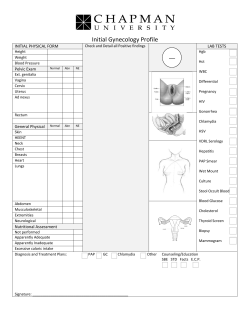


© Copyright 2026