
LABORATORY TESTS FOR THE DETECTION OF REPRODUCTIVE TRACT INFECTIONS 1999
WORLD HEALTH ORGANIZATION
Regional Office for the Western Pacific
LABORATORY TESTS
FOR THE DETECTION OF
REPRODUCTIVE TRACT INFECTIONS
1999
Laboratory Tests for the Detection of RTI
WHO
WPRO
WHO
WPRO
Laboratory Tests for the Detection of RTI
LABORATORY TESTS
FOR THE DETECTION OF
REPRODUCTIVE TRACT INFECTIONS
CONTENTS
Abbreviations
iii
Acknowledgements
iv
1 Introduction
2 Laboratory Tests for Specific Diseases
Candidiasis
Trichomoniasis
Bacterial vaginosis
Chlamydiosis
Gonorrhoea
Syphilis
Genital Herpes
Chancroid
Donovanosis
Human Papillomavirus
Human Immunodeficiency virus
3 Notes on Methods of Molecular Assay
4 References
1
3
3
5
7
9
12
15
18
20
22
24
26
28
33
i
ii
Laboratory Tests for the Detection of RTI
WHO Library Cataloguing in Publication Data
Laboratory Tests for the Detection of
Reproductive Tract Infections
1999
1.
2.
3.
4.
Genital diseases, Female - diagnosis
Genital diseases, Male - diagnosis
Sexually transmitted diseases - diagnosis
Laboratory techniques and procedures
ISBN 92 9061 148 0
The World Health Organization welcomes requests for permission to reproduce or translate
its publications, in part or in full. Applications and enquiries should be
addressed to the Office of Publications, World Health Organization, Geneva, Switzerland
or to the Regional Office for the Western Pacific, Manila, Philippines which will be glad
to provide the latest information on any changes made to the text, plans for new editions,
and reprints and translations already available.
© World Health Organization 1999
Publications of the World Health Organization enjoy copyright protection in accordance
with the provisions of Protocol 2 of the Universal Copyright Convention. All rights
reserved.
The designations employed and the presentation of the material in this publication do not
imply the expression of any opinion whatsoever on the part of the Secretariat of the
World Health Organization concerning the legal status of any country, territory, city or
area or of its authorities, or concerning the delimitation of its frontiers or boundaries.
The mention of specific companies or of certain manufacturers’products does not imply
that they are endorsed or recommended by the World Health Organization in preference
to others of a similar nature that are not mentioned. Errors and omissions excepted, the
names of proprietary products are distinguished by initial capital letters.
WHO
WPRO
WHO
WPRO
Laboratory Tests for the Detection of RTI
ABBREVIATIONS AND ACRONYMS
BV
DFA
DNA
dNTP
dUTP
EDTA
EIA
ELISA
FTA-ABS
GAC-EIA
HIV
HPV
HSV
IQS
LCR
LE
LIA
LPS
MHA-TP
NASBA
PCR
RNA
RPR
RTI
STI
STD
TMA
UNG
VDRL
WB
bacterial vaginosis
direct immunofluorescence assay
deoxyribunucleic acid
deoxynucleotide triphosphates
deoxyuridine triphosphate
ethylenediaminetetraacetic acid
enzyme immunoassay
enzyme-linked immuno-sorbent assay
fluorescent treponemal antibody absorption test
IgG antibody capture EIA
human immunodeficiency virus
human papillomavirus
herpes simplex virus
internal quantification standard
ligase chain reaction
leukocyte esterase assay
line immunoassay
lipopolysaccharide
microhaemagglutination assay
nucleic acid sequence-based amplification reaction
polymerase chain reaction
ribonucleic acid
rapid plasma reagin
reproductive tract infection
sexually transmitted infection
sexually transmitted disease
transcription mediated amplification
uracil-N-glycosylase
venereal disease research laboratory
western blot
iii
iv
Laboratory Tests for the Detection of RTI
WHO
WPRO
ACKNOWLEDGEMENTS
The Western Pacific Regional Office of the World Health Organization would
like to thank the principal contributor to this manual, Ms J Kuyper, and the
reviewers, Dr P Rowe, Dr P Shrestha, Dr S Lazarri, Dr Yoke-Fong Chiew and
Dr SM Garland.
WHO
WPRO
Laboratory Tests for the Detection of RTI
1
INTRODUCTION
Valid laboratory assays are necessary to:
• confirm suspected reproductive tract infections (RTI)
when laboratory facilities are available and accessible;
• detect infections in asymptomatic individuals;
• investigate cases of resistance to usual treatment;
• monitor the evolution of pathogen sensitivity to
antimicrobials; and
• conduct research on STI prevalence and incidence.
An optimal laboratory test for diagnosis of infection should be simple to perform, sensitive, specific, reproducible, objective, rapid and inexpensive, and
should require no special equipment.
In recent years, new methods to detect RTI have been developed, using sophisticated molecular biology techniques. Some of these new methods meet
many of the criteria for optimal tests and have improved the diagnosis of RTI. In
other cases, older, standard assays remain valid and should still be used.
Molecular detection techniques do not rely on the ability to culture or directly
observe intact organisms but are designed to detect specific cellular antigens
or nucleic acids. Therefore, the stringent transport requirements for clinical
samples are no longer needed. Viable and nonviable microorganisms can be
detected in samples. The development of molecular detection assays has led
to the ability to use one sample for the detection of many organisms, to test for
multiple organisms in one assay and to automate assays. Due to their extreme sensitivity, newly developed nucleic acid amplification systems allow the
use of samples obtained by noninvasive methods, including patient self-sampling and urine collection. In addition, some of these assays are used to determine the number of nucleic acid copies in a sample. The notes on methods of
molecular assay describe in detail the general techniques for the molecular
assays used to detect RTI.
This publication discusses state-of-the-art methods to detect eleven RTI. The
types of assay presented fall into several categories and include detection of
1
2
Laboratory Tests for the Detection of RTI
WHO
WPRO
the organism by direct microscopy, detection of metabolic products, culture,
and the detection of specific antibodies, antigens, DNA or RNA. Not all organisms can be detected using all types of assay, nor can all laboratories perform
all types of assay. Thus, the methods that are most useful in detecting each
organism are summarized, as well as their sampling procedures, sensitivity
and specificity, the advantages and disadvantages of laboratory testing, the
appropriate level of use, the training and equipment required, the ease of performance, and the indicative cost of reagents (at current rates in the United
States of America).
Detailed instructions for carrying out each test can be found in the manufacturer’s manual accompanying each test kit and should be strictly adhered to.
The sensitivity and specificity of an assay will vary depending on the method
used as the standard and the prevalence of the disease in the population
tested. The assay sensitivity and specificity figures in this publication are based
on a range of values taken from many different sources, including all types of
patient population.
WHO
WPRO
Laboratory Tests for the Detection of RTI
2
LABORATORY TESTS FOR SPECIFIC DISEASES
Candidiasis
Candida is a common commensal yeast found in the normal vaginal microbial flora. While most colonized women
show no symptoms of infection, in some, colonization
progresses to symptomatic disease. Laboratory confirmation is often necessary to diagnose candidiasis in cases
where symptoms are nonspecific. Infection is usually, but
not always, associated with higher numbers of yeast organisms and the presence of the mycelial form, together with an inflammatory
response (polymorph). The normal microbial flora, Gram-positive rods of lactobacillus and diptheroid species, are still present. However, the relationship
between the number of organisms and infection is not clear [1].
Methods of detection
Detection by
microscopy
A swab of the vaginal secretions is placed in saline and the evaluation for
yeast is carried out immediately by adding a drop to a slide and mixing it with a
drop of potassium hydroxide solution. The potassium hydroxide lyses the patient’s cells, making the yeast easier to see. Alternatively, the swab is rolled
onto a slide, fixed and Gram stained. This slide does not have to be stained or
evaluated immediately. Both slides are examined by a light microscope (400x)
for the presence of the yeast or mycelial form and inflammatory response.
Detection
by culture
A swab of the vaginal secretions or an aliquot of a vaginal wash is inoculated
into Sabouraud’s agar within a few hours of collection and incubated for up to
two days at 37° C. Colonies are identified as yeast by performing a Gram stain.
(Candida will also grow as dull white colonies on horse blood agar.) The quantity of yeast is determined, with more than 103 colony-forming units/ml of vaginal secretions usually being associated with disease.
Detection
of Candida
antigen
A swab of vaginal secretions or a drop of a vaginal wash is mixed on a slide
with a commercially available solution of latex beads that are coated with an
antibody to mannan, a soluble polysaccharide that is the major constituent of
the Candida cell wall and is found in the vagina when Candida are present.
The antigen-antibody complexes are observed as agglutination of the suspension. Positive and negative control should be used.
3
WHO
WPRO
Laboratory Tests for the Detection of RTI
4
Detection of
Candida DNA
A commercially available, 40-minute, nonisotopic, automated test [2], is a hybridization assay that uses DNA probes to directly detect Candida, Trichomonas
and Gardnerella in vaginal swab samples (Note 1). Samples are collected
with a swab, placed into the collection tubes supplied in the kit, stored at 4º C.
and tested within 24 hours.
Table 1: Characteristics of Candida detection assays
9
Microscopy
wet mount
Culture
>103cfu/ml
Antigen detection
DNA detection
1
35-45%
67%
61-81%
80%
1
Specificity
99%
66%
97%
98%
Advantages
rapid,
inexpensive
sensitive
rapid, also detects
Trichomonas
rapid, objective, also detects
Trichomonas and Gardnerella
Disadvantages
subjective
requires 24 hours
expensive
expensive, requires special
equipment and test read
immediately after completion
Level of use
exam room,
on-site lab
on-site lab,
intermediate lab
exam room,
on-site lab
intermediate lab, referral lab
Training
moderate
moderate
minimal
moderate
Equipment
light microscope
incubator,
light microscope
none
heat block, special processor
Ease of
performance
easy
moderate
easy
easy to moderate, automated
Cost
US$ 1.00
US$ 2.00
US$ 12.00
(includes detection
of Trichomonas)
US$ 12.00
(includes detection of
Trichomonas and Gardnerella)
Sensitivity
1
Sensitivity and specificity are for clinical signs and symptoms of vulvovaginal candidiasis.
WHO
WPRO
Laboratory Tests for the Detection of RTI
Trichomoniasis
Trichomonas vaginalis is a parasitic protozoan that causes vaginal inflammation. Infection with Trichomonas can range from severe vaginitis with discharge
to asymptomatic carriage. Since asymptomatic carriage can account for as
many as 50% of cases, diagnosis cannot be made solely on the basis of clinical presentation [3]. Laboratory detection of the parasite is required to confirm
diagnosis.
Methods of detection
Detection by
microscopy
Direct microscopic observation of motile characteristic parasites is made from
a swab of vaginal secretions taken from the posterior fornix and then placed
into a physiological saline solution and analysed immediately under a light microscope (100x).
Detection by
culture
A swab of secretions taken from the posterior vaginal fornix is used within six
hours of sample collection to inoculate a tube of Diamond’s modified medium.
The culture is incubated at 35°C. for up to four days with daily examination by
wet prep for motile trichomonas. A culture system with a two-chambered bag
is now available that, after inoculation with the swab, permits both incubation
of the culture and immediate wet prep by microscopic examination through the
upper chamber of the bag in one self-contained system [4].
Detection of
Trichomonas
vaginalis
antigens
T. vaginalis antigens are detected using the Trichomonas Direct Enzyme Immunoassay and Fluorescent Direct Immunoassay. A swab of vaginal secretions is rolled onto a glass slide and air-dried in the examination room. In this
one hour procedure, cocktails of peroxidase or fluorochrome-labelled
monoclonal antibodies to various T. vaginalis antigens are added to the slide,
incubated, washed and detected using substrates that change colour or fluoresce. Slides are examined under a light microscope for the colourimetric
assay and under a fluorescent microscope for the fluorescent assay.
Another method uses a swab of vaginal secretions or a drop of a vaginal wash
which is mixed on a slide with a commercially-available solution of latex beads
that are coated with antibodies to T. vaginalis antigens. The antigen-antibody
complexes are observed as agglutination of the suspension.
Detection of
T. vaginalis
DNA
A commercially available, 40-minute, nonisotopic, automated [2] hybridization
assay that uses DNA probes to directly detect Candida, Trichomonas and
Gardnerella in vaginal swab samples is now available (Note 1). The sample is
collected with a swab and placed into the collection tube supplied by the kit,
stored at 4º C. and tested within 24 hours.
5
6
WHO
WPRO
Laboratory Tests for the Detection of RTI
Amplification
and detection
of T. vaginalis
DNA
This assay is not commercially available [5]. Sample collection can be performed by obtaining a self-administered swab of the vaginal introitus or by a
technique in which the patient inserts, then immediately withdraws, a tampon
(commercially available). The sample is placed into transport media and can
be tested for up to one month following collection. The DNA is extracted and
added to a polymerase chain reaction (PCR) mixture (Note 2) containing primers TVA5 and TVA6 which amplify a 102 base-pair fragment of T. vaginalis
DNA. The specific T. vaginalis DNA fragment is labelled during amplification
and detected using DNA hybridization with probe TVB, a semi-automated
ELISA assay for detection of PCR-amplified products (Note 3).
Table 2: Characteristics of Trichomonas detection assays
Microscopy
Sensitivity1
Culture
Antigen
detection
DNA detection
Hybridization
assay
PCR
38-82%
98%
86%
88-91%
93%
100%
100%
99%
100%
96%
Advantages
rapid,
inexpensive
sensitive,
diagnosis in
men
rapid
rapid, objective,
also detects
Gardnerella and
Candida
very sensitive,
allows patient
self-sampling
Disadvantages
low sensitivity,
must be
performed
immediately,
subjective
takes 1-4
days
expensive
expensive, requires
special equipment
and test read
immediately after
completion
expensive,
requires
expertise
Level of use
exam room
on-site lab
on-site lab,
intermediate
lab
exam room,
on-site lab
intermediate,
referral lab
referral lab
Training
moderate
moderate
moderate/minimal
moderate
extensive
Equipment
light microscope
incubator,
light
microscope
light or
fluorescent
microscope/none
heat block, special
processor
thermal cycler,
microwell plate
reader
Ease of
performance
easy
easy
moderate/
easy
easy to moderate,
automated
complex,
automated
Cost
US$ 1.00
US$ 3.00
US$ 6.00-12.00
(includes
detection of
Candida)
US$ 12.00
(includes detection
of Candida and
Gardnerella)
$11.00
Specificity
1
1
Sensitivity and specificity are for detection of T. vaginalis by combined wet prep and culture results.
WHO
WPRO
Laboratory Tests for the Detection of RTI
Bacterial vaginosis
Bacterial vaginosis (BV) is a condition in which the natural balance of organisms found in the vagina is changed from a predominance of Lactobacillus to
an overgrowth of other bacteria including Gardnerella vaginalis, Mobiluncus
and other anaerobes [6].
Methods of detection
Detection by
microscopy
and/or
metabolic
products
The most widely accepted method for diagnosis of BV is the presence of three
of the following four criteria:
•
•
•
•
a homogeneous vaginal discharge;
a vaginal pH of greater than 4.5;
the presence of clue cells; and
a fishy odour after addition of potassium hydroxide to the vaginal
secretions (the amine test) [6].
A swab of vaginal fluid from the posterior fornix is placed in normal saline. This
sample is used immediately for the following tests. In the amine test, a drop is
placed on a slide and examined under a light microscope for the presence of
clue cells, which are epithelial cells heavily coated with bacteria so that the
peripheral borders are obscured. Samples are generally considered positive
for BV if clue cells comprise more than 20% of all epithelial cells. A drop is
placed on a slide with a drop of potassium hydroxide solution. The release of
amines produces a fishy odour. The pH value is determined using a pH dipstick onto secretions on a speculum.
A simpler and more specific assay is a Gram stain of a vaginal smear. A swab
of vaginal secretions is rolled onto a glass slide and air dried. The sample is
stable for days at ambient temperature. The slide is Gram stained and a standardized, 0-10 point scoring method is used to evaluate the smears [7]. Points
are given by estimating the number of three different bacterial morphotypes
from 0 to 4+, including large Gram-negative rods, small Gram-negative/variable rods, and curved Gram-negative/variable rods.
The proline aminopeptidase test [8] is an indirect test for a chemical produced
by the organisms associated with BV. Vaginal secretions are added to a
substrate and incubated for four hours. A colour change denotes the test
result.
The vaginal swab from the posterior fornix is placed into normal saline and can
be held at 4°C. for up to four hours and then frozen until testing. Samples can
be shipped on ice.
7
8
WHO
WPRO
Laboratory Tests for the Detection of RTI
Detection of
G. vaginalis
DNA
A commercially available, 40-minute, nonisotopic, automated [9] hybridization assay that uses DNA probes to directly detect Candida, Trichomonas and
high concentrations of Gardnerella in vaginal swab samples (Note 1). Samples are collected with a swab and placed into the collection tubes supplied
with the kit, stored at 4ºC. and tested within 24 hours.
Table 3: Characteristics of bacterial vaginosis detection assays
Microscopy and metabolic product detection
DNA detection
3 of 4 criteria
Gram stain
Proline
aminopeptidase
Hybridization assay
Sensitivity1
81%
89%
93%
94%
1
Specificity
94%
93%
93%
81%
Advantages
rapid, inexpensive
reproducible,
standardized,
inexpensive
objective
objective, can also
detect Candida and
Trichomonas
Disadvantages
subjective,
some criteria
nonspecific
requires
expertise
takes longer than
wet mount or stain
expensive, requires
special equipment,
test read
immediately after
completion
Level of use
exam room,
on-site lab
on-site lab
on-site lab,
intermediate lab
intermediate lab,
referral lab
Training
moderate
moderate
minimal
moderate
Equipment
light microscope
light microscope
centrifuge,
incubator
heat block, special
processor
Ease of
performance
easy
easy
easy
easy to moderate,
automated
Cost
US$ 1.00
US$ 0.50
US$ 1.00
US$ 12.00
(includes detection
of Candida and
Trichomonas)
1
Sensitivity and specificity are for diagnosis of BV by presence of 3 of 4 criteria and/or positive Gram stain.
WHO
WPRO
Laboratory Tests for the Detection of RTI
Chlamydiosis
Chlamydia trachomatis is an important cause of urethritis and cervicitis. Laboratory detection of C. trachomatis is necessary because as many as 70% to
80% of women and up to 50% of men who are infected do not experience any
symptoms. Untreated, infected individuals transmit chlamydial infections to
sexual partners and are at risk for sequelae such as epididymitis in men and
pelvic inflammatory disease and infertility in women [10].
Methods of detection
Detection
by culture
The conventional method for the laboratory diagnosis of C. trachomatis has
been inoculation of a cell culture with a genital specimen. This method is expensive, labour-intensive and time-consuming. It also requires considerable
expertise to perform it correctly and meticulous handling of the specimen during transport to maintain viable organisms. For these reasons, culture tests
are now used less frequently and antigen and nucleic acid detection techniques have become common methods for detection of C. trachomatis infection, allowing testing in laboratories that lack the facilities for tissue cell culture
[10].
Detection of
host response
The leukocyte esterase assay (LE) is a rapid, nonspecific urine dipstick test for
the presence of an enzyme found in urine when leukocytes are present due to
inflammation. The LE test can diagnose urethritis but cannot identify the specific cause of the infection. The sensitivity and specificity of LE for the detection of chlamydial and gonococcal infection are 54%-97% and 36%-95%, respectively. The LE test performs best as a screening test for chlamydial infection in asymptomatic adolescent and young men with positive tests confirmed
by enzyme immunoassay [11].
Detection by
microscopy
In the direct immunofluorescence assay (DFA), urethral or endocervical columnar epithelial cells collected on swabs are rolled onto glass slides, fixed
and stained with fluorescein-labelled monoclonal antibodies specific for the
major outer membrane protein of C. trachomatis. DFA allows for the visualization of the distinctive morphology and staining characteristics of chlamydial
inclusions and elementary bodies. It also permits simultaneous assessment
of the specimen adequacy.
For all chlamydial detection assays, samples must contain urethral or endocervical columnar cells and not exudate. The presence of ten or more elementary bodies is generally accepted for the test to be positive. DFA is useful as a
confirmatory test for samples found positive by antigen and DNA detection
assays.
9
10
Laboratory Tests for the Detection of RTI
Detection of
C. trachomatis
antigens
WHO
WPRO
A common test for genital C. trachomatis infection is the detection of chlamydial
genus-specific lipopolysaccharide (LPS) antigens in cervical and urethral swab
specimens and in urine samples from men using any of several commercially-available enzyme immunoassay (EIA) kits. Because chlamydial LPS
antibodies may cross-react with the LPS of other Gram-negative bacteria to
give false-positive results, blocking assays are needed to confirm positive EIA
results and thus improve specificity (Note 4). Confirmation can also be done
by performing direct fluorescent antibody assay on the centrifuged specimen.
Rapid immunodot or latex agglutination assays are simple tests in which the swab
sample is mixed with chlamydial LPS antibodies coated on a membrane or card.
The antigen-antibody complexes are observed visually as agglutination.
Detection of
C. trachomatis
RNA
C. trachomatis ribosomal RNA is detected by hybridization with a
chemoluminescent DNA probe (Note 5). A probe competition assay has been
developed to confirm positive samples. A similar kit can be used to detect N.
gonorrhoeae RNA in the same sample. Endocervical swabs, urethral swabs
and urine samples from men can be tested. Specimen collection kits containing swabs and transport medium that lyses the organisms and releases the
RNA are provided by the manufacturer. No stringent transportation conditions are required and the samples are stable at ambient temperature for one
month.
Amplification
and detection of
C. trachomatis
DNA or RNA
The polymerase chain reaction (PCR) amplification assay for C. trachomatis
DNA is available in a semi-automated and a fully automated format. A kit is
available that also amplifies N. gonorrhoeae DNA from the same sample and
detects it in a separate hybridization well. To prevent false-negative results
due to inhibitors of the amplification reaction in the patient samples, this kit
includes an internal standard that is amplified in the same reaction tube and
detected in a separate hybridization well (Note 6).
Another DNA amplification technique for C. trachomatis detection is ligase
chain reaction (LCR) (Note 7). The amplicons are detected in an automated
instrument that is designed to minimize false-positive samples due to contamination from carry-over. The enzymes used in LCR are not as sensitive as
the enzymes in PCR to inhibitors in the sample.
LCR and PCR are excellent tests for the detection of C. trachomatis in endocervical, urethral and urine samples from both men and women (these tests
do not have an internal control, such as ßglobin, to assess the adequacy of
sample collection). In addition, other noninvasive specimen collection methods such as self-collected vaginal introitus swabs or tampons can be tested.
Specimen collection kits are provided by the manufacturers. Swab and tampon samples can be kept at ambient temperature but urine samples should
be refrigerated and are stable for up to four days. They will remain stable for
up to 60 days when frozen.
WHO
WPRO
Laboratory Tests for the Detection of RTI
A third amplification assay for C. trachomatis uses transcription-mediated amplification (TMA) of the 16S ribosomal RNA (Note 8). After initial heating to
95oC, the reaction occurs at one temperature. Amplification and product detection take place in one tube [12].
LCR, PCR and TMA are all highly sensitive assays. Unless specimens are
carefully collected in the clinic, appropriately transported and handled carefully
in the laboratory, they are susceptible to contamination and may give falsepositive results. For kits, the manufacturer’s instructions must be carefully
followed and adhered to. For example, to ensure no contamination at the
laboratory level, positive-displacement pipettes (or plugged pipettes) should
be used. For “in-house”assays, there should be physical separation (in separate rooms) for the various steps of PCR, such as DNA extraction process,
detection and preparation of master mixes.
Table 4: Characteristics of Chlamydia detection assays
Microscopy
Antigen detection
RNA detection
Amplification & detection
DFA
EIA
Rapid
Chemoluminescent
DNA probe
PCR
LCR
Sensitivity1
74%-90%
71%-97%
52%-85%
75-%85%
90%
90%-97%
Specificity1
98%-99%
97%-99%
> 95%
98%-99%
99%-100%
99%-100%
Advantages
rapid, easy
can batch
samples
rapid, easy
also detects
N. gonorrhoeae,
automated
can detect
N. gonorrhoeae in
same sample,
less affected by
inhibitors
by inhibitors
Disadvantages
labour
intensive,
subjective
requires
confirmation
insensitive,
requires
confirmation
less sensitive than
PCR, requires
confirmation
false negatives
no test for
sample
inhibitors
Level of use
on-site lab,
intermediate
intermediate,
referral lab
exam room, onsite lab
intermediate,
referral lab
intermediate,
referral lab
intermediate,
referral lab
Training
moderate to
extensive
moderate
minimal
moderate
moderate to
extensive
moderate
Equipment
fluorescent
microscope
microwell
plate reader
none
heat block,
luminometer
thermal cycler,
incubator,
microwell plate
reader
thermal cycler,
LCx processor
Ease of
performance
moderate
moderate
easy
moderate
moderate to
difficult, automated
moderate,
automated
Cost
US$ 6.00
US$ 6.00
US$ 13.0016.00
US$ 8.00
US$ 11.00
(US$ 14.00 for
N. gonorrhoeae
detection also)
US$ 16.00
allow noninvasive sampling
1
Sensitivity and specificity are for detection of C. trachomatis by culture or by DNA amplification test.
11
12
Laboratory Tests for the Detection of RTI
WHO
WPRO
Gonorrhoea
Infection of the genital tract with Neisseria gonorrhoeae can cause urethritis,
cervicitis, proctitis, or Bartholinitis. Complications of untreated disease include
epididymitis, prostatitis and infertility in men and pelvic inflammatory disease
and infertility in women. Because most cases in females are asymptomatic,
detection of infection using laboratory tests is needed to prevent sequelae and
transmission to sexual partners and, for pregnant women, to neonates [13].
Methods of detection
Detection by
microscopy
A direct Gram smear is performed as soon as the specimen is collected by
rolling the swab onto a slide, staining, and viewing under an oil immersion lens
(1000X). The presence of Gram-negative diplococci inside polymorphonuclear leukocytes in urethral smears from symptomatic men is diagnostic for
presumptive gonorrhoea. However, the Gram stain is not as useful for endocervical smears (sensitivity of 50%-70%) because the presence of other
Gram-negative diplococci make interpretation difficult.
Detection of
host response
The leukocyte esterase assay (LE) is a rapid, nonspecific urine dipstick test
for the presence of an enzyme found in urine when leukocytes are present
due to inflammation. The LE test can diagnose urethritis but cannot identify
the specific cause of the infection. The sensitivity and specificity of LE as a
marker for chlamydial and gonococcal infection are 54%-97% and 36%-95%,
respectively. The LE test performs best as a screening test for gonococcal
infection in asymptomatic men with positive tests confirmed by DNA detection
[11].
Detection by
culture
The endocervical or urethral swab is used immediately after collection to inoculate a plate of selective media such as modified Thayer-Martin. The selective medium contains antimicrobial agents that allow the growth of N.
gonorrhoeae and inhibit the growth of other bacteria. The agar plate is placed
into a plastic zip-lock bag with a CO2 generating tablet for transport to the
laboratory, where the plate is incubated at 35oC for up to three days. Typical
colonies are tested with Gram-stain, oxidase and catalase and/or superoxal
tests for presumptive identification of N. gonorrhoeae. To confirm a presumptive culture, the isolated organism is tested for sugar fermentation by growth
in standard carbohydrate fermentation tubes, or by using any of several rapid
nongrowth methods for confirmation of N. gonorrhoeae isolates, including detection of preformed enzymes and agglutination with pools of mouse IgG
monoclonal antibodies to the outer membrane protein. The monoclonal antibodies can be bound to staphylococcal Protein A or to easy-to-read coloured
metal particles.
WHO
WPRO
Laboratory Tests for the Detection of RTI
Detection of
N. gonorrhoeae
antigen
Gonococcal antigen is detected by an enzyme immunoassay that is similar to
a Gram stain in sensitivity and specificity for presumptive diagnosis in men,
but is less sensitive for endocervical swabs.
Detection of
N. gonorrhoeae
RNA
A two-hour DNA probe hybridization assay is available (Note 5) [14] and a
probe competition assay has been developed to confirm positive samples. A
similar kit can be used to detect C. trachomatis RNA in the same sample.
Endocervical swabs and urethral swabs and urine samples from men can be
tested. Specimen collection kits, containing swabs and transport medium that
lyses the organisms and releases the RNA, are provided by the manufacturer.
No stringent transportation conditions are required and the samples are stable
at ambient temperature for one month.
Amplification
and detection of
N. gonorrhoeae
DNA
The polymerase chain reaction (PCR) amplification assay (Note 2) is designed
to also amplify C. trachomatis DNA from the same sample (Note 6). The reaction can be automated. To prevent false-negative results due to inhibitors of
the amplification reaction in the patient samples, the kit includes an internal
standard that is amplified in the same reaction tube and detected in a separate
hybridization well.
Another DNA amplification technique for N. gonorrhoeae detection is ligase
chain reaction (LCR) (Note 7). The amplicons are detected in an automated
instrument that is designed to minimize false-positive samples due to contamination from carry-over. The enzymes used in LCR are not as sensitive as the
enzymes in PCR to inhibitors in the sample [15].
LCR and PCR are excellent tests for the detection of N. gonorrhoeae in endocervical, urethral and urine samples from both men and women. In addition, other noninvasive specimen collection methods such as self-collected
vaginal introitus swabs or tampons can be tested. Specimen collection kits
are provided by the manufacturers. Swab samples and tampons can be kept
at ambient temperature, but urine samples should be refrigerated and are stable for up to four days. They will remain stable for up to 60 days when frozen.
13
WHO
WPRO
Laboratory Tests for the Detection of RTI
14
Table 5: Characteristics of N. gonorrhoeae detection assays
Microscopy
Sensitivity1
Culture
DNA Detection
Amplification & Detection
Hybridization assay
PCR
LCR
90%-95%
81%-100%
86%-100%
89%-97%
95%-100%
Specificity
98%-100%
100%
99%
94%-100%
98%-100%
Advantages
rapid,
inexpensive
gold standard, isolates
available for further
testing
rapid, viable organisms
not required
viable organisms not required,
extremely sensitive, allow non
invasive sampling can detect
C. trachomatis in same sample
Disadvantages
insensitive
for females
stringent handling,
requires up to 3 days
expensive
expensive, requires expertise
no test for sample inhibitors
Level of use
on-site lab
on-site lab, intermediate
intermediate, referral lab
intermediate,
referral lab
intermediate,
referral lab
Training
moderate
moderate
moderate
moderate to
extensive
moderate
Equipment
light
microscope
incubator, light
microscope, candle jar
water bath, luminometer
microfuge,
thermal cycler,
incubator,
microwell reader
heat block,
thermal cycler,
microfuge, Imx
processor
Ease of
performance
easy
moderate
moderate
moderate to
difficult,
automated
moderate,
automated
Cost
US$ 0.50
US$ 1.00 (+1-3 to
confirm positive isolates)
US$ 6.00
US$ 11.00
(US$ 14.00 for C.
trachomatis
detection also)
US$ 14.00
1
1
N. N.
gonorrhoeae
Sensitivity
specificityare
are for
for detection
in urethral,
Sensitivity
andand
specificity
detectionof of
gonorrhoeae
in urethral, endocervical and urine samples by
culture except for microscopy, which is for detection in urethral samples from symptomatic men.
WHO
WPRO
Laboratory Tests for the Detection of RTI
Syphilis
Syphilis, a chronic infection with clinical manifestations occurring in distinct
stages, is caused by the spirochete Treponema pallidum. This bacterium cannot be cultured in vitro or stained using standard techniques. The rabbit infectivity test is the oldest and most sensitive method to identify infection with
T. pallidum. While this test is not practical for routine laboratory use, it is the
standard for measuring the sensitivity of other methods. Routine laboratory
methods rely on demonstrating the presence of T. pallidum in a characteristic
lesion or the presence of antibodies in serum. Not all methods can be used to
diagnose all stages of syphilis. Antibodies are not present early in disease (1 to 4
weeks after a lesion has formed) and lesions are not present at all stages [16].
Methods of detection
Detection by
microscopy
T. pallidum is detected in primary or secondary lesions by dark-field microscopy.
A glass slide is touched to fluid expressed from a lesion or node aspirate,
coverslipped, and examined immediately under a light microscope fitted with a
dark-field condenser. T. pallidum is identified by its characteristic morphology
and motility. Oral specimens are not suitable for this method because other
spirochetes present in these specimens cannot be distinguished from T.
pallidum.
Direct fluorescent antibody assay for T. pallidum detects and differentiates T.
pallidum from nonpathogenic treponemes by an antigen-antibody reaction. The
organism does not have to be motile for identification. The specimen is obtained in the same way as for dark-field microscopy but the smear is air-dried
and fixed. A fluorescein-labelled polyclonal antibody to T. pallidum (IgG from
humans or rabbits with syphilis that is absorbed with Reiter treponemes) is
added to the smear. The slide is examined for fluorescing treponemes displaying typical morphology using a fluorescent microscope.
Detection of
nontreponemal
antibodies
Nontreponemal antibody tests for syphilis, which are used for screening patient serum, are based on detection of antibodies to a cardiolipid-cholesterollecithin antigen. Undiluted or serial two-fold dilutions of serum are added to
the antigen on a slide or card. The reagents are then mixed and rocked and
observed for flocculation. The rapid plasma reagin (RPR) test, in which the
antigen is mixed with charcoal so the antigen-antibody complexes can be seen
without a microscope, is the most common. The Venereal Disease Research
Laboratory (VDRL) slide test, which is read microscopically, can also be used
to detect antibodies in cerebral spinal fluid. For some immunoassays, the
VDRL antigen is coated onto microwells to capture the antibodies in the patient
serum which are then detected with anti-human immunoglobulins conjugated
15
16
Laboratory Tests for the Detection of RTI
WHO
WPRO
to an enzyme [16] or coated onto erythrocytes [17]. A positive test provides a
presumptive diagnosis for syphilis and must be confirmed using a specific
treponemal antibody test. It is also advantageous to gauge response to appropriate treatment, as specific tests, once positive, usually remain so for life.
Detection of
treponemal
antibodies
The fluorescent treponemal antibody absorption test (FTA-ABS) and the
microhaemagglutination assay for T. pallidum (MHA-TP) detect specific antibodies in serum to T. pallidum. For FTA-ABS, the Nichols strain of T. pallidum
is fixed on a slide. Patient serum is absorbed with an extract of Reiter
treponemes and added to the slide. Fluorescein-labelled anti-human immunoglobulin is added and the slide is examined for fluorescent spirochetes using a fluorescent microscope. In MHA-TP, erythrocytes are sensitized with T.
pallidum antigen, mixed with patient serum and observed for agglutination.
Immunoassays have also been developed that use T. pallidum antigen-coated
microwells to capture specific antibodies from the patient serum, which are
detected with enzyme-labelled anti-human IgG monoclonal antibodies [18].
Detection of
T. pallidum
antigens
T. pallidum antigens are detected using an enzyme immunoassay [19]. Antigens are extracted within 72 hours of collection from swabs containing lesion
exudate.
Amplification
and detection
of T. pallidum
DNA and RNA
T. pallidum DNA is amplified in a PCR (Note 2). This assay has been described as a multiplex-PCR for the simultaneous amplification of herpes simplex virus and H. ducreyi DNA in the same sample [20].
A highly sensitive reverse transcriptase PCR assay has been described for
the amplification and detection of T. pallidum 16S rRNA. This technique is
especially useful for diagnosis of neurosyphilis where very low numbers of
organisms exist [21].
WHO
WPRO
Laboratory Tests for the Detection of RTI
Table 6: Characteristics of syphilis detection assays
Microscopy
Antibody Detection
Antigen Detection
DNA Detection
dark-field
nontreponemal
RPR
treponemal
MHA-TP
Sensitivity1
74%-86%
72%-100%
69%-90%
81%
91%
Specificity1
97%-100%
93%-98%
98%-100%
89%
99%
Advantages
positive early,
rapid, specific,
inexpensive
inexpensive,
rapid, easy,
antibody titer to
follow treatment
specific, confirms
non treponemal
tests
detects T. Pallidum
before antibodies are
positive
sensitive, specific,
allows self-collected
sample
false positives,
less sensitive for
early disease
more difficult, more
expensive
time consuming,
expensive
inhibitors of PCR
reaction cause
false-negative
results, complex,
expensive
Disadvantages insensitive, no
oral sample,
requires live
treponemes
multiplex PCR
Level of use
exam room,
on-site lab
on-site lab,
intermediate lab
intermediate lab,
referral lab
intermediate lab,
referral lab
referral lab
Training
extensive
minimal
moderate
moderate
extensive
Equipment
light microscope
with dark-field
condenser
centrifuge, rotator
centrifuge
spectrophotometer
microfuge, thermal
cycler, incubator,
microwell plate
reader
Ease of
performance
easy
easy
moderate
moderate
complex
Cost
US$ 0.40
US$ 0.50
US$ 1.40
US$ 3.00
US$ 14.00 (includes
detection of
H. ducreyi and HSV)
1
Sensitivity and specificity are for detection of primary syphilis. The sensitivity of both nontreponemal and treponemal
antibody detection increases for detection of secondary syphilis. The sensitivity of nontreponemal antibody
detection decreases for detection of latent and tertiary syphilis. The tests for T. pallidum are only relevant
when lesions are present in primary and secondary syphilis. But these tests can detect latent untreated infection
which can be important for patient outcomes, such as in pregnancy.
17
18
Laboratory Tests for the Detection of RTI
WHO
WPRO
Genital Herpes
Herpes simplex virus (HSV) is one of the major causes of genital ulcer disease. Primary infection is followed by latency and variable periods of reactivation. Although clinical diagnosis may be accurate if based on the presence
of typical vesicles, up to two-thirds of individuals acquire HSV asymptomatically
and most infected persons shed virus during latent periods. Laboratory diagnosis is necessary to detect HSV in asymptomatically infected people to prevent
transmission to sexual partners and to children born to infected mothers [22].
Methods of detection
Culture of HSV
Inoculation of cells in tissue culture with infected secretions or tissues is considered the standard method. Lesions are rubbed at their base with cotton or
Dacron swabs (need to break intact vesticles), or fluid is extracted from vesicles, and immediately placed in transport medium. The sample can be stored
at 4oC for up to five days before it is used to inoculate a fibroblast cell-line,
although ideally this should be done as soon after collection as possible. Diagnosis is made by observation of a characteristic cytopathic effect on the
cells after incubation for up to one week (although most positives occur within
48 hours of cell inoculation) and confirmation of the virus by staining the infected cells with monoclonal antibodies specific for HSV. A rapid culture and
histochemical stain method uses a genetically engineered cell-line that is inoculated, incubated for 16 to 24 hours, and stained with a solution that causes
HSV infected cells to turn blue [23]. Another rapid tissue 24-hour culture method
is also available. Both of these rapid culture methods are as sensitive as
conventional culture [24].
Detection of
HSV antigen
In the direct immunofluorescence assay (DFA), cells collected from a lesion
(with the base rubbed well with a swab) are concentrated and spotted onto a
slide that is incubated with a fluorescein-conjugated monoclonal HSV typespecific antibody. The slide is observed under a fluorescent microscope for
the presence of intracellular fluorescence (this approach is not as sensitive as
a culture). In the immunoperoxidase assay, specimens are prepared as for
DFA and incubated with a monoclonal HSV antibody that is detected with a
second antibody conjugated to an enzyme and reacted with a colourimetric
substrate.
Amplification
and detection
of HSV DNA
HSV DNA is amplified in a PCR. This assay has been described as a multiplex-PCR for the simultaneous amplification of T. pallidum and H. ducreyi in
the same sample [20].
WHO
WPRO
Laboratory Tests for the Detection of RTI
Table 7: Characteristics of genital herpes detection assays
Culture
1
Sensitivity
S
DNA detection
multiplex PCR
gold standard
70%-95%
more sensitive than culture
Specificity
S
100%
90%-100%
98%-100%
Advantages
A
sensitive, specific
rapid, relatively inexpensive,
more sensitive than culture
for detection in late-stage
lesions
very sensitive, specific,
allows self-collected sample
Disadvantages
D
expensive, timeconsuming, requires
expertise
less sensitive
inhibitors of PCR cause
false-negative results,
complex, expensive
L
Level of use
referral lab
intermediate lab, referral lab
referral lab
Training
T
extensive
moderate
extensive
E
Equipment
CO2 incubator,
microscope, (centrifuge)
fluorescent microscope or
light microscope or microwell
plate reader
microfuge, thermal cylcer,
incubator, microwell plate
reader
Ease of
E
p
performance
complex
moderate
complex
Cost
C
US$ 40.00
US$ 4.00-8.00
US$ 14.00
(includes detection of
T. Pallidum and H. ducreyi)
1
1
Antigen detection
The sensitivity of culture varies depending on the type of medium used and can only be estimated since there is no
gold standard on which to base the diagnosis of chancroid.
19
20
Laboratory Tests for the Detection of RTI
WHO
WPRO
Chancroid
Chancroid is a genital ulcer disease caused by the bacterium Haemophilus
ducreyi. As also seen in other genital ulcer diseases, inguinal lymphadenopathy is present in about 50% of cases, which sometimes progresses to an
inguinal bubo. The accuracy of clinical diagnosis varies due to the atypical
presentation of the ulcer.
Methods of detection
Culture of
H. ducreyi
Before obtaining material for culture, the ulcer base should be exposed and
free of pus. Culture material should be obtained from the base or margins of
the ulcer with either a cotton or calcium alginate swab and immediately inoculated directly onto culture plates. H. ducreyi is a fastidious organism and requires special media for growth. An effective medium for H. ducreyi isolation
contains Columbia agar base, foetal bovine serum, haemoglobin, IsoVitalex,
activated charcoal and vancomycin. Plates are incubated for up to three days
at 33-35oC in 5% CO2 atmosphere. A Gram stain is performed on suspected
colonies [26]. Gram-negative bacilli from colonies compatible with H. ducreyi
can be identified based on their requirements for X but not V factor for growth,
fluorescent monoclonal antibody (see below), or by DNA hybridization on spot
blots [27].
Detection of
H. ducreyi
antigen
H. ducreyi can be detected in fixed smears of lesional material with a mouse
monoclonal antibody specific for this organism by adding fluorescein-labelled
antimouse immunoglobulin and examining the slide under a fluorescent microscope [26]. Rabbit polyclonal antibodies can detect the organism in genital lesion specimens using an enzyme immunoassay [28].
Amplification
and detection
of H. ducreyi
DNA
H. ducreyi DNA is amplified in a PCR [20]. This assay has been described as
a multiplex-PCR for the simultaneous amplification of T. pallidum and HSV in
the same sample. Swab samples collected from the genital ulcer or aspirated
from the bubo are placed in specimen transport medium. Samples are then
frozen until ready to test.
WHO
WPRO
Laboratory Tests for the Detection of RTI
Table 8: Characteristics of chancroid detection assays
Culture
Antigen detection
DNA detection
PCR
Sensitivity1
56%-90%1
not determined
77%-98%2
Specificity1
100%
not determined
98%-100%
Advantages
isolates available for
further testing
faster
very sensitive
Disadvantages
insensitive, proper
medium difficult to obtain
not commercially
available
inhibitors of PCR cause falsenegative results, complex,
expensive
Level of use
on-site lab
referral lab
referral lab
Training
moderate
moderate
extensive
Equipment
incubator, light
microscope, candle jar
fluorescent microscope
or microwell plate reader
microfuge, thermal cycler, incubator,
microwell plate reader
Ease of
performance
difficult
moderate
complex
Cost
US$ 2.00
(without confirmation)
not available
US$ 14.00
(also detects T. Pallidum and HSV)
1
2
The sensitivity of culture varies depending on the type of medium used and can only be estimated since there is
no gold standard on which to base the diagnosis of chancroid.
Resolved sensitivity of PCR vs H. ducreyi culture.
21
22
Laboratory Tests for the Detection of RTI
WHO
WPRO
Donovanosis
Donovanosis, or granuloma inguinale, is one cause of genital ulcer disease.
The disease is caused by Calymmatobacterium granulomatis which can be
seen in infected tissue as intracellular bacterial inclusions known as Donovan
bodies [29] but cannot be cultured on artificial media. Donovanosis is likely to
be confused with other diseases affecting the genital region, making clinical
diagnosis difficult. A limited number of laboratory techniques are available to
aid in the diagnosis. Some (immunofluoresence and PCR) are not commercially available but are promising methods for future development.
Methods of detection
Detection by
microscopy
Diagnosis of donovanosis is made by direct visualization under a light microscope of Donovan bodies in Giemsa or Warthin-Starry silver-stained thin sections made from biopsy specimens [30].
Alternatively, a piece of clean granulation tissue is removed from the leading
edge of the genital ulcer with a scalpel and crushed and spread on a slide, or
a smear is made by rolling a swab firmly over the ulcer surface and depositing
the material collected by rolling the swab across a glass slide. The slide is airdried and stained with Wright-Giemsa stain or a rapid staining technique called
RapiDiff [31]. RapiDiff is a one-minute staining technique using eosin and
thiazine dye solutions.
Detection of
C. granulomatis
antibodies
An indirect immunofluorescence technique has been developed [32]. Serological tests are not available for routine use but could be used as epidemiological tools.
Detection of
C. granulomatis
DNA
DNA extracted from biopsy specimens is added to PCR mixtures (Note 2)
[33]. Detection of C. granulomatis by PCR is only available as a research
tool.
WHO
WPRO
Laboratory Tests for the Detection of RTI
Table 9: Characteristics of donovanosis detection assays
Microscopy
Antibody detection
DNA detection
tissue
swab
RapiDiff
PCR
Sensitivity
60%-80%
60%-80%
not yet determined
not yet determined
Specificity
100%
100%
not yet determined
not yet determined
Advantages
very specific
very specific
sensitive
objective
Disadvantages
sample collection
painful, lengthy
rapid
limited sensitivity for early
detection
complex procedure, timeconsuming, usefulness
not yet determined
Level of use
intermediate lab/
referral lab
on-site lab,
intermediate lab
referral lab
referral lab
Training
extensive
moderate
extensive
extensive
Equipment
microtome,
light microscope
light microscope
microtome, fluorescent
microscope
thermal cycler, PAGE
apparatus, equipment for
DNA sequencing
Ease of
performance
difficult
easy
difficult
complex
Cost
US$ 7.00-8.00
US$ 0.50
not available
not available
23
24
Laboratory Tests for the Detection of RTI
WHO
WPRO
Human Papillomavirus
Human papillomavirus (HPV) is a common genital infection and some genotypes are associated with the development of several anogenital cancers.
The detection of HPV is complicated by the fact that it cannot be identified by
conventional viral detection methods such as growth in tissue cell culture or
serological assays. Cytological and histological methods have traditionally
provided indirect evidence of HPV infection by demonstrating cellular dysplasia, the result of an HPV infection. The most accurate way to demonstrate the
presence of an HPV infection is by detection of the viral DNA in the clinical
sample. Although over 100 distinct types of HPV have now been detected,
12-15 types are found in the majority of cervical lesions. DNA detection assays have been shown to be as good or better than cytology for the detection
of high-grade cervical dysplasia, the precursor lesion to cervical cancer [34].
Methods of detection
Detection by
cellular
morphology
Epithelial cells are collected from the endocervix and ectocervix using a plastic or wooden spatula, a plastic brush or a Dacron swab. Cells are either
rolled onto a glass slide, which is fixed with alcohol and transported to the
laboratory, or placed into a liquid cytology medium and sent to the laboratory
for processing into a Thin-prep slide using a special instrument. Cervical cell
samples are stained with the Papanicolaou stain and read by a cytotechnologist and/or pathologist. Particular abnormal cellular morphology is indicative
of an HPV infection. The sample can be used for both cytological analysis and
DNA detection.
Direct
detection of
HPV DNA
Hybrid Capture is a solution DNA-hybridization assay that detects DNA from
14 types of HPV using one probe mix. This mix contains probes to five lowrisk HPV (not usually found in precancerous lesions) and another probe mix
that contains probes to nine high-risk HPV (associated with precancerous
and cancerous lesions) (Note 9). For sample collection, the cervical swab
sample is placed into a tube of transport medium manufactured for the Hybrid
Capture kit or into a liquid-based cytology medium [35]. Both samples are
stable for days at room temperature.
In situ hybridization uses HPV DNA probes to hybridize HPV DNA present in
formalin-fixed, paraffin-embedded tissue sections mounted on slides (Note
10). Individual biotin-labelled, high-risk type HPV DNA probes (HPV 16, 18,
31, and 33) are commercially available for in situ hybridization assays. A kit
which provides probe mixes for high-risk HPV types 16 and 18, types 31, 33
and 51, and the in situ assay reagents is also available.
WHO
WPRO
Laboratory Tests for the Detection of RTI
Polymerase
chain reaction
amplification
and detection
of HPV DNA
For PCR amplification, DNA is extracted from the cervical cell sample (Note 2).
After amplification, the specific HPV DNA amplicons of five low-risk HPV types
and nine high-risk HPV types are identified (Note 11) [36]. Because of the high
analytical sensitivity of the PCR assay, a cervix/vaginal swab or tampon obtained by the patient can also be used, eliminating the need for a cervical swab
obtained with a speculum in place [37].
Table 10: Characteristics of HPV detection assays
Cellular morphology
High-risk HPV DNA detection
Hybrid Capture
In situ hybridization
PCR
Sensitivity
62%-85%1
74%-93%
60%-80%2
80%-100%3
Specificity3
33%-96%
64%-68%
80%-90%
40%-61%
Advantages
detects HPV
infection/disease
objective
confirm equivocal
histological evaluation
very sensitive
Disadvantages
false-negative results,
expertise required
no cellular
morphology
labour-intensive,
low sensitivity
low specificity for
disease
Level of use
intermediate lab, referral
lab
intermediate lab,
referral lab
referral lab
referral lab
Training
extensive
moderate
extensive
extensive
Equipment
light microscope
water bath, shaker,
luminometer
oven, slide incubator, light
microscope
heat block,
microcentrifuge,
thermal cycler,
microwell plate
reader
Ease of
performance
difficult
moderate
complex
complex
Cost
US$ 7.00-8.00
US$ 22.00
US$ 26.00
US$ 8.00
1
Sensitivity is for detection of biopsy-confirmed high-grade dysplasia.
2
Sensitivity is for detection of HPV in tissue confirmed by PCR amplification.
3
Specificity is for detection of biopsy-confirmed high-grade dysplasia.
25
26
Laboratory Tests for the Detection of RTI
WHO
WPRO
Human immunodeficiency virus
Most individuals can be diagnosed as infected with human immunodeficiency
virus (HIV) with an assay based on the detection of HIV specific IgG antibodies. For detection of early infection before seroconversion occurs, or to detect
HIV infection in neonates, assays that detect HIV p24 protein or HIV DNA or
RNA are used. Quantitative HIV RNA assays are not necessary for diagnosis
of infection but are useful for monitoring treatment.
Methods of detection
Detection of
HIV antibodies
(screening and
confirmation)
HIV antibodies can be detected in serum samples using any of more than 100
commercially available tests from more than 40 companies. The problem of
the “window phase” of early HIV infection, during which antibodies are not
detectable, and the variability of the virus have resulted in continual improvements in HIV antibody detection tests since 1985, with some assays being
marketed as third and fourth generation [38]. Assays for HIV antibody detection include enzyme immunoassay (EIA), simple immunodot assay, line immunoassay (LIA), particle agglutination assay, and western blot (WB). Most
HIV antibody screening tests are antigen sandwich EIA which detect both
HIV-1 and HIV-2 in one assay (Note 12) using mixtures of recombinant antigens and synthetic peptides common to all HIV types. New fourth generation
screening EIAs, which permit the simultaneous detection of HIV antigen and
antibodies, have been developed, reducing the diagnostic window for HIV
infection [40].
HIV antibody assays provide sensitivities and specificities with oral specimens
comparable to those achieved with serum samples. Oral fluid is collected by
dribbling or by using one of several commercially available devices that use a
chemically treated cotton fibre pad which is placed between the lower gum
and cheek. Comparative studies have shown no significant differences in sensitivity between these two methods of oral fluid collection. A special EIA called
IgG antibody capture EIA (GAC-EIA) has been developed to optimize antibody detection from oral fluid [41].
Whole blood can be collected and dried on filter paper and stored for up to six
weeks before testing. The blood is eluted from the paper and analysed by an
EIA [42].
Tests used to confirm the results of the screening assay and distinguish between HIV types 1 and 2 include WB, immunodot assay, LIA (Note 13), and a
combination of monospecific EIA tests [39].
WHO
WPRO
Laboratory Tests for the Detection of RTI
Detection of
HIV antigen
The presence of the p24 antigen of HIV-1 in serum is detected using an enzyme-labelled antibody. The p24 antigen assay becomes positive approximately 16 days after the subject becomes infectious.
Detection of
HIV DNA
HIV-1 DNA is detected in the peripheral blood mononuclear cells pelleted from
whole blood collected in EDTA tubes using a PCR assay (Note 2). This assay
is as sensitive as HIV culture for diagnosing perinatal HIV infection, and much
faster. The assay can be performed on as little as 0.1 ml of blood and on dried
blood spots. The primers used for amplification and the probes used for detection target a highly conserved region of the HIV genome. Two commercially available HIV-1 DNA detection assays use primers that amplify regions
of the HIV-1 gag gene (Note 14) [43].
Detection of
HIV RNA
Several commercial assays are available for the quantification of HIV-1 RNA in
peripheral blood. These assays are based on different techniques including
competitive reverse transcriptase-PCR, nucleic acid sequence-based amplification, branched-chain DNA assay (Note 15) and transcription-mediated amplification (Note 8). All the assays have similar reproducibility and equal reliability.
All the assays give similar results for HIV-1 clade B. However, some do not
recognize subtype A or E virus. Plasma is a better sample to use than serum,
and acid citrate dextrose or EDTA is a better anticoagulant than heparin [43].
Table 11: Characteristics of HIV detection assays
Antibody detection
EIA
Dot
Sensitivity
100%
100%
Specificity
95.8%-100%
Advantages
Antigen detection
DNA detection
RNA detection
PCR
Quantitative
detect earlier than
antibody tests
as sensitive as
culture
earliest
detection
99%-100%
100%
100%
100%
sensitive,
inexpensive
automated
sensitive, specific,
differentiate HIV-1
and 2
early detection
perinatal diagnosis,
sensitive
monitor HIV
levels
Disadvantages
false-positive
results, no
serotyping
expensive
insensitive
expensive, timeconsuming
expensive,
timeconsuming
Level of use
intermediate lab
on-site lab,
intermediate lab
referral lab
intermediate lab,
referral lab
referral lab
Training
moderate
minimal
moderate
moderate
extensive
Equipment
centrifuge,
microwell plate
reader
none
centrifuge, microwell
plate reader
microfuge, thermal
cycler, microwell
plate reader
depends on
1
method
Ease of
performance
moderate
easy
moderate
moderate
extensive
Cost
US$ 2.00-3.00
US$ 6.00-7.00
US$4.00-5.00
US$ 12.00
US$ 60.00
1
For RT-PCR: microfuge, thermal cycler, incubator, microwell plate reader. For bDNA: ultracentrifuge, luminometer.
For NASBA: microfuge, luminometer.
27
28
Laboratory Tests for the Detection of RTI
3
WHO
WPRO
NOTES ON METHODS OF MOLECULAR ASSAY
1
Pathogen-specific, oligonucleotide DNA probes are coupled to nylon beads
which are embedded in a dipstick to form a probe-analysis card. The sample
is placed in a lysis solution and heated to release DNA into the solution. A
hybridization buffer is added and the sample solution is hybridized to the probe
which is attached to the beads on the card. The card is moved through wells
containing a wash buffer, a second probe that is biotin-labelled and hybridizes to
the target DNA captured on the beads, and reagents that react with the biotin to
produce a colour change in positive samples.
2
A PCR mix contains salts, magnesium, Taq DNA polymerase, deoxynucleotide
triphosphates (dNTP), and paired oligonucleotide DNA primers designed so
that they hybridize to opposite strands of the target DNA, usually between 100
to 800 base-pairs apart. For some procedures, degenerate primers are used.
These are mixtures of oligonucleotides whose sequences differ from each
other by one or more bases. They are used to amplify consensus regions of
DNA found among related strains or variants of microorganisms. The PCR
takes place in a thermal cycler which incubates the reaction mix through 30 to
40 cycles of three temperature steps of denaturation, annealing and extension. DNA is synthesized in both directions to produce a double-stranded
piece that has one primer sequence on each end. One of the primers or one of
the dNTP’s can be labelled with a protein to facilitate later detection of the
synthesized fragment (amplicon). Each time a cycle is repeated, the number
of amplicon copies is doubled. In this way, one copy of a target DNA can be
amplified over a million-fold. The reaction can include a second primer pair to
amplify a human DNA sequence (internal control, such as ßglobin) to determine if the sample has an adequate amount of DNA for analysis and if the
sample contains inhibitors of PCR. Amplicons are usually detected using oligonucleotide probes that bind to sequences on the amplicon that are internal
to the primers.
3
The DNA detection assay uses digoxigenin-labelled dUTP in the reaction mixture to label the amplicons during PCR amplification. The labelled amplicons
are hybridized with a target-specific biotin-labelled probe, captured in microwells
coated with streptavidin, and detected with an enzyme-labelled anti-digoxigenin
antibody and a colourimetric substrate to the enzyme. The absorbance values of the wells are read in a microwell plate reader. The detection can be
automated using the ES 300 analyser.
4
In direct EIA, LPS extracted from elementary bodies in the specimen binds to
microwells. The bound antigen is detected with enzyme-labelled antibodies
that recognize all species of chlamydia. In indirect EIA, a primary murine anti-
WHO
WPRO
Laboratory Tests for the Detection of RTI
LPS antibody is used to bind the LPS extracted from the sample and then a
second enzyme-labelled anti-murine IgG antibody is added to the wells. Detection of the enzyme-labelled antibodies is performed using a substrate that
changes colour and by measuring the absorbance in a spectrophotometer. To
confirm a positive test, the EIA is repeated in the presence of non-labelled
monoclonal antibodies to chlamydial LPS, which block LPS binding to the enzyme-labelled antibody. A reduced signal with the addition of the blocking antibody is interpreted as confirmation of the positive EIA result.
5
Cells in the swab or urine samples are lysed to release 16S rRNA which is
hybridized to an acridine ester-labelled single-stranded DNA probe. The
RNA:DNA hybrids are captured on magnetic beads coated with anti-RNA:DNA
hybrid antibodies and an unhybridized probe is hydrolyzed in a hybridization
protection assay. The beads are separated from the hybridization solution and
washed. The chemoluminescence of the hybridized sample is read in a
luminometer.
6
The PCR mixture contains two biotinylated primer pairs, one each for amplification of C. trachomatis and N. gonorrhoeae DNA. The internal control DNA,
which is a synthetic DNA fragment containing identical primer binding sequences
and an internal sequence distinct from the C. trachomatis amplicon, is added
to each reaction. The amplicons are detected in three separate microwells,
each containing an immobilized oligonucleotide probe that captures a specific
amplicon. The detection kits are purchased separately. The biotinylated captured amplicons are detected using enzyme-labelled avidin and a colourimetric
substrate. A positive signal in the well containing the internal control DNA
probe indicates that the sample does not contain inhibitors of the polymerase
enzyme. To minimize carry-over contamination of the samples with previously
amplified DNA, the kit also contains uracil-N-glycosylase (UNG). During amplification, uracil is incorporated into the amplicons instead of thymidine. During the first step of the amplification reaction, UNG cleaves the uracil-containing DNA so that it can’t be used as a template for DNA synthesis. If the sample
is contaminated with DNA from a previous reaction, it will be destroyed and not
available for amplification. Both the chlamydia and the CT/NG kits are available in the microwell format and the fully automated format that performs amplification, detection and results reporting.
7
In the LCR test, two labelled oligonucleotide probes bind to adjacent sites on
one strand of the target DNA and two other labelled probes bind to adjacent
sites on the opposite strand. One probe of each pair is labelled with fluorescein
and the other is labelled with biotin. The gap of 1 to 2 nucleotides between the
adjacent probes is filled by DNA polymerase and the two probes are joined by
ligase. The two ligated probe pairs anneal to each other and form the templates for successive reactions. The reaction is done in a thermal cylcer for 3040 cycles of denaturation, annealing and ligation to produce a logarithmic amplification of the target sequences. The products are detected in the automated IMx instrument. Anti-fluorescein antibody-coated microparticles capture the amplicons that are labelled at one end with fluorescein. Anti-biotin
antibody conjugated to an enzyme binds to the opposite, biotin-labelled end of
29
30
Laboratory Tests for the Detection of RTI
WHO
WPRO
the captured amplicons. A fluorescent substrate is added and the samples
are read in the automated processor.
8
Transcription-mediated amplification (TMA) is very similar to NASBA (Note
15). The reaction contains RNA polymerase and reverse transcriptase and,
after initial heating to melt the nucleic acids in the sample, is isothermal. The
RNA amplicons are detected in the same tube as the amplification reaction
using a hybridization protection assay in which a single-stranded
chemoluminescent DNA probe is added and the RNA:DNA hybrids are captured with an anti-hybrid antibody (Note 5).
9
Hybrid Capture uses full-length genomic HPV RNA probes to hybridize to denatured target HPV DNA sequences in solution. Two probe mixes are supplied. Probe A consists of RNA transcripts of HPV types 6, 11, 42, 43 and 44.
Probe B consists of RNA transcripts of HPV types 16, 18, 31, 33, 35, 45, 51,
52 and 56. The RNA:DNA hybrids are captured by antibodies immobilized in
tubes. The captured hybrids are detected using antibodies conjugated to alkaline phosphatase and the enzyme is detected using a chemoluminescent
substrate. The results are read as light units in a luminometer.
10
Formalin-fixed, paraffin-embedded tissue is dewaxed and rehydrated, then
permeabilized with proteases and the probe, which is labelled with biotin, is
added and allowed to hybridize. The biotin is detected using avidin conjugated to horseradish peroxidase which is detected using a colorimetric
substrate. The rest of the tissue is counterstained. HPV-DNA-positive cells
are visualized using a light microscope.
11
The biotin-labelled PCR amplicons are denatured and added to a tube containing hybridization buffer and either of the two probe mixes. Probe A consists of full-length genomic HPV RNA transcripts of HPV types 6, 11, 42, 43
and 44. Probe B consists of full-length genomic HPV RNA transcripts of HPV
types 16, 18, 31, 33, 35, 45, 51, 52 and 56. After hybridization, each sample is
transferred to a microwell coated with streptavidin. The streptavidin captures
biotin-labelled amplicons and those that are hybridized to an RNA probe are
detected using an anti-RNA:DNA antibody that is conjugated to alkaline phosphatase. The enzyme is reacted with a colourimetric substrate and the absorbance values in the wells are read on the microwell plate reader.
12
Several variations of the sandwich-type capture EIA have been developed for
HIV-antibody-detection assays. Second generation assays are usually indirect assays in which the microwells are coated with anti-human IgG antibodies
that capture all IgG antibodies from the sample. The HIV-specific antibodies
are detected using HIV antigens that are conjugated to an enzyme. The enzyme is detected using a colourimetric substrate and the absorbance is read
in a microwell plate reader. Third generation tests are usually direct sandwich
EIA in which the microwells are coated with HIV recombinant antigens and
synthetic peptides from the core, env and p24 proteins. The immobilized antigens capture HIV-specific antibodies from the sample, which are then detected using enzyme-labelled antigens or anti-human IgG antibodies. In a
WHO
WPRO
Laboratory Tests for the Detection of RTI
double-antigen sandwich EIA, HIV antibodies bind to biotin-labelled and
digoxigenin-labelled antigens in solution. The immunocomplexes then bind to
streptavidin-coated microwells and are detected with an anti-digoxigenin antibody conjugated to an enzyme.
13
Rapid simple dot, strip or line immunoassays use membranes or nylon strips
or disks that are impregnated with HIV-1 and HIV-2 recombinant antigens and
synthetic peptides. The antigens can be combined into a screening assay for
the simultaneous detection of antibodies against HIV-1 and HIV-2 or coated
separately onto the membrane to distinguish between the types. The HIV
antibodies in the sample bind to the antigens and are detected in the same
way as for a western blot using enzyme-labelled anti-human IgG.
14
One test kit uses biotinylated primers that amplify a 124 base pair region of the
HIV-1 gag gene. Detection of amplicons is carried out in microwell plates
coated with a probe that hybridizes to the HIV-1 gag sequences, which are
detected using an enzyme-linked colourimetric absorbance assay. Another
test detects PCR-amplified HIV-1 gag sequences using a hybridization protection assay in which a chemoluminescent acridine ester-labelled RNA probe
binds to the amplicons. After hybridization, a base is added which hydrolyzes
any probe that is not hybridized to target DNA, destroying its ability to luminesce.
The intensity of the chemoluminescent signal, measured in a luminometer, is
proportional to the number of probe:target hybrids.
15
The RT-PCR assay uses rTth polymerase to reverse transcribe HIV RNA into
cDNA and PCR-amplify a 124 base pair fragment of the HIV-1 gag gene in the
same reaction tube. Quantification is accomplished using a competitive assay in which a known amount of an internal control RNA (IQS) is added to the
sample prior to RNA extraction. The HIV-1 primers amplify a similar size fragment from the IQS cDNA but the internal sequences are different. Detection
of amplicons is done in separate microwells containing probes for the HIV-1
fragment and for the IQS fragment. The absorbance values of the HIV and
IQS wells for each sample are compared and the amount of HIV RNA in the
sample can be calculated.
For the branched-chain DNA assay, the virions in the sample are formed into
pellets by ultracentrifugation. They are then lysed and the HIV-1 RNA is captured using a probe that hybridizes to the HIV-1 pol gene sequences. The
signal is amplified by hybridizing branched DNA-amplifier molecules to the
immobilized hybrids, then hybridizing multiple enzyme-labelled probes to each
branched DNA molecule. Detection is based on light emission of a
chemoluminescent substrate.
The nucleic acid sequence-based amplification reaction (NASBA) is able to
directly amplify specific sequences of single-stranded RNA. The NASBA reaction mixture contains T7 RNA polymerase, RNase H, reverse transcriptase,
nucleoside triphosphates, and two specific primers. One end of the first primer
is complementary to the target sequence while the other end contains a promoter for the T7 RNA polymerase. In the reaction, the target RNA is tran-
31
32
Laboratory Tests for the Detection of RTI
WHO
WPRO
scribed into cDNA, from which the RNA hybrid is hydrolyzed. The cDNA is
synthesized to double-stranded DNA and as many as 100 copies of RNA are
transcribed from the T7 promoter. The reactions take place at one temperature and become cyclic when each new RNA molecule is available to begin
the cycle again. The reaction is made quantitative by adding known amounts
of internal standard RNA to the sample. Amplified RNA is detected using
labelled probes in a chemoluminescent reaction. The amount of HIV-1 RNA
in the sample is calculated from the ratio of the target signal to the signal of
the standards.
WHO
WPRO
Laboratory Tests for the Detection of RTI
4
REFERENCES
1
Evans EGV. Diagnostic laboratory techniques in vaginal candidosis. British
Journal of Clinical Pathology, 1990, S71:70-72.
2
DeMeo LR, Draper DL, McGregor JA, Moore DF, Peter CR, Kapernick PS,
McCormack WM. Evaluation of a deoxyribonucleic acid probe for the
detection of Trichomonas vaginalis in vaginal secretions. American Journal
of Obstetrics and Gynecology, 1996,174:1339-42.
3
Petrin D, Delgaty K, Bhatt R, Garber G. Clinical and microbiologic aspects of
Trichomonas vaginalis. Clinical Microbiology Reviews, 1998,11:300-17.
4
Levi MH, Torres J, Pina C, Klein RS. Comparison of the InPouch TV culture
system and Diamond’s modified medium for detection of Trichomonas
vaginalis. Journal of Clinical Microbiology, 1997, 35:3308-10.
5
Tabrizi SN, Paterson B, Fairley CK, Bowden FJ, Garland SM. A self-administered technique for the detection of sexually transmitted diseases in remote
communities. International Journal of Infectious Disease, 1997,176:289-92.
6
Hillier SL. Diagnostic microbiology of bacterial vaginosis. American Journal
of Obstetrics and Gynecology, 1993, 169:455-59.
7
Nugent RP, Krohn MA, Hillier SL. Reliability of diagnosing bacterial vaginosis
is improved by a standard method of Gram stain interpretation. Journal of
Clinical Microbiology, 1991,29:297-301.
8
Schoonmaker JN, Lunt BD, Lawellin DW, French JI, Hillier SL, McGregor JA.
A new proline aminopeptidase assay for diagnosis of bacterial vaginosis.
American Journal of Obstetrics and Gynecology, 1991,165:737-42.
9
Briseldon AM, Hillier SL. Evaluation of Affirm VP microbial identification tests
for Gardnerella vaginalis and Trichomonas vaginalis. Journal of Clinical Microbiology, 1994,32:148-52.
10
Black CM. Current methods of laboratory diagnosis of Chlamydia trachomatis
infections. Clinical Microbiology Reviews, 1997,10:160-184.
11
Bowden FJ. Reappraising the value of urine leukocyte esterase testing in the
age of nucleic acid amplification. Sexually Transmitted Diseases, 1998,25:322-28.
33
34
Laboratory Tests for the Detection of RTI
WHO
WPRO
12
Pasternak R, Vuorinen P, Miettinen A. Evaluation of the Gen-Probe Chlamydia trachomatis transcription-mediated amplification assay with urine specimens from men. Journal of Clinical Microbiology, 1997, 35:676-78.
13
Gaydos CA, Quinn TC. Neisseria gonorrhoeae: Detection and typing by
probe hybridization, LCR and PCR, p. 15-32. In: Sexually transmitted diseases: Methods and protocols (Peeling RW, Sparling PF, editors) Totowa, NJ,
Humana Press, 1999.
14
Ciemens EL, Borenstein LA, Dyer IE, Cordero E, Courtney JG, Harvey SM,
Richwald GA. Comparisons of cost and accuracy of DNA probe test and
culture for the detection of Neisseria gonorrhoeae in patients attending public
sexually transmitted disease clinics in Los Angeles County. Sexually Transmitted Diseases, 1997,24:422-28.
15
Birkenmeyer L, Armstrong AS. Preliminary evaluation of the ligase chain reaction for specific detection of Neisseria gonorrhoeae. Journal of Clinical
Microbiology, 1992,30:3089-94.
16
Larsen SA, Steiner BM, Rudolph AH. Laboratory diagnosis and interpretation
of tests for syphilis. Clinical Microbiology Reviews, 1995,8:1-21.
17
Stone DL, Moheng MC, Rolich S, Sinor LT. Capture-S, a non-treponemal
solid phaseerythrocyte adherence assay for serological detection of syphilis.
Journal of Clinical Microbiology, 1997,35:217-22.
18
Silletti RP. Comparison of CAPTIA Syphilis G enzyme immunoassay with rapid
plasma reagin test for detection of syphilis. Journal of Clinical Microbiology,
1995,33:1829-31.
19
Cummings MC, Lukehart SA, Marra C, Smith BL, Shaffer J, Demeo LR, Castro
C, McCormack WM. Comparison of methods for the detection of Treponema
pallidum in lesions of early syphilis. Sexually Transmitted Diseases,
1996,23:366-69.
20
Orle KA, Gates CA, Martin DH, Body BA, Weiss JB. Simultaneous PCR detection of Haemophilus ducreyi, Treponema pallidum, and herpes simplex
virus types 1 and 2 from genital ulcers. Journal of Clinical Microbiology, 1996,
34:49-54.
21
Centurion-Lara AC, Castro C, Shaffer JM, vanVoorhis WC, Marra CM,
Lukehart SA. Detection of Treponema pallidum by a sensitive reverse transcriptase PCR. Journal of Clinical Microbiology, 1997,35:1348-52.
22
Woods GL. Update on laboratory diagnosis of sexually transmitted diseases.
International Journal of Gynecological Pathology, 1995, 15:665-84.
23
Stabell EC, O’Rourke SR, Storch GA, Olivo PD. Evaluation of a genetically
engineeredcell-line and a histochemical βb-galactosidase assay to detect
WHO
WPRO
Laboratory Tests for the Detection of RTI
herpes simplex virus in clinical specimens. Journal of Clinical Microbiology,
1993,31:2796-98.
24
Forman MS, Merz CS, Charache P. Detection of herpes simplex virus by a
nonradiometric spin-amplified in-situ hybridization assay. Journal of Clinical
Microbiology, 1992, 30:581-84.
25
Cone RW, Swenson PD, Hobson AC, Remington M, Corey L. Herpes simplex virus detection from genital lesions: a comparative study using antigen
detection (HerpChek) and culture. Journal of Clinical Microbiology,
1993,31:1774-76.
26
Ballard R, Morse S. Chancroid, p. 47-63. In: Atlas of STD and AIDS (Morse
SA, Moucland AA, Holmes KK, editors) Baltimore, MD, Mosby-Holtz, 1996.
27
Totten PA, Stamm WE. Clear broth and plate media for culture of Haemophilus
ducreyi. Journal of Clinical Microbiology, 1994,32:2019-23.
28
Roggen EL, Pansaerts R, VanDyck E, Piot P. Antigen detection and immunological typing of Haemophilus ducreyi with a specific rabbit polyclonal serum.
Journal of Clinical Microbiology, 1993,31:1820-25.
29
Joseph AK, Rosen T. Laboratory techniques used in the diagnosis of chancroid, granuloma inguinale and lymphogranuloma venereum. Dermatologic
Clinics 1994,12:1-8.
30
VanDyck E, Piot P. Laboratory techniques in the investigation of chancroid,
lymphogranuloma venereum and donovanosis. Genitourinary Medicine,
1992,68:130-33.
31
O’Farrell N, Hoosen AA, Coetzee K, vanden Ende J. A rapid stain for the
diagnosis of granuloma inguinale. Genitourinary Medicine, 1990,66:200-01.
32
Freinkel AL, Dangor Y, Koornhof HJ, Ballard RC. A serological test for
granuloma inguinale. Genitourinary Medicine, 1992,68:269-72.
33
Bastian I, Bowden FJ. Amplification of klebsiella-like sequences from biopsy
samples from patients with donovanosis. Clinical Microbiology and Infection,1996,23:1328-30.
34
Trofatter KF. Diagnosis of human papillomavirus genital tract infection.
American Journal of Medicine, 1997,102:21-27.
35
Ferenczy A, Franco E, Arseneau J, Wright TC, Richart RM. Diagnostic
performance of hybrid capture human papillomavirus deoxynucleic acid
assay combined with liquid-based cytologic study. American Journal of
Obstetrics and Gynecology, 1996,175:651-56.
36.
Lorincz AT. Molecular methods for the detection of human papillomavirus
infection. Obstetrics and Gynecology Clinics of North America ,
1996,23:707-30.
35
36
Laboratory Tests for the Detection of RTI
WHO
WPRO
37.
Moscicki AB. Comparison between methods for human papillomavirus
DNA testing: a model for self-testing in young women. International Journal
of Infectious Disease, 1993,167:723-25.
38.
Weber B. Multicenter evaluation of the new automated Enzymun-Test AntiHIV 1+2+Subtyp O. Journal of Clinical Microbiology, 1998,36:580-84.
39.
Weber B, Fall EHM, Berger A, Doerr HW. Reduction of diagnostic window
by new fourth-generation human immunodeficiency virus screening assays.
Journal of Clinical Microbiology, 1998,36:2235-39.
40.
Granade TC, Phillips SK, Parekh B, Gomez P, Kitson-Piggott W, Oleander
H, Mahabir B, Charles W, Lee-Thomas S. Detection of antibodies to human
immunodeficiency virus type 1 in oral fluids: a large-scale evaluation of
immunoassay performance. Clinical and Diagnostic Laboratory Immunology, 1998, 5:171-75.
41.
Pappaioanou M, Kashamuka M, Behets F, Mbala S, Biyala K, Davachi F,
George JR, Green TA, Dondero TJ, Heyward WL, Ryder RW. Accurate
detection of maternal antibodies to HIV in newborn whole blood dried on
filter paper. AIDS, 1993, 7:483-88.
42.
Nkengasong JN, Maurice C, Koblavi S, Kalou M, Bile C, Yavo D, Boateng
E, Wiktor SZ, Greenberg AE. Field evaluation of a combination of
monospecific enzyme-linked immunosorbent assays for type-specific diagnosis of human immunodeficiency virus type 1 (HIV-1) and HIV-2 infections
in HIV-seropositive persons in Abidjan, Ivory Coast. Journal of Clinical
Microbiology, 1998,36:123-27.
43.
Fiscus S. Molecular diagnosis of HIV-1 by PCR, p. 129-39. In: Sexually
transmitted diseases: Methods and protocols (Peeling RW, Sparling PF,
editors) Totowa, NJ, Humana Press, 1999.
WHO
WPRO
Laboratory Tests for the Detection of RTI
WORLD HEALTH ORGANIZATION
Regional Office for the Western Pacific
STI, HIV and AIDS Focus
United Nations Avenue
(P.O. Box 2932)
1000 Manila, Philippines
Fax: (63-2) 521-1036, 526-0279, 526-0362
Tel: (63-2) 528-800
E-mail: [email protected]
Web site: www.who.org.ph
Price: Sw.fr.5.00
In developing countries: Sw.fr.3.50
ISBN 92 9061 148 0


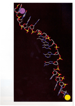

© Copyright 2026