
How to Grade Immunological Risk Using Sensitive HLA Abstract
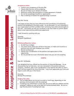
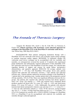
Trends in Transplant. 2010;4:3-10 Carmen Lefaucheur and Denis Glotz: Preformed Donor-Specific Antibodies
How to Grade Immunological Risk Using Sensitive HLA
Donor-Specific Antibodies Detection Techniques
Carmen Lefaucheur and Denis Glotz
Saint-Louis Hospital, Nephrology and Kidney Transplantation, Paris, France
Abstract
Since the pioneering work of Patel and Terazaki, the presence of an antidonor antibody of the
IgG isotype, as demonstrated by a lymphocytotoxic assay on T-cells, has been a contraindication
to transplantation due to the very high rate of graft loss reported. The advent of more sensitive
and specific techniques of detection of anti-HLA antibodies (ELISA or Luminex) has questioned
this dogma, with a number of reports showing that transplantation, despite the presence of
a donor-specific antibody, could be done without excessive graft losses, despite higher rates
of rejection.
We analyzed the results of a prospective observational study on the occurrence of acute
antibody-mediated rejection and survival of patients and grafts, in kidney transplant patients
with preexisting donor-specific HLA antibodies detected by Luminex. This study assess, for
the first time, kidney graft survival and the gradation of the risk of acute antibody-mediated
rejection according to the levels of donor-specific antibodies detected before transplantation.
We have shown a dramatic increase in the risk of antibody-mediated rejection with increasing
levels of preexisting donor-specific antibodies above 465 as detected by Luminex. We have also
shown that patients transplanted with donor-specific antibody mean fluorescence intensity
> 3,000 have a 3.8 increased risk of graft loss as compared to patients transplanted with
donor-specific antibody mean fluorescence intensity < 3,000 (95% CI: 3.5-18.4; p < 0.0001).
This stratification of immunological risk should be used to define acceptable graft and
therapeutic strategies in sensitized patients on the waiting list. Thus, the matching of
graft donors and recipients should take into account this precise analysis of the immunological profile of patients and the evaluation of the risk/benefit balance. (Trends in Transplant.
2010;4:3-10)
Corresponding author: Carmen Lefaucheur, [email protected]
Key words
HLA antibodies. Antibody-mediated rejection. Graft survival. Kidney transplantation.
Correspondence to:
Carmen Lefaucheur
Département de Néphrologie et Transplantation Rénale
Hôpital Saint-Louis
1 Av Claude Vellefaux
75010 Paris, France
E-mail: [email protected]
3
Trends in Transplantation 2010;4
Introduction
Anti-HLA immunization constitutes an immunogenetic hurdle to transplantation, leading
to protracted waiting times that continue to
increase for sensitized kidney transplant receivers1-3. In France, 25% of patients on the
waiting list have a panel-reactive antibody
level > 5%4, and in the USA, 32% of patients
awaiting transplant are sensitized1. Despite efforts to diminish the risk of sensitization, namely recombinant erythropoietin, leukocyte-depleted
transfusions, and the cessation of pre-graft transfusion protocols, the number of sensitized patients on transplant lists remains substantial.
Moreover, loss of a prior graft has become the
primary cause of anti-HLA sensitization.
Patel and Terasaki5, in 1969, demonstrated the efficacy of complement-dependent lymphocytotoxic crossmatch in defining immunological risk in renal transplantation. This became
the standard methodology, still used today, for
graft allocation. It became clear with time that
it did not identify all preexisting donor-specific HLA antibodies (DSA). In recent years, techniques for detection of HLA antibodies have
become more sensitive with the introduction of
solid-phase assays, including enzyme-linked
immunosorbent assays (ELISA), and multiple
bead-based technology, of which the Luminex-based assays are the most frequently used.
The clinical impact of the antibodies detected
by these more sensitive techniques has yet to
be fully evaluated in terms of graft survival and
definition of acceptable grafts3. Studies of the
clinical relevance of DSA in patients transplanted with negative crossmatches have been
contradictory6-8.
The ability to quantify these antibodies9
has added a further dimension of complexity
to the equation. The Luminex technique has
been used in recent studies to choose the type
of desensitization according to DSA strength10
and to determine acceptable DSA levels, allowing for successful kidney transplantation
4
following desensitization11. Forty years after
the initial definition of immunological risk by Terasaki and Patel, the introduction of these more
sensitive techniques revives and carries to a
new level the basic question of the clinical
relevance of donor-specific anti-HLA antibodies and their integration into current strategies
of transplantation. Indeed, no single study has
compared the sensitivity, specificity, and positive predictive value of classic or flow crossmatch, ELISA, and Luminex techniques in the
prediction of acute antibody-mediated rejection and graft survival.
Clinical relevance of preformed
HLA donor-specific antibodies
in kidney transplantation
To investigate the clinical relevance of
pre-graft DSA identified by strictly HLA-specific
assays, we undertook a first study in which we
retrospectively screened a series of 237 consecutive renal transplants performed in our
unit by high-definition ELISA for their presence12. Our study showed that kidney graft
survival at eight years was significantly worse
in patients with DSA. The incidence of antibodymediated rejection in patients with DSA was
nine-fold higher than in patients without DSA
and led to a significantly worse graft survival.
The prevalence of antibody-mediated rejection in patients with DSA detected on historical
serum was 32.3% and was significantly more
elevated in patients with strongly positive DSA
(score 6-8) and in patients with historical positive crossmatches. Thus, the presence of preformed DSA was strongly associated with increased graft loss in kidney transplants, related
to an increased risk of antibody-mediated rejection.
To appraise the full clinical potential of
DSA detected prior to transplantation, we conducted a prospective observational study
in which we employed the capacity of the
Luminex technique to identify with precision
Carmen Lefaucheur and Denis Glotz: Preformed Donor-Specific Antibodies
and to quantify HLA-specific antibodies in order to grade increasing immunological risk.
This study examined the impact of the strength
of preformed DSA on the risk of acute antibody-mediated rejection occurrence and graft
survival in deceased-donor kidney graft patients. The study included 402 consecutive deceased-donor single-organ kidney transplant
patients performed in our unit between January 1998 and June 2006. Our graft strategy
was the current worldwide strategy based on
the negative National Institutes of Health lymphocytotoxic crossmatch test using complement-dependant cytotoxicity on the day of
transplant.
Among the 402 consecutive deceaseddonor single-organ kidney transplant patients
performed in our unit between January 1998
and June 2006, 118 patients (29.4%) were
sensitized, having antibodies against class I
or class II HLA on any pretransplant sera. In
83 patients (20.6%), the antidonor specificity
was determined by Luminex single antigen
bead technique: 60 patients had DSA Luminex class I, 58 patients DSA Luminex class II, and
35 patients DSA Luminex class I and class II.
Patients with DSA had a mean of 2.4 DSA1-6
detected by Luminex on the peak serum. The
mean fluorescence intensity (MFI) of the highest ranked donor-specific bead (MFImax) was
6,189 (301-18,069) and the mean of total MFI
of the DSA detected by Luminex single antigen
bead technique was 10,724 (301-48,204).
Long-term outcomes of kidney
graft in patients with preexisting
donor-specific antibodies
are significantly worse
as compared to patients
transplanted without donor-specific
antibodies
Patients: The mean follow-up time was
51.4 ± 30.6 months (range 1-132). Patient
eight-year survival was similar in non-sensitized
patients, sensitized patients without DSA,
and patients with preformed DSA as assessed
by Luminex technique: 90.2 vs. 91.2 and 90.9%,
respectively (p = 0.98).
Grafts: Five- and eight-year death-censored graft survival was, respectively, 89.2 and
83.6% in non-sensitized patients, 92.5 and 92.5%
in sensitized patients without DSA, and finally 71.2 and 60.8% in patients with preformed DSA detected by Luminex technique.
Kaplan-Meier analysis revealed that patients
with preformed DSA had a significantly lower graft survival as compared to sensitized
patients without DSA and non-sensitized patients (p < 0.001) (Fig. 1A). There was no difference in graft survival analyzed according
to the class of the maximum DSA identified
pre-graft (p = 0.8). Patients with DSA had
poorer graft survival regardless of whether the
maximum DSA was class I or II (p < 0.0002)
(Fig. 1B).
Antibody-mediated rejection risk
according to quantification
of donor-specific anti-HLA antibodies
by Luminex
Acute antibody-mediated rejection occurred in 8% of kidney transplant patients.
Among the 32 patients with antibody-mediated rejection, were DSA-positive by Luminex single antigen bead technique. The patients
with antibody-mediated rejection had an average of three DSA detectable by Luminex
single antigen bead technique on peak sera
(range 0-6), with a mean MFI max of 7,852
(466-17,574) and a mean total MFI of the DSA
of 15,350 (823-42,472).
Receiver operating characteristic curve
analysis determined that an MFI of 465 is
associated with maximal specificity and sensitivity regarding the occurrence of antibody-mediated rejection (AUC 0.9; p < .0001)
(Fig. 2).
5
Trends in Transplantation 2010;4
Anti HLA–DSA–
Anti HLA+DSA–
Anti HLA+DSA+
A
100
90
Graft survival (%)
Graft survival (%)
100
80
70
60
50
40
p < 0.001
0
12 24
36 48
60
72
DSA–
DSA class I
DSA class II
B
90
80
70
60
50
40
84 96
p = 0.0002
0
12 24
Time posttransplant (months)
Number at risk
Anti HLA–DSA– 284 259 246 196 133 91
Anti HLA+DSA– 35 34 33 25 16 10
Anti HLA+DSA+ 83 73 67 60 37 30
64 43
10 7
23 18
36 48
60
72
84 96
Time (months)
29
6
13
Figure 1. Kaplan-Meier estimates of graft survival. According to the presence of anti-HLA antibodies and donor-specific antibodies
on the pre-graft serum in kidney transplant population (Panel A). According to the presence of Class I or Class II of the highest
pre-graft donor-specific antibodies on the pre-graft serum (Panel B). Donor-specific anti-HLA antibodies were detected by Luminex single antigen technique. P values were calculated with the use of the log-rank test. DSA: donor-specific antibodies.
1
0.9
0.8
Sensitivity
0.7
0.6
0.5
0.4
0.3
AUC 0.9
0.2
p < 0.001
0.1
0
0
0.1
0.2 0.3
0.4
0.5
0.6
0.7
0.8
0.9
1
1 - Specificity
Figure 2. Receiver operating characteristic (ROC) curve. For the mean intensity of fluorescence of highest pre-graft donor-specific
antibody detected by Luminex single antigen technique, associated with acute antibody-mediated rejection. AUC: area under
the ROC curve.
The prevalence of antibody-mediated
rejection rises significantly with increasing
MFI of highest pre-graft DSA detected by Luminex technique on peak pre-graft serum:
6
0.9% in patients with MFI < 465; 18.7% in
those with MFI of 466-3,000; 36.4% for MFI of
3,001-6,000; and 51.3% for patients with MFI
> 6,000 (Chi2 = 138.1; p < 0.0001).
Carmen Lefaucheur and Denis Glotz: Preformed Donor-Specific Antibodies
MFI ≤ 3,000
90
MFI > 3,000
Graft suvival (%)
100
80
70
60
50
40
p < 0.0001
0
12
24
36
48
60
72
84
96
Time posttransplantation (months)
Number at risk
MFI ≤ 3,000
MFI > 3,000
352 324 218 246 164 113 83
50 44 40 36 23 19 15
58
11
39
7
Figure 3. Kaplan-Meier estimates of graft survival according to the mean fluorescence intensity of highest pre-graft ranked donor-specific
antibodies detected by Luminex single antigen technique in the entire cohort of kidney transplant patients. DSA: donor-specific antibodies;
MFI: mean fluorescence intensity.
Graft survival according
to quantification of donor-specific
anti-HLA antibodies by Luminex
The eight-year graft survival decreases progressively with rising Luminex MFI:
82.5% in patients with MFI < 465; 78.4% for
patients with MFI of 466-3,000 ; 60.6% for
those with MFI of 3,001-6000; and 55.9 ±
9.6% for patients with MFI > 6,000 (p < 0.001).
As shown in figure 3, the graft survival in
patients with MFI > 3,000 was significantly
lower than that of patients with MFI ≤ 3,000
(p < 0.0001). The relative risk of graft loss
for patients transplanted with pre-graft DSA
higher than 3,000 was 3.8 (95% CI: 3.5-18.4;
p < 0.0001) as compared to with MFI DSA
lower than 3,000.
Estimation of the immunological
risk prior to transplantation
in clinical practice
Our study focuses on the estimation of
the immunological risk of a given patient based
on the antibody profile as defined prior to
transplant by Luminex analysis. It assess for the
first time, using the recent immunological techniques, kidney graft survival and the gradation
of the risk of acute antibody-mediated rejection according to the levels of DSA detected
before transplantation. This analysis has the
advantage of being performed much earlier
than any crossmatch assay and helps define
a transplant strategy for any patient on the waiting list. Based on this strategy, the clinician may
then decide to go on with the crossmatch or to
stop the process before any crossmatching.
So, the utilization of the Luminex single antigen
bead technique has expanded the role of histocompatibility testing beyond the traditional
one, identifying the contraindications to transplantation, to a personalized appraisal of immunological risk. In the present environment of
organ scarcity and financial and logistical constraints, the evaluation before transplantation
of the immunological profiles of patients on
the waiting lists helps to guide deceased-donor kidney allocation13. Stratification of immunological risk before transplantation should be
used not only for deceased-kidney allocation,
7
Trends in Transplantation 2010;4
but also to establish priority programs with the
aim of increasing graft access by highly sensitized patients and also to guide the immunosuppressive therapy and monitoring of recipients.
Thus, the transplantation strategy for each patient should weigh the risk/benefit ratio based on
defined immunological risk prior to transplant.
In recent years, a major change in renal
transplantation has come from the recognition
of the importance of antibody-mediated rejection, recognized initially only as the cause of
hyperacute rejection, but now known to be
responsible for acute and chronic lesions14.
Our study underlines the fact that acute antibody-mediated rejection is a major factor in
the evolution of HLA-incompatible kidney transplants and is associated with higher rates of
graft loss. For treatment of acute antibody-mediated rejection, we used a specific treatment
based on intravenous immune globulin products known to have powerful immunomodulatory effects15. Treatment of acute antibodymediated rejection has evolved from intravenous
immune globulin-based regimens to combination therapies, with plasmapheresis, intravenous immune globulin, and rituximab,
leading to an amelioration of the graft survival
of patients with acute antibody-mediated
rejection16-19.
Importantly, our study shows that even
in the absence of clinical acute antibody-mediated rejection, the long-term graft course is
worse in patients with preexisting DSA. The
recently described entity of subclinical antibody-mediated rejection20,21, in which progressive morphologic lesions are found on biopsy
in the absence of overt clinical rejection, may
account for this different course. These progressive lesions lead to chronic humoral rejection,
first described in 200122 and now recognized
to be a distinct cause of late graft dysfunction
and loss23,24.
Luminex analysis permits pre-graft characterization of the antibody profiles in sensitized
8
patients and gives improved definition of safe
(antibody-negative) and at-risk (antibody-positive) HLA specificities. The first step in the
transplant strategy for sensitized patients is
to define whether a graft with minimal immunological risk is possible. Whenever possible,
kidney transplantation should be performed in
the absence of DSA. Virtual cross-matching,
recently promoted by the United Network for
Organ Sharing25, consists in selecting potential donors without HLA-specificities against
receiver’s antibodies, predicting a negative
crossmatch. The use of the virtual crossmatch
permits expanding the geographic regions from
which kidneys can be drawn and reduces
waiting time and deaths on the waiting list26-28.
Furthermore, it is a good indicator to reduce
the risk of antibody-mediated rejection29.
In practice, we must evaluate, for each
sensitized patient on the waiting list, if the
listing of forbidden antigens permits a donor
pool sufficient to assure transplantation. For
some highly sensitized patients, use of virtual
crossmatching leads to an insufficient number of donors. Thus, alternatively, the access
to transplantation of these patients can be
augmented in three ways: priority programs,
desensitization regimens, or by increased immunological risk of transplantation with preexisting DSA.
Priority programs, such as the Eurotransplant “Acceptable Mismatch” program30, gives
priority to any hypersensitized patient when
there is no incompatibility between the class I
antigens of the donor and those of the recipient, including permitted antigens, and no more
than one donor/recipient incompatibility. This
program has shown that the probability for
a hyperimmunized patient to be grafted with a
kidney with a negative crossmatch rises from
17 to 58% over two years31. Class, et al.30 have
published graft survivals of such patients identical to that of non-immunized patients and superior to that of immunized patients grafted
without the aid of the permitted antigen.
Carmen Lefaucheur and Denis Glotz: Preformed Donor-Specific Antibodies
Desensitization protocols based on highdose intravenous immunoglobulins (IVIg)32-34
have been used with success. The desensitization protocol used by our group is based on
high-dose IVIg, given as three monthly treatments of IVIg 2 g/kg given over two days32.
Other possibilities, including plasmapheresis/
low-dose IVIg/anti-CD20 antibody35 or the combination IVIg/rituximab, can be efficacious in
desensitizing patients awaiting transplantation36.
Transplantation in the presence of DSA
requires a careful estimation of the immunological risk, as assessed by remote crossmatches and levels of antibodies detected by Luminex technique. It will then be up to the
transplant teams to decide, well before transplantation, if these patients are to be grafted
with historical DSA, and up to what level, or
possibly with remote positive crossmatch. For
such patients at high immunological risk, specific posttransplant protocols are used10,37-40.
In our experience, patients transplanted with
a remote positive crossmatch received, posttransplant, in addition to the standard induction with thymoglobulin, three monthly rounds
of high-dose IVIg. Nonetheless, the data in the
literature are, for the present, strictly observational. Little data is available on the latest advances in immunosuppressive drugs. The use
of rituximab in association with IVIg as prophylaxis against acute rejection in positive-crossmatch transplants has been disappointing so
far41. For such transplants at high immunologic
risk, close follow-up is indispensable, notably
with systematic quantification of anti-HLA antibodies posttransplantation and protocol biopsies including C4d staining.
Access to and results of transplantation
can therefore be improved in sensitized patients. The data presented in this paper emphasize the importance of precisely characterizing the status of the anti-HLA pretransplant
immunization using sensitive techniques. This
should allow optimization of donor immunological selection.
References
1. Organ Procurement and Transplantation network. Scientific
registry of transplant recipients. Available at http://www.
optn.org/data/ [accessed June 23, 2008].
2. Cho YW, Cecka J. Crossmatch tests: an analysis of UNOS data
from 1991 to 2000. In: Terasaki P (ed.) Clinical transplants. Los
Angeles: UCLA Immunogenetics Center. 2001:237-46.
3. Gebel HM, Bray RA, Nickerson P. Pre-transplant assessment of
donor-reactive, HLA-specific antibodies in renal transplantation: contraindication vs. risk. Am J Transplant. 2003;3:1488-500.
*Difficulty to assess the immunological risk using a single
technique. Stratification of the risk of antibody-mediated rejection occurrence based on the crossmatch tests.
4. Rapport d’activité de prélèvement et de greffe. Agence de
la Biomédecine. Available at http://www.agence-biomedecine.fr/agence/le-rapport-annuel.html [accessed December 19, 2008].
5. Patel R, Terasaki PI. Significance of the positive crossmatch
test in kidney transplantation. N Engl J Med. 1969;280:735-9.
*The first major demonstration of the clinical impact of DSA
detected by complement-dependent lymphocytotoxic crossmatch.
6. Vlad G, Ho EK, Vasilescu ER, et al. Relevance of different
antibody detection methods for the prediction of antibodymediated rejection and deceased-donor kidney allograft
survival. Hum Immunol. 2009;70:589-94.
7. Patel AM, Pancoska C, Mulgaonkar S, Weng FL. Renal transplantation in patients with pre-transplant donor-specific antibodies and negative flow cytometry crossmatches. Am J
Transplant. 2007;7:2371-7. *Provides the importance of the
detection of DSA by solid-phase methods.
8. Gupta A, Iveson V, Varagunam M, Bodger S, Sinnott P,
Thuraisingham RC. Pretransplant donor-specific antibodies
in cytotoxic negative crossmatch kidney transplants: are they
relevant? Transplantation. 2008;85:1200-4.
9. Mizutani K, Terasaki P, Hamdani E, et al. The importance of
anti-HLA-specific antibody strength in monitoring kidney
transplant patients. Am J Transplant. 2007;7:1027-31. **Introduces the capacity to quantify the strength of alloantibodies by Luminex assays.
10. Akalin E, Dinavahi R, Friedlander R, et al. Addition of plasmapheresis decreases the incidence of acute antibodymediated rejection in sensitized patients with strong donorspecific antibodies. Clin J Am Soc Nephrol. 2008;3:1160-7.
11. Reinsmoen NL, Lai CH, Vo A, et al. Acceptable donor-specific antibody levels allowing for successful deceased and
living donor kidney transplantation after desensitization therapy. Transplantation. 2008;86:820-5.**This paper points out
that, for desensitized patients, the association of high levels
of DSA by Luminex and flow cytometry correlates with high
risk of antibody-mediated rejection.
12. Lefaucheur C, Suberbielle-Boissel C, Hill GS, et al. Clinical
relevance of preformed HLA donor-specific antibodies in
kidney transplantation. Am J Transplant. 2008;8:324-31.
*This study demonstrates the importance of identifying and
characterizing the strength of pre-graft DSA by sensitive
techniques, as well as their association to a remote positive
crossmatch.
13. Bray RA, Nolen JD, Larsen C, et al. Transplanting the highly sensitized patient: The emory algorithm. Am J Transplant.
2006;6:2307-15.
14. Gloor J, Cosio F, Lager DJ, Stegall MD. The spectrum of
antibody-mediated renal allograft injury: implications for treatment. Am J Transplant. 2008;8:1367-73. *Important paper that
develops the concept of antibody-mediated rejection.
15. Kazatchkine MD, Kaveri SV. Immunomodulation of autoimmune and inflammatory diseases with intravenous immune
globulin. N Engl J Med. 2001;345:747-55.
16. Lefaucheur C, Nochy D, Andrade J, et al. Comparison of
combination Plasmapheresis/IVIg/anti-CD20 versus highdose IVIg in the treatment of antibody-mediated rejection.
Am J Transplant. 2009;9:1099-107. **Contemporary regimens
improve the success rate in the treatment of antibody-mediated rejection.
17. Becker YT, Becker BN, Pirsch JD, Sollinger HW. Rituximab
as treatment for refractory kidney transplant rejection. Am J
Transplant. 2004;4:996-1001.
9
Trends in Transplantation 2010;4
18. Faguer S, Kamar N, Guilbeaud-Frugier C, et al. Rituximab
therapy for acute humoral rejection after kidney transplantation. Transplantation. 2007;83:1277-80.
19. Mulley WR, Hudson FJ, Tait BD, et al. A single low-fixed dose
of rituximab to salvage renal transplants from refractory antibody-mediated rejection. Transplantation. 2009;87:286-9.
20. Gloor JM, Cosio FG, Rea DJ, et al. Histologic findings one
year after positive crossmatch or ABO blood group incompatible living donor kidney transplantation. Am J Transplant.
2006;6:1841-7.
21. Haas M, Montgomery RA, Segev DL, et al. Subclinical acute
antibody-mediated rejection in positive crossmatch renal allografts. Am J Transplant. 2007;7:576-85. *First complete histological definition of subclinical antibody-mediated rejection and
its evolution to development of chronic allograft nephropathy.
22. Mauiyyedi S, Pelle PD, Saidman S, et al. Chronic humoral
rejection: identification of antibody-mediated chronic renal
allograft rejection by C4d deposits in peritubular capillaries.
J Am Soc Nephrol. 2001;12:574-82. *The first demonstration
of the substantial prevalence of chronic allograft rejection
immunologically active.
23. Regele H, Bohmig GA, Habicht A, et al. Capillary deposition of
complement split product C4d in renal allografts is associated
with basement membrane injury in peritubular and glomerular capillaries: a contribution of humoral immunity to chronic
allograft rejection. J Am Soc Nephrol. 2002;13:2371-80. *Histological definition of chronic antibody-mediated rejection.
24. Gloor JM, Sethi S, Stegall MD, et al. Transplant glomerulopathy: subclinical incidence and association with alloantibody. Am J Transplant. 2007;7:2124-32.
25. Nikaein A, Cherikh W, Nelson K, et al. Organ procurement
and transplantation network/united network for organ sharing
histocompatibility committee collaborative study to evaluate
prediction of crossmatch results in highly sensitized patients.
Transplantation. 2009;87:557-62.
26. Zangwill SD, Ellis TM, Zlotocha J, et al. The virtual crossmatch--a screening tool for sensitized pediatric heart transplant recipients. Pediatr Transplant. 2006;10:38-41.
27. Zangwill S, Ellis T, Stendahl G, Zahn A, Berger S, Tweddell J.
Practical application of the virtual crossmatch. Pediatr Transplant. 2007;11:650-4.
28. Zachary AA, Sholander JT, Houp JA, Leffell MS. Using real
data for a virtual crossmatch. Hum Immunol. 2009;70:574-9.
29. Tambur AR, Ramon DS, Kaufman DB, et al. Perception
versus reality?: Virtual crossmatch--how to overcome some
of the technical and logistic limitations. Am J Transplant.
2009;9:1886-93. *Validates that the virtual crossmatch can
serve as an outstanding tool to predict HLA compatibility
between donor and recipient.
10
30. Claas FH, Witvliet MD, Duquesnoy RJ, Persijn GG, Doxiadis II. The acceptable mismatch program as a fast tool
for highly sensitized patients awaiting a cadaveric kidney
transplantation: short waiting time and excellent graft outcome. Transplantation. 2004;78:190-3. **The Eurotransplant
model of priority program for highly sensitized patients on
the waiting list.
31. Doxiadis II, Duquesnoy RJ, Claas FH. Extending options for
highly sensitized patients to receive a suitable kidney graft.
Curr Opin Immunol. 2005;17:536-40.
32. Glotz D, Antoine C, Julia P, et al. Desensitization and subsequent kidney transplantation of patients using intravenous
immunoglobulins (IVIg). Am J Transplant. 2002;2:758-60.
33. Jordan SC. Management of the highly HLA- sensitized patient. A novel role for intravenous gammaglobulin. Am J Transplant. 2002;2:691-2.
34. Jordan SC, Tyan D, Stablein D, et al. Evaluation of intravenous immunoglobulin as an agent to lower allosensitization
and improve transplantation in highly sensitized adult patients with end-stage renal disease: report of the NIH IG02
trial. J Am Soc Nephrol. 2004;15: 3256-62.
35. Stegall MD, Gloor J, Winters JL, Moore SB, Degoey S. A
comparison of plasmapheresis versus high-dose IVIG desensitization in renal allograft recipients with high levels
of donor specific alloantibody. Am J Transplant. 2006;6:
346-51.
36. Vo AA, Lukovsky M, Toyoda M, et al. Rituximab and intravenous immune globulin for desensitization during renal transplantation. N Engl J Med. 2008;359:242-51.
37. Anglicheau D, Loupy A, Suberbielle C, et al. Posttransplant
prophylactic intravenous immunoglobulin in kidney transplant
patients at high immunological risk: a pilot study. Am J Transplant. 2007;7:1185-92.
38. Akalin E, Ames S, Sehgal V, et al. Intravenous immunoglobulin and thymoglobulin induction treatment in immunologically high-risk kidney transplant recipients. Transplantation. 2005;79:742.
39. Burns JM, Cornell LD, Perry DK, et al. Alloantibody levels
and acute humoral rejection early after positive crossmatch kidney transplantation. Am J Transplant. 2008;8:2
684-94.
40. Akalin E, Bromberg JS. Intravenous immunoglobulin induction treatment in flow cytometry cross-match-positive kidney
transplant recipients. Hum Immunol. 2005;66:359-63.
41. Matignon M, Tagnaouti M, Audard V, Dahan K, Lang P,
Grimbert P. Failure of anti-CD20 monoclonal antibody therapy to prevent antibody-mediated rejection in three crossmatch-positive renal transplant recipients. Transplant Proc.
2007;39:2565-7.


© Copyright 2026